Embed Size (px)
Citation preview
AlgAl Biology And Biotechnology
Algal Biology and Biotechnology.indb 1 2/6/2009 1:59:24 PM

J. I. S. KhattarD. P. Singh
Gurpreet KaurDepartment of Botany
Punjabi UniversityPatiala, Punjab
India
I.K. International Publishing House Pvt. Ltd.New Delhi • BaNgalore
AlgAl Biology And Biotechnology
Algal Biology and Biotechnology.indb 3 2/6/2009 1:59:24 PM

Published byI.K. International Publishing House Pvt. Ltd.S-25, green Park extensionUphaar Cinema MarketNew Delhi–110 016 (India)e-mail: [email protected]
iSBN 978-93-80026-19-0
© 2009 i.K. international Publishing house Pvt. ltd.
10 9 8 7 6 5 4 3 2 1
all rights reserved. No part of this book may be reproduced or used in any form, electronic or mechanical, including photocopying, recording, or by any information storage and retrieval system, without written permission from the publisher.
Published by Krishan Makhijani for i.K. international Publishing house Pvt. ltd., S-25, green Park extension, Uphaar Cinema Market, New Delhi–110 016. Printed by rekha Printers Pvt. ltd., okhla industrial area, Phase ii, New Delhi–110 020.
Algal Biology and Biotechnology.indb 4 2/6/2009 1:59:24 PM
Preface
The algae are thallophytes that have chlorophyll a as their primary photosynthetic pigment and don’t have sterile jacket of cells around the reproductive cells. The term ‘algae’ used in this book includes blue-green algae (cyanobacteria) which are prokaryotic in nature and are evolutionary more closer to bacteria. Classification and taxonomy of algae has been mainly based on field characters which in-cluded mainly phenotypic characters. But recently, trend has changed to classify algae taking in to account molecular characters as well. recent advances in the taxonomy of cyanobacteria have been discussed in the first two chapters of the book. algae most commonly occur in water, be it fresh water, brackish or marine water. however, these organisms can be found in almost every environment on earth, from algae growing in snow of mountains, glaciers of antarctica, to hot springs, bare rocks or building facades. Microalgae found growing on buildings and their ecophysiological aspects have been discussed in chapter 3 of the book. Some cyanobacteria are unique algae in the sense that they inhabit extreme habitats like cold deserts, hot springs, acidic and alkaline environments etc. how these organisms are able to cope up with these environments is discussed in the next chapter. Cyanobacteria differentiate only three types of cells viz., vegetative cells, heterocysts and akinetes. Much is known about the mechanism of heterocyst differentiation. akinetes are consid-ered to be perennating structures but what triggers akinete differentiation in cyanobacteria is not properly understood. what is known about akinete differentiation in cyanobacteria is discussed in chapter 5. role of microalgae, especially cyanobacteria, in agriculture is well known. initially it was thought that cyanobacteria add to fertility of the soil only by fixing atmospheric nitrogen. But now it has been shown that besides fixing molecular nitrogen they also add growth hormones and other chemicals to the soil which help in increased growth/yield of the crop plants. role of cyanobacteria as biofertilizers, their association with the roots of crop plants other than rice, like wheat, and nitrogen metabolism in cyanobacteria are discussed in next four chapters. The second half of the book is devoted to biotechnological advances/applications of algae. These include algae as source of natural colours, production of bioactive compounds/secondary metabolites and role of microalgae in bioremediation etc. algae as source of natural colours and bioactive compounds are discussed in chapters 10 and 11, respectively. economic importance of sea weeds is well known. recent advances/researches in sea weed biotechnology are also discussed in one of the chapters. Cyanotoxins is another important area of research. water contamination by bloom forming algae is a serious threat to the environment and toxins released by some of these may cause serious health problems in animals and humans. These aspects and biotechnological
Algal Biology and Biotechnology.indb 5 2/6/2009 1:59:25 PM
vi l Preface
applications of secondary metabolites are discussed in next two chapters. algae as biofuels has generated much interest in the recent years. one chapter on this aspect is also there in the book. role of algae in bioremediation is discussed in the last two chapters. This book thus encompasses information on recent advances made in the fields of algal taxonomy, physiology and biotechnology. we hope that this book will be of interest to students of botany and to researchers. The authors worked very hard in preparing their chapters in a very short time. we are grateful to them for their efforts. Prof. T. a. Sarma, our worthy teacher, has always been a guiding force and source of inspiration for us. he has always supported our endeavours. we also sincerely thank head, Department of Botany, Punjabi University, Patiala for providing infrastructural facilities. Valuable suggestions, encouragement and support from all our teachers, colleagues and friends are also highly acknowledged. we sincerely thank Mr. Yadvinder Singh and Mr. Jeevesh Nadda for their invaluable help in preparing this book.
J. I. S. KhattarD. P. Singh
Gurpreet Kaur
Algal Biology and Biotechnology.indb 6 2/6/2009 1:59:25 PM
Contents
Preface vList of Contributors ix
1. Morpho-taxonomy of Coccoid Cyanobacteria 1 g. l. Tiwari, V. K. Dwivedi, richa Tandon, o. N. Tiwari and rama Kant
2. Cyanobacterial Ecology and Molecular Approaches for Biodiversity Analysis 27 o. avijeet Singh, o. N. Tiwari, K. laxmipriya, S. Deepa Devi and M. rohinikumar Singh
3. Cyanobacteria and Micro-algae on Building Facades and Stone Surface of Monuments – An Ecophysiological Prospective 41
l. K. Samad and S. P. adhikary
4. Some Secrets of Ubiquity in Cyanobacteria 57 S. P. Singh
5. Akinete Differentiation in Cyanobacteria 63 T. a. Sarma
6. Nitrogen Metabolism in Cyanobacteria 81 Mayashree B. Syiem, arvind K. Singh and a. N. rai
7. Developments in Cyanobacterial Biofertilizer 97 B. D. Kaushik
8. Distribution, Preservation and Maintenance of Cyanobacterial Diversity and their Application as Biofertilizer 109
l. Sophiya, gunapati oinam, M. K. leingaklemba, h. Boboy Singh, o. N. Tiwari and M. rohinikumar Singh
9. Cyanobacterial Diversity in Wheat Fields: Taxonomic and Functional Aspects 121
radha Prasanna, N. Kartheyan, M. Joshi, a. rana and lata
10. Microalgae: A Source of Natural Colours 129 gurpreet Kaur, J. i. S. Khattar, D. P. Singh, Yadvinder Singh and Jeevesh Nadda
Algal Biology and Biotechnology.indb 7 2/6/2009 1:59:25 PM

viii l Contents
11. A Decade Odyssey with Cyanobacteria: As a Source of Bioactive Molecules and a Model for Salt Stress Study 151
r. K. asthana
12. Seaweeds: A Survey of Research and Utilization 165 P. V. Subba rao, K. ganesan and K. Suresh Kumar
13. Cyanobacterial Toxins and Public Health Issues 179 N. K. Singh, S. Saxena, o. N. Tiwari and Dolly wattal Dhar
14. Secondary Metabolite Production in Cyanobacteria 205 r. ashwin Kumar and S. K. Verma
15. Renewable Energy Sources: Potential of Microalgae for Production of Environmental Friendly Biofuel 217
Jitender, anjuli Sood, amita Mahajan and a. S. ahluwalia
16. Bioremediation of Wastewater and Role of Microalgae 227 rajiv Kumar and Dinesh goyal
17. Role of Cyanobacterial Oxidases in Bioremediation – An Overview 251 l. Uma, D. Prabaharan, B. Priya and g. Subramanian
Index 263
Algal Biology and Biotechnology.indb 8 2/6/2009 1:59:25 PM

List of Contributors
Adhikary, S. P., P. g. Department of Botany and Biotechnology, Utkal University, Bhubane-swar-751004, orissaAhluwalia, A. S., Department of Botany, Pan-jab University, Chandigarh-160014. Asthana, R. K., Centre of advanced Study in Botany, Banaras hindu University, Varanasi-221005, UPDevi, S. Deepa, Microbial resources Divi-sion, institute of Bioresources and Sustainable Development, Takyelpat, imphal-795001, ManipurDhar, Dolly Wattal, Centre for Conservation and Utilization of Blue green algae, iari, New DelhiDwivedi, V. K., Department of Botany, Univer-sity of allahabad, allahabad-211002, UP Ganesan, K., Marine Biotechnology and ecology Discipline, Central Salt and Marine Chemicals research institute (CSir), Bhavna-gar-364 002, gujaratGoyal, Dinesh, Department of Biotechnology & environmental Sciences, Thapar University, Patiala-147004, PunjabJitender, Department of Botany, Panjab Uni-versity, Chandigarh-160014 Joshi, M. Division of Microbiology, indian agricultural research institute, New Delhi-110012Kant, Rama, Department of Botany, Univer-sity of allahabad, allahabad-211002, UP
Karthikeyan, N., Division of Microbiology, indian agricultural research institute, New Delhi-110012Kaur, Gurpreet, Department of Botany, Pun-jabi University, Patiala – 147 002, PunjabKaushik, B. D., Former head & Professor, Division of Microbiology, iari, New Delhi 110012Khattar, J. I. S., Department of Botany, Pun-jabi University, Patiala – 147 002, PunjabKumar, K. Suresh, Marine Biotechnology and ecology Discipline, Central Salt and Marine Chemicals research institute (CSir), Bhavna-gar-364 002, gujaratKumar, Rajiv., Department of Biotechnology & environmental Sciences, Thapar University, Patiala-147004, Punjab Kumar, R. A. Ashwin., Centre for Biotechnol-ogy, Biological Science group, BiTS, Pilani, rajasthan.Lata, Division of Microbiology, indian agri-cultural research institute, New Delhi-110012Laxmipriya, K., Microbial resources Divi-sion, institute of Bioresources and Sustainable Development, Takyelpat, imphal-795001, ManipurLeingaklemba, M. K., Microbial resources Division, institute of Bioresources and Sustain-able Development, (an autonomous institute under the DBT, goi), Takyelpat, imphal-795001, Manipur
Algal Biology and Biotechnology.indb 9 2/6/2009 1:59:25 PM

x l List of Contributors
Mahajan, Amita, Department of Biomedical engineering, rayat and Bahra College of en-gineering & Biotechnology, Kharar-140301, PunjabNadda, Jeevesh, Department of Botany, Pun-jabi University, Patiala – 147 002, PunjabOinam, Gunapati, Microbial resources Divi-sion, institute of Bioresources and Sustainable Development, (an autonomous institute un-der the DBT, goi), Takyelpat, imphal-795001, ManipurPrabaharan, D. National Facility for Marine Cyanobacteria (Sponsored by DBT, govt. of india), Bharathidasan University, Tiruchirap-palli-620024, Tamil NaduPrasanna, Radha, Division of Microbiology, indian agricultural research institute, New Delhi-110012Priya, B., National Facility for Marine Cy-anobacteria (Sponsored by DBT, govt. of india), Bharathidasan University, Tiruchirap-palli-620024, Tamil NaduRai, A. N., Vice-Chancellor, Mizoram Univer-sity, aizwal-796 009, MizoramRana, A., Division of Microbiology, indian agricultural research institute, New Delhi-110012Samad, L. K., P. g. Department of Botany and Biotechnology, Utkal University, Bhubaneswar - 751004, orissaSarma, T. A., Department of Biotechnology, MVr & Dr. h. S. M. i. C. College of Technol-ogy, Kanchikacherla-521 180, andhra PradeshSaxena, S., Centre for Conservation and Utili-zation of Blue green algae, iari, New DelhiSingh, Arvind Kumar, Department of Bio-chemistry, North-eastern University, Shillong, 793 022, MeghalayaSingh, D. P., Department of Botany, Punjabi University, Patiala – 147 002, Punjab
Singh, H. Boboy, Microbial resources Divi-sion, institute of Bioresources and Sustainable Development, (an autonomous institute un-der the DBT, goi), Takyelpat, imphal-795001, ManipurSingh, M. Rohinikumar, Microbial resources Division, institute of Bioresources and Sustain-able Development, (an autonomous institute under the DBT, goi), Takyelpat, imphal-795001, ManipurSingh, N. K., C. P. College of agriculture, S. D. a. U., S. K. Nagar, gujarat-3855506.Singh, O. Avijeet, Microbial resources Divi-sion, institute of Bioresources and Sustainable Development, Takyelpat, imphal-795001, ManipurSingh, S. P., Centre of advanced Study in Botany, Banaras hindu University, Varanasi-221005Singh, Yadvinder, Department of Botany, Pun-jabi University, Patiala – 147 002, PunjabSood, Anjuli, Department of Botany, Univer-sity of Delhi, Delhi-110007 Sophiya, L., Microbial resources Division, institute of Bioresources and Sustainable De-velopment, (an autonomous institute under the DBT, goi), Takyelpat, imphal-795001, ManipurSubba Rao, P. V., Marine Biotechnology and ecology Discipline, Central Salt and Marine Chemicals research institute (CSir), Bhavna-gar-364 002, gujaratSubramanian, G., National Facility for Ma-rine Cyanobacteria (Sponsored by DBT, govt. of india), Bharathidasan University, Tiruchirap-palli- 620024, Tamil NaduSyiem, Mayashree, Department of Biochemis-try, North-eastern University, Shillong - 793 022, MeghalayaTandan, Richa, Department of Botany, Uni-versity of allahabad, allahabad-211002, UP
Algal Biology and Biotechnology.indb 10 2/6/2009 1:59:25 PM

List of Contributors l xi
Tiwari, G. L., Department of Botany, Univer-sity of allahabad, allahabad-211002, UPTiwari, O. N., Microbial resources Division, institute of Bioresources and Sustainable De-velopment, (an autonomous institute under the DBT, goi), Takyelpat, imphal-795001, Manipur
Uma, L., National Facility for Marine Cy-anobacteria (Sponsored by DBT, govt. of india), Bharathidasan University, Tiruchirap-palli-620024, Tamil NaduVerma, S. K., Centre for Biotechnology, Biological Science group, BiTS, Pilani, rajasthan
Algal Biology and Biotechnology.indb 11 2/6/2009 1:59:25 PM
1C h a p t e r
Morpho-taxonomy of Coccoid Cyanobacteria
G. L. tiwari, V. K. Dwivedi, richa tandon, O. N. tiwari and rama Kant
Although starting point for names of unicellular and colonial Blue-green Algae is Linnaeus (1753) but Nägeli (1848) in a study entitled “Gatungen Einzelliger Algen” for the first time separated the family Chroococcaceae from more obviously green and red algae. He classified separate genera on the basis of cell division pattern and arrangement of cells in mucilaginous matrix of colonies. The criteria, which he set, are being used even in the present day taxonomy. Geitler (1932) summarized the Nägelian classification of Chroococcaceae and he created new series of families and genera. Desikachary (1959) described Indian coccoid cyanobacteria based on clas-sification of Geitler (1932) and Fritsch (1945). He divided the coccoid cyanobacteria into three orders viz. 1. Chroococcales, 2. Chamaesiphonales and 3. Pleurocapsales.
DrOUet’S CONCept
Drouet and Daily (1956) and Drouet (1981) observed that many accounts on unicellular and colonial forms of Blue-green Algae were prepared and many taxa were described, but presumed species or herbarium sheets were either not maintained at all or poorly stored. Therefore, validity of a species and the applications of its name could be judged only from the literary presentation of its description and the artistic excellence of its illustrations and not from the specimens which the authors studied. Drouet’s classifications (1981) are based on observations of thousands of collections of various species, living and preserved, axenic and xenic cultures from most regions and countries of the world. Drouet’s species represent a polymorphic cluster of ecophenes in a few broadly conceived genera. His classification of coccoid cyanobacteria is given below:
Algal Biology and Biotechnology.indb 1 2/6/2009 1:58:25 PM
2 l Algal Biology and Biotechnology
Myxophyceae
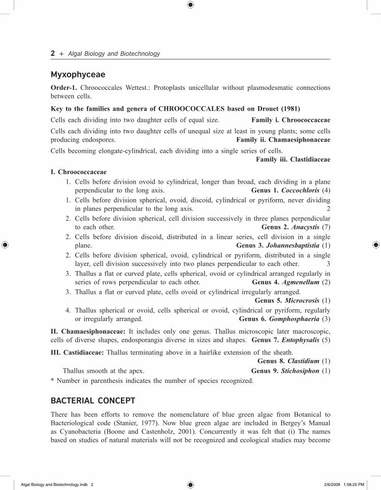
Order-1. Chroococcales Wettest.: Protoplasts unicellular without plasmodesmatic connections between cells.
Key to the families and genera of CHROOCOCCALES based on Drouet (1981)Cells each dividing into two daughter cells of equal size. Family i. ChroococcaceaeCells each dividing into two daughter cells of unequal size at least in young plants; some cells producing endospores. Family ii. Chamaesiphonaceae Cells becoming elongate-cylindrical, each dividing into a single series of cells. Family iii. Clastidiaceae
I. Chroococcaceae 1. Cells before division ovoid to cylindrical, longer than broad, each dividing in a plane
perpendicular to the long axis. Genus 1. Coccochloris (4) 1. Cells before division spherical, ovoid, discoid, cylindrical or pyriform, never dividing
in planes perpendicular to the long axis. 2 2. Cells before division spherical, cell division successively in three planes perpendicular
to each other. Genus 2. Anacystis (7) 2. Cells before division discoid, distributed in a linear series, cell division in a single
plane. Genus 3. Johannesbaptistia (1) 2. Cells before division spherical, ovoid, cylindrical or pyriform, distributed in a single
layer, cell division successively into two planes perpendicular to each other. 3 3. Thallus a flat or curved plate, cells spherical, ovoid or cylindrical arranged regularly in
series of rows perpendicular to each other. Genus 4. Agmenellum (2) 3. Thallus a flat or curved plate, cells ovoid or cylindrical irregularly arranged.
Genus 5. Microcrosis (1) 4. Thallus spherical or ovoid, cells spherical or ovoid, cylindrical or pyriform, regularly
or irregularly arranged. Genus 6. Gomphosphaeria (3)
II. Chamaesiphonaceae: It includes only one genus. Thallus microscopic later macroscopic, cells of diverse shapes, endosporangia diverse in sizes and shapes. Genus 7. Entophysalis (5)
III. Castidiaceae: Thallus terminating above in a hairlike extension of the sheath. Genus 8. Clastidium (1) Thallus smooth at the apex. Genus 9. Stichosiphon (1)* Number in parenthesis indicates the number of species recognized.
BaCterIaL CONCept
There has been efforts to remove the nomenclature of blue green algae from Botanical to Bacteriological code (Stanier, 1977). Now blue green algae are included in Bergey’s Manual as Cyanobacteria (Boone and Castenholz, 2001). Concurrently it was felt that (i) The names based on studies of natural materials will not be recognized and ecological studies may become
Algal Biology and Biotechnology.indb 2 2/6/2009 1:58:25 PM
Morpho-taxonomy of Coccoid Cyanobacteria l 3
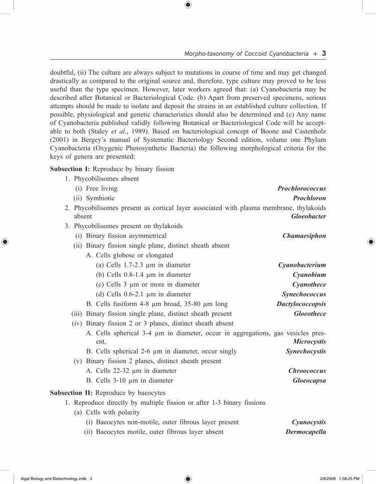
doubtful, (ii) The culture are always subject to mutations in course of time and may get changed drastically as compared to the original source and, therefore, type culture may proved to be less useful than the type specimen. However, later workers agreed that: (a) Cyanobacteria may be described after Botanical or Bacteriological Code. (b) Apart from preserved specimens, serious attempts should be made to isolate and deposit the strains in an established culture collection. If possible, physiological and genetic characteristics should also be determined and (c) Any name of Cyanobacteria published validly following Botanical or Bacteriological Code will be accept-able to both (Staley et al., 1989). Based on bacteriological concept of Boone and Castenholz (2001) in Bergey’s manual of Systematic Bacteriology Second edition, volume one Phylum Cyanobacteria (Oxygenic Photosynthetic Bacteria) the following morphological criteria for the keys of genera are presented:
Subsection I: Reproduce by binary fission 1. Phycobilisomes absent (i) Free living Prochlorococcus (ii) Symbiotic Prochloron 2. Phycobilisomes present as cortical layer associated with plasma membrane, thylakoids
absent Gloeobacter 3. Phycobilisomes present on thylakoids (i) Binary fission asymmetrical Chamaesiphon (ii) Binary fission single plane, distinct sheath absent A. Cells globose or elongated (a) Cells 1.7-2.3 µm in diameter Cyanobacterium (b) Cells 0.8-1.4 µm in diameter Cyanobium (c) Cells 3 µm or more in diameter Cyanothece (d) Cells 0.6-2.1 µm in diameter Synechococcus B. Cells fusiform 4-8 µm broad, 35-80 µm long Dactylococcopsis (iii) Binary fission single plane, distinct sheath present Gloeothece (iv) Binary fission 2 or 3 planes, distinct sheath absent A. Cells spherical 3-4 µm in diameter, occur in aggregations, gas vesicles pres-
ent, Microcystis B. Cells spherical 2-6 µm in diameter, occur singly Synechocystis (v) Binary fission 2 planes, distinct sheath present A. Cells 22-32 µm in diameter Chroococcus B. Cells 3-10 µm in diameter Gloeocapsa
Subsection II: Reproduce by baeocytes 1. Reproduce directly by multiple fission or after 1-3 binary fissions (a) Cells with polarity (i) Baeocytes non-motile, outer fibrous layer present Cyanocystis (ii) Baeocytes motile, outer fibrous layer absent Dermocapella
Algal Biology and Biotechnology.indb 3 2/6/2009 1:58:25 PM

4 l Algal Biology and Biotechnology
(b) Cells without polarity (i) Baeocytes non-motile, outer fibrous layer present Xenococcus (ii) Baeocytes motile, outer fibrous layer absent Stanieria 2. Reproduce by multiple fission but after extensive binary fissions (a) Cells arranged in cubical or irregular groups, all cells may undergo multiple fission
to produce baeocytes (i) Baeocytes non-motile, fibrous layer present Chroococcidiopsis (ii) Baeocytes motile, fibrous layer absent Myxosarcina (b) Binary fission in several planes results into pseudofilamentous and “heterotrichous”
thallus, only certain cells undergo multiple fission, baeocytes are motile (Pleurocapsa groups including Pleurocapsa, Hyella, Solentia)
KOMareK aND aNaGNOStIDIS (1998) CONCept
Based on modern approach, Komarek and Anagnostidis (1998) revised the entire group of coc-coid Cyanoprokaryotes (Cyanobacteria) in a new phylum Cyanoprokaryota and included all the forms in a single order Chroococcales. Their concept and classification up to genera level are given below based on the above work in the form of keys:Order Chroococcales: Unicells or colonies with loose, compact packets, pseudofilamentous or pseudoheterotrichous thalli reproducing by cell division or forming nanocytes baeocytes and exocytes etc.
Key to the families of Chroococcales: A. Prokaryotic cells without thylakoids and phycobilisomes 1-Gloeobacteraceae A. Prokaryotic cells with thylakoids and phycobilisomes B B. Cells or colonies, reproducing by exocytes, nannocytes, or baeocytes C B. Cells usually not reproducing by exocytes, nannocytes, or baeocytes F C. Cells reproducing by exocytes 2-Chamaesiphonales C. Cells or colonies usually reproducing by baeocytes or nanocytes D D. Thallus as solitary or groups of cells or packets E D. Thallus diversified and often represented by pseudo-heterotrichous stages
3-Hyellaeceae E. Cells solitary or in groups, multiple fission producing baeocytes 4-Dermocarpellaceae E. Cells solitary or in packets with distinct surrounding sheath, reproducing by nanocytes
or baeocytes 5-Xenococcaceae F. Unicells or colonies divisions in one or two planes G F. Colonies with cell divisions in three or more plane H G. Cell division in one plane 6-Synechococcaeae G. Cell division in two planes 7-Merismopediaceae
Algal Biology and Biotechnology.indb 4 2/6/2009 1:58:25 PM

Morpho-taxonomy of Coccoid Cyanobacteria l 5
H. Colonies without polarity I H. Colonies with polarity J I. Cell division in three planes, divided cells become rounded before next division
8-Microcystaceae I. Cell division in three planes, divided cell may not become rounded before next
division. 9-Chroococcaceae J. Colonies polarized, attached and irregular cell divisions in various planes, occasion-
ally one plane and form rows of cells. 10-Entophysalidaceae J. Radially differentiated colonies with linear arrangement of cells.
11-Hydrococcaceae
Chroococcales Wettst
Figures of all the genera mentioned below are included in plates 1 to 4 and their reference number is given before their name, figures of two genera Lithomyxa and Coccopedia were not available.
Family: 1. Gloeobacteraceae Komarek and AnagnostidisCells single or in mucilaginous groups, cells spherical or long cylindrical, mucilaginous envelope with laminations; cells pale blue-green, reddish or violet; cells without thylakoids and phycobil-lisomes, but with Chl-a and phycobilins 1. Gloeobacter Rippka et al.
Family: 2. Synechococcaceae Komárek and AnagnostidisCells single or in mucilaginous colonies, cells arranged irregularly or ± in one direction; cells mostly elongate, rarely spherical, ellipsoidal, fusiform, cylindrical; cell division in one plane, perpendicular to the long axis.
Sub-family: 1. Aphanothecoideae Komárek et AnagnostidisCells spherical or elongated up to 3 times of width, solitary or variously arranged into colonies, involution in cells present, nanocytes known.
Key to the genera of Sub-family Aphanothecoideae (after Komárek and Anagnostidis, 1998) A. Cells not in mucilaginous colonies B A. Cells in mucilaginous colonies D B. Cells oval or cylindrical, keritomization absent C B. Cells widely oval keritomization present 9. Cyanothece C. Cells oval, content differentiated in chromato-centroplasm 4. Cyanobium C. Cells cylindrical with lengthwise striations 3. Cyanobacterium D. Cells form spherical or irregular mucilaginous colonies E D. Cells associated in diverse types of mucilaginous stalks M
Algal Biology and Biotechnology.indb 5 2/6/2009 1:58:25 PM

6 l Algal Biology and Biotechnology
E. Cells ± in rows F E. Cells on or near the surface of mucilaginous spheres L F. Cells in spherical or amorphous colonies G F. Cells in spherical or irregular net like colonies or in rows J G. Colonies planktonic with iron precipitate H G. Colonies amorphous without iron precipitate I H. Cells spherical or wide oval, colonies somewhat spherical 6. Cyanogranis H. Colonies irregular, cells shortly cylindrical rounded at poles 7. Cyanocatena I. Colonies with calcareous incrustations Lithomyxa I. Colonies without calcareous incrustations N J. Colonies composed of mucilaginous strands K J. Colonies somewhat spherical, cells arranged in indistinct radiating rows
15. Radiocystis K. Colonies planktonic or endogloeic 5. Cyanodictyon K. Colonies endolithic 14. Lithococcus L. Cells spreaded on surface of mucilaginous colonies 10. Epigloeosphaera L. Cells arranged in periphery of mucilaginous colonies 13. Lemmermanniella M. Colonies spherical free floating with curved cells attached to radiating and branched
gelatinous stalks 8. Cyanonephron M. Colonies consist of gelatinous and lamellated stalks attached to substratum and cells
situated in apical ends 12. Hormothece N. Cells embedded in mucilaginous matrix 2. Aphanothece N. Cells have individual laminated envelopes and arranged in small groups
11. Gloeothece
Sub-family: 2. Synechococcoideae Komárek et AnagnostidisCells usually several times longer than broad, rarely short; colonies often arranged in one direc-tion, involuting cell long, nanocytes absent.
Key to the genera of Sub-family Synechococccoideae (after Komárek and Anagnostidis, 1998) A. Cells solitary or in groups, without mucilage B A. Cells in mucilaginous colonies C B. Cells ± fusiform, long 20. Myxobaktron B. Cells cylindrical 25. Synechococcus C. Cells scattered or arranged in rows D C. Cells ± in rows in mucilaginous structures F D. Macroscopic colonies, cells oval to cylindrical 18. Dzensia D. Microscopic colonies, cells elongate or fusiform E
Algal Biology and Biotechnology.indb 6 2/6/2009 1:58:25 PM

Morpho-taxonomy of Coccoid Cyanobacteria l 7
E. Cells fusiform 23. Rhabdogloea E. Cells cylindrical without rounded end 22. Rhabdoderma F. Cells arranged in their longer axis, in one or more rows G F. Cells arranged in uniseriate filamentous colonies 19. Johannesbaptistia G. Colonies ± spherical, free floating, branched rows radiating from center
17. Cyanothamnos G. Colonies with irregular or parallel row of cells in one layer H H. Tube like colonies I H. Mat forming, elongate colonies, cells arranged in parallel or zig-zag J I. Cells arranged in rows in narrow or wide sheath 27. Wolskyella I. Cells oriented in one direction not in rows, sheath tubular 16. Bacularia J. Colonies long, mucilaginous, spreaded over the soil surface 26. Tubiella J. Colonies soft, hemispherical or crustose K K. Colonies hemispherical, cells with individual envelope, in rows 21. Pseudoncobyrsa K. Colonies crustose, parallely arranged pseudofilamentous, cells without individual enve-
lopes 24. Rhodostichus
Family: 3 Merismopediaceae ElenkinCells solitary or in regular or irregular colonies, mucilage present, cells spherical to rod-shaped, cell division in two planes perpendicular to each other.
Sub-family: 1. Merismopedioideae ElenkinCells solitary or in colonies; colonies microscopic, usually free living, irregular or flat single layer; cells spherical to rod shaped; cell division in two planes, perpendicular to each other in successive generations.
Key to the genera of Sub-family Merismopedioideae (after Komárek and Anagnostidis, 1998) A. Cells solitary, free living 34. Synechocystis A. Cells in groups B B. Colonies mucilaginous, shapeless, cells irregularly disposed 28. Aphanocapsa B. Colonies flattened, single layered, free or attached C C. Colonies sessile, cells divide at right angle to substratum 30. Mantellum C. Colonies free D D. Cells elongate, oriented at right angle to plane of the colony 32. Microcrocis D. Cells spherical or slightly longer E E. Cells irregularly arranged in plane of the colony F E. Cells arranged in plane of the colony and in perpendicular rows G F. Colonies tabular flat Coccopedia F. Colonies irregular 33. Pannus
Algal Biology and Biotechnology.indb 7 2/6/2009 1:58:25 PM
8 l Algal Biology and Biotechnology
G. Cells spherical or hemispherical 31. Merismopedia G. Cells elongate, forming characteristic groups with in the colonies 29. Cyanotetras
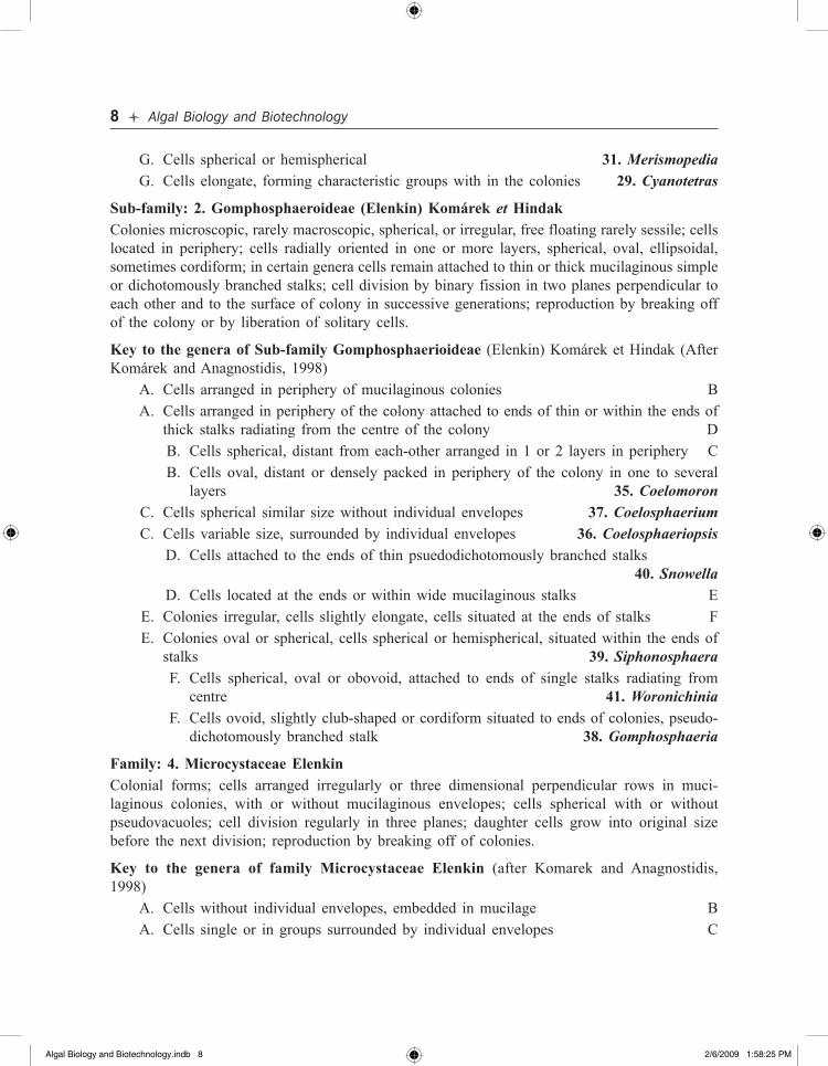
Sub-family: 2. Gomphosphaeroideae (Elenkin) Komárek et HindakColonies microscopic, rarely macroscopic, spherical, or irregular, free floating rarely sessile; cells located in periphery; cells radially oriented in one or more layers, spherical, oval, ellipsoidal, sometimes cordiform; in certain genera cells remain attached to thin or thick mucilaginous simple or dichotomously branched stalks; cell division by binary fission in two planes perpendicular to each other and to the surface of colony in successive generations; reproduction by breaking off of the colony or by liberation of solitary cells.
Key to the genera of Sub-family Gomphosphaerioideae (Elenkin) Komárek et Hindak (After Komárek and Anagnostidis, 1998) A. Cells arranged in periphery of mucilaginous colonies B A. Cells arranged in periphery of the colony attached to ends of thin or within the ends of
thick stalks radiating from the centre of the colony D B. Cells spherical, distant from each-other arranged in 1 or 2 layers in periphery C B. Cells oval, distant or densely packed in periphery of the colony in one to several
layers 35. Coelomoron C. Cells spherical similar size without individual envelopes 37. Coelosphaerium C. Cells variable size, surrounded by individual envelopes 36. Coelosphaeriopsis D. Cells attached to the ends of thin psuedodichotomously branched stalks
40. Snowella D. Cells located at the ends or within wide mucilaginous stalks E E. Colonies irregular, cells slightly elongate, cells situated at the ends of stalks F E. Colonies oval or spherical, cells spherical or hemispherical, situated within the ends of
stalks 39. Siphonosphaera F. Cells spherical, oval or obovoid, attached to ends of single stalks radiating from
centre 41. Woronichinia F. Cells ovoid, slightly club-shaped or cordiform situated to ends of colonies, pseudo-
dichotomously branched stalk 38. Gomphosphaeria
Family: 4. Microcystaceae ElenkinColonial forms; cells arranged irregularly or three dimensional perpendicular rows in muci-laginous colonies, with or without mucilaginous envelopes; cells spherical with or without pseudovacuoles; cell division regularly in three planes; daughter cells grow into original size before the next division; reproduction by breaking off of colonies.
Key to the genera of family Microcystaceae Elenkin (after Komarek and Anagnostidis, 1998) A. Cells without individual envelopes, embedded in mucilage B A. Cells single or in groups surrounded by individual envelopes C
Algal Biology and Biotechnology.indb 8 2/6/2009 1:58:25 PM
Morpho-taxonomy of Coccoid Cyanobacteria l 9
B. Cells irregularly arranged in formless colonies, with gas-vacuoles, planktonic 45. Microcystis
B. Cells arranged in three dimensional, cuboid colonies without pseudo-vacuoles 43. Eucapsis
C. Envelopes around individual cells or groups of cells often lamellated and coloured mainly epilithic or epiphytic 44. Gloeocapsa
C. Envelopes around individual cells thin colourless and widened, grow often submerged 42. Chondrocystis
Family: 5. Chroococcaceae NaegeliPacket like, microscopic, mostly colonial forms; colonies may aggregate to macroscopic layers; cells arranged radially or serially in cubic packets; cell division in three or more planes, divided cells may not become rounded before next division, nannocytes absent.
Key to the genera of family Chroococcaceae Naegeli (after Komarek and Anagnostidis, 1998) A. Cell division at right angle planes in first 2-3 generations, later irregular B A. Cell divisions begin irregular but later in one dominant way resulting into characteristic
patterns D B. Mucilaginous envelopes colourless 47. Chroococcus B. Mucilaginous envelopes coloured, firm and thin C C. Colonies small with one or a few irregularly arranged cells, envelope may be covered
by protuberances or warts 46. Asterocapsa C. Colonies many celled, densely packed, cells irregular shaped, surrounded by individual
stratified, coloured envelopes 51. Gloeocapsopsis D. Colonies spherical, without any mucilaginous stalks E D. Cells localized at ends of thick mucilaginous stalks 50. Cyanostylon E. Colonies always microscopic, cells arranged in two irregular, perpendicular rows, cells
distant from one another 48. Cyanokybus E. Colonies microscopic, cells grouped together in close contact with one another F F. Cells arranged in three-dimensional packets in old colonies, in more or less perpen-
dicular rows 49. Cyanosarcina F. Cells arranged radially or cruciately arranged G G. Cells radially arranged 53. Pseudocapsa G. Cells cruciately arranged 52. Nephrococcus
Family: 6. Entophysalidaceae GeitlerColonies polarized, mucilaginous, spherical, elongate or irregular, rarely pseudofilamentous cell orientation; thallus not differentiated in spite of certain degree of polarity, cell division in vari-ous planes, sometimes at margin by one plane; reproduction by solitary cells, monocytes or by cluster of cells; nannocytes absent.
Algal Biology and Biotechnology.indb 9 2/6/2009 1:58:26 PM

Algal Biology And Biotechnology
Publisher : IK International ISBN : 9789380026190 Author : JIS Khattar, DPSingh, Gurpreet
Type the URL : http://www.kopykitab.com/product/5636
Get this eBook
30%OFF