1
Alterna(ve Treatment Modality in the Management of Post-Surgical Esotropia with Anomalous Correspondence Alexandria Tilley, O.D., Tharanie Amarawardana, O.D., Kimberly Hamian O.D., M.H. Esther Han, O.D., F.C.O.V.D, F.A.A.O. IntroducBon The ves(bulo-ocular reflex (VOR) occurs when the semi-circular canals detect changes in angular accelera(on of the head. Upon cessa(on of a constant-velocity movement, post-rotary nystagmus occurs opposite the ini(al head rota(on. The direc(on of the post-rotary nystagmus can promote abduc(on in a pa(ent with esotropia. Techniques involving these subcor(cal reflexes are the basis for this treatment modality. VesBbulo-Ocular Reflex Keeps images stable on the re(na during brief head movements Serves to prevent re(nal image slip, which can lead to oscillopsia and visual blur The eyes rotate at the same velocity, but in the opposite direc(on, as head rota(on Example: if one is fixa(ng a target straight ahead and rotates their head to the leN, the eyes should move to the right Latency of 16 ms Made up of a slow component and a fast component The slow component, which is the subcor(cal por(on, is present at birth The fast saccadic return, which is cor(cally driven, may or may not be present at birth VesBbular Anatomy Peripheral ves(bular system (Fig. 1) Three semi-circular canals Otoliths: utricle, saccule Central ves(bular projec(ons Ves(bular nuclei Ascending tracts Descending tracts Semi-circular canals Anterior (Superior) Posterior (Inferior) Horizontal (Lateral) Each semi-circular canal is oriented 90 degrees away from each other Anterior and posterior canals: oriented 45 degrees on either side of the parasagi\al plane Horizontal canal: oriented 30 degrees above the axial plane The semi-circular canals act like func(onal pairs If you excite a semi-circular canal on one side of the head, you inhibit the other on the opposite side of the head that lies roughly within the same plane Physiology Each semi-circular canal contains endolymph, a viscous fluid. On one por(on of each semi-circular canal, there is a dila(on, known as an ampulla, in which a gela(nous mass, known as a cupula, is located (Figure 2). Within the cupula are ves(bular hair cells, innervated by bipolar sensory neurons of the ves(bular nerve. With rota(onal accelera(on of the head, endolymph, due to iner(a, does not move ini(ally with the head rota(on There is a net displacement of the endolymph that pushes on the cupula, and bends the embedded hair cells. This causes release of an ac(on poten(al and ac(va(on of the ves(bular nerve. References 1. Baxstrom C, Clopton J. COPE Course: Ves(bular processing – A key component of func(on and balance, January 2017. 2. Cohen HS. Neuroscience for rehabilita(on. Philadelphia: Lippinco\ Williams & Wilkins, 1999. 3. Leigh RJ, Zee DS. The neurology of eye movements: 4 th edi(on. New York, NY: Oxford Univ. Press, 2015. 4. Wong AM. Eye movement disorders. Oxford: Oxford Univ. Press, 2007. Fig 1: h\ps://www.nidcd.nih.gov/health/balance/balance_disorders.asp Fig 2: public domain image: original source from NASA’s ar(cle “The Effects of Space Flight on the Human Ves(bular System” h\ps://commons.wikimedia.org/wiki/ File:Inner_ear%27s_cupula_transmipng_indica(on_of_accelera(on.jpg Fig 3: “Push-pull principle in the ves(bular system, for a horizontal head-movmenet to the right: the right horizontal semicircular canal gets excited, while the leN horizontal canal gets inhibited” by Thomas Haslwanter. GFDL Permission. h\ps://commons.wikimedia.org/wiki/File:Ves(bular_PushPull.svg Table 1: Adapted from: Leigh RJ, Zee DS. The neurology of eye movements: 4 th edi(on- Table 3-2. New York, NY: Oxford Univ. Press, 2015. Case PresentaBon A 9 year old male presented to the University Eye Center with concerns of decreased vision in his right eye and a constant right esotropia. His mother reports that she ini(ally no(ced the eye turn prior to 6 months of age, and he had strabismus surgery on his right eye when he was five years old. Upon ini(al evalua(on, his mother reported she did not want to undergo another strabismus surgery and was interested in only pursuing vision therapy Post-Rotary Nystagmus A jerk nystagmus that occurs secondary to cessa(on of a constant-velocity rota(on. Occurs secondary to the con(nued movement of the endolymph secondary to iner(al forces The slow phase occurs in the direc(on of ini(al head rota(on, while the fast phase occurs in the opposite direc(on. For example: if one rotates to the leN at a constant velocity and then quickly stops movement, a jerk nystagmus would be observed with the slow phase to the leN and a fast phase to the right. Table 3: RelaBonship Between Semi-Circular Canals and Extra-Ocular Muscles Discussion of Techniques Monocular fixa2on with head rota2ons: In order to promote abduc(on of the right eye, the pa(ent wore a patch over his leN eye. He then fixated on a target in-front of him. He (lted his chin down by 30 degrees and rotated his head from side to side laterally while maintaining fixa(on straight ahead. When his head was towards the leN side, his right eye, by way of the ves(bulo-ocular reflex, was in a posi(on of abduc(on. Thereby, the visual-ves(bular interac(on was used in order to increase range of mo(on of the right esotropic eye. Spinning with post-rotary nystagmus: The pa(ent closed his eyes and (lted his head downwards by 30 degrees. He was then spun in a chair upwards of ten (mes to the right. ANer 10 chair spins, he had to quickly stop, open his eyes, and perform hart chart saccades. This technique was implemented because upon stop-rota(on both eyes con(nued to move with a jerk nystagmus, with the slow phase towards the right. The slow phase served to move both eyes to the right in a yoked fashion, promo(ng abduc(on of his right eye in a subcor(cal manner, while having him perform hart chart saccades introduced cor(cal processing to the technique. Conclusion S(mula(ng rota(onal input using post-rotary responses involving visual-ves(bular interac(ons were u(lized with this pa(ent since tradi(onal binocular techniques were contraindicated and strabismus surgery was not an op(on. The u(liza(on of the ves(bular ocular reflex and post-rotary responses can be considered as an alterna(ve to tradi(onal strabismus therapy. Ini(al evalua(on DVA cc: 20/200 OD, 20/20 OS Refrac(ve error: +3.50sph OD, +2.25sph OS DCT: 35 Δ V-pa\ern constant right esotropia NCT: 50 Δ V-pa\ern constant right esotropia Extra-ocular mo(li(es: abduc(on deficit OD, OS, DVD near his neutral point Worth 4 Dot: eso diplopia Re-evalua(on: session 8 DVA cc: 20/200 OD, 20/20 OS Amblyoscope: Horror fusionalis responses DCT: 35 Δ V-pa\ern constant right esotropia NCT: 50 Δ V-pa\ern constant right esotropia Re-evalua(on at conclusion of 21 sessions of in-office vision therapy DVA cc: 20/100 OD, 20/20 OS Refrac(ve error: +3.50sph OD, +2.25sph OS DCT: 25 Δ V-pa\ern constant right esotropia NCT: 25 Δ V-pa\ern constant right esotropia Three month re-evalua(on DVA cc: 20/100 OD, 20/20 OS DCT: 25 Δ V pa\ern constant right esotropia NCT: 25 Δ V pa\ern constant right esotropia Amblyoscope anomalous correspondence The pa(ent was instructed to discon(nue ves(bular-based exercises to see if size of the devia(on persisted. Canal Excita(on Right horizontal canal Right medial rectus LeN lateral rectus LeN horizontal canal LeN medial rectus Right lateral rectus LeN anterior canal LeN superior rectus Right inferior oblique LeN posterior canal LeN inferior oblique Right superior rectus Right anterior canal Right superior rectus LeN inferior oblique Right posterior canal Right superior oblique LeN inferior rectus Techniques used in vision therapy: sessions 1-7 Tradi(onal vision therapy techniques were a\empted: Brock string Luster apprecia(on Quoit vectograms GTVT Pola mirror Accommoda(ve rock VTS3 vergence Stepping stones to fusion Patch OS 2 hours/ day Techniques used in vision therapy: sessions 9-21 Ves(bular-based techniques were used: Post-rotary nystagmus with chair spins to promote abduc(on of the right eye Monocular fixa(ons while rota(ng the head laterally Turn and clap See video for demonstra(on of techniques Pathway of the Horizontal VOR Fig 4. A three to four neuron arc exists to connect the horizontal canals to the extra-ocular muscles, to cause yoked horizontal eye movements. LeN Horizontal Semi- Circular Canal VN AN ON Medial Rectus Lateral Rectus Figure Key: VN: Ves(bular Nucleus AN: Abducens Nucleus ON: Oculomotor Nucleus Figure 1 Figure 2 Figure 3 Table 1: ExaminaBon Findings Table 2: Therapy Protocol

Alexandria Tilley, O.D., Tharanie Amarawardana, O.D ...pubs.covd.org/VDR/17AM/24_Tilley-Amarawardana-Hamian-Han.pdf · Alternave Treatment Modality in the Management of Post-Surgical

Embed Size (px)

Citation preview

Alterna(veTreatmentModalityintheManagementofPost-SurgicalEsotropiawithAnomalousCorrespondence

AlexandriaTilley,O.D.,TharanieAmarawardana,O.D.,KimberlyHamianO.D.,M.H.EstherHan,O.D.,F.C.O.V.D,F.A.A.O.

IntroducBonTheves(bulo-ocularreflex(VOR)occurswhenthesemi-circularcanalsdetectchangesinangularaccelera(onofthehead.Uponcessa(onofaconstant-velocitymovement,post-rotarynystagmusoccursoppositetheini(alheadrota(on.Thedirec(onofthepost-rotarynystagmuscanpromoteabduc(oninapa(entwithesotropia.Techniquesinvolvingthesesubcor(calreflexesarethebasisforthistreatmentmodality.

VesBbulo-OcularReflex•   Keepsimagesstableonthere(naduringbriefheadmovements•   Servestopreventre(nalimageslip,whichcanleadtooscillopsiaandvisualblur•   Theeyesrotateatthesamevelocity,butintheoppositedirec(on,asheadrota(on•   Example:ifoneisfixa(ngatargetstraightaheadandrotatestheirheadtotheleN,theeyes

shouldmovetotheright•   Latencyof16ms•   Madeupofaslowcomponentandafastcomponent

•   Theslowcomponent,whichisthesubcor(calpor(on,ispresentatbirth•   Thefastsaccadicreturn,whichiscor(callydriven,mayormaynotbepresentatbirth

VesBbularAnatomyPeripheralves(bularsystem(Fig.1)•   Threesemi-circularcanals•   Otoliths:utricle,saccule

Centralves(bularprojec(ons•   Ves(bularnuclei•   Ascendingtracts•   Descendingtracts

Semi-circularcanals•   Anterior(Superior)•   Posterior(Inferior)•   Horizontal(Lateral)

•   Eachsemi-circularcanalisoriented90degreesawayfromeachother•   Anteriorandposteriorcanals:oriented45degreesoneithersideoftheparasagi\alplane•   Horizontalcanal:oriented30degreesabovetheaxialplane•   Thesemi-circularcanalsactlikefunc(onalpairs

•   Ifyouexciteasemi-circularcanalononesideofthehead,youinhibittheotherontheoppositesideoftheheadthatliesroughlywithinthesameplane

Physiology•   Eachsemi-circularcanalcontainsendolymph,aviscousfluid.•   Ononepor(onofeachsemi-circularcanal,thereisadila(on,knownasanampulla,inwhicha

gela(nousmass,knownasacupula,islocated(Figure2).•   Withinthecupulaareves(bularhaircells,innervatedbybipolarsensoryneuronsofthe

ves(bularnerve.•   Withrota(onalaccelera(onofthehead,endolymph,duetoiner(a,doesnotmoveini(ally

withtheheadrota(on•   Thereisanetdisplacementoftheendolymphthatpushesonthecupula,andbendsthe

embeddedhaircells.Thiscausesreleaseofanac(onpoten(alandac(va(onoftheves(bularnerve.

References1.BaxstromC,CloptonJ.COPECourse:Ves(bularprocessing–Akeycomponentoffunc(onandbalance,January2017.2.CohenHS.Neuroscienceforrehabilita(on.Philadelphia:Lippinco\Williams&Wilkins,1999.3.LeighRJ,ZeeDS.Theneurologyofeyemovements:4thedi(on.NewYork,NY:OxfordUniv.Press,2015.4.WongAM.Eyemovementdisorders.Oxford:OxfordUniv.Press,2007.Fig1:h\ps://www.nidcd.nih.gov/health/balance/balance_disorders.aspFig2:publicdomainimage:originalsourcefromNASA’sar(cle“TheEffectsofSpaceFlightontheHumanVes(bularSystem”h\ps://commons.wikimedia.org/wiki/File:Inner_ear%27s_cupula_transmipng_indica(on_of_accelera(on.jpgFig3:“Push-pullprincipleintheves(bularsystem,forahorizontalhead-movmenettotheright:therighthorizontalsemicircularcanalgetsexcited,whiletheleNhorizontalcanalgetsinhibited”byThomasHaslwanter.GFDLPermission.h\ps://commons.wikimedia.org/wiki/File:Ves(bular_PushPull.svgTable1:Adaptedfrom:LeighRJ,ZeeDS.Theneurologyofeyemovements:4thedi(on-Table3-2.NewYork,NY:OxfordUniv.Press,2015.

CasePresentaBonA9yearoldmalepresentedtotheUniversityEyeCenterwithconcernsofdecreasedvisioninhisrighteyeandaconstantrightesotropia.Hismotherreportsthatsheini(allyno(cedtheeyeturnpriorto6monthsofage,andhehadstrabismussurgeryonhisrighteyewhenhewasfiveyearsold.Uponini(alevalua(on,hismotherreportedshedidnotwanttoundergoanotherstrabismussurgeryandwasinterestedinonlypursuingvisiontherapyPost-RotaryNystagmus•   Ajerknystagmusthatoccurssecondarytocessa(onofaconstant-velocityrota(on.•   Occurssecondarytothecon(nuedmovementoftheendolymphsecondarytoiner(alforces•   Theslowphaseoccursinthedirec(onofini(alheadrota(on,whilethefastphaseoccursinthe

oppositedirec(on.•   Forexample:ifonerotatestotheleNataconstantvelocityandthenquicklystopsmovement,a

jerknystagmuswouldbeobservedwiththeslowphasetotheleNandafastphasetotheright.

Table3:RelaBonshipBetweenSemi-CircularCanalsandExtra-OcularMuscles

DiscussionofTechniquesMonocularfixa2onwithheadrota2ons:Inordertopromoteabduc(onoftherighteye,thepa(entworeapatchoverhisleNeye.Hethenfixatedonatargetin-frontofhim.He(ltedhischindownby30degreesandrotatedhisheadfromsidetosidelaterallywhilemaintainingfixa(onstraightahead.WhenhisheadwastowardstheleNside,hisrighteye,bywayoftheves(bulo-ocularreflex,wasinaposi(onofabduc(on.Thereby,thevisual-ves(bularinterac(onwasusedinordertoincreaserangeofmo(onoftherightesotropiceye.

Spinningwithpost-rotarynystagmus:Thepa(entclosedhiseyesand(ltedhisheaddownwardsby30degrees.Hewasthenspuninachairupwardsoften(mestotheright.ANer10chairspins,hehadtoquicklystop,openhiseyes,andperformhartchartsaccades.Thistechniquewasimplementedbecauseuponstop-rota(onbotheyescon(nuedtomovewithajerknystagmus,withtheslowphasetowardstheright.Theslowphaseservedtomovebotheyestotherightinayokedfashion,promo(ngabduc(onofhisrighteyeinasubcor(calmanner,whilehavinghimperformhartchartsaccadesintroducedcor(calprocessingtothetechnique.

ConclusionS(mula(ngrota(onalinputusingpost-rotaryresponsesinvolvingvisual-ves(bularinterac(onswereu(lizedwiththispa(entsincetradi(onalbinoculartechniqueswerecontraindicatedandstrabismussurgerywasnotanop(on.Theu(liza(onoftheves(bularocularreflexandpost-rotaryresponsescanbeconsideredasanalterna(vetotradi(onalstrabismustherapy.

Ini(alevalua(on

•   DVAcc:20/200OD,20/20OS•   Refrac(veerror:+3.50sphOD,+2.25sphOS•   DCT:35ΔV-pa\ernconstantrightesotropia•   NCT:50ΔV-pa\ernconstantrightesotropia•   Extra-ocularmo(li(es:abduc(ondeficitOD,OS,DVD

nearhisneutralpoint•   Worth4Dot:esodiplopia

Re-evalua(on:session8

•   DVAcc:20/200OD,20/20OS•   Amblyoscope:Horrorfusionalisresponses•   DCT:35ΔV-pa\ernconstantrightesotropia•   NCT:50ΔV-pa\ernconstantrightesotropia

Re-evalua(onatconclusionof21sessionsofin-officevisiontherapy

•   DVAcc:20/100OD,20/20OS•   Refrac(veerror:+3.50sphOD,+2.25sphOS•   DCT:25ΔV-pa\ernconstantrightesotropia•   NCT:25ΔV-pa\ernconstantrightesotropia

Threemonthre-evalua(on

•   DVAcc:20/100OD,20/20OS•   DCT:25ΔVpa\ernconstantrightesotropia•   NCT:25ΔVpa\ernconstantrightesotropia•   AmblyoscopeanomalouscorrespondenceThepa(entwasinstructedtodiscon(nueves(bular-basedexercisestoseeifsizeofthedevia(onpersisted.

Canal Excita(on

Righthorizontalcanal RightmedialrectusLeNlateralrectus

LeNhorizontalcanal LeNmedialrectusRightlateralrectus

LeNanteriorcanal LeNsuperiorrectusRightinferioroblique

LeNposteriorcanal LeNinferiorobliqueRightsuperiorrectus

Rightanteriorcanal RightsuperiorrectusLeNinferioroblique

Rightposteriorcanal RightsuperiorobliqueLeNinferiorrectus

Techniquesusedinvisiontherapy:sessions1-7

Tradi(onalvisiontherapytechniqueswerea\empted:•   Brockstring•   Lusterapprecia(on•   Quoitvectograms•   GTVT•   Polamirror•   Accommoda(verock•   VTS3vergence•   Steppingstonestofusion•   PatchOS2hours/day

Techniquesusedinvisiontherapy:sessions9-21

Ves(bular-basedtechniqueswereused:•   Post-rotarynystagmuswithchairspinstopromote

abduc(onoftherighteye•   Monocularfixa(onswhilerota(ngtheheadlaterally•   Turnandclap•   Seevideofordemonstra(onoftechniques

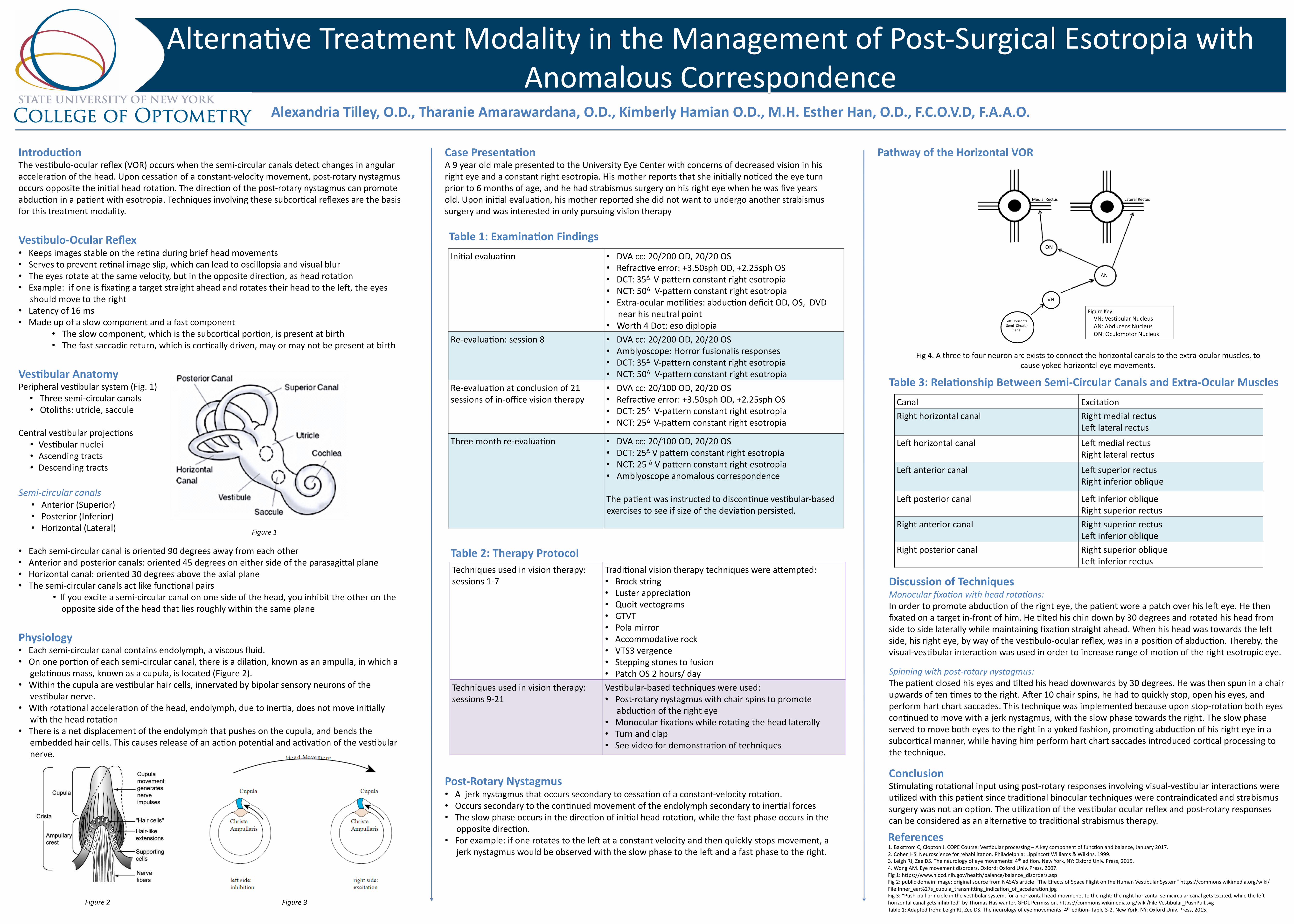
PathwayoftheHorizontalVOR

Fig4.Athreetofourneuronarcexiststoconnectthehorizontalcanalstotheextra-ocularmuscles,tocauseyokedhorizontaleyemovements.

LeNHorizontalSemi-Circular

Canal

VN

AN

ON

MedialRectus LateralRectus

FigureKey:VN:Ves(bularNucleusAN:AbducensNucleusON:OculomotorNucleus

Figure1

Figure2 Figure3

Table1:ExaminaBonFindings

Table2:TherapyProtocol