Embed Size (px)
Citation preview
8/16/2012
1
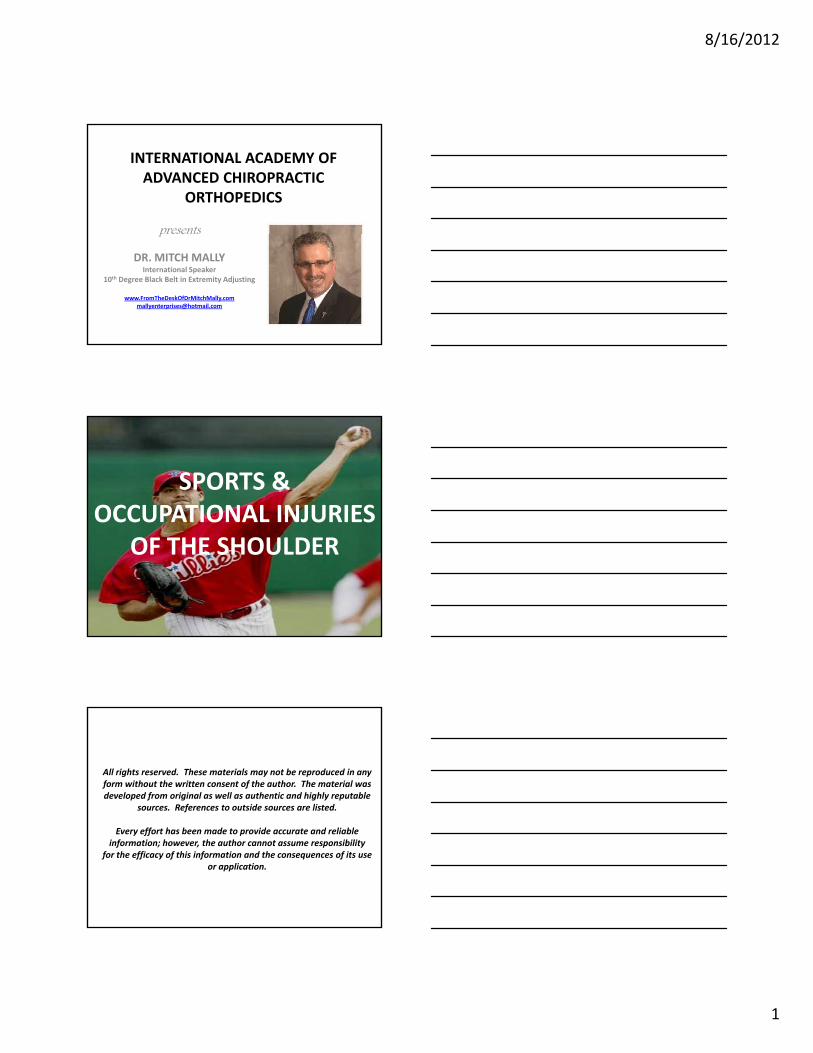
INTERNATIONAL ACADEMY OF ADVANCED CHIROPRACTIC
ORTHOPEDICS
presentsp
DR. MITCH MALLYInternational Speaker
10th Degree Black Belt in Extremity Adjusting
SPORTS & OCCUPATIONAL INJURIES
OF THE SHOULDER
All rights reserved. These materials may not be reproduced in any form without the written consent of the author. The material was developed from original as well as authentic and highly reputable
sources. References to outside sources are listed.
Every effort has been made to provide accurate and reliable information; however, the author cannot assume responsibility
for the efficacy of this information and the consequences of its use or application.

8/16/2012
2
The “Magnificent 7”
Shoulder
Acute Shoulder Injuries
“…shoulder is the most mobile joint in the human body…increased risk of injury…clavicle fractures are among the most common acute shoulder injuries, and more than 80 percent of them can be managed conservatively. Humeral head fractures are less common and usually occur in elderly persons; 85 percent of them can be managed non‐operatively…soft tissue injuries include shoulder dislocations, rotator cuff tears and acromio clavicular sprains Acromio clavicularcuff tears, and acromio‐clavicular sprains. Acromio‐clavicularinjuries…types I and II are treated conservatively, types III and IV are treated surgically…debate about the best approach for type III. Eighty percent of shoulder dislocations are anterior…nonsurgical technique…rotator cuff tears can be managed conservatively or surgically…”
American Family Physician. Vol. 70. Nov. 15, 2004. Quillen, D. M., Wuchner, M., and Hatch, R. L.
Childhood and Adolescent Sports‐Related Overuse Injuries
“Youth sports participation carries an inherent risk of injury, including overuse injuries. Little leaguer’s shoulder…usually is self‐limited. Little leaguer’s elbow…complete rest from throwing for four to six weeks followed by rehabilitation and a gradual throwing program…Treatment usually is conservative…Osgood‐Schlatter disease…most patients respond to conservative measures…United States, approximately 30 million children and teenagers participate in organized sports. Sports are the leading cause of injury in adolescents…one half of all sports injuries in children are preventable with proper education and use of protective equipment…at risk…as a result of impropereducation and use of protective equipment…at risk…as a result of improper technique, poorly fitting protective equipment, training errors, and muscle weakness and imbalance…injuries can be managed conservatively with proper and timely diagnosis…chronic overuse injuries in young athletes may be related to limited recovery time from longer competitive seasons and year‐round training…”
American Family Physician. Vol. 73, No. 6. March 15, 2006. Cassas, K. J. and Wayhs, A.
8/16/2012
3
MRI Findings in Throwing Shoulders: Abnormalities in
Professional Handball Players
“…shoulders of throwing athletes are highly stressed joints and likely to have more structural abnormalities seen on magnetic resonance imaging scans… an average of seven abnormal magnetic resonance imaging findings was observed in the throwing shoulders…93% of the throwing shoulders had abnormal magnetic resonance imagingthrowing shoulders had abnormal magnetic resonance imaging findings, only 37% were symptomatic. Partial rotator cuff tears…superolateral osteochondral defects of the humeral head…typical throwing lesions…this suggests that the evaluation of an athlete’s throwing shoulder should be done very thoroughly and should not be based mainly on abnormalities seen on magnetic resonance imaging scans…”
Clinical Orthopaedics & Related Research. 434: 130‐137, May 2005. Bernhard, J., Zumstein, M., Pfirrmann, C. W., Zanetti, M., and Gerber, C.
Identifying and Managing Shoulder Pain in Competitive Swimmers. How to Minimize Training
Flaws and other Risks.
“…shoulder pain resulting from glenohumeral instability is common among competitive swimmers. The biomechanics inherent to swimming promotes muscular imbalances that stresses the
l li t t t d t ib t t h ldcapsulo‐ligamentous structures and contribute to shoulder instability. Most swimmers respond favorably to conservative treatment of rest and rehabilitation … swimmers who respond well to rehabilitation have a better prognosis for a successful return to swimming that those who require surgery…”
The Physician and Sportsmedicine. Vol. 33, No. 9. Sept. 2005. O’Donnell, C. J., Bowen, J., and Fossati, J.
Upper extremity disorders in the workplace: Costs and outcomes beyond the
first return to work
“…Cumulative trauma disorders of the upper extremities (CTD) have become increasingly important in workers’ compensation caseloads over the last two decades…analyzed post‐injury employment patterns and return‐to‐work probabilities for a sample of Ontario
k ith CTD f t fi ft i j k ’ ith CTDworkers with CTD, for up to five years after injury…workers’ with CTD are compared to results for workers with back injuries or fractures…most workers with CTD return to work at least once…does not necessarily mark the end of work disability…26% with CTD report a second injury‐related absence…18% with back pain…12% with fractures…a substantial proportion of workers with CTD or work‐related back pain experience injury‐related absences after their first return to work…”
Journal of Occupational Rehabilitation. Vol. 16, No. 3. Sept. 2006. Baldwin, M. L. and Butler R. J.
8/16/2012
4
RESEARCH REPORTResting Position Variables at the Shoulder:
Evidence to Support a Posture‐Impairment Association
“…posture and impairment are not directly related, but linked by movement dysfunction…the relationships among posture, pectoralisminor muscle length, and movement alterations at the g ,shoulder…scapula orientation, thoracic kyphosis, and pectoralisminor muscle lengths were measured…significant group differences were demonstrated for several posture variables…thoracic spine kyphosis and scapular internal rotation…distance from the sternalnotch to the coracoid process demonstrated the highest correlation with pectoralis minor muscle length…”
Physical Therapy. Vol. 86, No. 4. April 2006. Borstad, J. D.
Postural Distortions
8/16/2012
5
The Effect of Long Versus Short Pectoralis Minor Resting Length on Scapular Kinematics in Healthy
Individuals
“…to compare scapular kinematics during arm elevation between groups distinguished by pectoralis minor resting length…individuals with subacromial impingement have altered scapular kinematics…loss of posterior tipping and increased internal rotation…with the scapula for the short group staying anteriorly tipped at higher angles…with g p y g y pp g gindividuals with a shorter pectoralis minor demonstrating a more internally rotated scapula…these results support the theory that an adaptively short pectoralis minor my influence scapular kinematics and is therefore a potential mechanism for subacromialimpingement…”
Journal of Orthopedic & Sports Physical Therapy. Vol. 35, No. 4. April 2005. Borstad, J. D. and Ludewig, P. M.
Shoulder function and 3‐dimensional scapular kinematics in people with and without shoulder
impingement syndrome.
“…several factors such as posture, muscle force, range of motion, and scapular dysfunction are commonly believed to contribute to shoulder impingement…impingement group…slightly greater scapular upward rotation and clavicular elevation during flexion and slightly greater scapular posterior tilt and clavicular retraction duringgreater scapular posterior tilt and clavicular retraction during scapular‐plane elevation…impingement group…less range of motion and force in all directions… no differences in resting posture… the kinematic differences found in subjects with impingement may represent scapulothoracic compensatory strategies for glenohumeral weakness or motion loss. The decreased range of motion and force found in subjects with impingement support rehabilitation approaches that focus on strengthening and restoring flexibility…”
Physical Therapy. 2006 Aug; 86(8): 1075‐90. McClure, P. W., Michener, L. A., and Karduna, A. R.
Scapulothoracic and Glenohumeral Kinematics Following an External
Rotation Fatigue Protocol
“…to determine the effects of shoulder external rotator muscle fatigue on 3‐dimensional scapulothoracic and glenohumeral kinematics. The external rotator muscles of the shoulder are important for normal shoulder f ti I i d f f th l h b b d ifunction. Impaired performance of these muscles has been observed in subjects with impingement syndrome and it is possible that external rotator muscle fatigue leads to altered kinematics of the shoulder girdle…after completing the fatigue protocol…demonstrated less external rotation of the humerus…less posterior tilt of the scapula in the beginning phase of arm elevation…more scapular upward rotation and clavicular retraction in the mid ranges of arm elevation…performance of an external rotation fatigue protocol results in altered scapulothoracicand glenohumeral kinematics..”
Journal of Orthopedic & Sports Physical Therapy. 2006; 36(8):557‐571. Ebaugh, D. D., McClure, P. W., and Karduna, A. R.
8/16/2012
6
Pathomechanics in Atraumatic Shoulder Instability: Scapular Positioning Correlates with
Humeral Head Centering
“…analyze three‐dimensional scapular positioning and glenohumeralcentering of normal atraumatic unstable shoulders…scapular plane was increased in nine of 14 patients and decreased in three patients…scapular internal rotation in the transverse plane was p p pincreased in all unstable shoulders…unstable shoulders also had malcentering…of the humeral head in the direction of instability during various arm positions… the high correlation suggests that scapular positioning is relevant for humeral head decentering … physiotherapeutic strategy should consider the malpositioning of the scapula and be adapted to the direction of instability…”
Clinical Orthopaedics & Related Research. 433: 82‐89, April 2005. von Eisenhart‐Rothe, R., Matsen, F. A., Eckstein, F., Vogl, T., and Graichen, H.
Anatomical and Biomechanical Mechanisms of Subacromial Impingement Syndrome.
“…subacromial impingement syndrome is the most common disorder of the shoulder, resulting in functional loss and disability … evidence exists to support the presence of the anatomical factors of inflammation of the tendons and bursa degeneration of the tendons weak or dysfunctionalbursa, degeneration of the tendons, weak or dysfunctional rotator cuff musculature, weak or dysfunctional scapular musculature, posterior glenohumeral capsule tightness, postural dysfunctions of the spinal column and scapula and bony or soft tissue abnormalities of the borders of the subacromial outlet…dysfunctional glenohumeral and scapulothoracic movement patterns…”
Clinical Biomechanics (Bristol, Avon). 2003 Jun; 18(5): 369‐79. Michener, L. A., McClure, P. W., and Karduna, A. R.
Has the management of shoulder dislocation changed over time?
“…Anterior shoulder dislocation…recently the treatment of traumatic shoulder dislocation has included immobilisation for varying periods of time followed by physiotherapy…most frequent mechanism of injury was a fall (65.66% of cases)…92.1% of the patients, the shoulder was reduced in the Emergency Department without the g y pneed for sedation or general anaesthesia…overall recurrence rate in all ages was 50%...88.9% in the 14‐20 year age group…duration of immobilisation did not affect the rate of re‐dislocation of the humeral head… conventional shoulder immobilisation in a sling offers no benefits, and it would be preferable not to immobilize the shoulder at all…”
International Orthopaedics (SICOT). April 2006. Chalidis, B., Sachinis, N., Dimitriou, C., Papadopoulos, P., Samoladas, E., and Pournaras, J.
8/16/2012
7
Current Concepts in the Recognition and Treatment of Superior Labral (SLAP) Lesions
“…Pathology of the superior aspect of the glenoid labrum (SLAP lesion) poses a significant challenge to the rehabilitation specialist due to the complex nature and wide variety of etiological factors associated with thesewide variety of etiological factors associated with these lesions… postoperative rehabilitation…emphasis is placed on protecting the healing labrum, while gradually restoring range of motion, strength, and dynamic stability of the glenohumeraljoint…”
Journal of Orthopaedic & Sports Physical Therapy. Vol. 35, No. 5. May 2005. Wilk, K. E., Reinold, M. M., Dugas, J. R., Arrigo, C. A., Moser, M. W., and Andrews, J. R.
A Retrospective, Descriptive Study of Shoulder Outcomes in Outpatient Physical Therapy
“…to describe the clinical and functional outcomes of clients with shoulder dysfunction following outpatient physical therapy and to compare the outcomes by type of shoulder dysfunction… 55.1% had shoulder impingement…18.3% had postoperative repair, 8.9% had a frozen p g p p pshoulder, 7.6% had a rotator cuff tear, 3.0% had shoulder instability, 2.1% were post fracture…demonstrated improvement in both clinical and functional measures at the conclusion of physical therapy…”
Journal of Orthopaedic & Sports Physical Therapy. Vol. 36, No. 6. June 2006. Millar, A. L., Jasheway, P. A., Eaton, W., and Christensen, F.
Management of Shoulder Hemiarthroplasty in a
Patient with Rheumatoid Arthritis
“…Rehabilitation after shoulder hemiarthroplasty for rotator cuff tear arthropathy (RCTA) represents a significant challenge…limited goals…no pain or slight pain at rest, moderate pain with vigorous activity…external rotation active range of motion (AROM) greater than 20 degrees and shoulder abduction AROM greater that 90 degrees…following physiotherapy…pain scale at rest was 0/10 vigorous activity 1/10 to 2/10 Shoulder AROM was normalat rest was 0/10…vigorous activity 1/10 to 2/10. Shoulder AROM was normal and shoulder rotation and elevation strength was good…despite limited expectations, this patient achieved normal shoulder ROM and near normal shoulder strength after 14 weeks of physical therapy…”
Journal of Orthopaedic & Sports Physical Therapy. Vol. 36, No. 8. Aug. 2006. Marsh, D. W.
8/16/2012
8
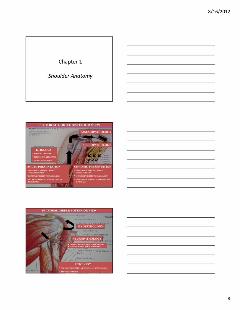
Chapter 1
Shoulder Anatomy
NEUROPATHOLOGY
c5, c6, c7
KINESIOPATHOLOGY
PECTORAL GIRDLE ANTERIOR VIEW
ETIOLOGY• WEIGHT LIFTERS
• THROWING ATHLETES
• HEAVY LABORERS
ACUTE PRESENTATION• INCREASE SENSATION LATERAL
ASPECT FOREARM
• PAINFUL/INABILITY TO FLEX ELBOW
• DECREASED STRENGTH OF BICEPS ANDBRACHIALIS
CHRONIC PRESENTATION• DECREASE SENSATION LATERAL
ASPECT FOREARM
• MOTORICINABILITY TO FLEX ELBOW
• DENERVATED STRENGTH OF BICEPS ANDBRACHIALIS
PECTORAL GIRDLE POSTERIOR VIEW
NEUROPATHOLOGY
MYOPATHOLOGY
LATERAL AXILLARY HIATUS SYNDROME QUADRILATERAL SPACE SYNDROME
ETIOLOGY• FORCIBLY ABDUCTED & EXTERNALLY ROTATED ARM
• THROWING SPORTS
8/16/2012
9
KINESIOPATHOLOGY
SHOULDER ARTHROKINEMATICS
FORCE COUPLE
CRITICAL AREA 1 CM PROXIMAL TO
GREATER TUBEROSITY
HYPOVASCULARITY SUBJECTIVE FOR TEAR OF SUPRASPINATUS TENDON
STERNOCLAVICULARJOINT
HISTOPATHOLOGY STERNOCLAVICULAR CAPSULITIS CHONDROMALACIA CREPITATION 1ST RIB SUBLUXATION
SCAPULOTHORACIC ARTICULATION
STRETCH REFLEX
MYOPATHOLOGY
8/16/2012
10
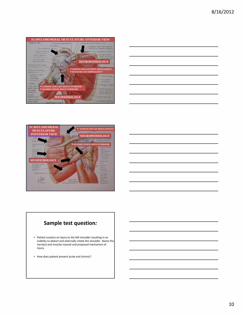
SCAPULOHUMERAL MUSCULATURE ANTERIOR VIEW
NEUROPATHOLOGY
SUPRASCAPULAR NERVE IMPINGEMENT ROTATOR CUFF IMPINGEMENT
LATERAL AXILLARY HIATUS SYNDROME QUADRILATERAL SPACE SYNDROME
MYOPATHOLOGY
SCAPULOHUMERALMUSCULATURE
POSTERIOR VIEWNEUROPATHOLOGY
SUPRASCAPULAR NERVE DAMAGE
QUADRILATERAL SPACE SYNDROME
MYOPATHOLOGY
Sample test question:
• Patient sustains an injury to the left shoulder resulting in an inability to abduct and externally rotate the shoulder. Name the nerve(s) and muscles injured and proposed mechanism of e e(s) a d usc es ju ed a d p oposed ec a s oinjury.
• How does patient present acute and chronic?

8/16/2012
11
Peripheral nerve injuries in athletes:
treatment and prevention.
• ABSTRACT: Peripheral nerve lesions are uncommon but serious injuries, which may delay or preclude an athlete’s safe return to sports. Early, accurate anatomical diagnosis is essential. Nerve lesions may be due to acute injury (i.e. from a direct blow) or chronic injury secondary to repetitive microtrauma(entrapment). Accurate diagnosis is based upon physical examination and knowledge of the relative anatomy. Palpation, neurological testing and provocative maneuvers are mainstays of physical diagnosis Diagnostic suspicion can bemaneuvers are mainstays of physical diagnosis. Diagnostic suspicion can be confirmed by electrophysiological testing, including electromyography and nerve conduction studies. Proper equipment, technique and conditioning are the keys to prevention. Rest, anti‐inflammatories, physical therapy and appropriate splinting are the mainstays of treatment.
Journal Source: Sports Medicine 16(2); 130-147; 1993.
Author(s): Lorwi, Matthew P. and Hershman, Elliott B.
Entrapment Neuropathies
Entrapment neuropathies are any condition in which a peripheral nerve is injured, irritated or otherwise compromised, by external pressure created by compression or impingement in its course through a fibrous or osseofibrous tunnel, or at a point that the nerve changes direction through or over a fibrous or muscular band.
Sympathetic Nerve Supply to Upper Extremities
Lateral Horn T2‐T9, 10
Anterior Roots
White Rami
Sympathetic Trunk
Stellate Ganglion T2,Ganglion
Synapse
Gray Rami
Join Brachial Plexus
8/16/2012
12
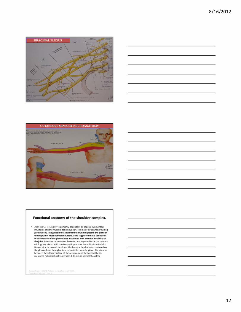
BRACHIAL PLEXUS
CUTANEOUS SENSORY NEUROANATOMY
Functional anatomy of the shoulder complex.
• ABSTRACT: Stability is primarily dependent on capsulo‐ligamentousstructures and the musculo‐tendinous cuff. The major structures providing joint stability. The glenoid fossa is retrotilted with respect to the plane of the scapula in most normal shoulders. Saha suggested that a ventral tilt or anteversion of the glenoid was associated with anterior instability of the joint. Excessive retroversion, however, was reported to be the primary j , , p p yetiology associated with non‐traumatic posterior instability in a study by Brewer et al. In normal shoulders, the humeral head remains centered on the glenoid fossa throughout elevation in the scapular plane. The distance between the inferior surface of the acromion and the humeral head, measured radiographically, averages 8‐10 mm in normal shoulders.
Journal Source: JOSPT; Volume 18; Number 1; July 1993.Author(s): Culham E.; Peat M..
8/16/2012
13
The role of the scapula.
• ABSTRACT: The scapular musculature is often neglected in designing a rehabilitation protocol for the shoulder. Weakness of the scapular stabilizers and resultant altered biomechanics could result in 1) Abnormal stresses to the anterior capsular structures of the shoulder, 2) increased possibility of rotator cuff compression, and 3)decreased
performance. This article presents known facts about the biomechanics of the scapula and
surrounding muscles and suggests methods for evaluation of scapular muscle weaknesssurrounding muscles and suggests methods for evaluation of scapular muscle weakness. Exercise techniques to maximally strengthen the scapular musculature are also described. As our ability to document strength of these muscles improves, we will be able to determine the effect of scapular strengthening on improving symptoms related to impingement and instability. Scapular strengthening exercises are usually nonstressful to the rotator cuff and are easily implemented into a rehabilitation program for the shoulder.
Journal Source: JOSPT; 1993 July; 18(1); P 386Author(s): Paine R; Voight M.
The role of the scapular stabilization in
overhead motion.
• ABSTRACT: Athletic activity that involves repetitive overhead movement such as throwing, swimming, or weight training places considerable stress on the glenohumeral joint. Therefore strength and conditioning programs for the overhead motion athlete generally emphasize the glenohumeral joint musculature (deltoids, rotator cuff muscles, pectoralis major, latissimus dorsi, teres major, triceps and biceps brachii) Frequently overlooked in designing the overheadtriceps, and biceps brachii). Frequently overlooked in designing the overhead motion athlete’s training program, however, is the conditioning of the scapular stabilizing muscles: the trapezius (upper, middle, lower), levator scapulae, rhomboids (major, minor), serratus anterior, and pectoralis minor. The scapular stabilizers are responsible for providing proper stability and mobility of the scapula against the thorax (scapulo‐thoracic joint). Many of the glenohumeral joint muscles attach to the scapula, thus control of the scapula on the thorax is critical in order for the glenohumeral joint musculature to function optimally during overhead movements.
Journal Source: Strength & Conditioning, (Feb 1996), pp 33-38.Author(s): Daniel Regan, ATC/R, CSCS.
The role of the scapular stabilization in overhead
motion. (cont.)
• If the muscles responsible for scapular stabilization are not properly conditioned, weakness and fatigue may occur during activity. This may interfere with optimal shoulder mechanics during overhead activities, in turn increasing the risk of injury or hindering performance. Therefore it is important to address the muscles responsible for scapular stabilization in the strength and conditioning program for an overhead motion athlete. This article discusses the role of the scapular stabilizers on the prevention and treatment of subacromial impingementstabilizers on the prevention and treatment of subacromial impingement syndrome, reviews the significance of these muscles on throwing performance, and illustrates exercises for strengthening the muscles responsible for scapular stabilization.
Journal Source: Strength & Conditioning, (Feb 1996), pp 33-38.Author(s): Daniel Regan, ATC/R, CSCS.
8/16/2012
14
The physical examination of the glenohumeral joint:
Emphasis on the stabilizing structures.
• ABSTRACT: Thorough descriptions of specific physical examination tests used to
determine gleno‐humeral instability are lacking in the scientific literature. The purpose of this paper was to discuss the importance of the subjective history and illustrate the physical examination of the gleno‐humeral joint. Additionally, the authors will illustrate specific stability assessment tests for the glenohumeral joint based on current basic science and clinical research The physical examination of a patient whose history suggests subtle glenohumeralresearch. The physical examination of a patient whose history suggests subtle glenohumeral joint instability may be extremely difficult for the clinician due to the normal amount of capsular laxity commonly present in most individuals. An essential component of the physical examination is a thorough and meticulous subjective history, which includes the mechanisms of injury and/or dysfunction, chief complaint, level of disability, and aggravating movements.The physical examination must include an assessment of motion, static stability testing, muscle testing, and a neurologic assessment. A comprehensive understanding of various stability testing maneuvers is important for the clinician to appreciate. The evaluation techniques discussed in this paper should assist the clinician in determining the passive stability of the glenohumeral joint.
Journal Source: JOSPT, Vol. 25, No. 6, June 1997, pp 380-389.Author(s): Kevin E. Wilk, PT, James R. Andrews, MD, Christopher A. Arrigo, MS, PT, ATC.
Chapter 2
Biomechanics
Force Couple
• A force couple is the action of two forces acting in opposite directions to imposeacting in opposite directions to impose rotation about the axis.
8/16/2012
15
Force Couple
• This coupling effect was confirmed by Mosely et.al. who performed electromyographic analyses during several scapular exercises. The force couple provides an extremely importantforce couple provides an extremely important function with upward rotation of the acromion away from the humerus in forward elevation of the shoulder, thereby preventing impingement.
Evaluation of Scapular Stability
• Kibler has described the lateral scapular slide measurement, which measures the ability of the scapular stabilizers to control the medialthe scapular stabilizers to control the medial border of the scapula. An increase of one centimeter or more in side‐to‐side measurements was reported to correlate directly with symptoms of pain and decreased shoulder function.
Chapter 3
Differential Diagnosis
8/16/2012
16
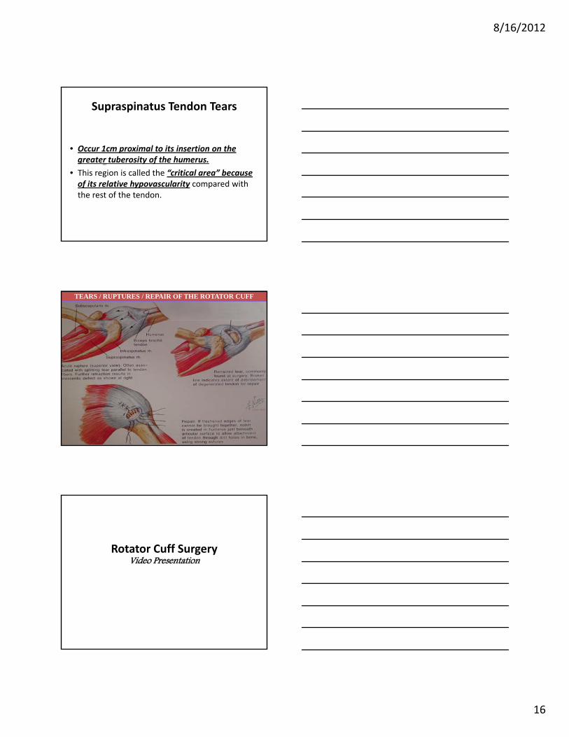
Supraspinatus Tendon Tears
• Occur 1cm proximal to its insertion on the greater tuberosity of the humerus.
125
• This region is called the “critical area” because of its relative hypovascularity compared with the rest of the tendon.
125
TEARS / RUPTURES / REPAIR OF THE ROTATOR CUFF
Rotator Cuff SurgeryVideo Presentation
8/16/2012
17
Wrap Up
• Become the Leading Extremity Expert in your Community
• Learn Dr. Mally’s “Sniper Specific” Techniques of Extremity Adjusting
• Learn how to help patients and exponentially p p p yincrease your business at the same time
The “Magnificent 7” featuring Dr. Mitch Mally
www.FromTheDeskOfDrMitchMally.com
or find me as Mitch Mally on Facebook
For more information on Dr. Mally’s seminars (group, Association and private
1‐on‐1 seminars) and products:Please email “PJ” Executive Director ofPlease email PJ Executive Director of
Mally Enterprises [email protected]
or email Dr. Mally [email protected]