Embed Size (px)
Citation preview
606
Biochimica et Biophysica Acta, 598 (1980) 606--615 © Elsevier/North-Holland Biomedical Press
BBA 78748
ALANINE TRANSPORT BY CHINESE HAMSTER OVARY CELLS WITH ALTERED PHOSPHOLIPID ACYL CHAIN COMPOSITION
JUDITH RYAN and ROBERT D. SIMONI
Department of Biological Sciences, Stanford University, Stanford, CA 94305 (U.S.A.)
(Received September l l th, 1979)
Key words: Alanine transport; Phase transition; Phospholipid concentration; (Kinetic parameter)
Summary
The Na*<lependent transport of alanine has been examined in Chinese hamster ovary (CHO) cells as a function of the fat ty acid composition of their membrane lipids. Significant changes in the fa t ty acid composition of the CHO cell phospholipids were achieved by supplementation of the growth medium with specific saturated (palmitate) or monoenoic (oleate) free fa t ty acids. Arrhenius plots of the temperature-dependent uptake of alanine were con- structed for cells of altered fa t ty acid composition. Alanine uptake was characterized by a single discontinuity in the Arrhenius plot. The temperature of this break was observed to be dependent upon the fat ty acid composition of the cell phospholipids, ranging from 16°C for cells enriched with oleate to 32 ° C for cells enriched in palmitate. Calculation of the Km value for the uptake process showed no significant change with temperature or fat ty acid supple- mentation. Correlations are made between the physical state of the membrane lipids and the temperature~lependence for alanine transport. The results are discussed in terms of membrane fat ty acid composition, ordered ~ fluid phase transitions and amino acid transport.
Introduction
Many reports exist in the literature which implicate a role for the physical state of the membrane phospholipids in the regulation of a variety of mem- brane-associated transport or catalytic functions [1--12]. In a number of cases the changes in biological activities have been correlated with the physical state of the membrane lipids. It is our goal in the present work to determine what effect alteration of the fat ty acid composition of Chinese hamster ovary (CHO) cell membranes would have on a membrane-associated process. Previous reports
607
from this laboratory have shown that CHO cells are capable of incorporating significant amounts of exogenous fatty acids into their cellular phospholipids [13,14]. Incorporation of free fatty acids into these membranes was independent of any apparent compensatory changes in polar head-group com- position or sterol/phospholipid ratios [13]. Furthermore, we have demon- strated that incorporation of saturated fatty acids into these membrane phos- pholipids resulted in altered physical properties of the phospholipids as detected by fluorescence intensity and polarization measurements using cis- and trans-parinaric acid [13]. Fluorescence depolarization studies indicated that the amount of ordered lipid could be increased by appropriate supplemen- tation with palmitic acid. Together, these data strongly suggest that the acyl chain composition of CHO cell membranes could be altered in such a manner as to influence the temperature of the ordered ~ fluid transition in these mem- brane phospholipids.
A logical extension of this work is to examine what effects changes in mem- brane fatty acid composition and the physical state of the membrane have on membrane-associated functional processes. A number of precedents exist for this type of study in procaryotic systems. The transport systems for ~-glucosides and ~-galactosides in unsaturated fatty acid auxotrophs of Escherichia coli have been correlated with changes in phospholipid fatty acid composition by several investigators [4--10]. These reports indicated that changes in the transport rate observed as a function of temperature could be related to changes in the physical state of the membrane. In eucaryotic systems the results, to date, have been less well<lefined. A number of studies on membrane-associated enzymes have suggested that the composition of the membrane phospholipids (either fatty acid or polar head-group modifications) play a regulatory role in enzyme activity [3,15,16]. However, it has been difficult in many of these systems to correlate changes in activity with an altered physical state of the membrane phospholipids. In other cases, alterations in the phospholipid composition resulted in little or no detectable change in functional properties [ 16,17 ].
The Na÷<lependent carrier-mediated transport of alanine has been well characterized in eucaryotic systems by a number of investigators [18--22]. Kaduce et al. [24] have examined the effect of fatty acid supplementation on the transport of a-aminoisobutyric acid, an amino acid analog, in Ehrlich ascites tumor cells. They suggested that changes in transport rates observed as a function of fatty acid supplementation were due to fluidity-induced changes in substrate binding affinities.
This paper will present our observations on the effect of altering the fatty acid composition of membrane phospholipids on alanine transport in CHO cells.
Materials and Methods
Cell culture and growth Chinese hamster ovary cells (CHO-K1) were obtained from the American
Type Culture Collection (ATCC CCL-61). Stock cultures were grown and maintained on Ham's F-12 (Gibco) or minimal Eagle's medium (Flow Labora- tories) in a 5% COx atmosphere at 37°C. The medium was supplemented with 5 or 6.6% delipidated serum prepared by the method of Rothblat et al. [25]. As
608
measured by gas-liquid chromatography, the delipidated serum was shown to contain less than 5% of the serum-associated acyl chains. For uptake experi- ments, cells were removed from stock flasks and plated at 1 . 1 0 6 cells per 100 X 15 mm petri dish containing 12 glass coverslips (9 × 22 mm). The cells were incubated for 48 h (approximately two cell~loublings) on media con- taining delipidated serum. At this t ime the cells had reached 5 0 - 6 0 % con- fluency. The media were then changed to one containing the specified con- centrations of fa t ty acids which were added from an ethanolic s tock solution. Cells were incubated for an additional 12--17 h. This feeding schedule was designed so that fa t ty acid incorporation and uptake experiments were com- pleted on sub~onf luen t cells. Incorporation of fat ty acids into the phospho- lipid fraction reached a maximum in 12 h and remained constant through to 1 7 h .
Uptake measurements Uptake of [1-14C]alanine (175 Ci/mol, New England Nuclear) was deter-
mined using the procedure of Foster and Pardee [26] as modified by Oxender et al. [19,21] for cells grown on glass coverslips. The uptake buffer contained 0.01 M potassium phosphate, pH 7.4, with 2.5 mM KC1, 0.1% glucose, 0.01% CaC12 and 0.01% MgC12. Alanine was present at 0.25 mM (10 pCi/1.1 ml) except as noted. To moni tor total uptake of alanine the uptake buffer also included 0.14 M NaC1. The Na÷-indepdent uptake of alanine was measured in the presence of 0.14 M choline chloride (in place of NaC1) which was added to maintain equivalent osmolarity. The Na+~lependent uptake was taken as the difference between total and Na÷-independent uptake. Prior to the uptake mea- surements, the cells were incubated for 10--15 min at room temperature in the above buffer to reduce intracellular amino acid levels. Coverslips were then washed three times in either Na ÷- or choline-uptake buffer to ensure removal of any non-adherent or non-viable cells. Uptake measurements were then initiated by placing the coverslips in uptake buffer containing labeled amino acid. After the specified t ime periods, coverslips were removed from the uptake buffer and rapidly washed three times by sequential immersion into beakers of ice-cold buffer. Excess buffer was removed by capillary action when the edge of the coverslip was touched onto a towel. Coverslips were placed in scintillation vials containing 1.0 ml of 0.2 M NaOH. Aliquots of the solubilized cells were removed for protein analysis by the method of Lowry et al. [26]. The cell suspension was neutralized with 0.2 ml of 3.0 M sodium citrate, pH 3, and counted in a Triton-toluene scintillation fluid.
Lipid analysis A port ion of the cells grown on coverslips was washed in phosphate-buffered
saline to remove any non-adherent cells. Cells were removed from coverslips by scraping with a rubber policeman, pooled and washed three times with phos- phate-buffered saline to ensure removal o f growth media. The resultant cell pellet was extracted with chloroform/methanol (2 : 1) and the phospholipid fraction was isolated by silicic acid chromatography. Fa t ty acid methyl esters were prepared by transesterification in 2% H2SO4 in methanol for 60 min at 70°C. Fa t ty acid methyl esters were identified and quanti tated by gas-liquid
609
chromatography using a model 7610-A Hewlett-Packard gas chromatograph equipped with a 6 ft column of 15% SP2340 on chromosorb P AW-DMCS. Standard fatty acid methyl esters were obtained from Supelco.
Results
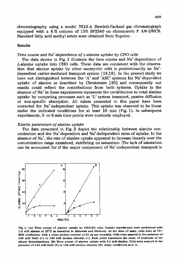
Time course and Na+-dependence o f L-alanine uptake by CHO cells The data shown in Fig. 1 illustrate the time course and Na÷-dependence of
L-alanine uptake into CHO cells. These data are consistent with the observa- tion that alanine uptake by other eucaryotic cells is predominantly an Na ÷- dependent carrier-mediated transport system [18,19]. In the present study we have not distinguished between the 'A' and 'ASC' systems for Na*-dependent uptake of alanine as described by Christensen [20] and consequently our results could reflect the contributions from both systems. Uptake in the absence of Na ÷ in these experiments represents the contribution to total alanine uptake by competing processes such as 'L' system transport, passive diffusion or non-specific absorption. All values presented in this paper have been corrected for Na÷-independent uptake. This uptake was observed to be linear under the indicated ~conditions for at least 10 min (Fig. 1). In subsequent experiments, 5- or 6-min time points were routinely employed.
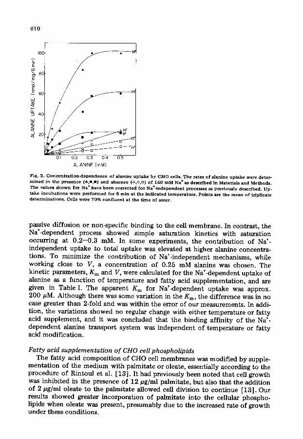
Kinetic parameters o f alanine uptake The data presented in Fig. 2 depict the relationship between alanine con-
centration and the Na÷<lependent and Na÷-independent rates of uptake. In the absence of Na ÷, the rate of alanine uptake appeared to increase linearly over the concentration range examined, exhibiting no saturation. The lack of saturation can be accounted for if the major component of Na÷-independent transport is
E
E_6o
W
I- 40 D-
bJ z ~ 2o _J
9 / /° ~ ° o
i i ,
A ' / B
12
4
li I 3 5 7 9 II
MINUTES
Fig. 1. (A) T i m e c o u r s e o f alanine u p t a k e b y CHO-K1 cells. U p t a k e e x p e r i m e n t s w e r e p e r f o r m e d with 1 .0 m M alanine a t 25°C as desc r ibed in Mater ia ls and M e t h o d s . A t t h e t i m e o f assay , ce l l s w e r e at 7 0 - - 80% c o n f l u e n c y wi th a m e a n p r o t e i n c o n t e n t of 31 / z g pe r covers l ip . Cells w e r e a s s a y e d in t h e p r e s e n c e o f 140 m M NaCl (~) or 140 m M c h o l i n e c h l o r i d e (o) . Each p o i n t r e p r e s e n t s t h e m e a n o f d u p l i c a t e or tri- p l ica te d e t e r m i n a t i o n s . (B) T i m e c o u r s e o f alanine u p t a k e wi th 0.1 m M alanine. Cells we re assayed in t h e p r e s e n c e o f 140 m M NaCI ( a ) or 140 m M c h o l i n e c h l o r i d e (e ) . Assay cond i t i ons as in A.
610
IOC ~ •
E 60 / ~ • 25'
40 •
~ . - - ~ - ~ . . . . OI 0 2 03 04 05
ALANINE (mM)
Fig. 2. C o n c e n t r a t i o n - d e p e n d e n c e o f alanine u p t a k e by Cl iO cells. The rates o f alanine u p t a k e were deter- m i n e d in the presence (A o,m) and ab se n c e (z~ o,D) of 140 m M Na + as descr ibed in Materials and Methods . The values s h o w n for Na + have b e e n c o r r e c t e d for Na+- independent proces se s as p rev ious ly descr ibed . Up- t ake i ncuba t i ons were p e r f o r m e d for 6 rain a t the i nd i ca t ed t emperat t tre . Po ints are the m e a n o f tr ipl icate d e t e r m i n a t i o n s . Cells were 70% c o n f l u e n t at the t ime of assay.
passive diffusion or non-specific binding to the cell membrane. In contrast, the Na÷<iependent process showed simple saturation kinetics with saturation occurring at 0.2--0.3 mM. In some experiments, the contr ibut ion of Na ÷- independent uptake to total uptake was elevated at higher alanine concentra- tions. To minimize the contr ibut ion of Na÷-independent mechanisms, while working close to V, a concentrat ion of 0.25 mM alanine was chosen. The kinetic parameters, Km and V, were calculated for the Na÷-dependent uptake of alanine as a function of temperature and fat ty acid supplementat ion, and are given in Table I. The apparent Km for Na+<lependent uptake was approx. 200 pM. Although there was some variation in the Kin, the difference was in no case greater than 2-fold and was within the error of our measurements. In addi- tion, the variations showed no regular change with either temperature or fat ty acid supplement, and it was concluded that the binding affinity of the Na ÷- dependent alanine transport system was independent o f temperature or fatty acid modification.
Fatty acid supplementation of CHO cell phospholipids The fat ty acid composi t ion of CHO cell membranes was modified by supple-
mentat ion of the medium with palmitate or oleate, essentially according to the procedure of Rintoul et al. [13]. It had previously been noted that cell growth was inhibited in the presence of 12 #g/ml palmitate, but also that the addition of 2 #g/ml oleate to the palmitate allowed cell division to continue [13]. Our results showed greater incorporation of palmitate into the cellular phospho- lipids when oleate was present, presumably due to the increased rate of growth under these conditions.
611
TABLE I
KINETIC PARAMETERS FOR Na+DEPENDENT ALANINE TRANSPORT
Kinetic parameters were determined from double-reciprocal plots of aianine uptake. Uptake measure-
ments are described in the legend to Fig. 2 and in Materials and Methods. Cells grown on coverslips to
50--60% confluency were supplemented with palmitate 12 of 17 h prior to the measurement of uptake as
described in Materials and Methods. Unsupplemented cells received fresh media with no added fatty acid
17 h prior to uptake measurements. K m values (raM) at 140 mM Na +, V values in nmol/mg per rain.
S u p p l e m e n t T e m p e r a t u r e ( °C)
15 25 35
K m
n o n e 0 . 1 5 0 .29 0 .22
n o n e 0 .16 0 .23 0 .24 1 6 : 0 0 .29 0 .26 0 .25
1 6 : 0 0 .14 0 .12 0 .14
V n o n e 4 .8 16.6 27.7
n o n e 7.2 19.2 25 1 6 : 0 4 .0 12.5 28
1 6 : 0 9 .5 18 .5 23
The fatty acid compositions of the phospholipids of CHO cells grown under these conditions are given in Table II. CHO cells are characteristic in their relatively simple acyl chain composition, with palmitate and oleate comprising over 70% of the total phospholipid acyl chains. It was observed that upon
T A B L E II
A C Y L C O M P O S I T I O N O F C H O C E L L P H O S P H O L I P I D S
Cor r e l a t i o n s b e t w e e n the t e m p e r a t u r e o f the change in s lope o f t he A r r h e n i u s p lo t for N a * - d e p e n d e n t
u p t a k e o f a lan ine and t h e t e m p e r a t u r e o f the phase t r a n s i t i o n for C H O cell p h o s p h o l i p i d s as d e t e r m i n e d
by fluorescence measttrements using trans-parinaric acid. Discontinuities were determined as described in
the legend to Fig. 3. Temperatures of the trans-parinaric acid (t-PnA) fluorescence transitions were extra- polated from the data of Rintottl et al. [13] for extracted CHO cell phospholipids having equivalent ratios
of unsaturated to saturated acyl chains. SAT, saturated fatty acid; UFA, unsaturated fatty acid.
S u p p l e m e n t F a t t y ac id c o m p o s i t i o n (%) S A T U F A
(pg/ml)
1 4 : 0 14 :1 1 6 : 0 1 6 : 1 1 8 : 0 1 8 : 1 1 8 : 2
Trans-
po~
break
point
point
(°C)
t-PnA
f luores-
cence b r e a k
p o i n t
(°C)
None 2
18:1 (12)
18:1 (12) 1
16:0 (10) +
18:1 (2)
16:0 ( 1 2 ) t r ace
1 6 : 0 (15) + 1 8 : 1 (2) 6
1 6 : 0 (15) + 18:1 (2) 3
19 14 12 53 - - 33 67 21 20
9 7 15 68 - - 24 75 18 15
11 9 20 59 - - 32 68 27 19 .5
28 7 20 44 - - 48 51 29 27 .5
31 20 12 37 - - 43 57 25 24 .5
t r ace 38 14 13 30 t r ace 57 43 31 32
37 10 17 32 -- 57 43 32 32
40 30 20 iO
B 80 ._~
L" 4o
t~ 20
rl
IO u.l Z
..J
40 30 20 I0
3:2 3:3 3:4 3:5
e v >_ 4C t -
~_ ~c z 0
z IC w -1-
612
3:2 3:3 3:4 3:5 ,b ~ 6b ~b 8o l /K (x IO 5) UNSATURATED ACYL CHAIN/
TOTAL ACYL CHAINS
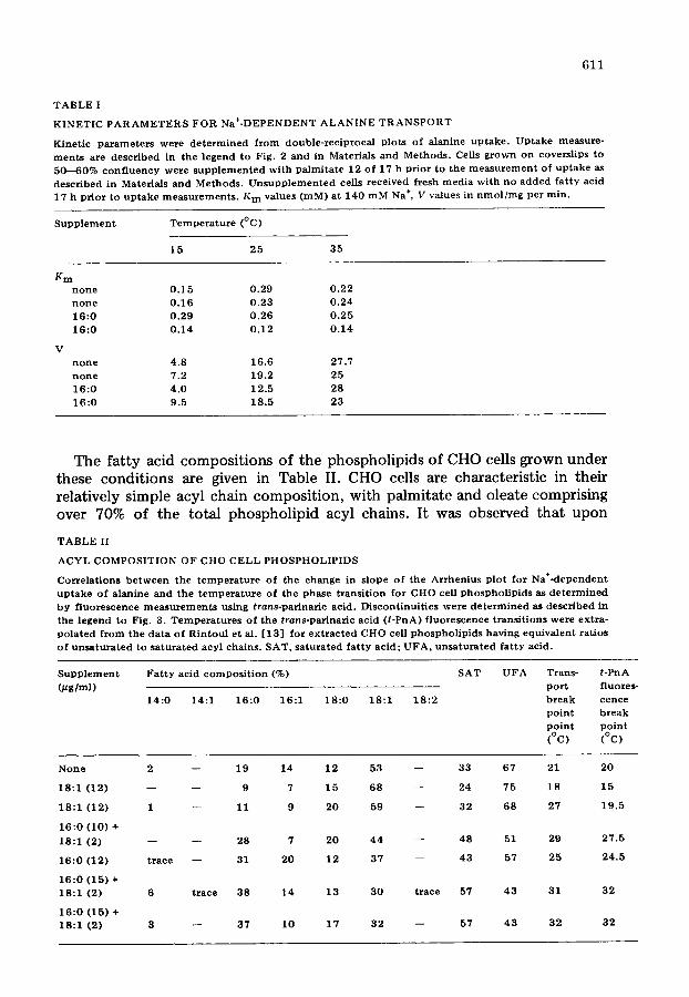
Fig. 3. Arrhenius p lots o f Na+-dependent alanine uptake . Alanine uptake was measured as described in Materials and Methods in buf fer containing 0 .25 m M alanine (10 #Ci /1 .1 ml ) . Uptake was measured from 10 to 44°C using 4-rain incubat ions . Values were corrected for Na÷-independent uptake at each tempera- ture as prev ious ly described. All po ints are the m e a n o f triplicate points . The data have been subjected to l inear regression analysis . The transi t ion temperature range was se lected to give the best fit o f l ines. Corre- lat ion coef f i c i ents de termined for these l ines were greater than - - 0 . 9 5 in all cases . A l though drawn as straight l ines, the data cou ld also be f i t ted to a s m o o t h curve. (A) Data obta ined from cells supp lemented for 15 h in the presence o f 15 # g / m l palmitate and 2 # g / m l o lea te . (B) Data obta ined from cells supple- m e n t e d wi th 12 # g / m l o leate for 14 h. All cells were 70% conf luent at the t ime of uptake measurements .
Fig. 4. Unsaturated acyl cha ins / to ta l acyl chains o f CHO cell phospho l lp ids versus the temperature o f the change in uptake rate o f the Arrhenius p lot for Na+-dependent alanine uptake . The acyl chain compos i - t ion was calculated b y gas-liquid c h r o m a t o g r a p h y as described in Materials and Methods . Uptake measure- ments and de terminat ion o f the temperature d i scont inui ty o f the Arrhenius pint are described in Materials and Methods and Fig. 3.
supplementation with either palmitate or oleate, the supplemented component was enriched at the expense o f the other major component . In this manner it was possible to cause significant changes in the ratio of unsaturated to satu- rated fatty acids present in the CHO cell membranes (see Table II).
Temperature-dependence of Na+-dependent alanine uptake The temperature<lependence of the Na÷<lependent alanine uptake was mea-
sured on unsupplemented and oleate- or palmitate-supplemented cells. Arrhenius plots were constructed for the uptake process and are shown for one set o f palmitate- and oleate-supplemented cells in Fig. 3A and B. In all cases, the Arrhenius plots for alanine uptake were characterized by a single discon- tinuity in the uptake rate. In the examples given, discontinuities in the Arrhenius plots were observed at approx. 30°C for palmitate-supplemented cells and approx. 20°C for oleate-supplemented cells.
The temperature of the thermal discontinuity was observed to vary with the acyl composi t ion of the CHO cell phospholipids, as summarized in Table II. An even more striking correlation could be made between the ratio of unsaturated acyl chains and the temperature of the Arrhenius break point. This correlation is illustrated in Fig. 4. As the percent o f unsaturated acyl chains increases in the CHO cell phospholipids, a decrease in the temperature of the Arrhenius discon-
613
tinuity is obtained. The anomalous behavior noted in Fig. 4 at 68% unsatura- tion is inexplicable.
A similar correlation has been made between the ratio of unsaturated acyl chains and the temperature of the Arrhenius break points as determined by trans.parinaric acid fluorescence measurements of CHO cell phospholipids [13]. Using this relationship and the fatty acid compositions shown, the tem- perature of the fluorescence break point was extrapolated as given in Table II. The Arrhenius break temperatures determined by these independent methods were found to be almost equal.
Discussion
The results demonstrate that the rate of Na÷-dependent alanine transport in CHO cells can be altered upon modification of the fatty acid composition of CHO cell phospholipids. Supplementation of the growth media of CHO cells with either oleate or palmitate resulted in the incorporation of these fatty acids into the cell phospholipids. The incorporation was not accompanied by any significant alterations in the polar head-group composition or the phospholipid to sterol ratio [13]. The consequence of this incorporation was a variation in the ratio of unsaturated to saturated acyl chains of 43--75% in these studies. A clear correlation could be made between this ratio and the discontinuity in the Arrhenius plot for Na÷<lependent alanine transport. Furthermore, the tem- perature of the discontinuity of the Arrhenius plot could be correlated with the thermal phase transition of CHO cell phospholipids as monitored by cis- or trans-parinaric acid fluorescence [13]. This correlation is particularly good in the case of control and palmitate-supplemented cells, with variation within 2 ° C between the temperatures of the Arrhenius discontinuity for transport and the thermal phase transition of the cell phospholipids. The correlation is less exact in the case of oleate-supplemented cells. The reason for this variation is not known. We feel these correlations provide evidence for a role of the physical state of the membrane in the function of this amino acid transport system.
Mechanistically there are numerous interpretations for a change in reaction rate described by the Arrhenius equation. These interpretations fall into two general categories for cases involving lipid modification. The first evokes fluid to ordered phase transitions of the phospholipids in the membrane. Changes in reaction rate could be accounted for by a partitioning of the protein into regions of altered mobility or restriction. An alternative explanation involves changes in the intrinsic properties of the protein (i.e., binding affinities, specific activation by fatty acid moiety). To test the latter possibility, the kinetic parameters of the uptake process were examined as a function of temperature and fatty acid supplementation. The Km value for uptake did not vary signifi- cantly. In cases where changes in the binding affinity have been related to dis- continuities in the Arrhenius plot, the temperature-induced changes in Km have been over 10-fold [28,29].
In only a few cases have eucaryotic transport systems been examined as a function of in vivo fatty acid modifications. Kanduce et al. [24] have examined ~-aminoisobutyric acid uptake in Ehrlich ascites cells with altered fatty acid composition. They observed single discontinuities in the Arrhenius plot at
614
approx. 30°C regardless of supplement. Although they achieved significant alteration in the ratio of monenoic to polyenoic fatty acids upon dietary supplementation, there was no appreciable change in the ratio of saturated to unsaturated fatty acids in these membranes. Based on our results for the correlation between unsaturated/total acyl chain composition to changes in the Arrhenius discontinuity, this result is not unexpected. Correlation with physical measurements has not been made on these membranes. In comparison, Wisnieski et al. [30] observed six breaks in the Arrhenius plot for a-aminoiso- butyric acid transport in LM cells. These breaks were correlated with five characteristic temperatures detected by ESR. The physical interpretation of these results remains unclear.
In summary, the results presented here provide evidence for an interrela- tionship among the acyl chain composition of the membrane, amino acid trans- port and physical state of the membrane phospholipids as reported previously [13]. The agreement in the temperature<lependent changes in transport and physical properties suggests an intimate association of the carrier protein with the phospholipid of the bilayer. Since the fluorescence measurements were determined on phospholipid preparations in the absence of neutral lipids, it might be suggested that the carrier protein is localized in a cholesterol-deficient region of the membrane.
Verification of this idea and the precise protein-lipid interactions involved must await the resolution of this transport system and its reconstitution into a more<iefined lipid environment. Our results suggest that the Na+-dependent transport system for alanine uptake would be a reasonable candidate for further investigation.
Acknowledgements
The authors gratefully acknowledge many instructive discussions with Dr. Dale Oxender and Dr. Gary Cecchini on the transport assay and Dr. David Rintoul on the lipid and fluorescence analysis. We are indebted to Ms. Pare Hoy for her expert technical assistance in many of these experiments. This work was supported by NIH grants 5T32-GM07026 (JR) and GM 18539 (RDS).
References
1 Zakim, D. and Vessey , D.A. (1975) J. Biol. Chem. 250 ,342 - -343 2 Solomonson, L.P., Liepkalns, V.A. and Spector, A.A. (1976) Biochemistry 15 ,892 - -897 3 Kimelberg, H.K. and Papahadjopoulos, D. (1974) J. Biol. Chem. 249, 1071--1080 4 0 v e r a t h , P. and Tr~/uble, H. (1973) Biochemistry 12, 2625--2634 5 Sackmann, E., Tr//uble, H., Galla, H J . and Overath, P. (1973) Biochemistry 12, 5360----5369 6 Esfahani, M., Limbriek, A.R., Knut ton , S., Oka, T. and Wakil, S~I. (1971) Proc. Natl. Acad. Sci.
U.S.A. 68, 3180--3184 7 Shechter, E., Letellier, L. and Gulik-Krzywicki, T. (1974) Eur. J. Biochem. 49, 61--76 8 Letellier, L., Weft, R. and Shechter, E. (1977) Biochemistry 16, 3777--3780 9 0 v e r a t h , P. and Thflo, L. (1978) in Biochemistry of Cell Walls and Membranes II (Metcalfe, J.C., ed.),
pp. 1--44, University Park Press, Baltimore, MD 10 Thilo, L., Tr/iuble, H. and Overath, P. (1977) Biochemistry 16, 1283--1290 11 Stier, A. and Sackmarm, E. (1973) Biochim. Biophys. Acta 311 , 400 - -408 12 Lipelliello, P.M., Holloway, C.T., Garfield, S.A. and Hal loway, P.W. (1979) J. Biol. Chem. 254,
2004--2009 13 Rintoul , D.A., Sklar, L.A. and Simoni, R.D. (1978) J. Biol. Chem. 253, 7447--7452
6 1 5
14 Rintoul , D.A. and Simoni, R.D. (1977) J. Biol. Chem. 252, 7916--7918 15 Engelhard, V.H., Esko, J.D., Storm, D.R. and Glaser, M. (1976) Proc. Natl. Acad. Sci. U.S.A. 73,
4482--4486 16 Hale, A.H., Pessin, J.E., Palmer, F., Weber, M~J. and Glaser, M. (1979) J. Biol. Chem. 6190--6200 17 Cronan, J.E., Jr. and Gelman, E.P. (1975) Bacteriol. Rev. 3 9 , 2 3 2 - - 2 5 6 18 Oxender, D.L. and Christensen, H.N. (1963) J. Biol. Chem. 238, 3686--3699 19 Oxender, D.L., Lee, M., Moore, P.A. and Cecchini, G. (1977) J. Biol. Chem. 252, 2675--2679 20 Christensen, H. (1979) in Advances in Enzymology (Meister, A., ed.). Vol. 49, pp. 41--101, John
Wiley and Sons, New York 21 Oxender, D.L., Lee, M. and Cecchini, G. (1977) J. Biol. Chem. 252, 2680--2683 22 Finkelstein, M.C. and Adelberg, E.A. (1977) J. Biol. Chem. 252, 7101--7108 23 Sigrist-Nelson, K., Murer, H. and Hopfer, U. (1975) J. Biol. Chem. 250, 5674---5680 24 Kaduce, T.L., Awad, A.B., Fontenel le , L~ . and Spector, A.A. (1977) J. Biol. Chem. 252, 6624--6630 25 Rothbla t , G., Arbogast, L., OueUene, L. and Howard, B. (1976) In Vitro 12 , 554 - -557 26 Foster, D.O. and Pardee, A.B. (1969) J. Biol. Chem. 244, 2675--2681 27 Lowry, O.H., Rosebrough, N J . , Farr, A.L. and Randall , R.J. (1951) J. Biol. Chem. 193 , 265 - -275 28 Jinks, D.C., Silvius, J.R. and McElhaney, R.N. (1978) J. Bacteriol. 136, 1027--1036 29 Silvius, J.R., Read, B.D. and MeElhaney, R.N. (1976) Science 1 9 9 , 9 0 2 - - 9 0 4 30 Wisnieski, BJ . , Parkes, J.G., Huang, Y.O. and Fox, C.F. (1974) Proc. Natl. Aead. Sci. U.S.A. 71,
4381--4385


![Hamster[1] (3)rt](https://img.pdfslide.us/doc/110x75/5453f07faf7959856d8b512d/hamster1-3rt-5584af5997318.jpg)