Embed Size (px)
Citation preview

ARTICLETranslational Therapeutics
AKR1C enzymes sustain therapy resistance in paediatricT-ALLRoberta Bortolozzi1, Silvia Bresolin1, Elena Rampazzo1,2, Maddalena Paganin3, Francesca Maule1, Elena Mariotto1, Daniele Boso1,Sonia Minuzzo4, Valentina Agnusdei5, Giampietro Viola1, Geertruy te Kronnie1, Giovanni Cazzaniga6, Giuseppe Basso1,3 andLuca Persano2
BACKGROUND: Despite chemotherapy intensification, a subgroup of high-risk paediatric T-cell acute lymphoblastic leukemia (T-ALL) patients still experience treatment failure. In this context, we hypothesised that therapy resistance in T-ALL might involve aldo-keto reductase 1C (AKR1C) enzymes as previously reported for solid tumors.METHODS: Expression of NRF2-AKR1C signaling components has been analysed in paediatric T-ALL samples endowed withdifferent treatment outcomes as well as in patient-derived xenografts of T-ALL. The effects of AKR1C enzyme modulation has beeninvestigated in T-ALL cell lines and primary cultures by combining AKR1C inhibition, overexpression, and gene silencingapproaches.RESULTS: We show that T-ALL cells overexpress AKR1C1-3 enzymes in therapy-resistant patients. We report that AKR1C1-3enzymes play a role in the response to vincristine (VCR) treatment, also ex vivo in patient-derived xenografts. Moreover, wedemonstrate that the modulation of AKR1C1-3 levels is sufficient to sensitise T-ALL cells to VCR. Finally, we show that T-ALLchemotherapeutics induce overactivation of AKR1C enzymes independent of therapy resistance, thus establishing a potentialresistance loop during T-ALL combination treatment.CONCLUSIONS: Here, we demonstrate that expression and activity of AKR1C enzymes correlate with response tochemotherapeutics in T-ALL, posing AKR1C1-3 as potential targets for combination treatments during T-ALL therapy.
British Journal of Cancer (2018) 118:985–994; https://doi.org/10.1038/s41416-018-0014-0
INTRODUCTIONThe nuclear factor erythroid 2 (NF-E2)-related factor 2(NRF2)–Kelch-like (ECH)-associated protein 1 (KEAP1) signalingpathway is a cellular system that protects cells from oxidativestress and insults from toxic xenobiotics.1 Indeed, the NRF2 axis isfinely regulated in normal cells by acting as a surveillance systemto prevent the accumulation of endogenous metabolites orhazardous substrates from the microenvironment. Nonetheless,in the past decade, multiple studies reported a significantoveractivation of this pathway in many tumors suggesting apositive correlation between its enhanced activity and poorprognosis in cancer patients.1,2 Under basal conditions, NRF2activity is repressed by binding to its inhibitor KEAP1, which leadsto NRF2 proteasomal degradation through the CUL3-dependentubiquitin ligase complex.3 Oxidation of KEAP1 mediated byoxidative stress or electrophilic agents induces the release ofNRF2 and its translocation into the nucleus where it binds to AREconsensus sequences and transactivates a series of cytoprotectivetarget genes, including aldo-keto reductase (AKR) familymembers.4
The superfamily of AKR enzymes catalyze the NADPH-dependent conversion of aldehydes and ketones to theircorresponding alcohols. Thus, a wide variety of substrates thatinclude simple carbohydrates, cellular metabolites, steroid hor-mones, endogenous prostaglandins, xenobiotic compounds, andchemotherapeutics are potential targets of these enzymes.5 TheAKR1C subfamily includes four isoenzymes (AKR1C1-4) widelyexpressed in different human tissues, except for AKR1C4 that isliver-specific.6 Recent studies demonstrated that AKR1C1-3 arehighly expressed in many human cancers including prostate,7
breast,4 glioma,8,9 neuroblastoma,10 lung,11,12 and acute myeloidleukemia (AML), where they mediate drug resistance, regulate celldifferentiation, and promote cancer cell proliferation.13,14
In this context, it has been demonstrated that the pan AKR1Cinhibitor medroxyprogesterone acetate (MPA), greatly enhancesthe anti-leukemic activity of bezafibrate, by inhibiting theprostaglandin D2 11b-ketoreductase activity of AKR1C enzymes,thus promoting growth arrest, apoptosis, and cell differentiation inAML cells.15 Moreover, different studies aimed to selectivelyinhibit specific AKR1C isoforms (i.e., AKR1C3) and revealed that
www.nature.com/bjc
Received: 22 September 2017 Revised: 8 January 2018 Accepted: 9 January 2018Published online: 8 March 2018
1Department of Women’s and Children’s Health, University of Padova, Padova 35128, Italy; 2Istituto di Ricerca paediatrica Città della Speranza–IRP, corso Stati Uniti 4, Padova35127, Italy; 3Clinic of paediatric Oncohematology, University Hospital of Padova, Padova 35128, Italy; 4Department of Surgery, Oncology and Gastroenterology, University ofPadova, Padova 35128, Italy; 5Istituto Oncologico Veneto - IRCCS, Padova 35128, Italy and 6Department of paediatric, Centro Ricerca M. Tettamanti, University of Milano Bicocca,Fondazione MBBM, Monza 20900, ItalyCorrespondence: Luca Persano ([email protected])
© Cancer Research UK 2018

inhibition of AKR1C3 alone is not adequate to exert an anti-leukemic effect in AML cells,16 thus reinforcing the hypothesis thatredundant activity of AKR1C enzymes supports intracellularantioxidant response.In acute lymphoblastic leukemia (ALL), AKR1C1-4 messenger
RNA (mRNA) and protein expression has been correlated withcellular sensitivity to the mustard pro-drug PR-104A,17 the latterwas reported to be converted and activated by AKR1C3 in a subsetof cancer cell lines.18 Although Moradi Manesh et al.17 nicelydemonstrated that AKR1C3 expression and enzymatic activity aremore abundant in T-ALL compared to BCP-ALL xenografts, andpositively correlate with the response to the mustard pro-drug PR-104A, little is known about the potential correlation betweenAKR1C1-3 expression and the response to standard treatmentregimens in ALLs.Recent improvements in chemotherapeutic protocols for child-
hood ALL achieve a 5-year survival rate of about 80%.19 However,different studies identified a slower clearance of cancer cellsduring treatment in T-ALL compared to B-ALL,20 with moreresistant tumors (high-risk T-ALL) showing a 7-year survival of only40%.19 Therefore, the study of the mechanisms underlying drugresponse and the development of new therapeutic strategies forpatients who poorly respond to current treatment protocolsremains an important challenge in T-ALL. In this context, wehypothesised that AKR1C enzymes could perturb therapeuticsuccess in T-lineage leukemia. We evaluated the expression andenzymatic activity of AKR1Cs in a large cohort of T-ALLs, finding asignificant upregulation of AKR1C1-3 in “resistant” or “poorlyresponding” tumors. Moreover, we functionally validated the roleexerted by these enzymes in controlling T-ALL cell response tochemotherapeutics by pharmacologically or genetically modulat-ing their activity. Finally, we directly correlated the expression ofAKR1C1-3 to chemotherapy response in patient-derived xenograft(PDX)-T-ALL samples.
MATERIALS AND METHODSPrimary leukemia cell culturesT-cell acute lymphoblastic leukemia cells derived from bonemarrow (BM) of patients were obtained after informed consentfollowing the tenets of the Declaration of Helsinki andaccording to the guidelines of the ethics committee of theUniversity of Padova, the Padova Academic Hospital, and theItalian Association of paediatrics Onco-Hematology (AIEOP).Diagnosis was obtained according to standard cytomorphol-ogy, cytochemistry, and immunophenotypic criteria.21 Allanalysed T-ALL samples were obtained after hemolysis of redblood cells at the time of diagnosis, before treatment. DerivedT-ALL cells have then been subjected to total RNA and/orprotein extraction according to standard procedures or wereused for drug testing and injected in mice to generate PDX-T-ALL as described hereafter. T-ALL patients have been classifiedas therapy “responders” or “resistant” according to the minimalresidual disease (MRD) molecularly detected at day 78 from thestart of therapy.In some experiments, primary and PDX-T-ALL cells were seeded
at a concentration of 105 cells per well in 96-well microtiter platesand cultured in α-MEM medium supplemented with 10% FBS, 1%penicillin/streptomycin, 1% glutamine (all from Thermo FisherScientific, Waltham, MA), 10% human heat inactivated AB+ serum(Sigma-Aldrich S.r.l., Milan, Italy), human IL7 (20 ng/ml; R&DSystems, Minneapolis, MN), human Stem Cell Factor (SCF) (50ng/ml), human FLT3-ligand (20 ng/ml; both from Peprotech,London, UK), and insulin (20 nM; Sigma-Aldrich S.r.l.). Cells wereimmediately exposed to the test compounds and cell survival wasevaluated by MTT assay after 48 h. Clinical information of T-ALLpatients, from whom cells included in this study derived, areprovided as Suppl. Table S1.
Measurement of AKR1C enzymatic activityBone marrow or peripheral blood derived T-ALL blasts wereobtained from cryopreserved samples from our collection. AKR1Cenzymatic activity was measured as previously described.22,23
Briefly, cells were added to white 96-well plates at 2 × 10⁵ cells perwell in phenol red-free media and equilibrated at 37 °C in a 5%CO2 incubator for 1 h in the presence of coumberone (10 µM).Coumberol-derived fluorescence intensity was detected after 1–3h of coumberone addition with a VICTOR3™ Multilabel PlateReader (excitation: 385 nm; emission: 510 nm; Perkin Elmer,Waltham, MA) and normalised to cell density. The slopes of linearenzymatic reactions obtained by regression analyses have beenconsidered as good surrogates for measuring enzymatic activityand thus reported in the manuscript graphs.
Generation of T-ALL xenograftsTo establish xenografts, 6- to 9-week-old mice were injectedintravenously (i.v.) with 107 T-ALL cells in 300 μl of Dulbecco’sphosphate buffered saline as previously described.24 NOD/SCIDmice were purchased from Charles River (Wilmington, MA).Procedures involving animals and their care conformed toinstitutional guidelines that comply with national and interna-tional laws and policies (EEC Council Directive 86/609, OJ L 358, 12December 1987) and were authorised by the local ethicalcommittee. T-cell acute lymphoblastic leukemia cell engraftmentwas monitored by periodic blood drawings and flow cytometryanalysis of CD5 and CD7 markers over a 5-month period or untilclear leukemia initiation. T-cell acute lymphoblastic leukemiaxenograft cells have been derived from engrafted mice spleensand then used for ex vivo experiments. Information regarding T-ALL patients from whom xenografts have been generated aresummarised in Suppl. Table S1.
Combined drug analysisLeukemia cell lines and primary cultures were treated withcytarabine (Aractyn; AraC), daunorubicin (Dauno), vincristine (VCR)(all from Pfizer, New York, NY) or L-asparaginase (Asp) (Sigma-Aldrich S.r.l.) in the presence or absence of MPA (Sigma-Aldrich S.r.l.), added to each drug solution at fixed combination ratios. Cellviability was determined after 48–72 h of treatment by MTT assayas described above. To determine the synergistic, additive, orantagonistic effects of drug combinations, we used CompuSynsoftware (ComboSyn Inc., Paramus, NJ; www.combosyn.com)based on the method of the combination index (CI) describedby Chou.25 Synergy, additivity, and antagonism were defined by aCI <1, CI = 1, or CI >1, respectively. Where indicated for someexperiments, T-ALL cell lines have been treated with tert-buthylhydroxyquinone (t-BHQ; Sigma-Aldrich S.r.l.) for 18 h at afinal concentration of 5 μM.
RESULTSAKR1C1-3 are overexpressed in T-ALL cells from therapy-resistantpatientsIn order to evaluate a potential role of AKR1C1-4 on thephenomenon of therapy resistance in T-ALL, we analysed theexpression levels, at diagnosis, of a series of NRF2-AKR1C axiscomponents including NRF2, KEAP1, CUL3, and AKR1C1-4 in 48patients for which the transcriptional profile was generated(Suppl. Table S1). T-cell acute lymphoblastic leukemia sampleswere divided into two subgroups based on their response to first-line treatment (details in Suppl. Materials and Methods). Inparticular, T-ALL patients have been defined as therapy “respon-ders” when showing absent MRD (MRDneg), molecularly detectedat day +78 from induction therapy, or “resistant” if MRD >5 × 10⁻⁴
(MRDpos) was detected at the same time point. Indeed, evaluationof MRD levels allows the identification of disease persistence andis considered a powerful predictor of therapy response.19,20 We
AKR1C enzymes modulate VCR susceptibility in T-ALLR. Bortolozzi et al.
986
1234567890();,:

found that MRDpos patients display a significant higher expressionof both NRF2 and AKR1C1-3 transcripts, together with a lowerexpression of the NRF2 inhibitors KEAP1 and CUL3 (Fig. 1a). Asexpected,6 AKR1C4 displayed low/absent expression in T-ALLsamples (Suppl. Fig. S1). The expression of AKR1C isoenzymes(1–3) was significantly correlated (Fig. 1b). We then confirmedprotein expression of AKR1C1-3 in a smaller set of MRDpos vs.MRDneg patients, showing that AKR1C1-3 proteins are over-expressed in MRDpos patient samples (Fig. 1c). Moreover, as afurther validation, we measured the speed of the specificconversion of the ketone coumberone metabolite to coumberol(slope of linear enzymatic reaction) as a reliable surrogate ofAKR1C1-3 enzyme activity.22,23 We found that AKR1C1-3 enzy-matic function is enhanced in resistant MRDpos compared to
MRDneg T-ALLs (Fig. 1d). Finally, to better characterise the extentof AKR1C1-3 activation in resistant T-ALL samples, we correlatedthe enzymatic activity of AKR1C isoenzymes (1–3) to their cognatemRNA expression levels obtained for the same patients, showing ahighly concordance (Pearson r > 0.7) (Fig. 1e). These data supportour hypothesis that AKR1C1-3 enzymes could modulate drugsensitivity in T-ALL cells as demonstrated by a clear-cut over-expression and activation in MRDpos T-ALL patients.
Inhibition of AKR1Cs sensitise T-ALL cell lines to vincristinetreatmentIn order to demonstrate the involvement of AKR1C1-3 in thephenomenon of drug resistance, we evaluated if AKR1C1-3inhibition could enhance drug response in terms of cell viability.
13
9
8
7
6
5
9
8
7
6
5
9
8
7
9
10
9
MRD ne
g
MRD ne
g
MRD po
s
MRD po
s
MRD ne
g
MRD po
s
MRD ne
g
MRD po
s
MRD ne
g
MRD po
s
MRD ne
g
MRD po
s
10
mR
NA
exp
ress
ion
(log 2
) 11
A
8
7
6
p= 0.0306p= 0.0104AKR1C3AKR1C2
p= 0.0061AKR1C1
p= 0.0009CUL3
p= 0.0111KEAP1
p= 0.0162NRF2
12
11
10
9
8
7
6
5
10
9
9
8
8
7
7
6
65
5
AK
R1C
2 m
RN
A e
xpre
ssio
n (lo
g 2)
AKR1C1 mRNA expression (log2)
B
p< 0.0001
r= 0.9608
1011121314
98765
98765AK
R1C
3 m
RN
A e
xpre
ssio
n (lo
g 2)
AKR1C1 mRNA expression (log2)
p< 0.0001
r= 0.7861 PT46
1
1
1
0.83
0.18
1.2 0.65 1.39 3.81 2.25 2.01 1.5
2.66 2.61
1.97 1.6 1.49 1.16
2.11 1.05 2.79
0.13 0.54
1.62
PT44 PT49 PT4 PT7 PT28 PT45 PT1
1011121314
98765
9 108765AK
R1C
3 m
RN
A e
xpre
ssio
n (lo
g 2)
AKR1C2 mRNA expression (log2)
C
p< 0.0001
r= 0.8092MRDneg MRDpos
AKR1C1
AKR1C2
AKR1C3
GAPDH
AKR1C3 mRNA expression (log2)AKR1C1 mRNA expression (log2)
D E
p= 0.0037
Slo
pe o
f enz
ymat
ic r
eact
ion
(AU
)
Slo
pe o
f enz
ymat
ic r
eact
ion
(AU
)
Slo
pe o
f enz
ymat
ic r
eact
ion
(AU
)
r= 0.7203p= 0.0016
p= 0.031
MRDneg MRDpos
r= 0.76144 × 10⁵
3 × 10⁵
2 × 10⁵
1 × 10⁵
0
4 × 10⁵
3 × 10⁵
2 × 10⁵
1 × 10⁵
0
4 × 10⁵
3 × 10⁵
2 × 10⁵
1 × 10⁵
085.55.0
AKR1C2 mRNA expression (log2)
Slo
pe o
f enz
ymat
ic r
eact
ion
(AU
)
p= 0.002r= 0.750
4 × 10⁵
3 × 10⁵
2 × 10⁵
1 × 10⁵
0
5.5 7.57.06.56.06.0 6.5 9 10 11
Fig. 1 AKR1C1-3 are overexpressed/activated in T-ALLs from therapy-resistant patients. a Box plots showing expression of selected transcripts(as indicated) in MRDneg (n= 29) and MRDpos (n= 19) T-ALL samples at diagnosis. b Graphs reporting the linear correlation existing betweenthe expression values of the single AKR1C isoenzymes. Correlation between the mRNA expression of AKR1C1 vs. AKR1C2 (left panel), AKR1C1 vs.AKR1C3 (middle panel), and AKR1C2 vs. AKR1C3 (right panel) are shown. c Western blot analysis of AKR1C1-3 protein expression in MRDsubgroups. Relative densitometric values of bands normalised to GAPDH expression are reported below each protein analysed. d, e Box plotsummarising the slope of linear coumberone conversion during time of T-ALL cells from MRDneg (n= 11) and MRDpos (n= 9) patients d andrelative correlation with the mRNA expression of each AKR1C isoenzymes (AKR1C1-3) (n= 14). mRNA expression of AKR1C1 (left panel), AKR1C2(middle panel), and AKR1C3 (right panel) vs. the calculated slope of linear coumberone conversion reactions are shown. e In correlationgraphs, 95% confidence interval is indicated by dotted lines. Moreover, Pearson r and relative p values are reported
AKR1C enzymes modulate VCR susceptibility in T-ALLR. Bortolozzi et al.
987

To this end, we used a panel of three T-ALL cell lines (CCRF-CEM,DND-41, and LOUCY, all expressing varied levels of AKR1C1-3isoforms; Suppl. Fig S2A) that were treated with vincristine (VCR),L-asparaginase (ASP), daunorubicin (Dauno), and cytarabine (AraC),all compounds employed during T-ALL therapy,19 in combinationwith MPA, a pan AKR1C inhibitor.15,26 As expected, inhibition ofAKR1C1-3 by MPA dramatically reduced AKR1C-dependent enzy-matic activity as assessed by measurement of coumberoneconversion (Suppl. Fig. S2B). Moreover, MPA administration wassufficient to sensitise all T-ALL cell lines examined for VCR
treatment, displaying a calculated CI <1, and thus a synergisticaction according to the Chou method25 (Fig. 2a, b, Suppl. Table S2,and Suppl. Fig. S2C–E). On the contrary, we did not observe anysignificant increase in efficacy of Dauno, AraC, and ASP treatmentswhen combined with MPA (Fig. 2c–h and Suppl. Table S2).Interestingly, treatment with MPA alone demonstrated a strongreduction of cell viability of CCRF-CEM and DND-41 when used atvery high concentrations (i.e., 100 µM), and showed efficacy toonly some extent in LOUCY T-ALL cells, even if synergisticpotential with VCR was maintained (Fig. 2a–h, Suppl. Table S2, and
ED50 ED75
CCRF-CEMDND-41LOUCY
ED50 ED75
CCRF-CEMDND-41LOUCY
ED50 ED75
CCRF-CEMDND-41LOUCY
ED50 ED75
CCRF-CEMDND-41LOUCY
2–11 2–10 2–9 2–8 2–7 2–6
1.25
1.00
0.75
0.50
0.25
0.00
Com
bina
tion
inde
x
22 23 24 25 26 27
VCRMPA
BLOUCY
MPA+VCR
μM
1.251.501.75
1.000.750.500.250.00
Com
bina
tion
inde
x
2–5 2–4 2–3 2–2 2–1 20
22 23 24 25 2621
MPADaunoMPA+Dauno
D
μM
1.251.501.75
1.000.750.500.250.00
Com
bina
tion
inde
x
2–5 2–4 2–3 2–2 2–1 20
21 22 23 24 25 26
MPAAraCMPA+AraC
F
μM
5.55.04.54.03.53.02.52.0
Com
bina
tion
inde
x
1.51.00.50.0
2–5 2–4 2–3 2–2 2–1 20
21 22 23 24 25 26
MPAAspMPA+Asp
H
UI/ml-μM
VCRMPA
Cel
l via
bilit
y (%
)
100
80
60
40
20
0
VCRMPA
MPA+VCR
DND-41
μM
10–7 10–6 10–5 10–4 10–3 10–2
10–3 10–2 10–1 100 101 102
Cel
l via
bilit
y (%
)
100
80
60
40
20
0
MPADaunoMPA+Dauno
μM
DaunoMPA
10–5 10–4 10–3 10–2 10–1 100
Cel
l via
bilit
y (%
)
10–3 10–2 10–1 100 101 102
100
80
60
40
20
0
MPAAraCMPA+AraC
μM
AraCMPA
Cel
l via
bilit
y (%
)
100
80
60
40
20
0
MPAAspMPA+Asp
UI/ml-μM
10–5 10–4 10–3 10–2 10–1 100
10–3 10–2 10–1 100 101 102
Cel
l via
bilit
y (%
)
10–7 10–6 10–5 10–4 10–3 10–2
10–3 10–2 10–1 100 101 102
100
80
60
40
20
0
VCRMPA
MPA+VCR
CCRF-CEM
μM
VCRMPA
Cel
l via
bilit
y (%
)100
80
60
40
20
0
MPADaunoMPA+Dauno
μM
10–5 10–4 10–3 10–2 10–1 100
10–3 10–2 10–1 100 101 102Dauno
MPA
Cel
l via
bilit
y (%
)
100
80
60
40
20
0
MPAAraCMPA+AraC
μM
10–5 10–4 10–3 10–2 10–1 100
10–3 10–2 10–1 100 101 102AraCMPA
Cel
l via
bilit
y (%
)
100
80
60
40
20
0
MPAAspMPA+Asp
UI/ml-μM
10–4 10–3 10–2 10–1 100 101
10–3 10–2 10–1 100 101 102
100
80
60
40
Cel
l via
bilit
y (%
)
20
0VCRMPA
AC
ell v
iabi
lity
(%)
100
80
60
40
20
0
C
DaunoMPA
Cel
l via
bilit
y (%
)
100
80
60
40
20
0
E
AraCMPA
Cel
l via
bilit
y (%
)
100
80
60
40
20
0
G
AspMPA
AspMPA
AspMPA
10–6 10–5 10–4 10–3 10–2 10–1
10–3 10–2 10–1 100 101 102
Fig. 2 Pan inhibition of AKR1C1-3 enzymes sensitises T-ALL cell lines to vincristine. Dose–response curves of MPA and its combination atconstant molar ratio with vincristine (VCR; a), daunorubicin (Dauno; c), cytarabine (AraC; e), and L-asparaginase (Asp; g) in T-ALL cell lines. Cellviability was determined by MTT assay after 72 h of drug exposure. Data are expressed as mean± S.E.M. of at least three independentexperiments. Combination index (CI) values were calculated for each drug combination at effective dose (ED) 50 and ED75, respectively (b, d, f,and h)
AKR1C enzymes modulate VCR susceptibility in T-ALLR. Bortolozzi et al.
988

Suppl. Fig. S2E). We then further characterised the synergisticeffect mediated by MPA/VCR combination by analyzing themechanism of cell death induction after treatment with sub-lethal doses of the single drugs. Indeed, only the combination ofboth drugs (MPA + VCR) was able to induce a potent pro-
apoptotic response in T-ALL cells, which showed a significantincrease of apoptotic cells (by Annexin-V/PI staining) aftertreatment (Fig. 3a, Suppl. Fig. S3A, and Suppl. Fig. S4A).Since VCR treatment induces cell death via a mitochondrial
pathway, thus generating a concurrent production of Reactive
CCRF-CEM
p< 0.01
100
A
80
% A
nnex
in/P
I+ c
ells
60
40
20
0
Control
MPA(15 μM)
MPA+VCR
VCR(1.5 nM)
p< 0.05
p< 0.05
p< 0.05
B
% J
C-1
mon
omer
s
60
50
40
30
20
10
0
Control
MPA(15 μM)
MPA+VCR
VCR(1.5 nM)
p< 0.05
C
% D
CF
+ c
ells
40
30
20
10
0
Control
MPA(15 μM)
MPA+VCR
VCR(1.5 nM)
DND-41
p< 0.05100
80
60
40
20
0
Control
MPA(15 μM)
MPA+VCR
VCR(1.5 nM)
p< 0.0140
30
20
10
0
Control
MPA(15 μM)
MPA+VCR
VCR(1.5 nM)
p< 0.05
p< 0.05
60
50
40
30
20
10
0
Control
MPA(15 μM)
MPA+VCR
VCR(1.5 nM)
LOUCYp< 0.01
100
80
60
40
20
0
Control
MPA(5 μM)
MPA+VCR
VCR(0.5 nM)
p< 0.01
40
30
20
10
0
Control
MPA(5 μM)
MPA+VCR
VCR(0.5 nM)
p< 0.05
p< 0.05
60
50
40
30
20
10
0
Control
MPA(5 μM)
MPA+VCR
VCR(0.5 nM)
Fig. 3 The combined MPA/VCR treatment increases apoptosis in T-ALL cell lines. a Analysis of apoptosis (by Annexin-V/PI staining) induced byMPA, VCR, and their combination (at same molar ratios as in Fig. 2) at the indicated concentrations 72 h post treatment. In particular,representative drug dosages shown in graphs have been selected in order to achieve a lethal effect of VCR/MPA combination around 75%. Weconsidered as apoptotic/dead cells all cells being alternatively stained for Annexin-V, PI, or both. b Assessment of mitochondrial membranepotential after treatments. Cells were treated with the indicated concentration of compounds for 72 h, then stained with the fluorescent probeJC-1. c Evaluation of ROS production after treatments. Cells were treated with the indicated concentration of compound for 72 h and thenstained with H2-DCFDA. All data are expressed as mean± S.E.M. of at least three independent experiments. Statistical analysis was assessed byone-way ANOVA with Newman–Keuls multiple comparison post-test
AKR1C enzymes modulate VCR susceptibility in T-ALLR. Bortolozzi et al.
989

Oxygen Species (ROS),27 we evaluated if the combination withMPA produced a significant increase of mitochondrial mem-brane depolarisation (as measured by JC-1 probe) and ROSaccumulation (as measured by DCF+ cells). VCR treatment,combined with MPA, induced a strong imbalance of mitochon-drial potential (Fig. 3b, Suppl Fig. S3B, and Suppl. Fig. S4B) and asignificant increase of cellular ROS content (Fig. 3c, Suppl. Fig.S3C, and Suppl. Fig. S4C), supporting the hypothesis thatinhibition of the AKR1C1-3-dependent detoxifying mechanismmay enhance the anti-cancer effect exerted by some che-motherapics (in particular of VCR).
Inhibition of AKR1C1-3 sensitise primary T-ALL cells to VCRtreatmentTo further validate the sensitisation effect exerted by AKR1C1-3inhibition on VCR treatment, we treated 10 primary T-ALL cultures(derived from leukemia patients at diagnosis) with VCR alone or incombination with MPA. All primary cells tested exhibited a strongsynergistic effect mediated by MPA/VCR combination, indepen-dently from their response to therapy at day 78, as shown by CIcalculation in Fig. 4a (exact CI calculated at ED50 and ED75 andMRD status for each primary T-ALL culture are reported in Suppl.Table S3). Representative complete dose–response curve areshown for PT55, PT58, and PT60 (Fig. 4b–d).
Modulation of AKR1C1-3 expression regulates T-ALL response toVCR treatmentIn order to verify if the enhanced anti-cancer effects observedafter combination treatment with MPA/VCR is dependent on theinhibition of a specific AKR1C enzyme or either by their combinedknockdown due to a shared redundant function, we transfectedCCRF-CEM with specific siRNAs against each AKR1C (1–3)isoenzyme or obtained the combined gene silencing of all thethree AKR1C isoforms by using two different siRNAs thatconsistently reduced all their protein expression in T-ALL cells,respectively (Fig. 5a and Suppl. Fig. S5A–C, left panels). Supportingthe hypothesised redundancy of AKR1C1-3 enzymes16 andconfirming the effects obtained with MPA treatment (Fig. 2a),only the combined silencing of all the AKR1C genes (C1, C2, andC3) strongly sensitised CCRF-CEM to VCR treatment (Fig. 5b andSuppl. Fig. S5A–C, right panels), however without directly affectingcell proliferation or viability (Suppl. Fig. S5D,E).In a complementary way, we treated CCRF-CEM with the redox-
cycling agent t-BHQ, known to induce transcriptional activation ofNRF2, subsequent ARE-driven gene expression and consequentantioxidant protection.4 A short treatment of T-ALL cells with t-BHQ was sufficient to induce the overexpression of the threeAKR1C isoenzymes (Fig. 5c) and a significant increase of theirenzymatic activity assessed by coumberone conversion (Fig. 5d).In agreement with previous results, activation of AKR1C1-3
enzymes significantly protected CCRF-CEM from VCR treatment(Fig. 5e). Together, these data demonstrate that modulation ofAKR1C1-3 might play a fundamental role in the mechanism of T-ALL response to VCR treatment.
AKR1C1-3 expression is directly correlated to VCR resistanceex vivo of T-ALL xenograftsIn order to functionally correlate the overexpression of AKR1C1-3enzymes with the response to VCR also in T-ALL primary cells, wegenerated seven T-ALL xenografts (PDX-T-ALL) by injectingpatient-derived cells into NOD/SCID mice as described.24 GraftedT-ALL cells were subjected to gene expression profiling in order tocharacterize the transcriptional levels of AKR1C1-4. As previouslyshown for T-ALL primary samples (Suppl. Fig. S1), also T-ALL-derived xenografts showed absent expression of AKR1C4 gene(data not shown). Moreover, confirming previous results, themRNA levels of the single AKR1C isoenzymes were strictlycorrelated (Suppl. Fig. S6).It has been previously suggested that the outcome of T-ALL
patients could partially predict the response of PDXs to differentchemotherapics, including VCR, both in vivo and ex vivo.28 Basedon this suggestion, we decided to correlate AKR1C1-3 expressionand VCR response separately in the group of PDX-T-ALLsgenerated from prednisone good responder (PGR) or prednisonepoor responder (PPR, commonly stratified as high-risk) patients. Asexpected, PGR patients-derived PDX-T-ALLs displayed a significanthigher ex vivo sensitivity to VCR treatment than PPR xenografts(Fig. 5f). In addition, both PDX-T-ALL subgroups displayed a directcorrelation between AKR1C1-3 expression levels and the amountof surviving cells after VCR treatment (Fig. 5g), with the onlyexception for AKR1C3 correlation in PGR xenografts (Fig. 5g, rightpanel). Although based on a small number of samples, theseresults suggest the presence of a direct link between AKR1C1-3activity and VCR resistance in T-ALL.
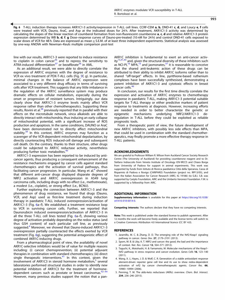
T-ALL induction chemotherapy promotes activation of AKR1C1-3and resistance to VCRIt has been recently demonstrated that cancer cells cantranscriptionally regulate NRF2 and, consequently, AKR1C1-3activity as a pro-survival response against drug treatments.4 Thus,in order to evaluate the potential establishment of such aresistance loop in our experimental setting, we measured theamount of coumberone conversion rate in all T-ALL cell lines after24 h of treatment with ASP, VCR, Ara-C, and Dauno at sub-lethalconcentrations. Only Dauno induced a significant increase in theproduction of fluorescent coumberol in all T-ALL cell lines. Instead,VCR treatment increased coumberone conversion rate only inDND-41 cells and Asp significantly augmented AKR1C-dependentenzymatic activity in both DND-41 and LOUCY cell lines. Ara-C didnot induce any increase of AKR1C1-3 activity (Fig. 6a, c, e). We
ED75ED50
Cel
l via
bilit
y (%
)
PT58
MPA
MPA + VCRVCR
VCR 10–5 10–4 10–3 10–2 10–1 100
μM
MPA 10–3 10–2 100 10 10010–1
100
80
60
40
20
0
C 100
80
Cel
l via
bilit
y (%
)
60
40
20
PT60
MPA
MPA
MPA + VCRVCR
VCR 10–5
10–310–4
10–210–3 10–2 10–1
100100
10
μM
10010–1
0
DA1.00
Com
bina
tion
inde
x
0.75
0.50
0.25
0.00
AntagonismSynergism
Cel
l via
bilit
y (%
)
PT55
MPA
MPA + VCRVCR
VCR 10–5 10–4 10–3 10–2 10–1 100
μM
MPA 10–3 10–2 100 10 10010–1
100B
80
60
40
20
0
Fig. 4 MPA sensitise primary T-ALL cells to vincristine. a CI values calculated at ED50 and ED75 for the combination of MPA and VCR in 10primary cell lines. Cell viability was evaluated by MTT assay after 48 h of treatment with VCR and MPA. b–d Representative completedose–response curves of MPA and its combination with VCR for PT55, PT58, and PT60, respectively
AKR1C enzymes modulate VCR susceptibility in T-ALLR. Bortolozzi et al.
990

confirmed these results by Western Blot (WB), showing thatDauno-induced overexpression of AKR1C1-3 proteins in all thethree T-ALL cell lines tested (Fig. 6b, d, f). In order to functionallyvalidate these data, we pretreated DND-41 cells with Dauno, withconsequent AKR1C1-3 enzymes overexpression and activation(Fig. 6c, d), and tested the response to VCR treatment. Indeed, a24 h pretreatment of T-ALL cells with Dauno was sufficient tomake these cells significantly more resistant to VCR treatment(Fig. 6g). These data corroborate the “open” hypothesis thattreatment with certain drugs can influence the response to otheragents by activating a NRF2-dependent surveillance system andestablish a potential AKR1C-mediated drug resistance loop duringT-ALL therapy and highlight the relevance of potential pharma-cological inhibition of AKR1C1-3 as adjuvant treatment to currentchemotherapy protocols.
DISCUSSIONDespite of significant improvements in intensive combinationchemotherapy and hematopoietic stem cell transplantationachieved in the last decades,19,29 a number of childhood T-ALLpatients only partially respond to treatment, thus experiencingrelapse and poor disease outcome.19,30 In this context, AKR1C1-3,being part of the NRF2-KEAP1 signaling pathway,4 are intimatelylinked with cancer biology and could participate in sustainingresistance to anti-cancer treatments.2,31 Given their intrinsicpromiscuity of substrates,32 AKR1C enzymes have been exten-sively reported to increase cancer cell resistance to therapeutics invarious human cancers, by reducing the intracellular levels of drugproducts, adducts, or compounds themselves. In this study, weinvestigated if AKR1C1-3 could contribute to therapy resistance inT-ALL by evaluating the potential impact of AKR1C1-3 expressionand inhibition on drug sensitivity in T-ALL cell lines, primarycultures, and PDXs.
MRD levels molecularly detected after induction therapy (day78) are a good marker of treatment response and predictive ofsubsequent relapse of T-ALL patients.19 For this reason, we choseto analyse NRF2-AKR1C1-3 signaling activation in MRDneg vs.MRDpos (MRD >5 × 10⁻⁴) T-ALL patients at diagnosis as a reliableand representative distinction of therapy “sensitive” or “resistant”tumors, respectively. Our data clearly show that therapy-resistantT-ALL samples are endowed with a significant overexpression and/or activation of three AKR1C (1–3) isoenzymes, with augmentedenzymatic activity being dependent on increased gene transcrip-tion (Fig. 1).Exploring the relationship between NRF2 overactivation and
drug resistance in cancer, Wang et al.33 previously demonstratedthat the transient knockdown of NRF2, or its specific inhibition byKEAP1 overexpression, both strongly increased the susceptibilityof lung cancer cells to different chemotherapics, includingcisplatin, doxorubicin, and etoposide. Along this line, in our study,we demonstrate that inhibition of AKR1C1-3 function by the pan-AKR1C inhibitor MPA (Figs. 2, 3, and 4) or AKR1C-specific genesilencing (Fig. 5a, b) are sufficient to increase T-ALL cell sensitivityto VCR. This sensitisation effect was obtained by the specificinhibition of AKR1C1-3, without the need to counteract additionaldetoxifying genes, generally regulated by NRF2, thus indicating amajor role of AKR1C family members in modulating drug responsein T-ALL. In order to further sustain the direct involvement ofAKR1C1-3 enzymes in chemotherapy resistance, we compared themRNA expression of additional NRF2 targets, previously involvedin drug metabolism, such as the glutamate-cysteine ligasecatalytic subunit (GCLC), the glutamate-cysteine ligase modifiersubunit (GCLM), or the UDP-glucuronosyltransferase 1A634,35 inMRDpos vs. MRDneg T-ALL patients. Gene expression data disclosedthat the above mentioned genes were not overexpressed inMRDpos relative to MRDneg patients, thus questioning a potentialrole in sustaining therapy resistance in T-ALL (data not shown). In
AKR1C1
A
1 0.29
siNeg
siAKR1C
#1
siAKR1C
#2
0.41
1 0.46 0.71
1 0.17 0.31
AKR1C2
AKR1C3
β-Actin
120B
100
80
60
Cel
l via
bilit
y (%
)
40 siNEGsiAKR1C#1siAKR1C#2
p< 0.000120
VCR (μM)
10–6
10–5
10–4
10–3
10–2 10
–1
0
C
1
1
1
2.07
1.18
3.49
Contro
l
tBHQ
AKR1C1
AKR1C2
AKR1C3
β-Actin
D4 × 10
5
3 × 10⁵
2 × 10⁵
1 × 10⁵
p=0.0007
Slo
pe o
f enz
ymat
ic r
eact
ion
(AU
)
Control
tBHQ (5 μM)
E
VCRVCR+tBHQ (5 μM)
100
80
60
40
20p= 0.0007
2–1
20
21
22
23
24
25
Cel
l via
bilit
y (%
)
μM
F 100
80
60
40
Via
bilit
y at
100
nM
VC
R (
%)
20
100
80
60
40
Via
bilit
y at
100
nM
VC
R (
%)
20
0PPR
PPR r= 0.9962 p= 0.0038PGR r= 0.9700 p= 0.1563
PDTALL8
PDTALL9
PDTALL5
PDTALL6PDTALL15
PDTALL25
PDTALL43
9.0 10.09.5 10.5
AKR1C3 mRNA expression (log2)
100
80
60
40
Via
bilit
y at
100
nM
VC
R (
%)
20
0
PGR r= 0.9985 p= 0.0354PPR r= 0.9896 p= 0.0104
PDTALL8
PDTALL9
PDTALL5
PDTALL6
PDTALL15
PDTALL25
PDTALL43
5.0 5.5 6.0 6.5 7.0 7.5
AKR1C2 mRNA expression (log2)
11.0 11.5PGR
p= 0.0002 G100
80
60
40
Via
bilit
y at
100
nM
VC
R (
%)
20
05.0 5.5
PPR r= 0.9925 p= 0.0075PGR r= 0.9981 p= 0.0391
PDTALL8
PDTALL9
PDTALL5
PDTALL6PDTALL15
PDTALL25 PDTALL43
6.0 6.5 7.0
AKR1C1 mRNA expression (log2)
Fig. 5 AKR1C1-3 levels affect response to vincristine in T-ALL cells. a, b After 48 h from electroporation with two different siRNAs againstAKR1C1-3, CCRF-CEM cell lysates were analysed by immunoblotting with AKR1C1, AKR1C2, AKR1C3, and β-Actin-specific antibodies, showingthe effective gene silencing of AKR1C enzymes a. Response of AKR1C-silenced CCRF-CEM cells to scalar doses of VCR (48 h) is reported b. c, dAKR1C1-3 activity was stimulated in CCRF-CEM by t-BHQ treatment (5 μM) for 18 h and then evaluated by WB c and calculation of the slope oflinear coumberone conversion reaction d. e Dose–response curves of t-BHQ-stimulated and control CCRF-CEM exposed to scalar doses of VCRfor 48 h. f Box plot showing the ex vivo response to VCR treatment in terms of cell viability of cells obtained from PPR (n= 4) and PGR (N= 3) T-ALL patient-derived xenografts (PDX-T-ALLs). g Correlation of AKR1C1-3 mRNA expression with the viability of PDTALL cells after ex vivotreatment with 100 nM VCR for 48 h is reported. 95% confidence interval is indicated by dotted lines. All data are expressed as mean± S.E.M.of three independent experiments. In correlation graphs, Pearson r and relative p values are reported independently for PGR and PPR patient-derived PDX-T-ALLs
AKR1C enzymes modulate VCR susceptibility in T-ALLR. Bortolozzi et al.
991

4 × 10⁵A
p< 0.05
Contro
l
VCR (5 n
M)
Dauno
(50
nM)
Asp (0
.5 U
l/ml)
AraC (1
μM)
3 × 10⁵
2 × 10⁵
1 × 10⁵
0
CC
RF
-CE
MS
lope
of e
nzym
atic
rea
ctio
n (A
U)
C 1 × 10⁶
8 × 10⁵
6 × 10⁵
4 × 10⁵
2 × 10⁵
p< 0.0001p< 0.01
p< 0.001
Contro
l
VCR (5 n
M)
Dauno
(50
nM)
Asp (0
.5 U
l/ml)
AraC (1
μM)
0
DN
D-4
1S
lope
of e
nzym
atic
rea
ctio
n (A
U)
B
AKR1C11 1.81 2.12 1.982.14
1 1.8 1.9 1.631.81
1 1.45 1.74 1.571.34
AKR1C2
AKR1C3
β-actin
Contro
l
VCR (5 n
M)
Dauno
(50
nM)
Asp (0
.5 U
l/ml)
AraC (1
μM)
D
1 2.03 2.27 2.923.98
1 1 0.86 1.151.35
1 2.01 1.99 0.862.24
AKR1C1
AKR1C2
AKR1C3
β-actin
Contro
l
VCR (5 n
M)
Dauno
(50
nM)
Asp (0
.5 U
l/ml)
AraC (1
μM)
E 5 × 10⁵
4 × 10⁵
3 × 10⁵
2 × 10⁵
1 × 10⁵
0
p< 0.001
p< 0.001 F
1 1.07 1.39 1.821.58
1 1.26 1.64 0.740.92
1 1.43 1.80 1.311.47
AKR1C1
AKR1C2
AKR1C3
β-actin
Contro
l
VCR (5 n
M)
Dauno
(50
nM)
Asp (0
.5 U
l/ml)
AraC (1
μM)
Contro
l
VCR (5 n
M)
Dauno
(50
nM)
Asp (0
.5 U
l/ml)
AraC (1
μM)
LOU
CY
Slo
pe o
f enz
ymat
ic r
eact
ion
(AU
)
G 120
100
No treatment
Dauno (10 nM)
p< 0.0001
80
60
40
20
0
10–8 10–5
VCR (μM)10–4 10–3 10–210–7 10–6
Cel
l via
bilit
y (%
)
AKR1C enzymes modulate VCR susceptibility in T-ALLR. Bortolozzi et al.
992
line with our results, AKR1C1-3 were reported to induce resistanceto cisplatin in colon cancer36 and to repress the sensitivity toATRA-induced differentiation37 or bezafibrate26 in AML.As an additional result, we were able to directly correlate the
expression of AKR1C enzymes with the degree of resistance toVCR ex vivo treatment of PDX-T-ALL cells (Fig. 5f, g). In particular,minimal changes in the balance of AKR1C expression wereassociated to a very different drug efficacy in terms of survivingcells after VCR treatment. This suggests that any little imbalance inthe regulation of the AKR1C surveillance system may producedramatic effects on cellular metabolism, especially during theonset or progression of resistant tumors. Intriguingly, our dataclearly show that AKR1C1-3 enzyme levels mainly affect VCRresponse rather than other chemotherapeutics. Supporting theseresults, Rovini et al.38 previously reported that in parallel with theireffects on the microtubule network, vinca alkaloids are able todirectly interact with mitochondria, thus inducing an early collapseof mitochondrial potential, with a significant increase of ROSproduction and apoptosis. In the same conditions, DAUNO or AraChave been demonstrated not to directly affect mitochondrialstability.39 In this context, AKR1C enzymes may function as ascavenger of the VCR-dependent mitochondrial depolarisation, bydirectly counteracting ROS induced-cell damage and subsequentcell death. On the contrary, thanks to their structure, other drugscould be subjected to AKR1C reductase activity, neverthelessproducing further toxic metabolites.40
AKR1C1-3 expression has been reported to be induced by anti-cancer agents, thus producing a consequent enhancement of theresistance mechanisms engaged by cancer cells against standardchemotherapics and the activation of survival pathways, thusfacilitating cancer progression. In particular, Wang et al.4 showedthat different anti-cancer drugs displayed disparate degrees ofNRF2 activation and AKR1C overexpression in A549 lungcarcinoma cells, including drugs with no effect (i.e., methotrexate),a modest (i.e., cisplatin), or strong effect (i.e., BCNU).Further exploring the connection between AKR1C1-3 and the
phenomenon of drug resistance, we found that drugs (Dauno,VCR, and Asp) used as first-line treatment during inductiontherapy in paediatric T-ALL induced overexpression/activation ofAKR1C1-3 (Fig. 6a–f). We established a treatment resistance loopto VCR in surviving cancer cells. Further, we reported thatDaunorubicin induced overexpression/activation of AKR1C1-3 inall the three T-ALL cell lines tested (Fig. 6a–f), showing variousdegree of activation probably depending on the redox status (andthus GSH content) of each particular cell line, as previouslysuggested.4 Moreover, we showed that Dauno-induced AKR1C1-3overexpression partially counteracted the effects exerted by VCRtreatment (Fig. 6g), suggesting the potential antagonistic effect ofcombined AKR1C-inducing drugs.From a pharmacological point of view, the availability of novel
AKR1C-selective inhibitors would be of value for multiple reasonsincluding: (i) cancer chemoprevention; (ii) potential adjuvanttherapies in combination with standard chemotherapics; (iii) directsingle therapeutic interventions.41 In this context, given theinvolvement of AKR1C3 in steroid hormone metabolism,5 severallaboratories performed structural studies in order to identify newpotential inhibitors of AKR1C3 for the treatment of hormone-dependent cancers such as prostate or breast carcinomas.42–44
However, many previous studies support the notion that a pan-
AKR1C inhibition is fundamental to exert an anti-cancer activ-ity15,41,45 and, given the structural diversity of these inhibitors suchas NCI-PI,16 MPA,15 and jasmonates,37 it is reasonable to conceivethat the shared anti-leukemic action of these drugs can beattributed to their ability to inhibit AKR1C isoforms rather than toshared “off-target” effects. In line, pyrithione-based rutheniumcomplexes have been successfully synthesised, demonstrating apotent inhibition of AKR1C1-3 and cytotoxic effects in breastcancer cells.46
In conclusion, our results for the first time directly correlate theexpression and activation of AKR1C enzymes to chemotherapyresponse in paediatric T-ALL, making AKR1C1-3 potential suitabletargets for T-ALL therapy or either predictive markers of patientresponse to treatments at diagnosis. However, increasing effortsare needed in order to better characterize the genetic orepigenetic mechanisms underlying NRF2/AKR1C1-3 de-regulation in T-ALL before they could be exploited as reliableprognostic tools.From a therapeutic point of view, the future development of
new AKR1C inhibitors, with possibly less side effects than MPA,that could be used in combination with the standard chemother-apeutics, may finally promote a more effective therapy response inT-ALL patients.
ACKNOWLEDGEMENTSWe are grateful to Professor William R. Wilson from Auckland Cancer Society ResearchCentre (The University of Auckland) for providing coumberone reagent and to Dr.Stefano Indraccolo from Veneto Institute of Oncology IOV-IRCCS and Chiara Borgafrom University of Padova for support in animal experiments. This work wassupported by funds from Istituto di Ricerca paediatrica Città della Speranza, Cassa diRisparmio di Padova e Rovigo (CARIPARO) Foundation (project no. IRP13/05), andfrom the Italian Association for Cancer Research (AIRC; IG 19186) (to G.B.). S.B. wassupported by Cariplo Foundation, AIRC and the Umberto Veronesi Foundation. F.M. issupported by a fellowship from AIRC.
ADDITIONAL INFORMATIONSupplementary information is available for this paper at https://doi.org/10.1038/s41416-018-0014-0.
Competing interests: The authors declare that they have no competing interests.
Note: This work is published under the standard license to publish agreement. After12 months the work will become freely available and the license terms will switch toa Creative Commons Attribution 4.0 International licence (CC BY 4.0).
REFERENCES1. Jaramillo, M. C. & Zhang, D. D. The emerging role of the Nrf2-Keap1 signaling
pathway in cancer. Genes Dev. 27, 2179–2191 (2013).2. Sporn, M. B. & Liby, K. T. NRF2 and cancer: the good, the bad and the importance
of context. Nat. Rev. Cancer 12, 564–571 (2012).3. Taguchi, K., Motohashi, H. & Yamamoto, M. Molecular mechanisms of the Keap1-
Nrf2 pathway in stress response and cancer evolution. Genes Cells 16, 123–140(2011).
4. Wang, X. J., Hayes, J. D. & Wolf, C. R. Generation of a stable antioxidant responseelement-driven reporter gene cell line and its use to show redox-dependentactivation of nrf2 by cancer chemotherapeutic agents. Cancer Res. 66,10983–10994 (2006).
5. Penning, T. M. The aldo-keto reductases (AKRs): overview. Chem. Biol. Interact.234, 236–246 (2015).
Fig. 6 T-ALL induction therapy increases AKR1C1-3 activity/expression in T-ALL cell lines. CCRF-CEM a, b, DND-41 c, d, and Loucy e, f cellswere treated with VCR, Dauno, AraC, and Asp at the indicated doses for 24 h. After treatment, AKR1C1-3 activity was determined bycalculating the slopes of the linear reaction of coumberol formation from non-fluorescent coumberone a, c, d and relative AKR1C1-3 proteinexpression determined by WB b, d, f. g Dose–response curves of Dauno-pre-treated (10 nM for 24 h) and control DND-41 cells exposed toscalar doses of VCR for 48 h. Data are expressed as mean ± S.E.M. of at least three independent experiments. Statistical analysis was assessedby one-way ANOVA with Newman–Keuls multiple comparison post-test
AKR1C enzymes modulate VCR susceptibility in T-ALLR. Bortolozzi et al.
993

6. O’Connor, T., Ireland, L. S., Harrison, D. J. & Hayes, J. D. Major differences exist inthe function and tissue-specific expression of human aflatoxin B1 aldehydereductase and the principal human aldo-keto reductase AKR1 family members.Biochem. J. 343, 487–504 (1999).
7. Dozmorov, M. G. et al. Elevated AKR1C3 expression promotes prostate cancer cellsurvival and prostate cell-mediated endothelial cell tube formation: implicationsfor prostate cancer progression. BMC Cancer 10, 672 (2010).
8. Le Calve, B. et al. Long-term in vitro treatment of human glioblastoma cells withtemozolomide increases resistance in vivo through up-regulation of GLUTtransporter and aldo-keto reductase enzyme AKR1C expression. Neoplasia 12,727–739 (2010).
9. Li, D. & Ellis, E. M. Inducible protection of human astrocytoma 1321N1 cellsagainst hydrogen peroxide and aldehyde toxicity by 7-hydroxycoumarin isassociated with the upregulation of aldo-keto reductases. Neurotoxicology 33,1368–1374 (2012).
10. Lyon, R. C., Li, D., McGarvie, G. & Ellis, E. M. Aldo-keto reductases mediate con-stitutive and inducible protection against aldehyde toxicity in human neuro-blastoma SH-SY5Y cells. Neurochem. Int. 62, 113–121 (2013).
11. Miller, V. L. et al. Aldo-keto reductase family 1 member C3 (AKR1C3) is expressedin adenocarcinoma and squamous cell carcinoma but not small cell carcinoma.Int. J. Clin. Exp. Pathol. 5, 278–289 (2012).
12. Palackal, N. T., Lee, S. H., Harvey, R. G., Blair, I. A. & Penning, T. M. Activation ofpolycyclic aromatic hydrocarbon trans-dihydrodiol proximate carcinogens by humanaldo-keto reductase (AKR1C) enzymes and their functional overexpression in humanlung carcinoma (A549) cells. J. Biol. Chem. 277, 24799–24808 (2002).
13. Mahadevan, D. et al. Transcriptosome and serum cytokine profiling of an atypicalcase of myelodysplastic syndrome with progression to acute myelogenous leu-kemia. Am. J. Hematol. 81, 779–786 (2006).
14. Birtwistle, J. et al. The aldo-keto reductase AKR1C3 contributes to 7,12-dime-thylbenz(a)anthracene-3,4-dihydrodiol mediated oxidative DNA damage inmyeloid cells: implications for leukemogenesis. Mutat. Res. 662, 67–74 (2009).
15. Khanim, F. L. et al. Combined bezafibrate and medroxyprogesterone acetate:potential novel therapy for acute myeloid leukaemia. PLoS ONE 4, e8147 (2009).
16. Khanim, F. et al. Selective AKR1C3 inhibitors do not recapitulate the anti-leukaemic activities of the pan-AKR1C inhibitor medroxyprogesterone acetate. Br.J. Cancer 110, 1506–1516 (2014).
17. Moradi Manesh, D. et al. AKR1C3 is a biomarker of sensitivity to PR-104 in preclinicalmodels of T-cell acute lymphoblastic leukemia. Blood 126, 1193–1202 (2015).
18. Guise, C. P. et al. The bioreductive prodrug PR-104A is activated under aerobicconditions by human aldo-keto reductase 1C3. Cancer Res. 70, 1573–1584 (2010).
19. Schrappe, M. et al. Late MRD response determines relapse risk overall and insubsets of childhood T-cell ALL: results of the AIEOP-BFM-ALL 2000 study. Blood118, 2077–2084 (2011).
20. Willemse, M. J. et al. Detection of minimal residual disease identifies differences intreatment response between T-ALL and precursor B-ALL. Blood 99, 4386–4393 (2002).
21. Basso, G., Buldini, B., De Zen, L. & Orfao, A. New methodologic approaches forimmunophenotyping acute leukemias. Haematologica 86, 675–692 (2001).
22. Jamieson, S. M. et al. A novel fluorometric assay for aldo-keto reductase 1C3predicts metabolic activation of the nitrogen mustard prodrug PR-104A in humanleukaemia cells. Biochem. Pharmacol. 88, 36–45 (2014).
23. Yee, D. J., Balsanek, V., Bauman, D. R., Penning, T. M. & Sames, D. Fluorogenicmetabolic probes for direct activity readout of redox enzymes: selective mea-surement of human AKR1C2 in living cells. Proc. Natl Acad. Sci. USA 103,13304–13309 (2006).
24. Agnusdei, V. et al. Therapeutic antibody targeting of Notch1 in T-acute lym-phoblastic leukemia xenografts. Leukemia 28, 278–288 (2014).
25. Chou, T. C. Theoretical basis, experimental design, and computerized simulationof synergism and antagonism in drug combination studies. Pharmacol. Rev. 58,621–681 (2006).
26. Murray, J. A. et al. Combined bezafibrate and medroxyprogesterone acetate haveefficacy without haematological toxicity in elderly and relapsed acute myeloidleukaemia (AML). Br. J. Haematol. 149, 65–69 (2010).
27. Zamzami, N. et al. Sequential reduction of mitochondrial transmembranepotential and generation of reactive oxygen species in early programmed celldeath. J. Exp. Med. 182, 367–377 (1995).
28. Liem, N. L. et al. Characterization of childhood acute lymphoblastic leukemiaxenograft models for the preclinical evaluation of new therapies. Blood 103,3905–3914 (2004).
29. Locatelli, F., Schrappe, M., Bernardo, M. E. & Rutella, S. How I treat relapsedchildhood acute lymphoblastic leukemia. Blood 120, 2807–2816 (2012).
30. Nguyen, K. et al. Factors influencing survival after relapse from acute lympho-blastic leukemia: a Children’s Oncology Group study. Leukemia 22, 2142–2150(2008).
31. Zeng, C. M. et al. Aldo-keto reductase AKR1C1-AKR1C4: functions, regulation, andintervention for anti-cancer therapy. Front. Pharmacol. 8, 119 (2017).
32. Jez, J. M., Bennett, M. J., Schlegel, B. P., Lewis, M. & Penning, T. M. Comparativeanatomy of the aldo-keto reductase superfamily. Biochem. J. 326, 625–636 (1997).
33. Wang, X. J. et al. Nrf2 enhances resistance of cancer cells to chemotherapeuticdrugs, the dark side of Nrf2. Carcinogenesis 29, 1235–1243 (2008).
34. Satoh, T., McKercher, S. R. & Lipton, S. A. Nrf2/ARE-mediated antioxidant actionsof pro-electrophilic drugs. Free Radic. Biol. Med. 65, 645–657 (2013).
35. Bock, K. W. & Kohle, C. UDP-glucuronosyltransferase 1A6: structural, functional,and regulatory aspects. Methods Enzymol. 400, 57–75 (2005).
36. Matsunaga, T. et al. Pathophysiological roles of aldo-keto reductases (AKR1C1and AKR1C3) in development of cisplatin resistance in human colon cancers.Chem. Biol. Interact. 202, 234–242 (2013).
37. Desmond, J. C. et al. The aldo-keto reductase AKR1C3 is a novel suppressor of celldifferentiation that provides a plausible target for the non-cyclooxygenase-dependent antineoplastic actions of nonsteroidal anti-inflammatory drugs. Can-cer Res. 63, 505–512 (2003).
38. Rovini, A., Savry, A., Braguer, D. & Carre, M. Microtubule-targeted agents: whenmitochondria become essential to chemotherapy. Biochim. Biophys. Acta 1807,679–688 (2011).
39. Andre, N. et al. Paclitaxel induces release of cytochrome c from mitochondriaisolated from human neuroblastoma cells. Cancer Res. 60, 5349–5353 (2000).
40. Bains, O. S. et al. A correlation between cytotoxicity and reductase-mediatedmetabolism in cell lines treated with doxorubicin and daunorubicin. J. Pharmacol.Exp. Ther. 347, 375–387 (2013).
41. Davies, N. J. et al. AKR1C isoforms represent a novel cellular target for jasmonatesalongside their mitochondrial-mediated effects. Cancer Res. 69, 4769–4775(2009).
42. Day, J. M., Tutill, H. J., Purohit, A. & Reed, M. J. Design and validation of specificinhibitors of 17beta-hydroxysteroid dehydrogenases for therapeutic applicationin breast and prostate cancer, and in endometriosis. Endocr. Relat. Cancer 15,665–692 (2008).
43. Penning, T. M. et al. Aldo-keto reductase (AKR) 1C3: role in prostate disease andthe development of specific inhibitors. Mol. Cell. Endocrinol. 248, 182–191 (2006).
44. Byrns, M. C., Steckelbroeck, S. & Penning, T. M. An indomethacin analogue, N-(4-chlorobenzoyl)-melatonin, is a selective inhibitor of aldo-keto reductase 1C3(type 2 3alpha-HSD, type 5 17beta-HSD, and prostaglandin F synthase), apotential target for the treatment of hormone dependent and hormone inde-pendent malignancies. Biochem. Pharmacol. 75, 484–493 (2008).
45. Bunce, C. M. et al. Indomethacin potentiates the induction of HL60 differentiationto neutrophils, by retinoic acid and granulocyte colony-stimulating factor, and tomonocytes, by vitamin D3. Leukemia 8, 595–604 (1994).
46. Kljun, J. et al. Pyrithione-based ruthenium complexes as inhibitors of aldo-ketoreductase 1C enzymes and anticancer agents. Dalton Trans. 45, 11791–11800(2016).
AKR1C enzymes modulate VCR susceptibility in T-ALLR. Bortolozzi et al.
994