Embed Size (px)
Citation preview

National Center for Emerging and Zoonotic Infectious Diseases
American Journal of Infection Control NHSN Case Studies: A Case Study
Kathy Allen-Bridson, BSN, MScPH, CICScott Decker, MPH
NHSN Training 2019
National Center for Emerging and Zoonotic Infectious Diseases
An NHSN Case Study: Respiratory Illness and Positive Blood Cultures- What’s the Correct Determination?
Kathy Allen-Bridson, BSN, MScPH, CICLead- NHSN Protocol and Validation TeamDivision of Healthcare Quality Promotion
Greater Atlanta APICAugust 1, 2018

Agenda
Review Case
Straw Polls
Answers, Rationales
Questions

This is a…..
Judgement free zone!
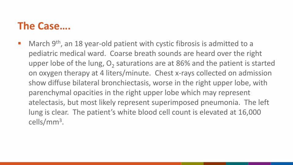
The Case…. March 9th, an 18 year-old patient with cystic fibrosis is admitted to a
pediatric medical ward. Coarse breath sounds are heard over the right upper lobe of the lung, O2 saturations are at 86% and the patient is started on oxygen therapy at 4 liters/minute. Chest x-rays collected on admission show diffuse bilateral bronchiectasis, worse in the right upper lobe, with parenchymal opacities in the right upper lobe which may represent atelectasis, but most likely represent superimposed pneumonia. The left lung is clear. The patient’s white blood cell count is elevated at 16,000 cells/mm3.

The Case…. March 10th, the patient develops a fever of 101.4°F, and pulmonary
crackles are heard over the upper right lung. Sputum, which is now blood-tinged, is collected for culture. The patient is tachypneic and is placed on a conventional mode of mechanical ventilation.
March 11th, fever, crackles and tachypnea continue, and the patient is coughing up large amounts of green sputum. FiO2 settings range between 30-100%. A triple lumen catheter is placed in the subclavian vein.

The Case…. March 12th, the sputum culture collected on March 10th is reported
positive for Staphylococcus aureus and Pseudomonas aeruginosa.
March 13th, another chest x-ray is obtained, which shows an increased, confluent opacity in the right upper lobe, and the radiologist states that pneumonia is likely. Patient remains on mechanical ventilation.

Poll Everywhere Question: Which of the following is most true?a. Patient meets PNU1 criterion, clinically defined pneumonia, date of event on
March 9th and the event is not ventilator associated.
b. Patient meets PNU1 criterion, clinically defined pneumonia, date of even on March 9th and the event is ventilator associated.
c. Patient meets PNU2 criterion, pneumonia, with common bacterial or filamentous pathogens and specific laboratory findings, with date of event on March 10th.
d. Patient meets PNU2 criterion, pneumonia with common bacterial or filamentous pathogen and specific laboratory findings, with date of event on March 12th.
Correct Answer: A
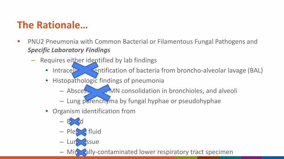
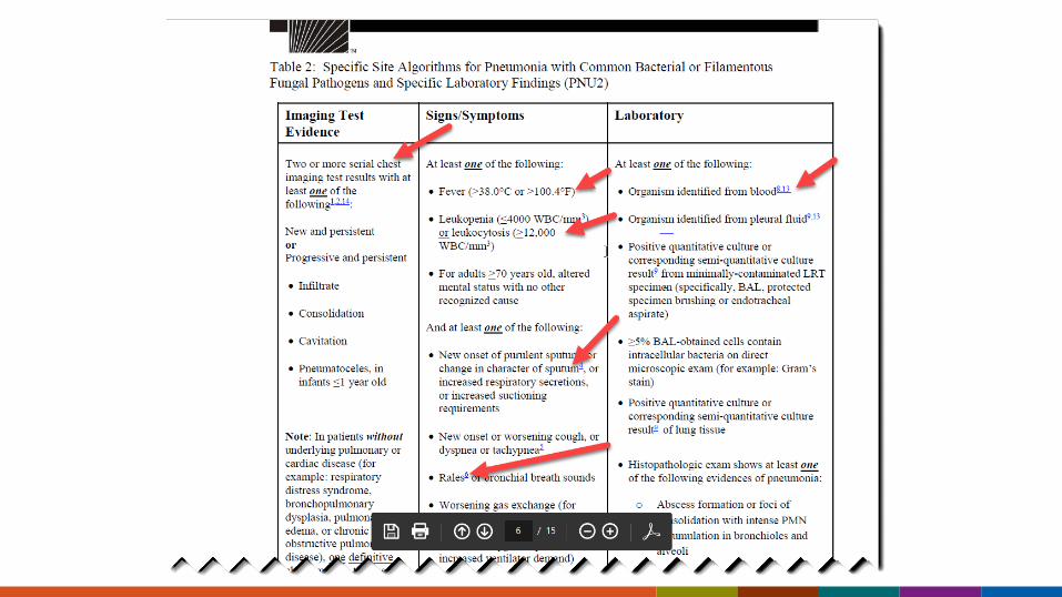
The Rationale… PNU2 Pneumonia with Common Bacterial or Filamentous Fungal Pathogens and
Specific Laboratory Findings– Requires either identified by lab findings
• Intracellular identification of bacteria from broncho-alveolar lavage (BAL)• Histopathologic findings of pneumonia
– Abscess with PMN consolidation in bronchioles, and alveoli– Lung parenchyma by fungal hyphae or pseudohyphae
• Organism identification from – Blood– Pleural fluid– Lung tissue– Minimally-contaminated lower respiratory tract specimen
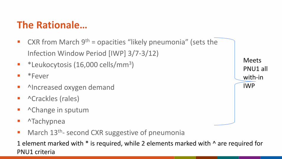
The Rationale… CXR from March 9th = opacities “likely pneumonia” (sets the
Infection Window Period [IWP] 3/7-3/12) *Leukocytosis (16,000 cells/mm3) *Fever ^Increased oxygen demand ^Crackles (rales) ^Change in sputum ^Tachypnea March 13th- second CXR suggestive of pneumonia
Meets PNU1 all with-in IWP
1 element marked with * is required, while 2 elements marked with ^ are required for PNU1 criteria


The Rationale… The patient was not ventilated for more than 2 days on the DOE.
Therefore this is not a VAP and answer “b” is incorrect.
Note: Even though this patient is 18 years old, he is housed in a pediatric location and therefore if pedVAP was selected in the monthly reporting plan the patient would be eligible for pedVAP surveillance.
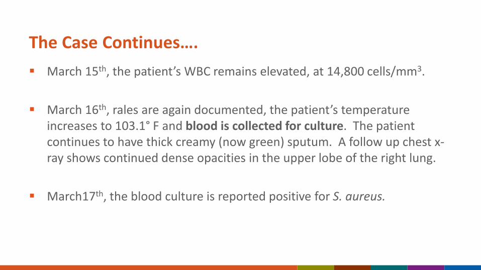
The Case Continues…. March 15th, the patient’s WBC remains elevated, at 14,800 cells/mm3.
March 16th, rales are again documented, the patient’s temperature increases to 103.1° F and blood is collected for culture. The patient continues to have thick creamy (now green) sputum. A follow up chest x-ray shows continued dense opacities in the upper lobe of the right lung.
March17th, the blood culture is reported positive for S. aureus.
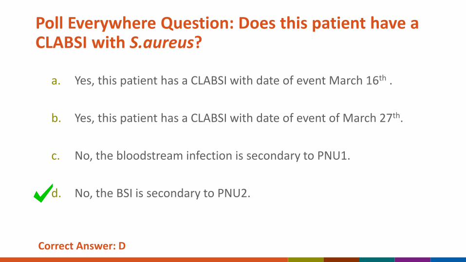
a. Yes, this patient has a CLABSI with date of event March 16th .
b. Yes, this patient has a CLABSI with date of event of March 27th.
c. No, the bloodstream infection is secondary to PNU1.
d. No, the BSI is secondary to PNU2.
Poll Everywhere Question: Does this patient have a CLABSI with S.aureus?
Correct Answer: D

Poll Everywhere Question: What is the date of event and the Repeat Infection Timeframe (RIT) for the PNEU event now?
a. The date of event and repeat infection timeframe do not change.
b. The date of event from PNU2 is 3/10 and the RIT becomes 3/10-3/23.
c. The date of event for PNU2 is 3/13 and the RIT becomes 3/13-3/26.
Let’s explore the answersCorrect Answer: A

Scenario 1A positive blood specimen must contain at least one eligible matching organism to the site-specific specimen
And the blood specimen is collected in the site-specific secondary BSI attribution
And an eligible organism identified from the site-specific specimen is used as an element to meet the site-specific definition
The Rationale… PNEU Repeat Infection Timeframe 3/9-3/22 PNEU Secondary BSI Attribution Period 3/7 – 3/22 Blood culture collected during RIT and SBAP; pneumonia symptoms
remain
Scenario 2Positive blood specimen must contain an element of the site-specific definition
And blood specimen is collected in the site-specific infection window period
And an eligible organism in a blood specimen is used as an element to meet the site-specific definition
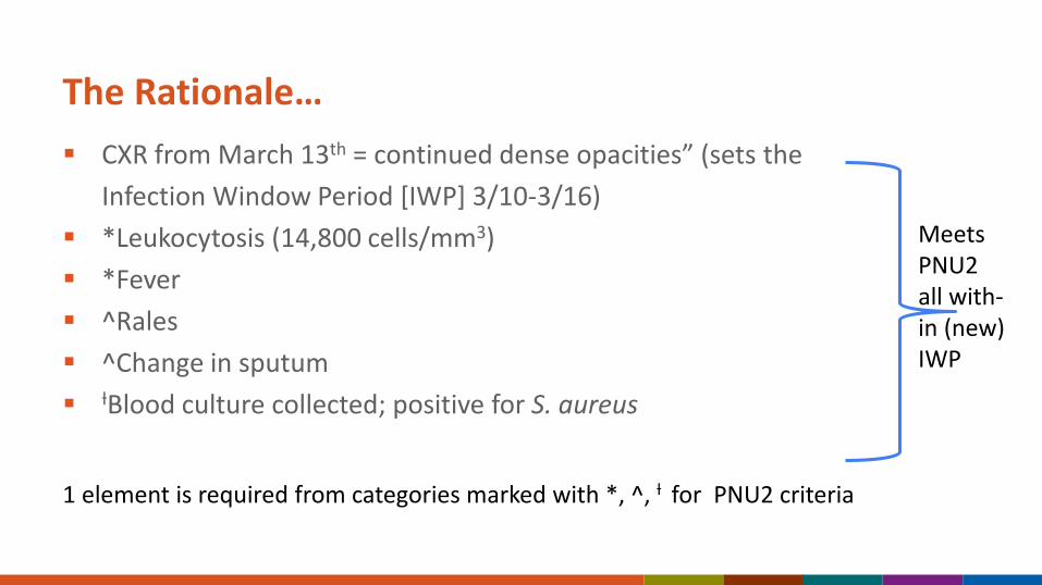
The Rationale… CXR from March 13th = continued dense opacities” (sets the
Infection Window Period [IWP] 3/10-3/16) *Leukocytosis (14,800 cells/mm3) *Fever ^Rales ^Change in sputum ƚBlood culture collected; positive for S. aureus
Meets PNU2 all with-in (new) IWP
1 element is required from categories marked with *, ^, ƚ for PNU2 criteria
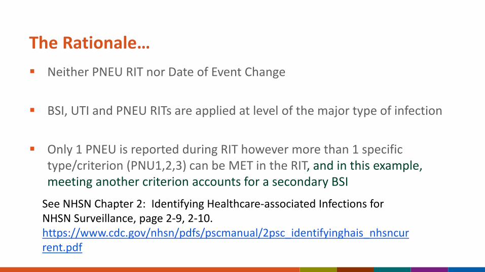
The Rationale… Neither PNEU RIT nor Date of Event Change
BSI, UTI and PNEU RITs are applied at level of the major type of infection
Only 1 PNEU is reported during RIT however more than 1 specific type/criterion (PNU1,2,3) can be MET in the RIT, and in this example, meeting another criterion accounts for a secondary BSI
See NHSN Chapter 2: Identifying Healthcare-associated Infections for NHSN Surveillance, page 2-9, 2-10. https://www.cdc.gov/nhsn/pdfs/pscmanual/2psc_identifyinghais_nhsncurrent.pdf

The Takeaways and Additional Pearls…. Simply because a patient met a criterion for one type of infection without
secondary BSI, does not mean that he may not meet another criterion for that infection for which the BSI may be secondary.– If there is a BSI which you believe may be secondary, consider other
criteria of the same infection type or other infection types. PNEU definition can be used for secondary BSI attribution when
conducting BSI/CLABSI surveillance for all patients, all locations, ventilated or not ventilated.
Patients for whom Ventilator-associated Event criteria does not account for a BSI, may meet a Pneumonia criterion as primary cause.
The Takeaways and Additional Pearls…. Repeat Infection Timeframes and Secondary BSI Attribution Periods do not
“roll forward”. One must end before another is eligible to begin for the same type of infection.
Published case studies are tools for confirming the accuracy of surveillance within a facility.
For more information, contact CDC1-800-CDC-INFO (232-4636)TTY: 1-888-232-6348 www.cdc.gov
The findings and conclusions in this report are those of the authors and do not necessarily represent the official position of the Centers for Disease Control and Prevention.
Thank You !
Questions
National Center for Emerging and Zoonotic Infectious Diseases
An NHSN Case Study: Location Mapping
Scott Decker, MPHNHSN Methods and Analytics TeamDivision of Healthcare Quality Promotion
NHSN Training 2019

The primary teaching points of this case study are:
Defining unit acuity according to established criteria
Determining service type for locations with mixed populations
Managing interim physical relocations in NHSN
***We strongly recommend that you review/reference the following section of the NHSN Patient Safety Component Manual: https://www.cdc.gov/nhsn/PDFs/pscManual/15LocationsDescriptions_current.pdf
NHSN 80% Rule The NHSN 80% rule translates to the requirement that for a given location,
80% of the patients share a common acuity level (for critical vs non-critical care categorization).
Similarly, subspecialization (e.g. orthopedic, neurosurgical, medical) requires that 80% of the patients admitted to the location share the same specialty service.
Services may be combined into more generic categories such as neurosurgical, cardiac surgery and general surgery combining to a total of 80% and being categorized as “surgical.”
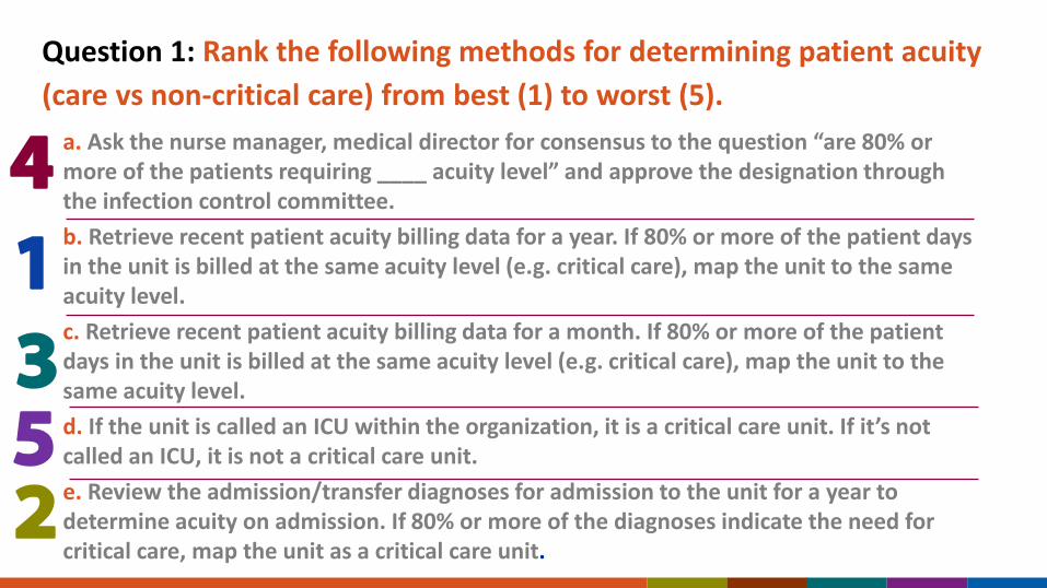
Question 1: Rank the following methods for determining patient acuity (care vs non-critical care) from best (1) to worst (5).
a. Ask the nurse manager, medical director for consensus to the question “are 80% or more of the patients requiring ____ acuity level” and approve the designation through the infection control committee. b. Retrieve recent patient acuity billing data for a year. If 80% or more of the patient days in the unit is billed at the same acuity level (e.g. critical care), map the unit to the same acuity level. c. Retrieve recent patient acuity billing data for a month. If 80% or more of the patient days in the unit is billed at the same acuity level (e.g. critical care), map the unit to the same acuity level. d. If the unit is called an ICU within the organization, it is a critical care unit. If it’s not called an ICU, it is not a critical care unit. e. Review the admission/transfer diagnoses for admission to the unit for a year to determine acuity on admission. If 80% or more of the diagnoses indicate the need for critical care, map the unit as a critical care unit.
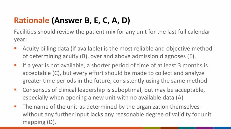
Rationale (Answer B, E, C, A, D) Facilities should review the patient mix for any unit for the last full calendar year: Acuity billing data (if available) is the most reliable and objective method
of determining acuity (B), over and above admission diagnoses (E). If a year is not available, a shorter period of time of at least 3 months is
acceptable (C), but every effort should be made to collect and analyze greater time periods in the future, consistently using the same method
Consensus of clinical leadership is suboptimal, but may be acceptable, especially when opening a new unit with no available data (A)
The name of the unit-as determined by the organization themselves-without any further input lacks any reasonable degree of validity for unit mapping (D).

Question 2: A given unit is 60% adult critical care and 40% adult ward as determined by a year’s worth of admission diagnoses. Of the following, which is NOT an acceptable choice to designate a location in NHSN?
a. Re-analyze the data using an equally acceptable but alternate measure (such as billing data) and if this analysis reveals a single acuity level for >80% of the patient population, map to that acuity level.
b. Map the unit as adult critical care-60% is the majority.
c. Map as a CDC mixed acuity location.
d. Split the unit into two virtual locations. Correct Answer: B

Rationale The method of determining acuity is not definitive, alternative data
sources are an acceptable (albeit potentially suboptimal) solution. If this is not an option, the user may choose to create virtual locations,
which if properly mapped can be reported separately and allow reporting and benchmarking for two or more locations.
Mixed acuity location is also acceptable, but benchmarking efforts are more heterogeneous and may not offer a benchmark as comparable as virtual locations

Virtual Locations
Created in NHSN when a facility is unable to meet the 80% rule for location designation in a single physical unit, but would like to report their NHSN surveillance data for each of the major specific patient types in that unit
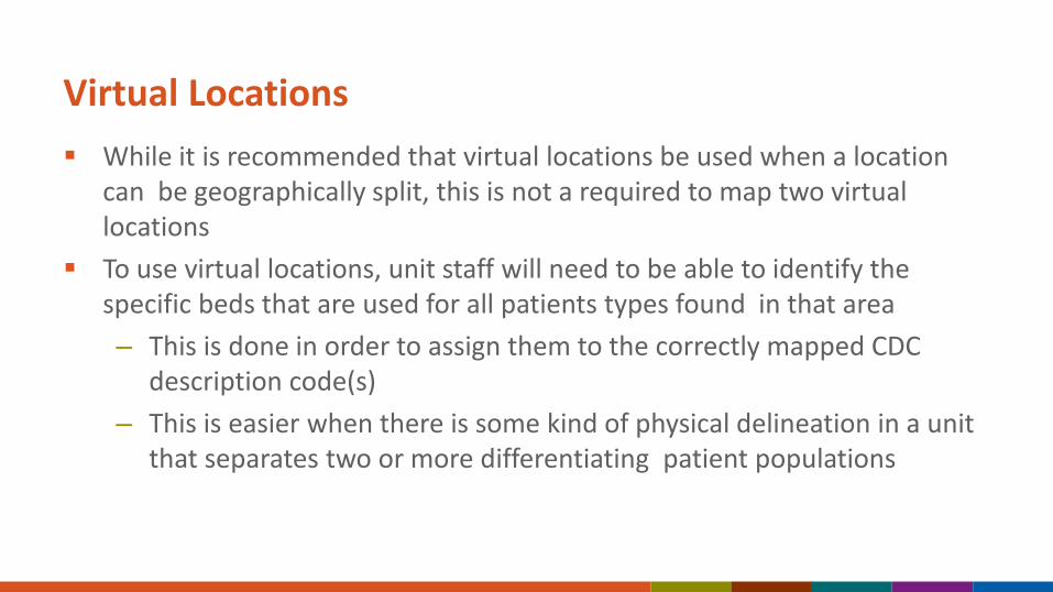
Virtual Locations While it is recommended that virtual locations be used when a location
can be geographically split, this is not a required to map two virtual locations
To use virtual locations, unit staff will need to be able to identify the specific beds that are used for all patients types found in that area– This is done in order to assign them to the correctly mapped CDC
description code(s)– This is easier when there is some kind of physical delineation in a unit
that separates two or more differentiating patient populations
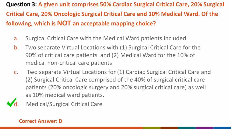
Question 3: A given unit comprises 50% Cardiac Surgical Critical Care, 20% Surgical Critical Care, 20% Oncologic Surgical Critical Care and 10% Medical Ward. Of the following, which is NOT an acceptable mapping choice?
a. Surgical Critical Care with the Medical Ward patients includedb. Two separate Virtual Locations with (1) Surgical Critical Care for the
90% of critical care patients and (2) Medical Ward for the 10% of medical non-critical care patients
c. Two separate Virtual Locations for (1) Cardiac Surgical Critical Care and (2) Surgical Critical Care comprised of the 40% of surgical critical care patients (20% oncologic surgery and 20% surgical critical care) as well as 10% medical ward patients.
d. Medical/Surgical Critical Care
Correct Answer: D

Question 3 Rationale This unit’s population comprises 90% critical care and 90% surgical
services
Medical/Surgical cannot be selected because > 80% of the patients are surgical
(50% cardiac surgical critical care + 20% surgical critical care + 20% oncologic surgical critical care)
Selecting the lowest common denominator of the surgical services (“surgical critical care”) as is acceptable, the ward level patients will be considered Surgical ICU Patients
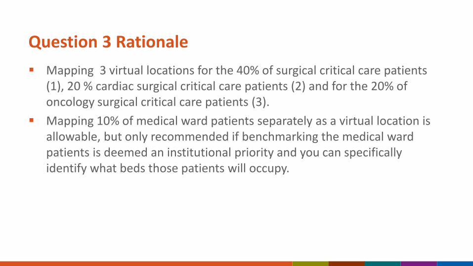
Question 3 Rationale Mapping 3 virtual locations for the 40% of surgical critical care patients
(1), 20 % cardiac surgical critical care patients (2) and for the 20% of oncology surgical critical care patients (3).
Mapping 10% of medical ward patients separately as a virtual location is allowable, but only recommended if benchmarking the medical ward patients is deemed an institutional priority and you can specifically identify what beds those patients will occupy.
Question 4An inpatient location for the Level III Neonatal Critical Care Unit located on the 3rd floor (Label = NICU3) which is currently part of your reporting plan is being temporarily relocated to the 4th floor (no existing label) while the 3rd
floor location is being remodeled. Construction is expected to last 6 months.
The patient type and bed size will remain the same. Which method(s) can you use to continue to report data for this mapped unit in NHSN?

Question 4: Answer optionsa. Change the existing location “Your Code” to match the new code assigned (e.g., NICU4) on the day of the relocation and change back to the original (NICU3) on the day the unit returns to its original physical location 6 months later. b. Create a new location code and location type NICU4/Level III NICU, inactivate NICU3/Level III NICU. When the unit reopens, inactivate NICU4 and reactivate NICU3. c. Either of the above. d. Neither a nor b
Correct Answer: C
Question 4 Rationale Option “a” ensures that data from this unit will not be interrupted with
only a modification to “Your Code” as opposed to creating a newly mapped unit.
If option “b” is selected, users will need to adjust their monthly reporting plans to reflect the location changes

Question 5: Your acute care facility performs FacWideIN reporting for LabIDevents and owns and operates 2 separate off-site emergency departments which admit patients to your acute care facility. Of the following mapping, which is not acceptable?
a. Map each ED location separately using the CDC location code “OUT: ACUTE: ED”.
b. Map the emergency departments together as 1 off site ED using the CDC location code ‘OUT: ACUTE: ED’.
c. Do not map these ED locations to the acute care facility
Correct Answer: C

Question 5 Rationale Each emergency department affiliated to the acute care facility should be
mapped for location attribution in LabID event reporting If the acute care facility combines visit information for the 2 off-site
emergency departments, it’s allowable to map these as a single location for reporting purposes.
Otherwise, each emergency department should be mapped individually.

For more information, contact CDC1-800-CDC-INFO (232-4636)TTY: 1-888-232-6348 www.cdc.gov
The findings and conclusions in this report are those of the authors and do not necessarily represent the official position of the Centers for Disease Control and Prevention.
Thank You !
Questions