Embed Size (px)
Citation preview
Airway Management:Airway Management:An Introduction and OverviewAn Introduction and Overview
&&Massive HemoptysisMassive Hemoptysis
Division of Critical Care MedicineDivision of Critical Care MedicineUniversity of AlbertaUniversity of Alberta
Airway ManagementAirway Management
OutlineOutline
OverviewOverview Normal airwayNormal airway Difficult intubationDifficult intubation Structured approach to airway Structured approach to airway
managementmanagement Causes of failed intubationCauses of failed intubation
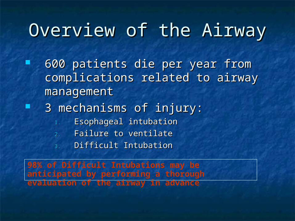
Overview of the AirwayOverview of the Airway
600 patients die per year from 600 patients die per year from complications related to airway complications related to airway managementmanagement
3 mechanisms of injury:3 mechanisms of injury:1.1. Esophageal intubationEsophageal intubation
2.2. Failure to ventilateFailure to ventilate
3.3. Difficult IntubationDifficult Intubation
98% of Difficult Intubations may be anticipated by performing a thorough evaluation of the airway in advance
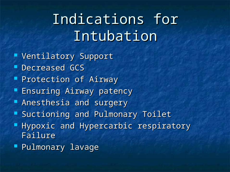
Indications for IntubationIndications for Intubation
Ventilatory SupportVentilatory Support Decreased GCSDecreased GCS Protection of AirwayProtection of Airway Ensuring Airway patencyEnsuring Airway patency Anesthesia and surgeryAnesthesia and surgery Suctioning and Pulmonary ToiletSuctioning and Pulmonary Toilet Hypoxic and Hypercarbic respiratory FailureHypoxic and Hypercarbic respiratory Failure Pulmonary lavagePulmonary lavage
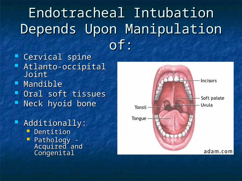
Endotracheal Intubation Endotracheal Intubation Depends Upon Manipulation of:Depends Upon Manipulation of: Cervical spineCervical spine Atlanto-occipital JointAtlanto-occipital Joint MandibleMandible Oral soft tissuesOral soft tissues Neck hyoid boneNeck hyoid bone
Additionally:Additionally: DentitionDentition Pathology - Acquired Pathology - Acquired
and Congenitaland Congenital
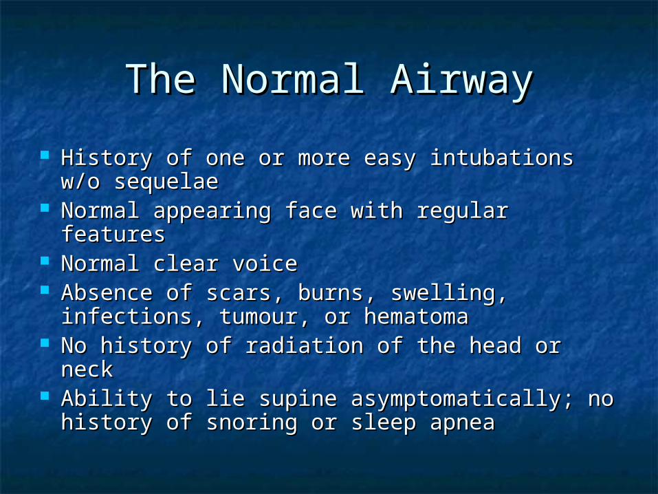
The Normal AirwayThe Normal Airway
History of one or more easy intubations w/o History of one or more easy intubations w/o sequelaesequelae
Normal appearing face with regular featuresNormal appearing face with regular features Normal clear voiceNormal clear voice Absence of scars, burns, swelling, Absence of scars, burns, swelling,
infections, tumour, or hematomainfections, tumour, or hematoma No history of radiation of the head or neckNo history of radiation of the head or neck Ability to lie supine asymptomatically; no Ability to lie supine asymptomatically; no
history of snoring or sleep apneahistory of snoring or sleep apnea

The Normal AirwayThe Normal Airway Patent naresPatent nares Ability to open mouth widely Ability to open mouth widely
with TMJ rotation and with TMJ rotation and subluxation (3 – 4 cm or two subluxation (3 – 4 cm or two finger breaths)finger breaths)
Mallampati Class IMallampati Class I Patient sitting straight up, Patient sitting straight up,
opening mouth as wide as opening mouth as wide as possible, with protruding possible, with protruding tongue; the uvula, posterior tongue; the uvula, posterior pharyngeal wall, entire pharyngeal wall, entire tonsillar pillars, and fauces can tonsillar pillars, and fauces can be seenbe seen
At least 6 cm (3 finger At least 6 cm (3 finger breaths) from tip of mandible breaths) from tip of mandible to thyroid notch with neck to thyroid notch with neck extensionextension
At least 9 cm from symphysis At least 9 cm from symphysis of mandible to mandible angleof mandible to mandible angle
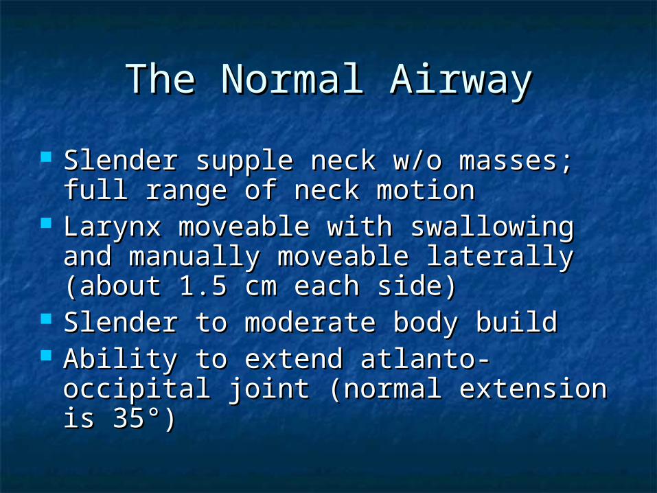
Slender supple neck w/o masses; full Slender supple neck w/o masses; full range of neck motionrange of neck motion
Larynx moveable with swallowing Larynx moveable with swallowing and manually moveable laterally and manually moveable laterally (about 1.5 cm each side)(about 1.5 cm each side)
Slender to moderate body buildSlender to moderate body build Ability to extend atlanto-occipital Ability to extend atlanto-occipital
joint (normal extension is 35joint (normal extension is 35°)°)
The Normal AirwayThe Normal Airway
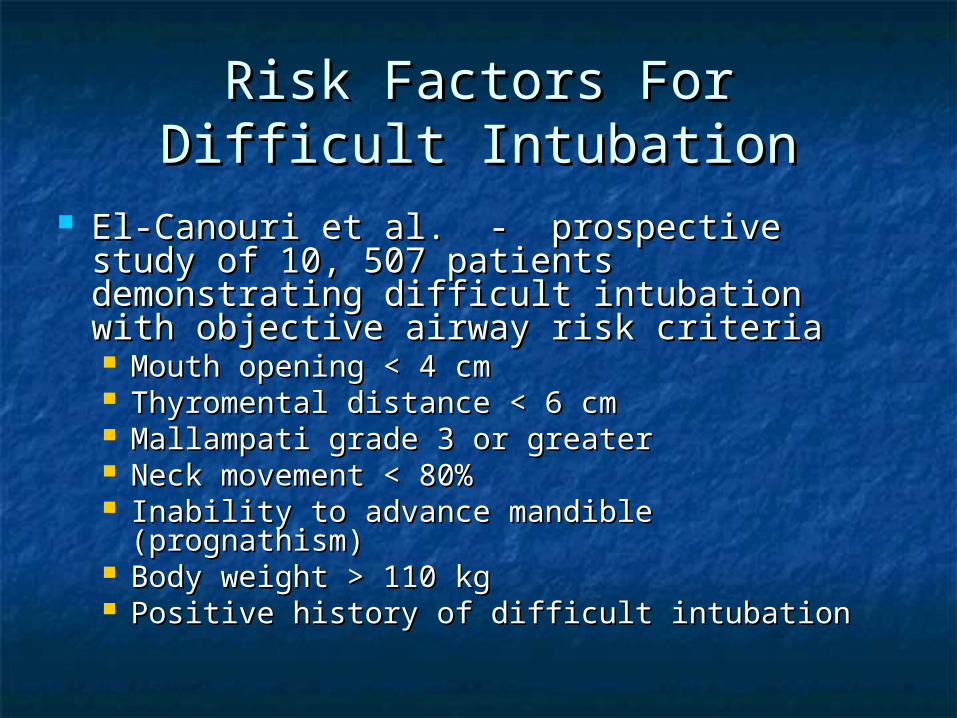
Risk Factors For Difficult Risk Factors For Difficult IntubationIntubation
El-Canouri et al. - prospective study of El-Canouri et al. - prospective study of 10, 507 patients demonstrating difficult 10, 507 patients demonstrating difficult intubation with objective airway risk intubation with objective airway risk criteriacriteria Mouth opening < 4 cmMouth opening < 4 cm Thyromental distance < 6 cmThyromental distance < 6 cm Mallampati grade 3 or greaterMallampati grade 3 or greater Neck movement < 80%Neck movement < 80% Inability to advance mandible (prognathism)Inability to advance mandible (prognathism) Body weight > 110 kg Body weight > 110 kg Positive history of difficult intubationPositive history of difficult intubation
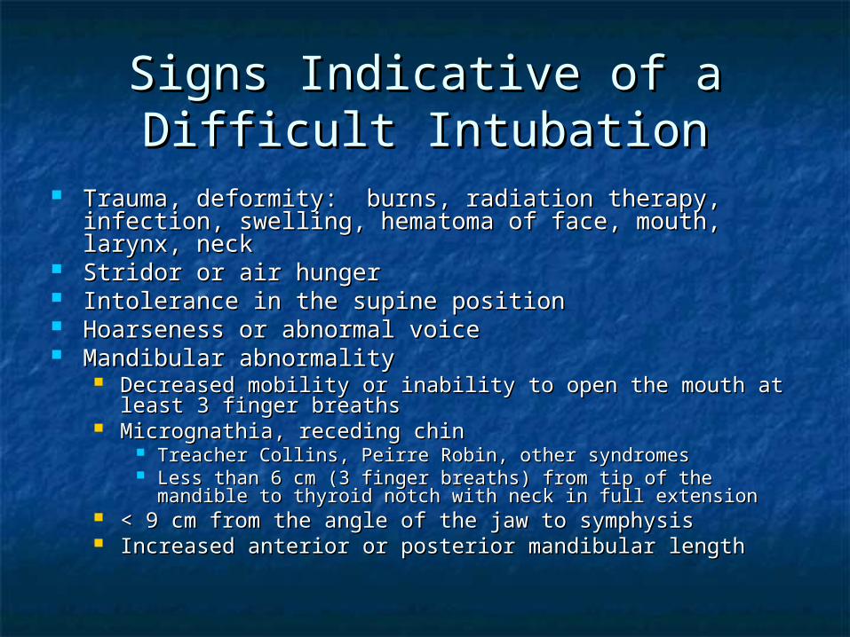
Signs Indicative of a Difficult Signs Indicative of a Difficult IntubationIntubation
Trauma, deformity: burns, radiation therapy, infection, Trauma, deformity: burns, radiation therapy, infection, swelling, hematoma of face, mouth, larynx, neckswelling, hematoma of face, mouth, larynx, neck
Stridor or air hungerStridor or air hunger Intolerance in the supine positionIntolerance in the supine position Hoarseness or abnormal voiceHoarseness or abnormal voice Mandibular abnormalityMandibular abnormality
Decreased mobility or inability to open the mouth at least 3 Decreased mobility or inability to open the mouth at least 3 finger breathsfinger breaths
Micrognathia, receding chinMicrognathia, receding chin Treacher Collins, Peirre Robin, other syndromesTreacher Collins, Peirre Robin, other syndromes Less than 6 cm (3 finger breaths) from tip of the mandible to Less than 6 cm (3 finger breaths) from tip of the mandible to
thyroid notch with neck in full extensionthyroid notch with neck in full extension < 9 cm from the angle of the jaw to symphysis< 9 cm from the angle of the jaw to symphysis Increased anterior or posterior mandibular lengthIncreased anterior or posterior mandibular length
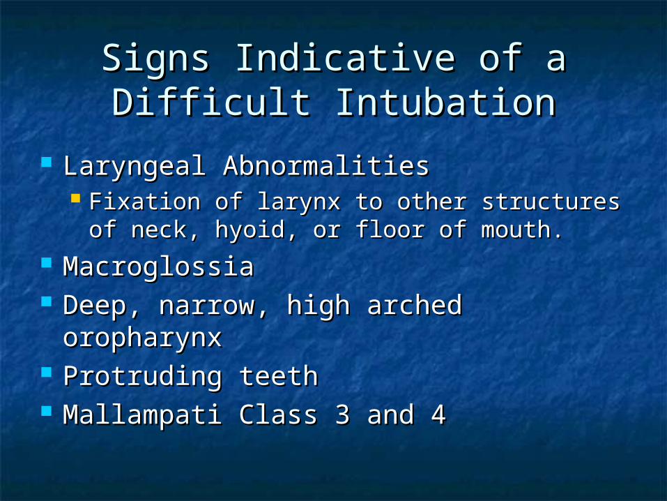
Laryngeal AbnormalitiesLaryngeal Abnormalities Fixation of larynx to other structures of Fixation of larynx to other structures of
neck, hyoid, or floor of mouth.neck, hyoid, or floor of mouth. MacroglossiaMacroglossia Deep, narrow, high arched oropharynxDeep, narrow, high arched oropharynx Protruding teethProtruding teeth Mallampati Class 3 and 4Mallampati Class 3 and 4
Signs Indicative of a Difficult Signs Indicative of a Difficult IntubationIntubation

Neck AbnormalitiesNeck Abnormalities Short and thickShort and thick Decreased range of motion (arthritis, spondylitis, Decreased range of motion (arthritis, spondylitis,
disk disease)disk disease) Fracture (subluxation)Fracture (subluxation) TraumaTrauma
Thoracoabdominal abnormalitiesThoracoabdominal abnormalities KyphoscoliosisKyphoscoliosis Prominent chest or large breastsProminent chest or large breasts Morbid obesityMorbid obesity Term or near term pregnancyTerm or near term pregnancy
Age 50 – 59Age 50 – 59 Male genderMale gender
Signs Indicative of a Difficult Signs Indicative of a Difficult IntubationIntubation
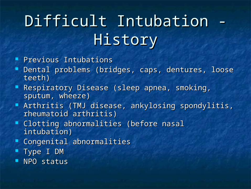
Previous IntubationsPrevious Intubations Dental problems (bridges, caps, dentures, loose Dental problems (bridges, caps, dentures, loose
teeth)teeth) Respiratory Disease (sleep apnea, smoking, Respiratory Disease (sleep apnea, smoking,
sputum, wheeze)sputum, wheeze) Arthritis (TMJ disease, ankylosing spondylitis, Arthritis (TMJ disease, ankylosing spondylitis,
rheumatoid arthritis)rheumatoid arthritis) Clotting abnormalities (before nasal intubation)Clotting abnormalities (before nasal intubation) Congenital abnormalitiesCongenital abnormalities Type I DMType I DM NPO statusNPO status
Difficult Intubation - HistoryDifficult Intubation - History
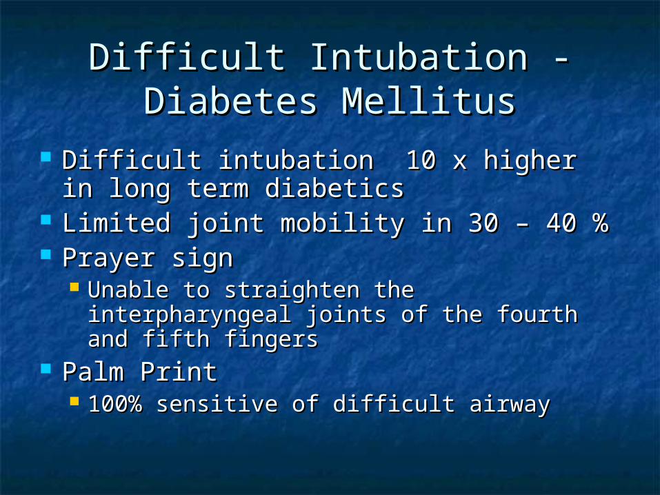
Difficult Intubation - Diabetes Difficult Intubation - Diabetes MellitusMellitus
Difficult intubation 10 x higher in Difficult intubation 10 x higher in long term diabeticslong term diabetics
Limited joint mobility in 30 – 40 %Limited joint mobility in 30 – 40 % Prayer sign Prayer sign
Unable to straighten the interpharyngeal Unable to straighten the interpharyngeal joints of the fourth and fifth fingersjoints of the fourth and fifth fingers
Palm Print Palm Print 100% sensitive of difficult airway100% sensitive of difficult airway
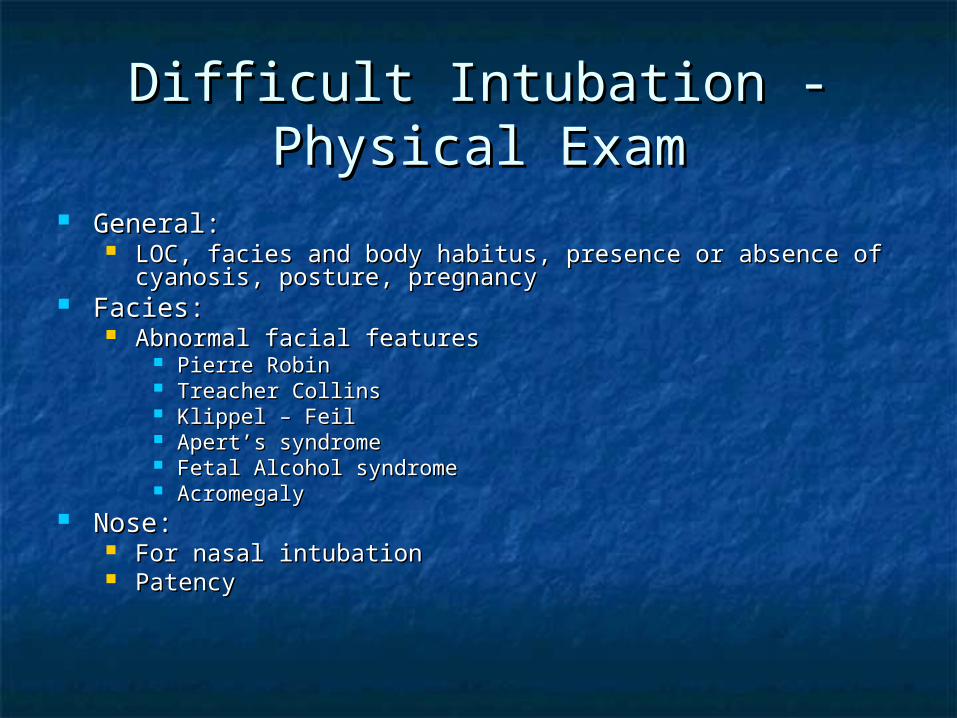
Difficult Intubation - Physical Difficult Intubation - Physical ExamExam
General:General: LOC, facies and body habitus, presence or absence of cyanosis, LOC, facies and body habitus, presence or absence of cyanosis,
posture, pregnancyposture, pregnancy Facies:Facies:
Abnormal facial featuresAbnormal facial features Pierre RobinPierre Robin Treacher CollinsTreacher Collins Klippel – FeilKlippel – Feil Apert’s syndromeApert’s syndrome Fetal Alcohol syndromeFetal Alcohol syndrome AcromegalyAcromegaly
Nose:Nose: For nasal intubationFor nasal intubation PatencyPatency
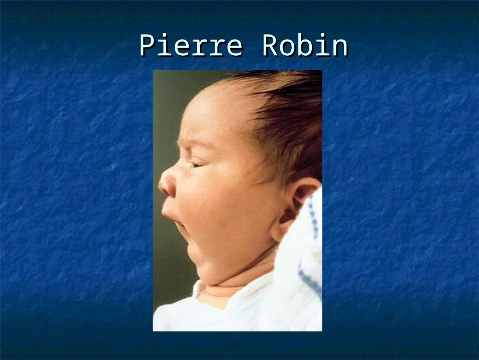
Pierre RobinPierre Robin
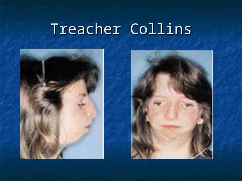
Treacher CollinsTreacher Collins
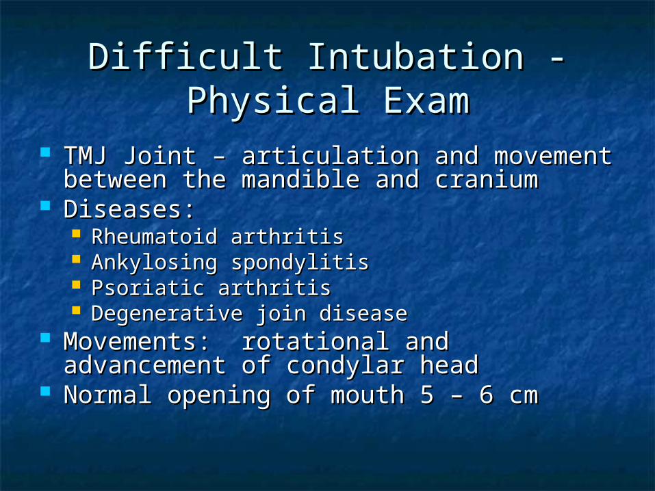
TMJ Joint – articulation and movement TMJ Joint – articulation and movement between the mandible and craniumbetween the mandible and cranium
Diseases:Diseases: Rheumatoid arthritisRheumatoid arthritis Ankylosing spondylitisAnkylosing spondylitis Psoriatic arthritisPsoriatic arthritis Degenerative join diseaseDegenerative join disease
Movements: rotational and advancement Movements: rotational and advancement of condylar headof condylar head
Normal opening of mouth 5 – 6 cmNormal opening of mouth 5 – 6 cm
Difficult Intubation - Physical Difficult Intubation - Physical ExamExam
Difficult Intubation - Physical Difficult Intubation - Physical ExamExam
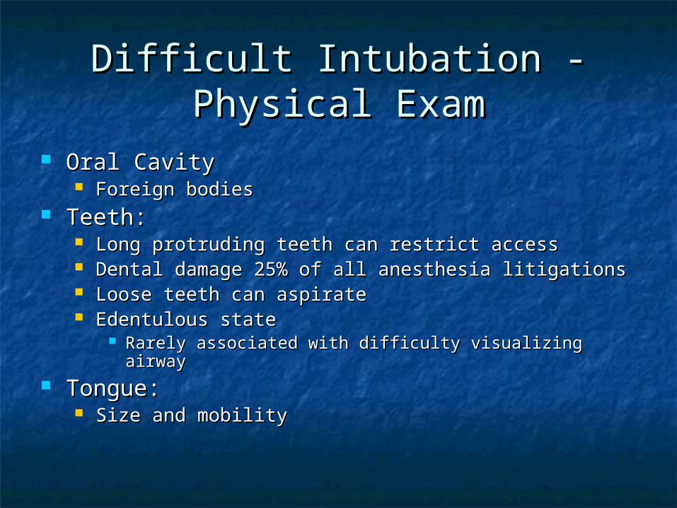
Oral Cavity Oral Cavity Foreign bodiesForeign bodies
Teeth:Teeth: Long protruding teeth can restrict accessLong protruding teeth can restrict access Dental damage 25% of all anesthesia litigationsDental damage 25% of all anesthesia litigations Loose teeth can aspirateLoose teeth can aspirate Edentulous stateEdentulous state
Rarely associated with difficulty visualizing airwayRarely associated with difficulty visualizing airway Tongue:Tongue:
Size and mobilitySize and mobility
Mallampati ClassificationMallampati Classification
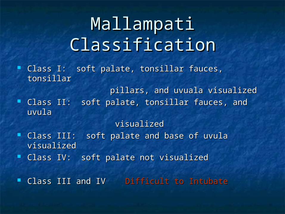
Class I: soft palate, tonsillar fauces, tonsillarClass I: soft palate, tonsillar fauces, tonsillar
pillars, and uvuala visualizedpillars, and uvuala visualized Class II: soft palate, tonsillar fauces, and uvula Class II: soft palate, tonsillar fauces, and uvula
visualizedvisualized Class III: soft palate and base of uvula Class III: soft palate and base of uvula
visualizedvisualized Class IV: soft palate not visualizedClass IV: soft palate not visualized
Class III and IV Class III and IV Difficult to IntubateDifficult to Intubate

Mallampati ClassificationMallampati Classification
Structured Approach to Structured Approach to Airway ManagementAirway Management
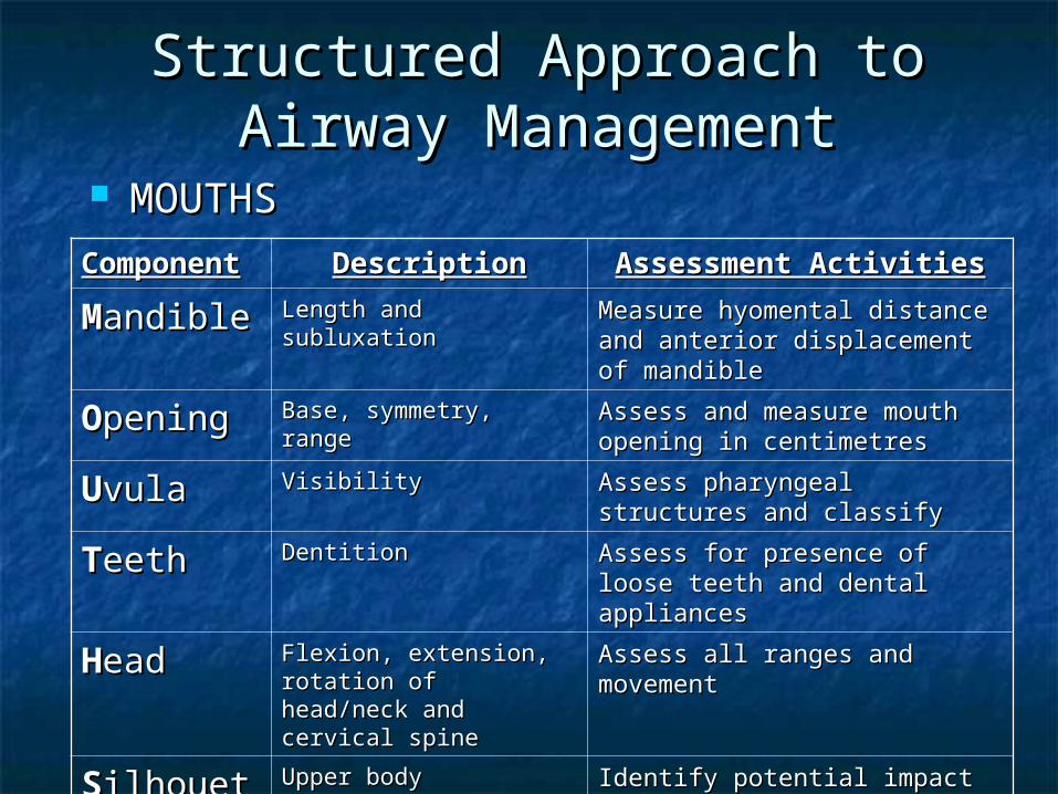
MOUTHSMOUTHS
ComponenComponentt
DescriptionDescription Assessment ActivitiesAssessment Activities
MMandibleandible Length and subluxationLength and subluxation Measure hyomental distance Measure hyomental distance and anterior displacement of and anterior displacement of mandiblemandible
OOpeningpening Base, symmetry, rangeBase, symmetry, range Assess and measure mouth Assess and measure mouth opening in centimetresopening in centimetres
UUvulavula VisibilityVisibility Assess pharyngeal structures Assess pharyngeal structures and classifyand classify
TTeetheeth DentitionDentition Assess for presence of loose Assess for presence of loose teeth and dental appliancesteeth and dental appliances
HHeadead Flexion, extension, Flexion, extension, rotation of head/neck rotation of head/neck and cervical spineand cervical spine
Assess all ranges and Assess all ranges and movementmovement
SSilhouettilhouettee
Upper body Upper body abnormalities, both abnormalities, both anterior and posterioranterior and posterior
Identify potential impact on Identify potential impact on control of airway of large control of airway of large breasts, buffalo hump, breasts, buffalo hump, kyphosis, etc.kyphosis, etc.

Bag/Valve/Mask VentilationBag/Valve/Mask Ventilation
Always need to anticipate difficult mask ventilationAlways need to anticipate difficult mask ventilation Langeron et al. 1502 patients reported a 5% incidence of Langeron et al. 1502 patients reported a 5% incidence of
difficult mask ventilationdifficult mask ventilation 5 independent risk factors of difficult mask ventilation:5 independent risk factors of difficult mask ventilation:
BeardBeard BMI > 26BMI > 26 EdentulousEdentulous Age > 55 years of ageAge > 55 years of age History of snoring (obstruction)History of snoring (obstruction)
Two of these predictors of DMVTwo of these predictors of DMV Sensitivity and specificity > 70%Sensitivity and specificity > 70%
DMV Difficult Intubation in 30% of casesDMV Difficult Intubation in 30% of cases

Intubation TechniqueIntubation Technique Preparation:Preparation:
Equipment CheckEquipment Check 100% oxygen at high flows (> 10 Lpm) 100% oxygen at high flows (> 10 Lpm)
during bask/mask ventilationduring bask/mask ventilation Suction apparatusSuction apparatus Intubation trayIntubation tray
Two laryngoscopic handles and bladesTwo laryngoscopic handles and blades AirwaysAirways ET tubesET tubes Needles and syringesNeedles and syringes StyletStylet KY JellyKY Jelly Suction YankauerSuction Yankauer Magill ForcepsMagill Forceps LMA’sLMA’s

Pre - oxygenationPre - oxygenation
Traditional:Traditional: 3 minutes of tidal volume breathing at 5 ml/kg 3 minutes of tidal volume breathing at 5 ml/kg
100% O100% O22
RapidRapid 8 deep breaths within 60 seconds at 10 L/min8 deep breaths within 60 seconds at 10 L/min
Always ensure pulse oximetry on Always ensure pulse oximetry on patientpatient
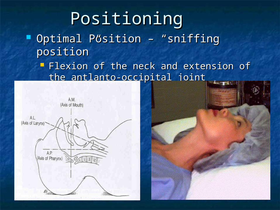
PositioningPositioning Optimal Position – “sniffing position”Optimal Position – “sniffing position”
Flexion of the neck and extension of the Flexion of the neck and extension of the antlanto-occipital jointantlanto-occipital joint
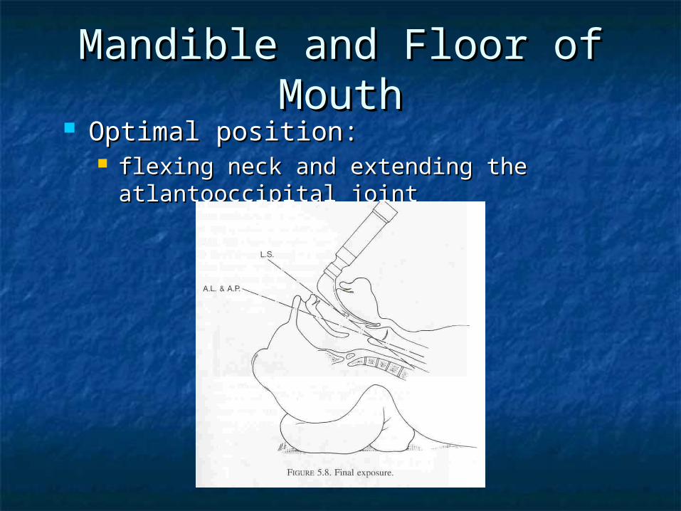
Mandible and Floor of Mandible and Floor of MouthMouth
Optimal position:Optimal position: flexing neck and extending the flexing neck and extending the
atlantooccipital jointatlantooccipital joint
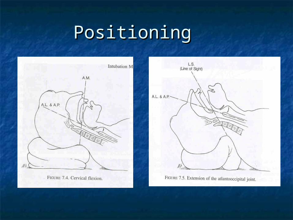
PositioningPositioning
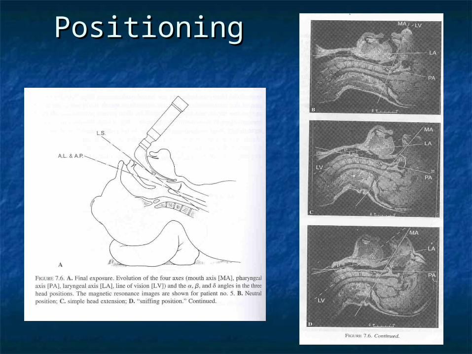
PositioningPositioning

Factors that Interfere with Factors that Interfere with AlignmentAlignment
Large teeth or Large teeth or tethered tonguetethered tongue
Short mandibleShort mandible Protruding upper Protruding upper
incisorsincisors Pathology in floor Pathology in floor
of mouthof mouth Reduced size of Reduced size of
intra and sub intra and sub mandibular spacemandibular space
Practical Note: Thyromental distance 6 cm or 3 finger breaths should show Normal mandible
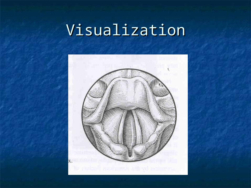
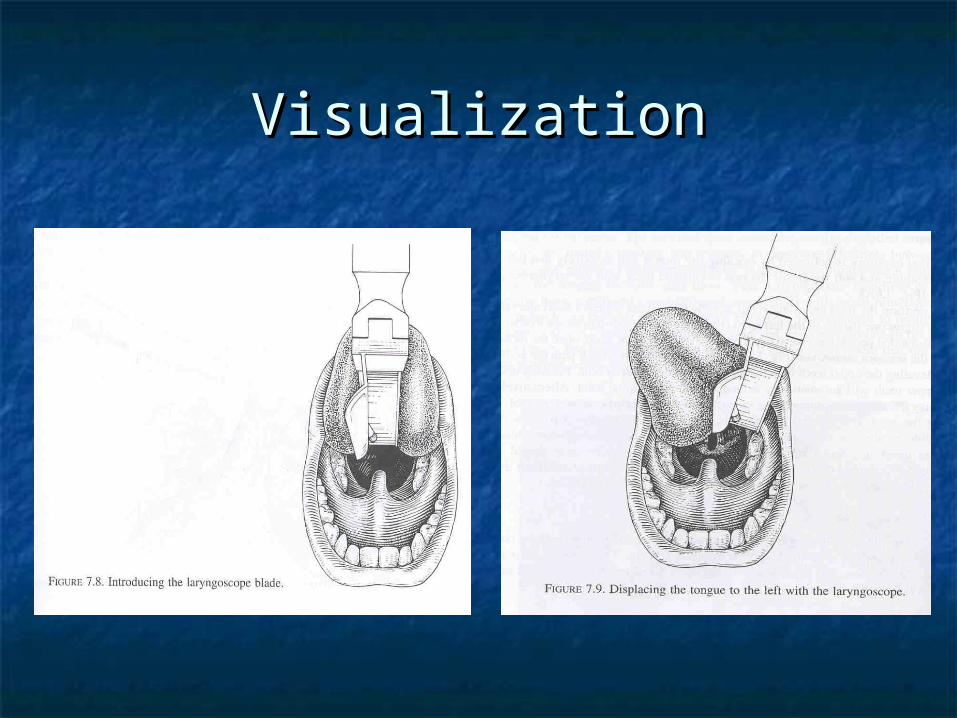
VisualizationVisualization
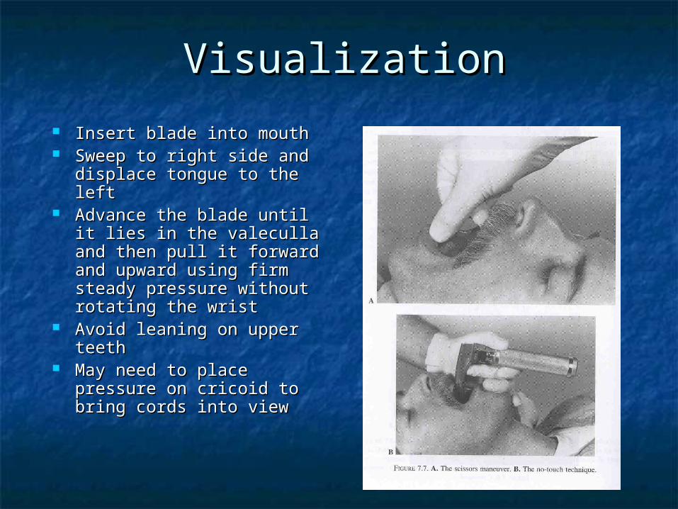
VisualizationVisualization
Insert blade into mouthInsert blade into mouth Sweep to right side and Sweep to right side and
displace tongue to the leftdisplace tongue to the left Advance the blade until it Advance the blade until it
lies in the valeculla and lies in the valeculla and then pull it forward and then pull it forward and upward using firm steady upward using firm steady pressure without rotating pressure without rotating the wristthe wrist
Avoid leaning on upper Avoid leaning on upper teethteeth
May need to place May need to place pressure on cricoid to pressure on cricoid to bring cords into viewbring cords into view
VisualizationVisualization

VisualizationVisualization
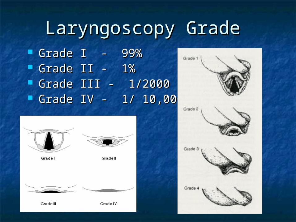
Laryngoscopy Grade Laryngoscopy Grade Grade I - 99%Grade I - 99% Grade II - 1%Grade II - 1% Grade III - 1/2000Grade III - 1/2000 Grade IV - 1/ 10,000Grade IV - 1/ 10,000
InsertionInsertion
Insert cuff to Insert cuff to ~ 3 cm beyond cords~ 3 cm beyond cords Tendency to advance cuff too farTendency to advance cuff too far
Right mainstem intubationRight mainstem intubation
Cuff InflationCuff Inflation Inflate to 20 cm HInflate to 20 cm H22OO Listen for leak at patients mouthListen for leak at patients mouth Over inflation can lead to ischemia of tracheaOver inflation can lead to ischemia of trachea

Confirmation ETT PositionConfirmation ETT Position Continuous COContinuous CO22 monitoring or capnometry monitoring or capnometry
Gold standardGold standard Must have at least 3 continuous readings Must have at least 3 continuous readings
without declining COwithout declining CO22
False Negative ResultsFalse Negative Results
Tube in Trachea, Capnogram Suggests Tube in Trachea, Capnogram Suggests Tube in EsophagusTube in Esophagus Concurrent PEEP with ETT cuff leakConcurrent PEEP with ETT cuff leak Severe Airway obstructionSevere Airway obstruction Low Cardiac OutputLow Cardiac Output Severe hypotensionSevere hypotension Pulmonary embolusPulmonary embolus Advanced pulmonary diseaseAdvanced pulmonary disease
False Positive ResultsFalse Positive Results
Tube Tube NOTNOT in trachea, capnogram in trachea, capnogram suggests tube in tracheasuggests tube in trachea Bag/valve/mask ventilation prior to Bag/valve/mask ventilation prior to
intubationintubation Antacids in stomachAntacids in stomach Recent ingestion of carbonated Recent ingestion of carbonated
beveragesbeverages Tube in pharynxTube in pharynx
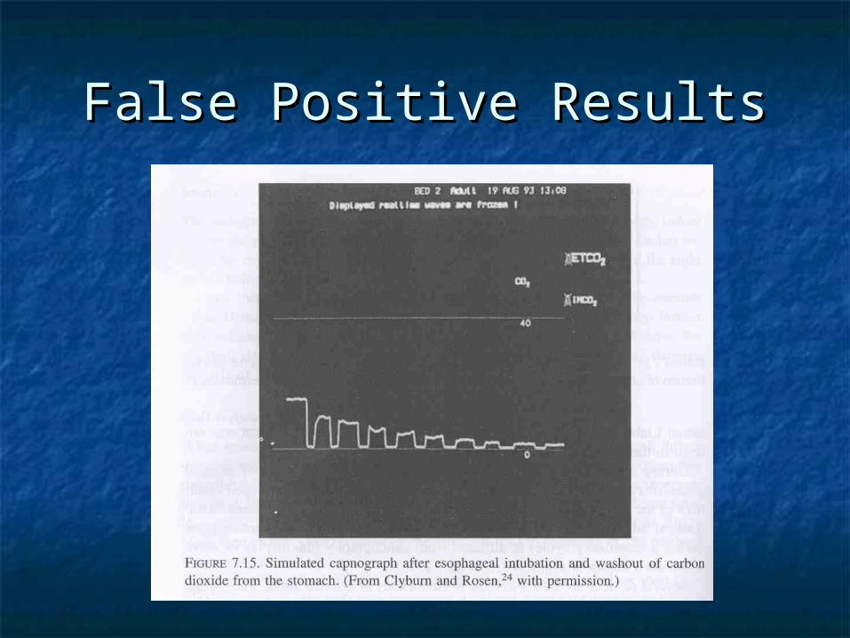
False Positive ResultsFalse Positive Results
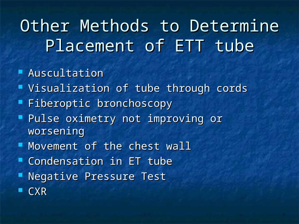
Other Methods to Determine Other Methods to Determine Placement of ETT tubePlacement of ETT tube
AuscultationAuscultation Visualization of tube through cordsVisualization of tube through cords Fiberoptic bronchoscopyFiberoptic bronchoscopy Pulse oximetry not improving or worseningPulse oximetry not improving or worsening Movement of the chest wallMovement of the chest wall Condensation in ET tubeCondensation in ET tube Negative Pressure TestNegative Pressure Test CXRCXR
Airway ManeuversAirway Maneuvers
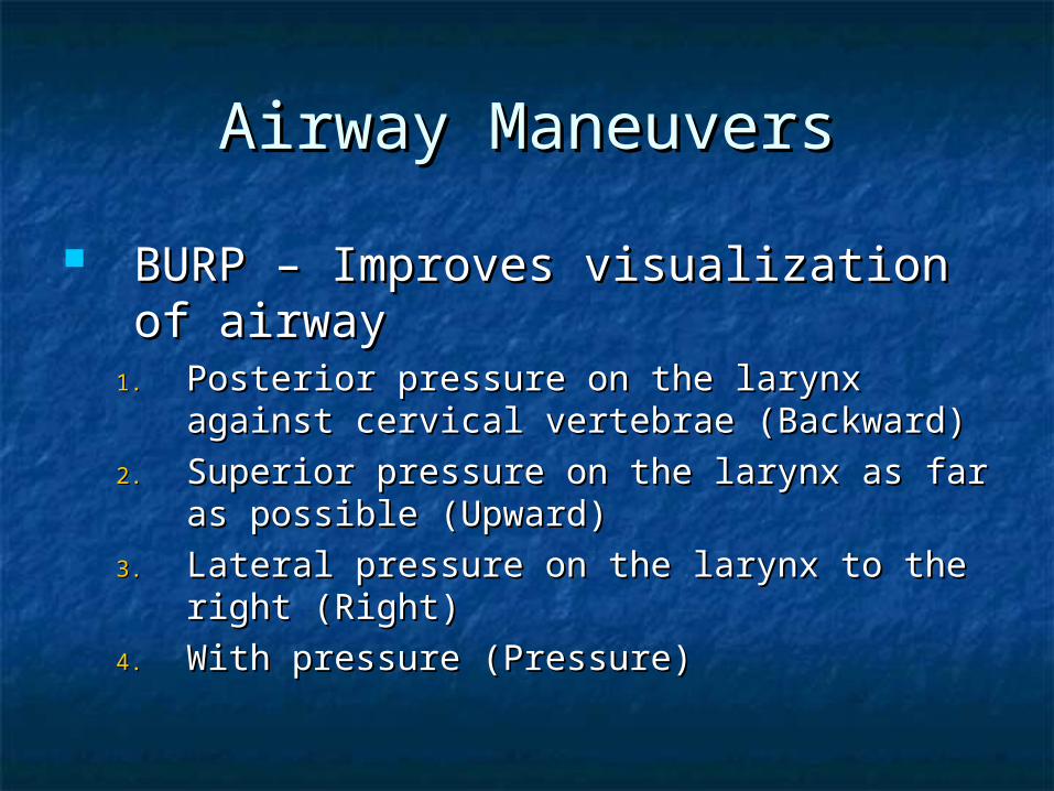
BURP – Improves visualization of BURP – Improves visualization of airwayairway
1.1. Posterior pressure on the larynx against Posterior pressure on the larynx against cervical vertebrae (Backward)cervical vertebrae (Backward)
2.2. Superior pressure on the larynx as far as Superior pressure on the larynx as far as possible (Upward)possible (Upward)
3.3. Lateral pressure on the larynx to the right Lateral pressure on the larynx to the right (Right)(Right)
4.4. With pressure (Pressure)With pressure (Pressure)
Causes of Failed IntubationCauses of Failed Intubation
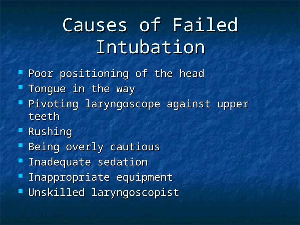
Poor positioning of the headPoor positioning of the head Tongue in the wayTongue in the way Pivoting laryngoscope against upper teethPivoting laryngoscope against upper teeth RushingRushing Being overly cautiousBeing overly cautious Inadequate sedationInadequate sedation Inappropriate equipmentInappropriate equipment Unskilled laryngoscopistUnskilled laryngoscopist
SummarySummary
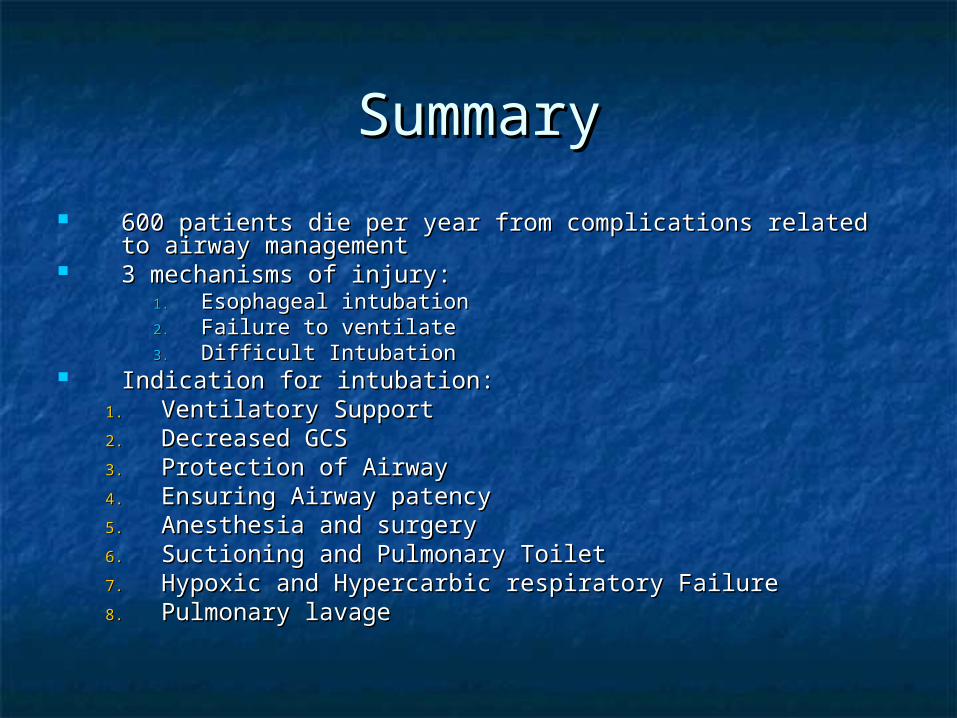
600 patients die per year from complications related to airway 600 patients die per year from complications related to airway managementmanagement
3 mechanisms of injury:3 mechanisms of injury:1.1. Esophageal intubationEsophageal intubation2.2. Failure to ventilateFailure to ventilate3.3. Difficult IntubationDifficult Intubation
Indication for intubation:Indication for intubation:1.1. Ventilatory SupportVentilatory Support2.2. Decreased GCSDecreased GCS3.3. Protection of AirwayProtection of Airway4.4. Ensuring Airway patencyEnsuring Airway patency5.5. Anesthesia and surgeryAnesthesia and surgery6.6. Suctioning and Pulmonary ToiletSuctioning and Pulmonary Toilet7.7. Hypoxic and Hypercarbic respiratory FailureHypoxic and Hypercarbic respiratory Failure8.8. Pulmonary lavagePulmonary lavage
Massive HemoptysisMassive Hemoptysis
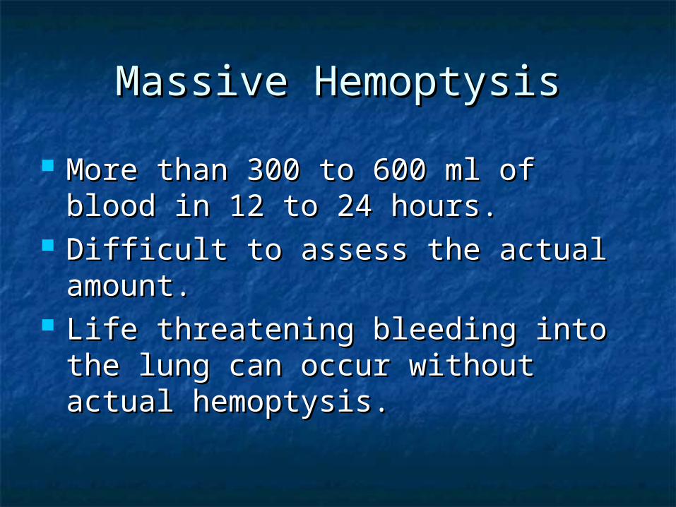
Massive HemoptysisMassive Hemoptysis
More than 300 to 600 ml of blood in More than 300 to 600 ml of blood in 12 to 24 hours.12 to 24 hours.
Difficult to assess the actual amount.Difficult to assess the actual amount. Life threatening bleeding into the Life threatening bleeding into the
lung can occur without actual lung can occur without actual hemoptysis.hemoptysis.
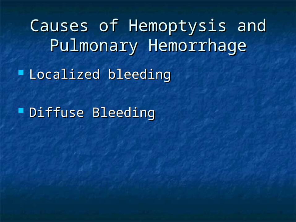
Causes of Hemoptysis and Causes of Hemoptysis and Pulmonary HemorrhagePulmonary Hemorrhage
Localized bleedingLocalized bleeding
Diffuse BleedingDiffuse Bleeding
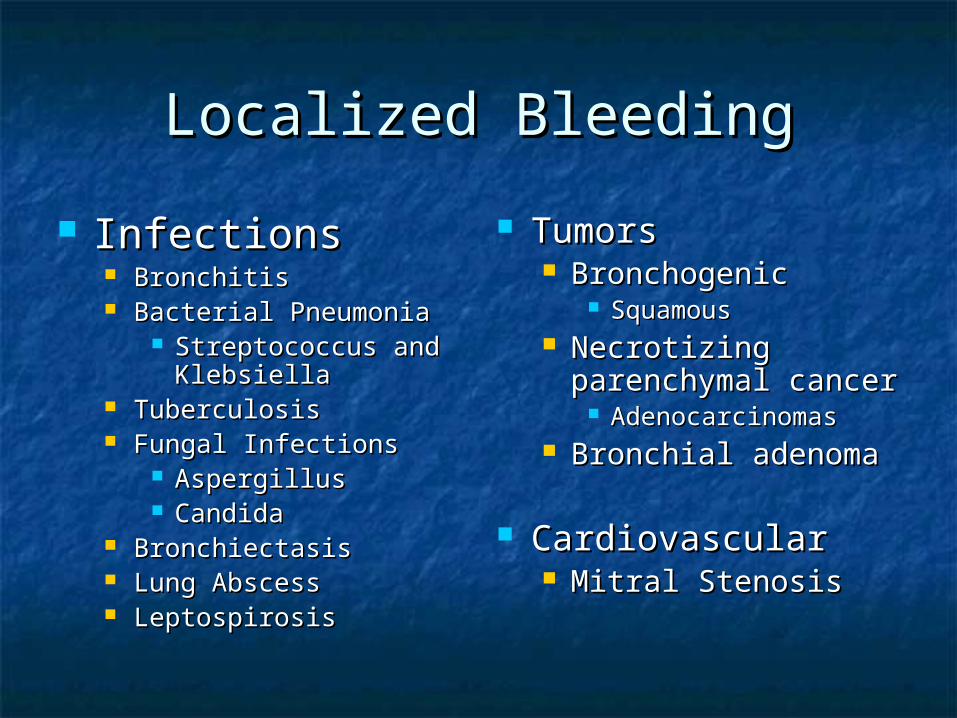
Localized BleedingLocalized Bleeding
InfectionsInfections BronchitisBronchitis Bacterial PneumoniaBacterial Pneumonia
Streptococcus and Streptococcus and KlebsiellaKlebsiella
TuberculosisTuberculosis Fungal InfectionsFungal Infections
AspergillusAspergillus CandidaCandida
BronchiectasisBronchiectasis Lung AbscessLung Abscess LeptospirosisLeptospirosis
TumorsTumors BronchogenicBronchogenic
SquamousSquamous Necrotizing Necrotizing
parenchymal cancerparenchymal cancer AdenocarcinomasAdenocarcinomas
Bronchial adenomaBronchial adenoma
CardiovascularCardiovascular Mitral StenosisMitral Stenosis
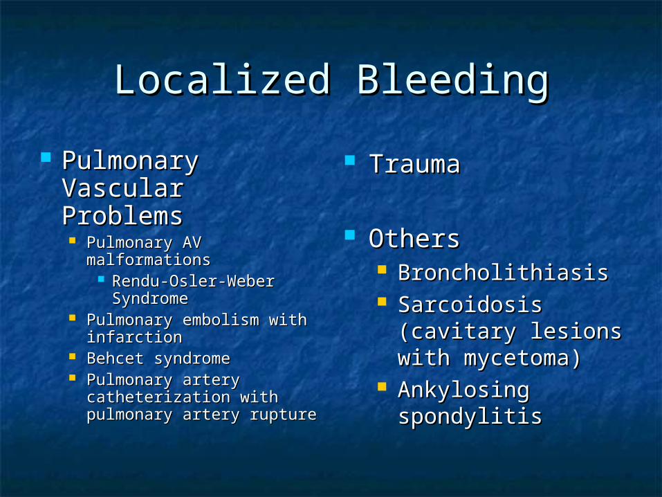
Localized BleedingLocalized Bleeding
Pulmonary Pulmonary Vascular Vascular ProblemsProblems Pulmonary AV Pulmonary AV
malformationsmalformations Rendu-Osler-Weber Rendu-Osler-Weber
SyndromeSyndrome Pulmonary embolism with Pulmonary embolism with
infarctioninfarction Behcet syndromeBehcet syndrome Pulmonary artery Pulmonary artery
catheterization with catheterization with pulmonary artery rupturepulmonary artery rupture
TraumaTrauma
OthersOthers BroncholithiasisBroncholithiasis Sarcoidosis Sarcoidosis
(cavitary lesions (cavitary lesions with mycetoma)with mycetoma)
Ankylosing Ankylosing spondylitisspondylitis
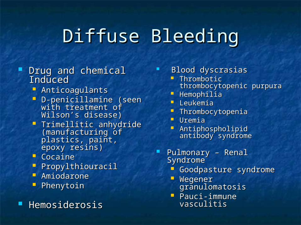
Diffuse BleedingDiffuse Bleeding
Drug and chemical Drug and chemical InducedInduced AnticoagulantsAnticoagulants D-penicillamine (seen D-penicillamine (seen
with treatment of with treatment of Wilson’s disease)Wilson’s disease)
Trimellitic anhydride Trimellitic anhydride (manufacturing of (manufacturing of plastics, paint, epoxy plastics, paint, epoxy resins)resins)
CocaineCocaine PropylthiouracilPropylthiouracil AmiodaroneAmiodarone PhenytoinPhenytoin
HemosiderosisHemosiderosis
Blood dyscrasiasBlood dyscrasias Thrombotic Thrombotic
thrombocytopenic purpurathrombocytopenic purpura HemophiliaHemophilia LeukemiaLeukemia ThrombocytopeniaThrombocytopenia UremiaUremia Antiphospholipid antibody Antiphospholipid antibody
syndromesyndrome
Pulmonary – Renal Pulmonary – Renal SyndromeSyndrome Goodpasture syndromeGoodpasture syndrome Wegener Wegener
granulomatosisgranulomatosis Pauci-immune vasculitisPauci-immune vasculitis
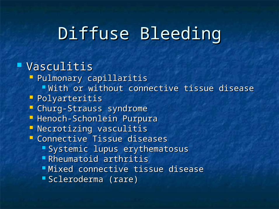
Diffuse BleedingDiffuse Bleeding
VasculitisVasculitis Pulmonary capillaritisPulmonary capillaritis
With or without connective tissue diseaseWith or without connective tissue disease PolyarteritisPolyarteritis Churg-Strauss syndromeChurg-Strauss syndrome Henoch-Schonlein PurpuraHenoch-Schonlein Purpura Necrotizing vasculitisNecrotizing vasculitis Connective Tissue diseasesConnective Tissue diseases
Systemic lupus erythematosusSystemic lupus erythematosus Rheumatoid arthritisRheumatoid arthritis Mixed connective tissue diseaseMixed connective tissue disease Scleroderma (rare)Scleroderma (rare)
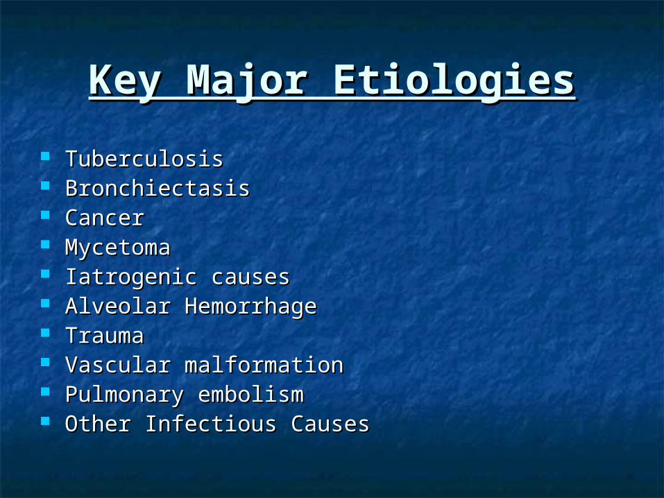
Key Major EtiologiesKey Major Etiologies
TuberculosisTuberculosis BronchiectasisBronchiectasis CancerCancer MycetomaMycetoma Iatrogenic causesIatrogenic causes Alveolar HemorrhageAlveolar Hemorrhage TraumaTrauma Vascular malformationVascular malformation Pulmonary embolismPulmonary embolism Other Infectious CausesOther Infectious Causes
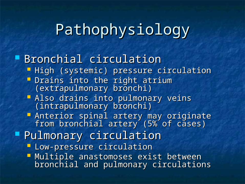
PathophysiologyPathophysiology
Bronchial circulationBronchial circulation High (systemic) pressure circulationHigh (systemic) pressure circulation Drains into the right atrium (extrapulmonary Drains into the right atrium (extrapulmonary
bronchi)bronchi) Also drains into pulmonary veins Also drains into pulmonary veins
(intrapulmonary bronchi)(intrapulmonary bronchi) Anterior spinal artery may originate from Anterior spinal artery may originate from
bronchial artery (5% of cases)bronchial artery (5% of cases) Pulmonary circulationPulmonary circulation
Low-pressure circulationLow-pressure circulation Multiple anastomoses exist between bronchial Multiple anastomoses exist between bronchial
and pulmonary circulationsand pulmonary circulations

Clinical FindingsClinical Findings
Hemoptysis, Dyspnea, Cough, AnxietyHemoptysis, Dyspnea, Cough, Anxiety Fever, weight lossFever, weight loss Smoking and Travel historySmoking and Travel history Bloody sputumBloody sputum
Frothy blood – sputum mixtureFrothy blood – sputum mixture Bright redBright red AlkalineAlkaline
Tachypnea, respiratory distressTachypnea, respiratory distress Localized wheezing, rales, poor dentitionLocalized wheezing, rales, poor dentition Digital clubbingDigital clubbing HematuriaHematuria
Differential DiagnosisDifferential Diagnosis
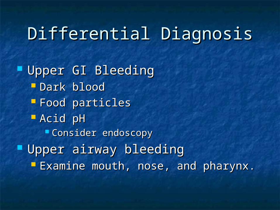
Upper GI BleedingUpper GI Bleeding Dark bloodDark blood Food particlesFood particles Acid pHAcid pH
Consider endoscopyConsider endoscopy
Upper airway bleedingUpper airway bleeding Examine mouth, nose, and pharynx.Examine mouth, nose, and pharynx.

Laboratory TestsLaboratory Tests
No specific testsNo specific tests CBC, diff, INR, PTT, platelet countCBC, diff, INR, PTT, platelet count Electrolytes, BUN, CrElectrolytes, BUN, Cr Sputum culture and AFBSputum culture and AFB UrinalysisUrinalysis ECGECG ABG’sABG’s Type and ScreenType and Screen
Imaging StudiesImaging Studies
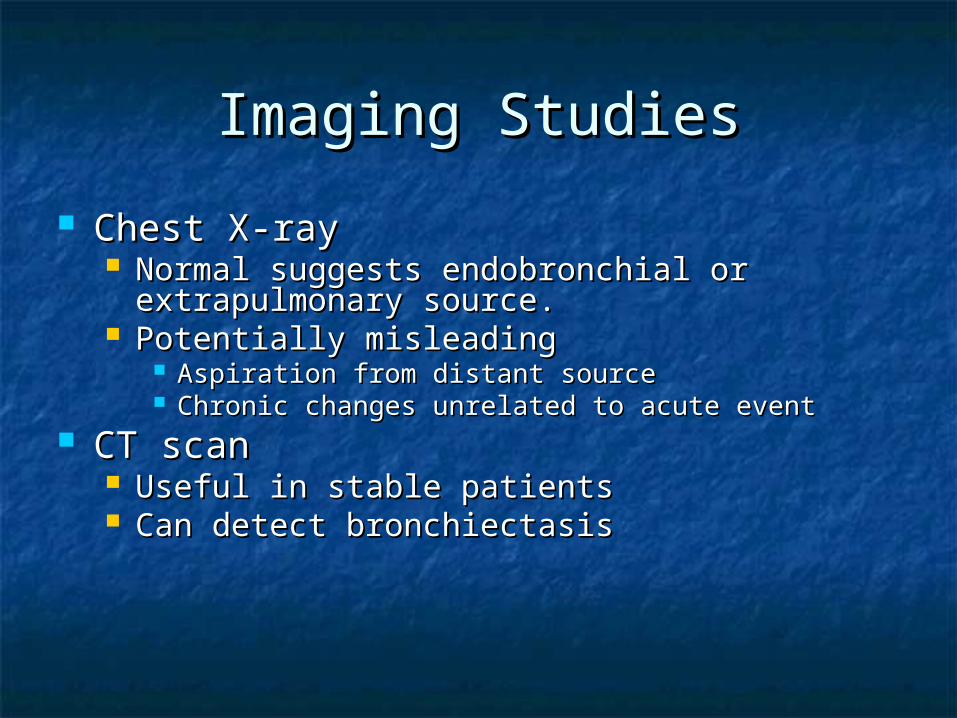
Chest X-rayChest X-ray Normal suggests endobronchial or Normal suggests endobronchial or
extrapulmonary source.extrapulmonary source. Potentially misleadingPotentially misleading
Aspiration from distant sourceAspiration from distant source Chronic changes unrelated to acute eventChronic changes unrelated to acute event
CT scanCT scan Useful in stable patientsUseful in stable patients Can detect bronchiectasisCan detect bronchiectasis
StabilizationStabilization
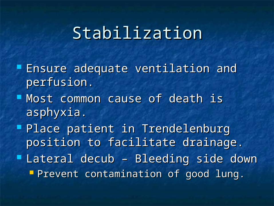
Ensure adequate ventilation and Ensure adequate ventilation and perfusion.perfusion.
Most common cause of death is Most common cause of death is asphyxia.asphyxia.
Place patient in Trendelenburg Place patient in Trendelenburg position to facilitate drainage.position to facilitate drainage.
Lateral decub – Bleeding side downLateral decub – Bleeding side down Prevent contamination of good lung.Prevent contamination of good lung.
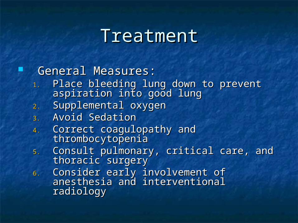
TreatmentTreatment
General Measures:General Measures:1.1. Place bleeding lung down to prevent Place bleeding lung down to prevent
aspiration into good lungaspiration into good lung2.2. Supplemental oxygenSupplemental oxygen3.3. Avoid SedationAvoid Sedation4.4. Correct coagulopathy and thrombocytopeniaCorrect coagulopathy and thrombocytopenia5.5. Consult pulmonary, critical care, and thoracic Consult pulmonary, critical care, and thoracic
surgerysurgery6.6. Consider early involvement of anesthesia Consider early involvement of anesthesia
and interventional radiologyand interventional radiology

Primary Goal is Airway Primary Goal is Airway ControlControl
Asphyxiation, not blood loss, is the cause Asphyxiation, not blood loss, is the cause of death.of death.
Only stable patients with ability to protect Only stable patients with ability to protect and clear their own airway should be and clear their own airway should be managed without intubation.managed without intubation.
Intubation:Intubation: Performed by experienced personnel.Performed by experienced personnel. Large bore tube for bronchoscopy and Large bore tube for bronchoscopy and
suctioning.suctioning. Consider bronchial blocker or double lumen Consider bronchial blocker or double lumen
tube if bleeding site is known.tube if bleeding site is known.
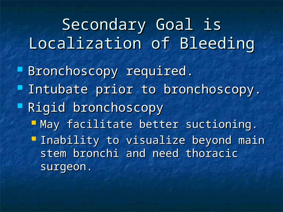
Secondary Goal is Localization Secondary Goal is Localization of Bleedingof Bleeding
Bronchoscopy required.Bronchoscopy required. Intubate prior to bronchoscopy.Intubate prior to bronchoscopy. Rigid bronchoscopyRigid bronchoscopy
May facilitate better suctioning.May facilitate better suctioning. Inability to visualize beyond main stem Inability to visualize beyond main stem
bronchi and need thoracic surgeon.bronchi and need thoracic surgeon.
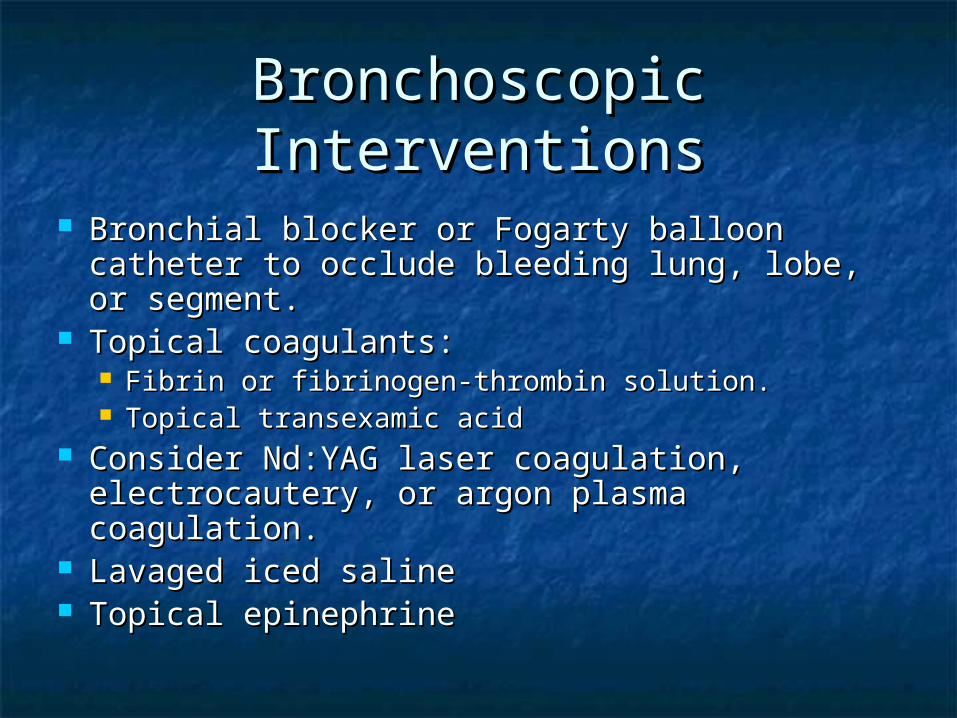
Bronchoscopic InterventionsBronchoscopic Interventions
Bronchial blocker or Fogarty balloon catheter Bronchial blocker or Fogarty balloon catheter to occlude bleeding lung, lobe, or segment.to occlude bleeding lung, lobe, or segment.
Topical coagulants:Topical coagulants: Fibrin or fibrinogen-thrombin solution.Fibrin or fibrinogen-thrombin solution. Topical transexamic acidTopical transexamic acid
Consider Nd:YAG laser coagulation, Consider Nd:YAG laser coagulation, electrocautery, or argon plasma coagulation.electrocautery, or argon plasma coagulation.
Lavaged iced salineLavaged iced saline Topical epinephrineTopical epinephrine
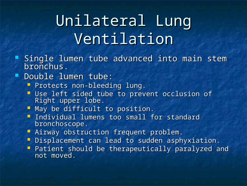
Unilateral Lung VentilationUnilateral Lung Ventilation
Single lumen tube advanced into main stem Single lumen tube advanced into main stem bronchus.bronchus.
Double lumen tube:Double lumen tube: Protects non-bleeding lung.Protects non-bleeding lung. Use left sided tube to prevent occlusion of Right upper Use left sided tube to prevent occlusion of Right upper
lobe.lobe. May be difficult to position.May be difficult to position. Individual lumens too small for standard bronchoscope.Individual lumens too small for standard bronchoscope. Airway obstruction frequent problem.Airway obstruction frequent problem. Displacement can lead to sudden asphyxiation.Displacement can lead to sudden asphyxiation. Patient should be therapeutically paralyzed and not Patient should be therapeutically paralyzed and not
moved.moved.
Bronchial Arteriography and Bronchial Arteriography and EmbolizationEmbolization
Favored initial approach if facilities and expertise Favored initial approach if facilities and expertise available.available.
High success rate: approximately 90% when a High success rate: approximately 90% when a bleeding vessel is identified.bleeding vessel is identified.
Recurrence rate: 10 – 27%Recurrence rate: 10 – 27% 10% of patients bleed from the pulmonary 10% of patients bleed from the pulmonary
circulation (TB or mycetoma).circulation (TB or mycetoma). Serious complications:Serious complications:
Occlusion of the anterior spinal artery with paraplegia.Occlusion of the anterior spinal artery with paraplegia. Embolic infarction of distal organs.Embolic infarction of distal organs.

Early Surgical TreatmentEarly Surgical Treatment
Offers definitive treatment.Offers definitive treatment. Indicated for lateralized massive life-Indicated for lateralized massive life-
threatening hemoptysis, or failure or threatening hemoptysis, or failure or recurrence after other interventions.recurrence after other interventions.
Contraindications:Contraindications: Poor baseline respiratory function.Poor baseline respiratory function. Inoperable lung carcinoma.Inoperable lung carcinoma. Inability to localize bleeding site.Inability to localize bleeding site. Diffuse lung disease (relative) eg. CF.Diffuse lung disease (relative) eg. CF.
Mortality is higher if bleeding is acuteMortality is higher if bleeding is acute
Late Surgical TreatmentLate Surgical Treatment
Indicated for definitive treatment of Indicated for definitive treatment of underlying lesion, once bleeding underlying lesion, once bleeding subsided.subsided.
Indications:Indications: MycetomaMycetoma Resectable carcinomaResectable carcinoma Localized bronchiectasisLocalized bronchiectasis
PrognosisPrognosis
Factors likely affecting outcomeFactors likely affecting outcome Etiology of hemoptysisEtiology of hemoptysis Underlying co-morbid illnessesUnderlying co-morbid illnesses Surgical vs. medical treatmentSurgical vs. medical treatment
MortalityMortality Medical mortality: 17 – 85%Medical mortality: 17 – 85% Estimated early surgical mortality: 0 – 50%Estimated early surgical mortality: 0 – 50% Most case series reports preceded the Most case series reports preceded the
development of angiographic embolization.development of angiographic embolization.
ConclusionConclusion
More than 300 to 600 ml of blood in 12 to More than 300 to 600 ml of blood in 12 to 24 hours.24 hours.
Major causes:Major causes: TuberculosisTuberculosis BronchiectasisBronchiectasis CancerCancer MycetomaMycetoma Iatrogenic causesIatrogenic causes Alveolar HemorrhageAlveolar Hemorrhage TraumaTrauma Vascular malformationVascular malformation Pulmonary embolismPulmonary embolism
Primary goal is airway control followed by Primary goal is airway control followed by bleeding localization.bleeding localization.