Embed Size (px)
Citation preview

TSD for Noncancer RELs March 2016
Appendix D1 Methylene diphenyl diisocyanate
Methylene Diphenyl Diisocyanate (Monomer and Polymeric Forms) Reference Exposure Levels
Technical Support Document for the Derivation of Noncancer Reference Exposure Levels
Appendix D1 Final March 2016
Air, Community, and Environmental Research Branch Office of Environmental Health Hazard Assessment California Environmental Protection Agency
OFFICE OF ENVIRONMENTAL HEALTH HAZARD ASSESSMENT
Air Toxics Hot Spots Program
TSD for Noncancer RELs March 2016
Appendix D1 Methylene diphenyl diisocyanate
Page Intentionally Left Blank

TSD for Noncancer RELs March 2016
Appendix D1 Methylene diphenyl diisocyanate
Methylene Diphenyl Diisocyanate Reference Exposure Levels
(Monomer and Polymeric Forms) Technical Support Document for the Derivation of
Noncancer Reference Exposure Levels Appendix D1
Final
Prepared by the
Office of Environmental Health Hazard Assessment Lauren Zeise, Ph.D., Acting Director
Authors Daryn E. Dodge, Ph.D.
Rona Silva, Ph.D.
Technical Reviewers Lauren Zeise, Ph.D.
David M. Siegel, Ph.D. Melanie A. Marty, Ph.D. James F. Collins, Ph.D
Rajpal Tomar, Ph.D. Marlissa Campbell, Ph.D.
March 2016

TSD for Noncancer RELs March 2016
Appendix D1 Methylene diphenyl diisocyanate
Page Intentionally Left Blank

TSD for Noncancer RELs March 2016
Appendix D1 1 Methylene diphenyl diisocyanate
Methylene Diphenyl Diisocyanate Reference Exposure Levels
(Monomer and Polymeric Forms)
(Diphenylmethane diisocyanate, Methylene bisphenyl diisocyanate,
4,4’-Methylenediphenyl diisocyanate, Diphenylmethane-4,4’-diisocyanate)
CAS: 101-68-8 (Monomer)
1. Summary The Office of Environmental Health Hazard Assessment (OEHHA) is required to develop guidelines for conducting health risk assessments under the Air Toxics Hot Spots Program (Health and Safety Code Section 44360 (b) (2)). In response to this statutory requirement, OEHHA developed a Technical Support Document (TSD) that was adopted in 2008 and describes acute, 8 hour and chronic Reference Exposure Levels (RELs). The TSD presents methodology for deriving Reference Exposure Levels. In particular, the methodology explicitly considers possible differential effects on the health of infants, children and other sensitive subpopulations, in accordance with the mandate of the Children’s Environmental Health Protection Act (Senate Bill 25, Escutia, chapter 731, statutes of 1999, Health and Safety Code Sections 39669.5 et seq.). These guidelines have been used to develop the following RELs for methylene diphenyl diisocyanate; this document will be added to Appendix D of the TSD. Exposure to monomeric methylene diphenyl diisocyanate (MDI) and polymeric MDI (PMDI), has been found to cause adverse effects on the respiratory system in both animals and humans. These effects include, 1) acute impacts such as sensory irritation and respiratory inflammation, 2) sensitization and induction of asthma with repeated exposures, and 3) decrements in lung function without evidence of sensitization with chronic exposure. Once asthma has been induced in sensitized individuals, triggering of asthmatic attacks can occur following very low exposures to MDI or PMDI (≤1 ppb). The RELs are intended to reasonably protect the general population from these health effects resulting from exposure to MDI and PMDI, but may not protect all individuals previously sensitized to MDI or PMDI. The RELs are applicable for both MDI and PMDI due to similar toxicological effects and potencies, and similar regional deposition in the lungs in key studies. Literature summarized and referenced in this document covers the relevant published literature for MDI through Spring 2015.

TSD for Noncancer RELs March 2016
Appendix D1 2 Methylene diphenyl diisocyanate
1.1 MDI/PMDI Acute REL
Reference Exposure Level 12 µg/m3 (1.2 ppb)
Critical effect(s) Increased total protein in bronchoalveolar lavage fluid of rats - marker of pulmonary irritation
Hazard index target(s) Respiratory system
1.2 MDI/PMDI 8-hour REL
Reference Exposure Level 0.16 µg/m3 (0.015 ppb)
Critical effect(s) Bronchiolo-alveolar hyperplasia and pulmonary interstitial fibrosis
Hazard index target(s) Respiratory system
1.3 MDI/PMDI Chronic REL
Reference Exposure Level 0.08 µg/m3 (0.008 ppb)
Critical effect(s) Pulmonary interstitial fibrosis Hazard index target(s) Respiratory system

TSD for Noncancer RELs March 2016
Appendix D1 3 Methylene diphenyl diisocyanate
List of Acronyms AEC Asymptomatic exposed controls AIC Akaike information criterion ACE Angiotensin converting enzyme ANOVA Analysis of variance BALF Bronchoalveolar lavage fluid BMC Benchmark Concentration BMC05 Benchmark concentration
producing a 5% response rate BMCL05 the 95% lower confidence limit of
the dose producing a 5% response rate BMD Benchmark Dose BMDL estimation of the BMD 95% lower
confidence limit DA Diisocyanate-induced asthma DLco Carbon monoxide diffusion test ELISA Enzyme-linked immunosorbent
assay FEF25-75% Forced respiratory flow (25-75%
of forced vital capacity) FEV1 Forced expiratory volume in 1
second FVC Forced vital capacity GSH Glutathione GST glutathione-S-transferase HDI Hexamethylene diisocyanate HEC Human equivalent concentration HLA Human leucocyte antigen HPLC High pressure liquid
chromatography HSA Human serum albumin IPDI Isophorone diisocyanate IgE Immunoglobulin E antibody type IgG Immunoglobulin G antibody type LC50 Median lethal concentration LDH Lactate dehydrogenase LOAEL Lowest observed adverse effect
level LOQ Limit of quantitation
MDA 4,4’-methylenedianiline MDI Methylene diphenyl diisocyanate MMAD Mass median aerodynamic
diameter MMEF Maximum mid-expiratory flow NAG N-acetyl glucosaminidase NAT N-acetyl transferase NDI Naphthylene diisocyanate NOAEL No observed adverse effect level OA Occupational asthma OR Odds Ratio PD20 Provocation dose of methacholine
(in mg) to cause a 20% drop in FEV1 PEFR Peak expiratory flow rate PEL Permissible exposure limit PMDI Polymeric methylene diphenyl
diisocyanate PMN Neutrophilic granulocytes POD Point of departure ppb Parts per billion ppm Parts per million RADS Reactive airways dysfunction
syndrome RAST Radioallergosorbent test RDDR Regional deposited dose ratio REL Reference exposure level RGDR Regional gas deposition ratio SNP Single nucleotide polymorphism TAC Toxic air contaminant TDI Toluene diisocyanate TLV Threshold limit value TRPA transient receptor potential A TSD Technical support document TWA Time-weighted average UF Uncertainty factor VC Vital capacity VOC Volatile organic compound
TSD for Noncancer RELs March 2016
Appendix D1 4 Methylene diphenyl diisocyanate
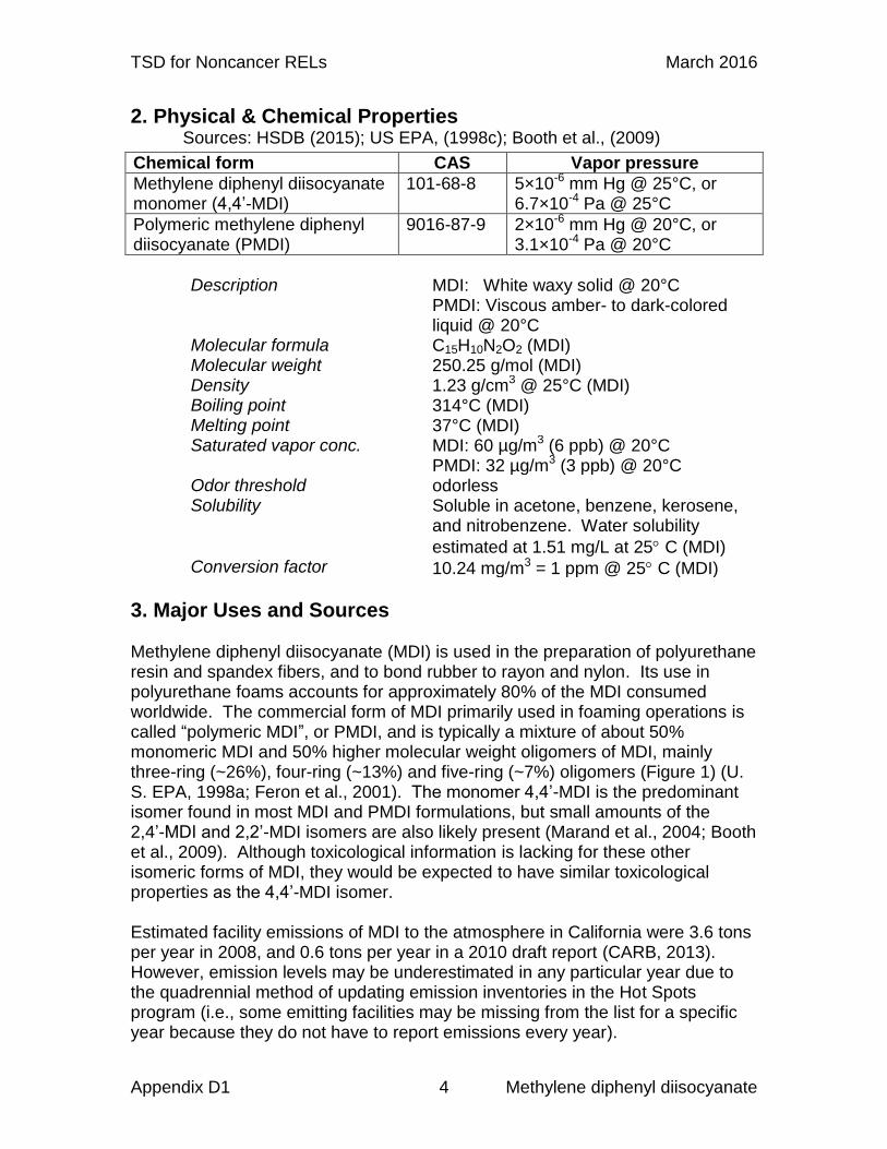
2. Physical & Chemical Properties Sources: HSDB (2015); US EPA, (1998c); Booth et al., (2009)
Chemical form CAS Vapor pressure
Methylene diphenyl diisocyanate monomer (4,4’-MDI)
101-68-8 5×10⁻⁶ mm Hg @ 25°C, or 6.7×10⁻⁴ Pa @ 25°C
Polymeric methylene diphenyl diisocyanate (PMDI)
9016-87-9 2×10⁻⁶ mm Hg @ 20°C, or 3.1×10⁻⁴ Pa @ 20°C
Description MDI: White waxy solid @ 20°C
PMDI: Viscous amber- to dark-colored liquid @ 20°C
Molecular formula C15H10N2O2 (MDI) Molecular weight 250.25 g/mol (MDI) Density 1.23 g/cm3 @ 25°C (MDI) Boiling point 314°C (MDI) Melting point 37°C (MDI) Saturated vapor conc. MDI: 60 µg/m3 (6 ppb) @ 20°C
PMDI: 32 µg/m3 (3 ppb) @ 20°C Odor threshold odorless Solubility Soluble in acetone, benzene, kerosene,
and nitrobenzene. Water solubility
estimated at 1.51 mg/L at 25 C (MDI) Conversion factor 10.24 mg/m3 = 1 ppm @ 25 C (MDI)
3. Major Uses and Sources Methylene diphenyl diisocyanate (MDI) is used in the preparation of polyurethane resin and spandex fibers, and to bond rubber to rayon and nylon. Its use in polyurethane foams accounts for approximately 80% of the MDI consumed worldwide. The commercial form of MDI primarily used in foaming operations is called “polymeric MDI”, or PMDI, and is typically a mixture of about 50% monomeric MDI and 50% higher molecular weight oligomers of MDI, mainly three-ring (~26%), four-ring (~13%) and five-ring (~7%) oligomers (Figure 1) (U. S. EPA, 1998a; Feron et al., 2001). The monomer 4,4’-MDI is the predominant isomer found in most MDI and PMDI formulations, but small amounts of the 2,4’-MDI and 2,2’-MDI isomers are also likely present (Marand et al., 2004; Booth et al., 2009). Although toxicological information is lacking for these other isomeric forms of MDI, they would be expected to have similar toxicological properties as the 4,4’-MDI isomer. Estimated facility emissions of MDI to the atmosphere in California were 3.6 tons per year in 2008, and 0.6 tons per year in a 2010 draft report (CARB, 2013). However, emission levels may be underestimated in any particular year due to the quadrennial method of updating emission inventories in the Hot Spots program (i.e., some emitting facilities may be missing from the list for a specific year because they do not have to report emissions every year).

TSD for Noncancer RELs March 2016
Appendix D1 5 Methylene diphenyl diisocyanate
Figure 1. Structure of Polymeric MDI (Tury et al., 2003)
MDI was introduced in the 1960s because it has a lower vapor pressure than toluene diisocyanate (TDI), resulting in lower air concentrations relative to TDI during flexible foam operations. About 90% of world production is based on MDI and TDI, with MDI used for the production of rigid polyurethane items (Redlich et al., 2007). TDI is also listed as a Toxic Air Contaminant and is described in a separate REL document. Occupational exposure most commonly occurs during processes or applications in which the chemical is sprayed (mainly as an aerosol) or heated. With a vapor pressure of 5.0 x10⁻⁶ mm Hg at 25°C, MDI will exist in both the vapor and particulate phases in the ambient atmosphere. Polyurethane spraying processes include spray-on truck bed lining, building insulation with sprayed-in-place polyurethane foam, and foam injection (Crespo and Galan, 1999; Ulvestad et al., 1999; Lofgren et al., 2003; Bonauto et al., 2005). MDI is also used in particle board bonding and production of mold cores in the foundry industry (Liss et al., 1988; Woellner et al., 1997). Most studies that have collected personal breathing zone samples in the polyurethane foam industry have measured very low (often <1 µg/m3) to non-detectable levels of MDI (Liljelind et al., 2010). In a study of a large body of industry air sampling data (8,134 samples), most (74.6%) of the airborne MDI concentrations measured were below the limit of quantitation (LOQ) (Booth et al., 2009). Depending on the quantitation method, the LOQ was 0.04 to 0.5 µg/sample. However, only the monomer is typically quantified during air monitoring. Even though the higher molecular weight oligomers are also airborne during spray operations, analytical standards are not available to most laboratories to quantify these oligomers. Use of an impinger-filter sampling technique enables sampling of the MDI monomers and oligomers in vapor and different particulate phases either together or separately (Marand et al., 2004). Large particles (>1.5 µm) and the gas phase are collected in the impinger and all particles <1.5 µm are collected on the filter. In-field experiments with spray foam insulation conducted by Lesage and colleagues (2007) showed that the concentrations of MDI monomer and oligomer in the air during application were greater than the OSHA PEL at distances ≤6 m

TSD for Noncancer RELs March 2016
Appendix D1 6 Methylene diphenyl diisocyanate
and ≤2 m from the application site, respectively. The majority of particulates generated during spray application were larger than 10 µm (≤30% were under 10 µm, and ~20% were respirable). By 45 min post application of MDI spray foam, the highest indoor airborne MDI monomer and oligomer concentrations were 0.003 and 0.004 mg/m3 (3 and 4 µg/m3), respectively. By the time the foam had fully cured 24 hours post application, the concentration of MDI was below the limit of quantification (0.0012 mg/m3, <0.2 ppb). In another spray foam insulation exposure study, personal sampling using only an impinger measured MDI concentrations in the range of 0.077-0.400 mg/m3 during spraying operations (Crespo and Galan, 1999). In a spray booth operation producing rigid polyurethane foam with PMDI, the monomer and oligomer compositions in the air corresponded well to the ones in the technical products (Marand et al., 2004). The median monomer concentration was 622 µg/m3, and the median oligomer concentration was 498 µg/m3. Vapor pressure studies using simultaneous torsion and mass loss effusion techniques showed that the molecular mass of the vapor phase above PMDI at 110°C was 250 (±7%), the same molecular weight as that of monomeric MDI (Tury et al., 2003). This finding suggests that vapor released from heating processes in the manufacture of polyurethane from PMDI would consist primarily of monomeric MDI. This finding was corroborated in a workplace study in which polyurethane elastomers were produced by pouring heated PMDI into preheated molds (Marand et al., 2004). Using an impinger-filter method, only monomeric MDI was found in air above the molds. An average of 27% of the MDI was collected on the filter, indicating the presence of a condensation aerosol. Vapor-phase MDI may be degraded in the atmosphere by reaction with photochemically produced hydroxyl radicals with an estimated half-life of 15 hours (Tury et al., 2003). Particulate-phase MDI is removed from the atmosphere by both wet and dry deposition. Mainly by analogy with studies on TDI, MDI is not expected to react significantly with atmospheric water vapor. When added to water (e.g., environmental spill), PMDI does not readily disperse and reacts slowly due to its high viscosity and low water solubility to form insoluble polyureas and only a very small amount of methylenediamine (Yakabe et al., 1999). When added and mixed in water at very low concentrations (≤1 mg/L), higher conversions to diamines may be found. However, any diamines produced would be at relatively low concentrations, aerobically biodegradable, and capable of binding strongly and irreversibly to soil (Tury et al., 2003). No information could be found indicating the presence of aromatic diamine impurities in MDI and PMDI formulations. Few studies could be found that investigated exposure of residential or commercial areas to MDI/PMDI emissions. Jan et al. (2008) reports irritant and asthma-like symptoms in children exposed to emissions from a MDI-xylene

TSD for Noncancer RELs March 2016
Appendix D1 7 Methylene diphenyl diisocyanate
mixture during a track paving/spraying operation. This study is summarized in Section 5.2. Kullman et al. (1998) reported on a variety of building health complaints and an elevated prevalence of asthma at a Texas middle school. Based on sampling results from a test application of roofing materials, NIOSH investigators concluded that the potential for MDI exposure (and possibly TDI exposure) existed through entrainment into the school during roofing or following periodic roofing repair. Occupational exposure occurs through inhalation of vapors and aerosols, and through dermal contact with compounds containing MDI (Bello et al., 2007). Exposure to particulate and/or vapor phase MDI may also result from thermal decomposition of MDI-containing polyurethane foam as may occur, for example, during manufacturing, structural fires, or welding of polyurethane insulated pipe. Research into the thermal degradation products of polyurethane foam strips (240 mm x 10 mm x 1 mm) containing polymerized MDI has shown that at temperatures ≥ 300 °C (572 °F), MDI was emitted (Lastbom et al., 2003). Of the emitted MDI, 75% was in the particulate phase with diameters in the respirable range (i.e., <1.5 µm in diameter). In other studies, small-scale cone calorimeter combustion of rigid and flexible polyurethane foams, particle board or cables yielded variable MDI concentrations, with the highest proportion of particles (by mass) in the 0.1-0.3 µm size-range (Hertzberg et al., 2003; Blomqvist et al., 2014). In some types of polyurethane foam, concentrations of MDI up to 16 ppb (160 µg/m3) were measured in cone calorimeter exhaust. Isocyanic acid, which is a final breakdown product of the polyurethane chain structure, comprised the largest fraction of the emissions. Emissions of amines and aminoisocyanates were measured at very low or undetectable concentrations.
4. Metabolism Isocyanates, including MDI, are characterized by the N=C=O group which contains two double bonds and exhibits strong chemical reactivity (Raulf-Heimsoth and Baur, 1998). Given its high chemical reactivity, inhaled MDI is expected to react initially with glutathione prior to being absorbed as the glutathione conjugate. Alternatively, a portion of the inhaled MDI may be cleared from the lungs and swallowed. If swallowed, conditions in the gastrointestinal tract favor spontaneous formation of polyureas, the smaller of which may be absorbed and excreted in the bile, while the larger urea polymers remain in the intestinal tract to be eliminated with the feces. The enzyme-catalyzed pathway of the proposed metabolic scheme (Figure 2) is expected to occur in the lungs, liver and/or kidneys following absorption and systemic distribution of MDI (Gledhill et al., 2005) with N-acetylation occurring prior to the hydroxylation step. The metabolic pathway shown in Figure 2 features monomeric MDI. It is not clear how and to what extent the metabolism of the polymeric and monomeric forms may be different.

TSD for Noncancer RELs March 2016
Appendix D1 8 Methylene diphenyl diisocyanate
In a biomonitoring study of patients undergoing inhalation challenge tests with isocyanates, urinary MDI metabolites were collected and quantified following acid hydrolysis of the urine samples to form diphenylmethane diamines (Budnik et al., 2011). The urinary excretion peak of the MDI metabolites occurs 12-14 hrs post exposure. The urinary elimination of MDI metabolites was significantly slower than for other isocyanates, and excretion of the metabolites was not complete after 24 hrs. In another biomonitoring study, MDI metabolites in urine were found to reflect recent MDI exposure in workers during the past few days (Skarping et al., 1996). However, MDI metabolites in plasma reflected several weeks of exposure, likely a result of isocyanate adduct formation with blood proteins. A study of genotypic variation in enzymes involved in the metabolism of MDI, specifically N-acetyltransferases (NATs) and glutathione transferases (GSTs), among occupationally exposed workers revealed a complex picture (Littorin et al., 2008). For example, two different polymorphisms of GSTP1, GSTP1114 and GSTP1105, were associated with higher levels of urinary metabolites of MDI than were the other two. At the same time, GSTP1105 was associated with lower levels of serum MDI-specific IgG and fewer eye symptoms, but with an increased risk of symptoms in the airways, as well as with atopy. The allergic symptomatology appears to be affected by how rapidly MDI is conjugated to glutathione for excretion. By comparison, among workers with slow NAT2 acetylating capacity, lower plasma and urinary levels of MDI metabolites, lower MDI-specific IgG levels, and better lung function were observed, but with a higher risk of airway and eye symptoms. Thus the variations among workers in the manifestation of pulmonary and allergic symptoms following MDI exposure reflect the complex genotypic variation in metabolic enzymes, and the speed with which MDI is removed from the system.

TSD for Noncancer RELs March 2016
Appendix D1 9 Methylene diphenyl diisocyanate
Figure 2 Metabolic Scheme for Monomeric MDI
As described above MDI reacts with GSH in lung lining fluid, which is reversible. GSH may then act as a shuttle for systemic distribution of inhaled MDI. In vitro studies have shown that GSH can act as a “shuttle” for MDI, in that once MDI-GSH is absorbed, MDI-albumin conjugates, which exhibit distinct changes in conformation and charge, are generated via GSH-mediated transcarbamoylation (Wisnewski et al., 2013). These MDI-albumin conjugates were specifically recognized by serum IgG of MDI workers with diisocyanate-induced asthma, suggesting one possible pathway for MDI in promoting immune responses. As with other isocyanates, MDI could also react with hydroxyl, sulfhydryl and amine groups on other macromolecules found in airway epithelial cells, serum and skin, including hemoglobin, laminin, keratin and tubulin (Skarping et al., 1996; Bello et al., 2004).

TSD for Noncancer RELs March 2016
Appendix D1 10 Methylene diphenyl diisocyanate
In another study, hybridomas secreting anti-MDI monoclonal antibodies were derived from mice immunized with self (serum)-proteins, which had been conjugated with MDI ex vivo (Wisnewski and Liu, 2013). Molecular characterization of the hybridomas’ rearranged cDNA identified clonally distinct antibody heavy and light chain combinations that encode MDI recognition. The secreting clones were identified in initial screening ELISAs, based on differential binding to MDI conjugated human albumin vs. mock exposed albumin. The monoclonal antibodies secreted by the hybridomas also recognized MDI conjugated to other model proteins (e.g., ovalbumin, transferrin), but did not bind unconjugated proteins, or protein conjugates prepared with TDI or HDI. These data provide insight into the molecular determinants of humoral MDI specificity, and characterize anti-MDI IgG1 monoclonal antibodies that may be developed into useful diagnostic reagents.
In mice immunologically sensitized to MDI via prior skin exposure, GSH-MDI reaction products delivered intra-nasally induced significantly greater airway eosinophilia and mucus production, both hallmarks of asthma, than naïve mice without prior MDI skin exposure (Wisnewski et al., 2015). Local airway inflammatory response to GSH-MDI were characterized by markers of alternative macrophage activation and selective increases in the shared beta subunit of IL-12/IL-23 but not the respective alpha subunits or other asthma associated Th2-type cytokines. The IL-12/IL-23β subunit is produced largely by macrophages/dendritic cells and, to a lesser extent, B-cells. These findings describe a GSH mediated pathway that may distinguish the pathogenesis of isocyanate asthma from that triggered by other allergens. Kim et al. (2010) observed that the expression of ferritin light chain (FTL) was decreased in both BALF and serum of workers with TDI-induced asthma (n=74) compared to asymptomatic exposed controls (n=144) and nonexposed controls (n=92). Ferritin is an iron storage protein consisting of two subunits, a heavy chain and light chain that sequester iron in the ferric (Fe3+) state. Ferritin expression is regulated by oxidative stress via modifications of iron regulatory protein activity. The ability of cells to induce rapid ferritin synthesis prevents the effects of free radical damage to cellular components. Alternatively, transferrin was increased in serum of workers with TDI-induced asthma compared to asymptomatic exposed controls and nonexposed controls. Hypotransferrinemia is associated with resistance to oxidant injury. Culture of A549 cells, a human epithelial cell line, with TDI resulted in a down regulation of FTL in a time- and dose-dependent manner (Kim et al., 2010). Although these were in vitro studies, they suggest that TDI may down regulate FTL expression in airway epithelial cells directly. Kim and colleagues also investigated the effects of TDI on heme oxygenase-1 (HO-1), which catalyzes the degradation of heme, a potent oxidant. HO-1 activity is linked to FTL expression, in that ferritin is regulated in part by intracellular iron levels at both transcriptional and translational levels. TDI was also found to down-regulate HO-1 expression in A549 cells in a time- and dose-dependent manner. TDI also down-regulated
TSD for Noncancer RELs March 2016
Appendix D1 11 Methylene diphenyl diisocyanate
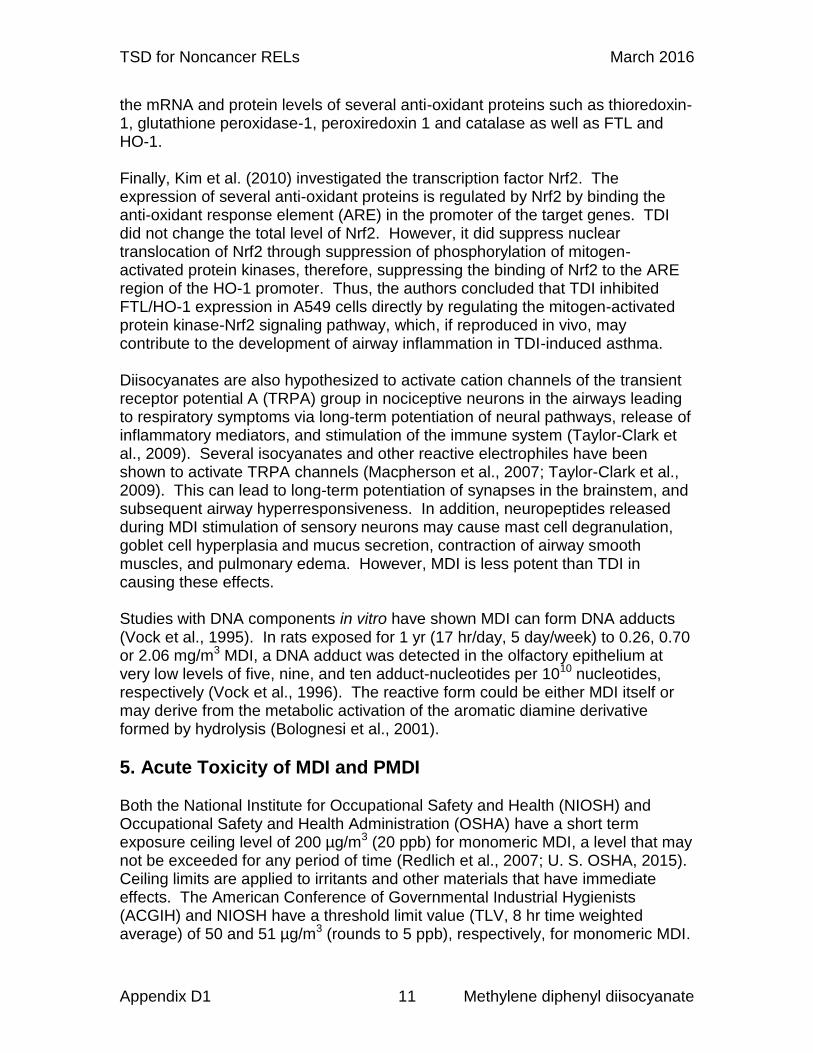
the mRNA and protein levels of several anti-oxidant proteins such as thioredoxin-1, glutathione peroxidase-1, peroxiredoxin 1 and catalase as well as FTL and HO-1. Finally, Kim et al. (2010) investigated the transcription factor Nrf2. The expression of several anti-oxidant proteins is regulated by Nrf2 by binding the anti-oxidant response element (ARE) in the promoter of the target genes. TDI did not change the total level of Nrf2. However, it did suppress nuclear translocation of Nrf2 through suppression of phosphorylation of mitogen-activated protein kinases, therefore, suppressing the binding of Nrf2 to the ARE region of the HO-1 promoter. Thus, the authors concluded that TDI inhibited FTL/HO-1 expression in A549 cells directly by regulating the mitogen-activated protein kinase-Nrf2 signaling pathway, which, if reproduced in vivo, may contribute to the development of airway inflammation in TDI-induced asthma. Diisocyanates are also hypothesized to activate cation channels of the transient receptor potential A (TRPA) group in nociceptive neurons in the airways leading to respiratory symptoms via long-term potentiation of neural pathways, release of inflammatory mediators, and stimulation of the immune system (Taylor-Clark et al., 2009). Several isocyanates and other reactive electrophiles have been shown to activate TRPA channels (Macpherson et al., 2007; Taylor-Clark et al., 2009). This can lead to long-term potentiation of synapses in the brainstem, and subsequent airway hyperresponsiveness. In addition, neuropeptides released during MDI stimulation of sensory neurons may cause mast cell degranulation, goblet cell hyperplasia and mucus secretion, contraction of airway smooth muscles, and pulmonary edema. However, MDI is less potent than TDI in causing these effects. Studies with DNA components in vitro have shown MDI can form DNA adducts (Vock et al., 1995). In rats exposed for 1 yr (17 hr/day, 5 day/week) to 0.26, 0.70 or 2.06 mg/m3 MDI, a DNA adduct was detected in the olfactory epithelium at very low levels of five, nine, and ten adduct-nucleotides per 1010 nucleotides, respectively (Vock et al., 1996). The reactive form could be either MDI itself or may derive from the metabolic activation of the aromatic diamine derivative formed by hydrolysis (Bolognesi et al., 2001).
5. Acute Toxicity of MDI and PMDI Both the National Institute for Occupational Safety and Health (NIOSH) and Occupational Safety and Health Administration (OSHA) have a short term exposure ceiling level of 200 µg/m3 (20 ppb) for monomeric MDI, a level that may not be exceeded for any period of time (Redlich et al., 2007; U. S. OSHA, 2015). Ceiling limits are applied to irritants and other materials that have immediate effects. The American Conference of Governmental Industrial Hygienists (ACGIH) and NIOSH have a threshold limit value (TLV, 8 hr time weighted average) of 50 and 51 µg/m3 (rounds to 5 ppb), respectively, for monomeric MDI.
TSD for Noncancer RELs March 2016
Appendix D1 12 Methylene diphenyl diisocyanate
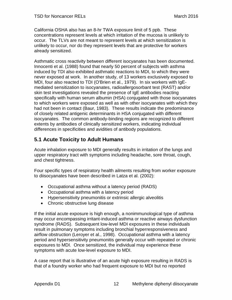
California OSHA also has an 8-hr TWA exposure limit of 5 ppb. These concentrations represent levels at which irritation of the mucosa is unlikely to occur. The TLVs are not meant to represent levels at which sensitization is unlikely to occur, nor do they represent levels that are protective for workers already sensitized. Asthmatic cross reactivity between different isocyanates has been documented. Innocenti et al. (1988) found that nearly 50 percent of subjects with asthma induced by TDI also exhibited asthmatic reactions to MDI, to which they were never exposed at work. In another study, of 13 workers exclusively exposed to MDI, four also reacted to TDI (O'Brien et al., 1979). In six workers with IgE-mediated sensitization to isocyanates, radioallergosorbant test (RAST) and/or skin test investigations revealed the presence of IgE antibodies reacting specifically with human serum albumin (HSA) conjugated with those isocyanates to which workers were exposed as well as with other isocyanates with which they had not been in contact (Baur, 1983). These results indicate the predominance of closely related antigenic determinants in HSA conjugated with different isocyanates. The common antibody-binding regions are recognized to different extents by antibodies of clinically sensitized workers, indicating individual differences in specificities and avidities of antibody populations.
5.1 Acute Toxicity to Adult Humans
Acute inhalation exposure to MDI generally results in irritation of the lungs and upper respiratory tract with symptoms including headache, sore throat, cough, and chest tightness. Four specific types of respiratory health ailments resulting from worker exposure to diisocyanates have been described in Latza et al. (2002):
Occupational asthma without a latency period (RADS)
Occupational asthma with a latency period
Hypersensitivity pneumonitis or extrinsic allergic alveolitis
Chronic obstructive lung disease If the initial acute exposure is high enough, a nonimmunological type of asthma may occur encompassing irritant-induced asthma or reactive airways dysfunction syndrome (RADS). Subsequent low-level MDI exposures in these individuals result in pulmonary symptoms including bronchial hyperresponsiveness and airflow obstruction (Leroyer et al., 1998). Occupational asthma with a latency period and hypersensitivity pneumonitis generally occur with repeated or chronic exposures to MDI. Once sensitized, the individual may experience these symptoms with acute low-level exposure to MDI. A case report that is illustrative of an acute high exposure resulting in RADS is that of a foundry worker who had frequent exposure to MDI but no reported

TSD for Noncancer RELs March 2016
Appendix D1 13 Methylene diphenyl diisocyanate
respiratory or other symptoms (Leroyer et al., 1998). After three years he received an intense acute inhalation exposure as a result of an MDI spill in his work area. Within one hour he experienced headache, sore throat, cough and chest tightness. Other workers in the area experienced similar symptoms but only transiently. These initial symptoms were consistent with a diagnosis of RADS. However, over the course of the subsequent month his chest symptoms and wheezing worsened, especially at work, with some remission during weekends. Spirometric testing revealed moderate airflow obstruction, a forced expiratory volume in 1 second (FEV1) of 2.5 L (83% predicted), and a forced vital capacity (FVC) of 4.5 L (121% predicted). Symptoms persisted despite treatment with budesonide, a glucocorticoid anti-inflammatory. After salbutamol inhalation (a ß2-adrenergic receptor agonist to treat bronchospasm), FEV1 increased 12%. Bronchoprovocation tests with 15 ppb MDI were performed for 4, 30 and 60 min. An isolated late reaction was associated with the 60 min exposure, with a 22% fall in FEV1 seven hours after exposure. The authors suggest his symptoms were consistent with occupational asthma caused by the acute high level exposure (Leroyer et al., 1998). However, it is not clear what role the low level exposures prior to the acute exposure may have played in the etiology of this case’s symptomatology. Provocation tests have been used to confirm a diagnosis of MDI-induced occupational asthma in polyurethane workers. However control groups consisting of normal or asthmatic individuals without previous exposure to isocyanates were not included in these studies to quantitatively elucidate potential acute toxicity of MDI exposure. In the provocation studies, concentrations of MDI or PMDI used in challenge tests ranged between 1 and 20 ppb with exposure durations of seconds (i.e., one breath) to up to 4 hrs (O'Brien et al., 1979; Burge, 1982; Zammit-Tabona et al., 1983; Cartier et al., 1989; Vandenplas et al., 1992; Leroyer et al., 1998; Piirila et al., 2000; Lemiere et al., 2002). These studies have observed asthmatic responses to exposures of 1 ppb MDI or lower in sensitized workers undergoing challenge testing. Lemiere et al. (2002) exposed eight subjects with occupational asthma induced by specific diisocyanates to 1 ppb MDI, TDI or hexamethylene diisocyanate (HDI) using a closed circuit apparatus. The authors considered a positive result to be a 20% or greater reduction in FEV1. By this criterion asthma was induced in two of the subjects with a 30 min exposure, one to MDI and the other to HDI. A third subject had asthma induced with a 45 min exposure to TDI. There was also a significant correlation (Spearman rank order test ρ=0.8, P<0.001) between the percentage of maximum decrease in FEV1 after exposure to 1 ppb and the increase in sputum neutrophil count, indicating inflammatory changes as well. In another study, Burge (1982) found that 2 of 24 MDI-sensitized workers showed a positive reaction with exposure to MDI as low as 1 ppb. The criterion in this study for a positive reaction was a 15% or greater reduction in FEV1.

TSD for Noncancer RELs March 2016
Appendix D1 14 Methylene diphenyl diisocyanate
The lowest concentration of MDI resulting in an asthmatic response in a sensitized worker occurred following a 15 min exposure to 0.51 µg/m3 (0.05 ppb) monomeric MDI (Suojalehto et al., 2011). The subject’s FEV1 fell by a maximum of 25% from base line after 1 hr, requiring use of a bronchodilator and an oral steroid. The worker had a history of severe reactions during work, in which she was occasionally exposed to synthetic plastic containing MDI during orthopedic plaster casting. The levels of MDI in the air were measured in the exposure chamber using filter collection and subsequent liquid chromatography-mass spectrometry analysis of the MDI isocyanate groups. Sampling of breathing zones of nurses when applying and removing the casts was below 1 ppb MDI. The authors noted that dermal exposure to the unhardened MDI-containing plastic material could have been a factor in development of respiratory sensitization. The study by Suojalehto et al. (2011) provides evidence that it may not be possible to set a REL that can protect all individuals that have acquired specific hypersensitivity to MDI, due to some sensitized individuals having a positive response at extremely low concentrations. In a study by Zammit-Tabona et al. (1983), exposure to an average MDI concentration of 12 ppb over 60 min resulted in a ≥20% fall in FEV1 in 7 of 11 foundry workers suspected to have MDI-induced asthma. One of the responders developed cough and wheeze after 10 min of exposure to MDI but recovered within 5 min after leaving the chamber. The MDI concentration in the chamber had only reached 10 ppb when the subject had a positive response. This subject also responded in a similar fashion to 2.5 ppm formaldehyde after 10 min of exposure, and recovered within 5 min of cessation of exposure. None of the other 10 subjects responded with an asthmatic reaction to formaldehyde exposure. This subject had a longstanding history of asthma before employment at the foundry and had a marked degree of bronchial hyperreactivity to methacholine. The authors concluded the cause of bronchoconstriction in this subject from both MDI and formaldehyde was likely irritation and not sensitization. This study suggests that MDI can induce an asthmatic response in non-sensitized individuals with asthma due to MDI’s pulmonary irritant qualities.
5.2 Acute Toxicity to Infants and Children
Asthma-like symptoms were observed among 203 Taiwanese school children during a school track paving/spraying operation of an MDI mixture at 870 ppm w/w in xylene (Jan et al., 2008). The concentration of the MDI and xylene that the children were exposed to is unknown. Acute symptoms were observed when the wind direction suddenly changed direction and blew the emissions towards nearby school classrooms. Of the exposed children, 70.9% reported headache, 67.5% had persistent cough, 63.5% had dyspnea, and 62.6% had nausea. Chest discomfort was reported by 23.6% of the students but chest X-rays were normal. Bronchodilators were administered to 15.8% who experienced wheezing and difficulty breathing. The authors observed an inverse linear relationship

TSD for Noncancer RELs March 2016
Appendix D1 15 Methylene diphenyl diisocyanate
between the incidence of affected students in various classrooms and the distance from the site of MDI spillage (r = -0.48, p < 0.05) suggesting a dose-response. During follow-up surveillance three days after the incident, the prevalence of residual symptoms was cough 30.0%, headache 19.7%, dyspnea 15.3%, sore throat 10.3%, and nausea 3.9% (Jan et al., 2008). A positive history of asthma among 10.8% of the students was strongly correlated with the incidence of dyspnea (OR 4.09; 95% CI 1.17-14.32) and an abnormal pulmonary function test (OR 3.84; 95% CI 1.09-13.5). However, none of the other symptoms during the episode were correlated with either asthma history or abnormal lung function tests. In addition, 60.8% of the children without a history of asthma also complained of dyspnea, and 16.2% required bronchodilators for symptomatic relief. Acute exposure to high levels of MDI was thus associated with an asthma-like syndrome among previously unexposed individuals. A spot urine test did not reveal a positive reaction for MDA after hydrolysis of the urine samples. The authors attributed this finding as characteristic of a brief exposure to MDI. The authors did not discuss effects seen in exposed adults, so it is unclear if children were more prone to the acute effects of MDI than adults. Also, no apparent follow-up was performed to determine if the children had been immunologically sensitized as a result of the high acute exposure. Jan et al. (2008) assumed all the symptomology was due to MDI even though xylenes also are known to cause acute eye and respiratory symptoms. Controlled acute exposures of human adult volunteers to 460-690 ppm xylenes resulted in transient eye and throat irritation and dizziness (Carpenter et al., 1975). In children living in high traffic density regions, upper airway or asthma symptom episodes were found to be associated with combustion-related xylene exposure (Buchdahl et al., 2000; Delfino et al., 2003). However, similar associations were found with other VOCs, and combustion-related gases. A proportion of the eye and respiratory effects could have been caused by xylene exposure, since xylenes are more volatile than MDI, and the formulation applied to the track was composed primarily of xylenes. However, TDI (and possibly MDI) is about 10,000 times more potent (50 ppb for TDI vs. 460 ppm for xylenes) in causing acute eye and throat irritation compared xylenes. Krone and associates have postulated that a relationship exists between exposure to polyurethane products made from isocyanates and childhood asthma (Krone and Klingner, 2005). Further discussion is presented in Section 6.2. One animal study was found that investigated the differential sensitivity of young rats to PMDI. In a subacute study, four week old and six week old rats (20 rats/group/sex) were exposed to 14.1 mg/m3 PMDI for 6 hr/day, five days/week for 2 weeks (Reuzel et al., 1994b). Only mortality was recorded. Four-week-old rats died earlier and in greater numbers than did rats that were six weeks old.

TSD for Noncancer RELs March 2016
Appendix D1 16 Methylene diphenyl diisocyanate
This early-life susceptibility was greater for females than for males. The reason for differential sensitivity was unknown to the authors. However, in humans the relative minute volume to surface area in the pulmonary region is greater in infants by 3-4-fold compared to adults (OEHHA, 2008). Chemicals that are pulmonary irritants, such as MDI, are predicted to have greater pulmonary effects in the young.
5.3 Acute Toxicity to Experimental Animals
During a single 4 hour exposure to high concentrations of PMDI aerosols (376 – 638 mg/m3, particle size < 5 µm), Wistar rats displayed labored respiration and mouth breathing (Reuzel et al., 1994b). Deaths occurred at all exposure levels within two days following the end of exposure, with an LC50 of 490 mg/m3. Among animals that survived, transient weight loss was observed during the second and fourth days after exposure. At higher aerosol levels, hemorrhagic nasal discharge was observed and the lungs of rats euthanized immediately after exposure were grayish and wet, with some pulmonary hemorrhaging. A nose-only exposure 4-hr lethal potency comparison study of PMDI and monomeric MDI was carried out by Pauluhn (2011). The LC50 of rats exposed to PMDI (mean particle size 1.8 µm) was 310 mg/m3, with males and females equally sensitive. The LC50 of MDI (mean particle size 3.3 µm) was 367 mg/m3 (males and females combined), with males approximately twice as susceptible as females. As with the acute exposures above, subacute exposure to PMDI aerosols (0, 2.2, 4.9, 13.6 mg/m3; 10 rats/sex/dose) for six hr/day, five days per week for two weeks led to labored respiration and mouth breathing in the high exposure group (13.6 mg/m3) starting on day four (Reuzel et al., 1994b). Other clinical changes reported for this group included slow movements, dyspnea, piloerection, salivation, bleeding from the nares and swollen abdomens. While rats in the 2.2 mg/m3 group were not visibly affected, those in the 4.9 mg/m3 group were restless, slightly dyspneic, and showed piloerection. In all treatment groups, lung weights were elevated relative to body weights. The effects of PMDI were mainly on the respiratory tract with males more severely affected than females. The adverse effects of acute exposures to PMDI manifest mainly in the lungs as pulmonary inflammation characterized by increased immune cell infiltration, protein production, and organ weight. Kilgour et al. (2002) examined the appearance and resolution of these effects over a 30 day period in rats following an acute 6 hr exposure to 10, 30, or 100 mg/m3 PMDI. Immediately following a single, 6-hr acute exposure, lung lavage fluid showed massive increases in neutrophils (37% of total cells at 10 mg/m3, 78% at 100 mg/m3), but a reduced absolute number of alveolar macrophages. Protein content was elevated in lavage fluid, and enzyme activities increased for N-acetyl glucosaminidase (NAG), alkaline phosphatase, and lactate dehydrogenase (LDH). The accumulation of crystalline surfactant and cellular debris in alveolar lumina

TSD for Noncancer RELs March 2016
Appendix D1 17 Methylene diphenyl diisocyanate
through day three at all PMDI concentrations suggests PMDI is cytotoxic to macrophages. By day three post-exposure, neutrophil numbers were still markedly elevated in the 100 mg/m3 group, but had dropped substantially in the lower dose groups, while macrophage numbers increased. LDH continued to rise but the NAG and alkaline phosphatase activities had returned to control levels. By day ten, most of the measured parameters had returned to control levels, although epithelialization of alveoli was observed in animals at 30 and 100 mg/m3. Thirty days following the last exposure, lung weights, lung lavage parameters, cell proliferation and ultrastructural appearance had returned to normal. These results suggest that even at relatively high acute exposure levels, recovery from PMDI-associated toxic effects in the lung is relatively rapid. Many of the effects observed by Kilgour et al. (2002) may be related to MDI-induced changes in pulmonary epithelium that forms the blood-air barrier in lungs. Pauluhn (2000) examined bronchoalveolar lavage fluid (BALF) of female Wistar rats (n=6 or 7) for markers of damage to pulmonary epithelium following an acute 6-hr exposure to MDI at 0.7, 2.4, 8, or 20 mg/m3. These markers included angiotensin converting enzyme (ACE), protein levels, alkaline phosphatase, LDH, γ-glutamyltranspeptidase, and sialic acid, and were assayed 3 hrs, 1, 3, and 7 days following exposure. PMDI at all dose levels caused an immediate significant increase in alkaline phosphatase activity (p < 0.05) that returned to control levels by day three. This was deemed to be consistent with an adaptive increase in pulmonary surfactant that is rich in alkaline phosphatase from type II pneumocytes. Plasma protein levels in BALF similarly were immediately and significantly elevated at all PMDI concentrations suggesting dysfunction in the epithelial barrier. The activity of ACE was also significantly elevated but only at 2.4 mg/m3 and above. However, there was no dose-dependent effect on LDH levels, suggesting that cytotoxicity was not a cause of elevated protein and ACE levels. Glutathione levels measured in BALF peaked on day one following exposure, and returned to control levels by day seven. However, GSH levels in lung tissue remained elevated through day seven. Based on these results, Pauluhn (2000) proposed that MDI interferes with the pulmonary epithelial barrier leading to pulmonary surfactant dysfunction and increased alveolar surface tension. This surface tension in turn enhances transudation of fluid and solutes from the capillaries, contributing to pulmonary edema that is characteristic of MDI exposure. While this effect appears to be transitory at these dose levels, it was observed to occur at concentrations as low as 0.7 mg/m3. Pauluhn (2002) conducted a concentration × time (C × t) study in which male rats were exposed to 3.4 to 58.1 mg PMDI/m3 and exposure durations of 6 hr to 23 min, respectively, so that C × t was approximately 1200 mg/m3-min. Using total protein content in BALF one day post-exposure as the endpoint for response, total protein was increased 50 percent for all PMDI exposure groups compared to the control group. Thus, the magnitude of BALF protein matched

TSD for Noncancer RELs March 2016
Appendix D1 18 Methylene diphenyl diisocyanate
the exposure intensity over the entire range of concentrations investigated when C × t was kept constant. A conclusion was that changes in BALF protein were dose-dependent (i.e., equally dependent on changes in concentration and duration of exposure). In a separate study, groups of female Wistar rats were exposed nose-only to a range of PMDI concentrations (0.7, 2.3, 8 and 20 mg/m3) for 6 hrs (Pauluhn, 2000; 2002). The MMAD of the PMDI aerosols generated were approximately 1.5 µm (geometric standard deviation ~ 1.6 µm). Earlier results showed total protein and ACE in BALF were among the most sensitive indicators for acute irritant effects of PMDI. Total protein and ACE in BALF were determined at 3 hrs and 1, 3 and 7 days post-exposure. Total protein at 1 day post-exposure and ACE at 3 hr and 1 day post exposure were statistically significantly increased above control following exposure to 2.3 mg/m3 PMDI (Table 1). Total protein 3 hr post-exposure was statistically significantly increased above control at the lowest exposure of 0.7 mg/m3 PMDI. At 1 day post-exposure, total protein in BALF had returned to control levels in rats exposed to 0.7 mg/m3 PMDI, while total protein levels were still increased and essentially unchanged in rats exposed to higher PMDI concentrations. At three days post-exposure, total protein and ACE had returned to control levels for all exposure groups. Table 1. BALF results for total protein and ACE at 3 hours and 1 day post-exposure following 6 hour exposure of rats to PMDI
Endpoint Dose (mg/m3)a
0 0.7 2.3 8 20
Total protein – 3 hr post-exposure (g/l) mean±SD
0.152 ±0.034
0.224 ±0.021
0.215 ±0.037
0.363 ±0.062
0.484 ±0.131
Total protein – 1 d post-exposure (g/l) mean ± SD
0.153 ± 0.018
0.164 ±0.016
0.222 ±0.030
0.336 ±0.066
0.464 ±0.139
ACE – 3 hr post-exposure (nmol/min/ml) mean ± SD
0.099 ±0.047
0.106 ±0.038
0.209 ±0.017
0.369 ±0.069
0.411 ±0.202
ACE – 1 d post exposure (nmol/min/ml) mean ± SD
0.102 ±0.043
0.096 ±0.011
0.210 ±0.055
0.249 ±0.055
0.436 ±0.134
a n per dose group: n=12 for 0 mg/m3; n=6 for all other exposure groups.
Double-logarithmic analysis by Pauluhn (2002) of the concentration-effect relationship for these two endpoints estimated an acute irritant benchmark no-effect threshold concentration of 0.5 mg/m3 for 6 hr exposure to PMDI aerosol. The no-effect threshold was considered a relative change of 100 percent total protein and ACE from control values. We applied continuous modeling methodology in U.S EPA’s benchmark dose software, version 2.3.1 (U. S. EPA, 2012) for benchmark dose analysis on the statistical results for increased total protein and ACE in BALF with increasing PMDI exposure. The data in Table 1 were kindly provided to OEHHA by Dr. Pauluhn in order to run BMD modeling and originates from Pauluhn (2002) in which the data had been presented only in graphical form.

TSD for Noncancer RELs March 2016
Appendix D1 19 Methylene diphenyl diisocyanate
No model in the U.S. EPA BMD suite was able to fit an acceptable line to the data points for ACE 3 hrs and 1 day post-exposure, and total protein 3 hrs post-exposure (the most sensitive indicator of cellular dysfunction). Although a statistically significant dose-response was found for these endpoints (p<0.05), the small n and variations in means and variances at the exposure levels resulted in unacceptable fits with the models provided. Although total protein had returned to control levels at the lowest concentration (0.7 mg/m3) 1-day post-exposure, acceptable non-homogeneous variance model fits were found for total protein for this time point. The effects of 4 hr exposures to MDI aerosol (mass median aerodynamic diameter 0.7 µm and geometric SD 1.6 µm) on pulmonary and sensory irritation at concentrations ranging from 7 to 59 mg/m3 were measured as respiratory rate depression in mice (Weyel and Schaffer, 1985). The concentration required to reduce the respiratory rate 50 percent (RD50) was 32 mg/m3. Respiratory depression progressed slowly with MDI exposure, not reaching a plateau until 3-4 hrs into the exposures. The decline in respiratory rate was dependent on both concentration and duration. Unlike other isocyanates such as TDI and HDI, MDI acted primarily as a pulmonary irritant evoking little or no sensory irritation (i.e., stimulation of lower respiratory tract receptors and not the trigeminal nerves in the upper respiratory tract). Increased lung wet weight was observed at every concentration 24 hrs after the exposures. Several animal models of asthma have been developed for both respiratory and dermal sensitization to MDI or PMDI (Rattray et al., 1994; Pauluhn et al., 2000; Pauluhn and Poole, 2011; Wisnewski et al., 2011). In guinea pigs, one high level 15 min inhalation exposure to 135 mg/m3 PMDI resulted in respiratory sensitization when challenged three weeks later with 15 and 49 mg/m3 PMDI (Pauluhn et al., 2000). Respiratory sensitization was assessed by a change in respiratory rate and an influx of eosinophilic granulocytes into bronchial tissues. Another study in guinea pigs using a lower dose of 19.4 to 23.7 mg/m3 MDI 3 hrs/day for 5 consecutive days did not lead to sensitization (Rattray et al., 1994). In a Brown Norway rat asthma model, the C × t relationship for PMDI sensitization was examined using a 5-day exposure sensitization protocol (Pauluhn and Poole, 2011). Consistent with the sensitization protocol used, the most sensitive endpoints characterizing an allergic pulmonary inflammation were BALF-neutrophils and delayed-onset respiratory changes. A high exposure concentration of PMDI during 10 min exposures (e.g., 100 mg/m3 for 10 min) elicited a more vigorous response than the similar C × t at 360 min (e.g., 3 mg/m3 for 360 min). The C × t study also showed that the dose that triggers an elicitation response in the rat asthma model is slightly below that causing acute pulmonary irritation in naïve rats. This finding suggests that allergic responses via inhalation of PMDI appear to be linked with pulmonary irritation of the lower airways. The NOAEL dose for elicitation of the most sensitive indicator of an

TSD for Noncancer RELs March 2016
Appendix D1 20 Methylene diphenyl diisocyanate
“asthmatic” response (increased PMNs) in inhalation-sensitized rats was calculated by the authors to be 5 mg/m3 × 30 min. Animal studies have shown that MDI skin exposure can induce MDI sensitivity with subsequent challenges via the respiratory tract, suggesting that skin contact may be an important cause of occupational respiratory asthma. In mice and guinea pigs with previous MDI skin exposure (≥1% MDI in solution) significant airway inflammatory responses to respiratory MDI challenge have been demonstrated (Rattray et al., 1994; Wisnewski et al., 2011). These inflammatory responses included influxes of eosinophils and lymphocytes in BALF samples and serum antibody responses.
6. Chronic Toxicity of MDI and PMDI
6.1 Chronic Toxicity to Adult Humans
The effects of chronic inhalation exposure to MDI are largely reflected in decrements in pulmonary function and exacerbation of MDI-induced asthma. Impairment of lung function is mainly a function of allergic inflammation. Variable airflow restriction of the airways and bronchial hypersensitivity are associated with asthma. On rare occasions isocyanates can also cause extrinsic alveolitis, or hypersensitivity pneumonitis, which involves the air sacs and lung parenchyma. Symptoms of hypersensitivity pneumonitis include headache, nausea, muscle aches, fever and chills, significant falls in both FEV1 and FVC, hypoxia, audible moist rales, increased blood neutrophils, increased neutrophils and lymphocytes in bronchoalveolar lavage, and significant levels of IgG and IgE antibodies to MDI-human serum albumin (Baur et al., 1984; Vandenplas et al., 1993b). MDI appears to have a greater propensity to cause hypersensitivity pneumonitis than TDI (Vandenplas et al., 1993b; Baur, 1995). At least 4.7 percent (8/167) of workers exposed to PMDI resin in the manufacture of woodchip boards developed hypersensitivity pneumonitis (Vandenplas et al., 1993b). The latency period before symptoms appear ranged from weeks to 1 year following beginning of exposure. It is not clear from occupational studies whether asthma and hypersensitivity pneumonitis are the result of chronic low-level exposure, acute exposure to high levels, or both. It is clear, however, that once individuals are sensitized to MDI, further exposure generally exacerbates respiratory symptoms (Piirila et al., 2000; Redlich et al., 2007). A 10-year follow-up of 245 workers that had been diagnosed with asthma induced by diisocyanates, 96 of which were due to MDI exposure, was conducted by Piirila et al. (2000). The average duration of symptoms before
TSD for Noncancer RELs March 2016
Appendix D1 21 Methylene diphenyl diisocyanate
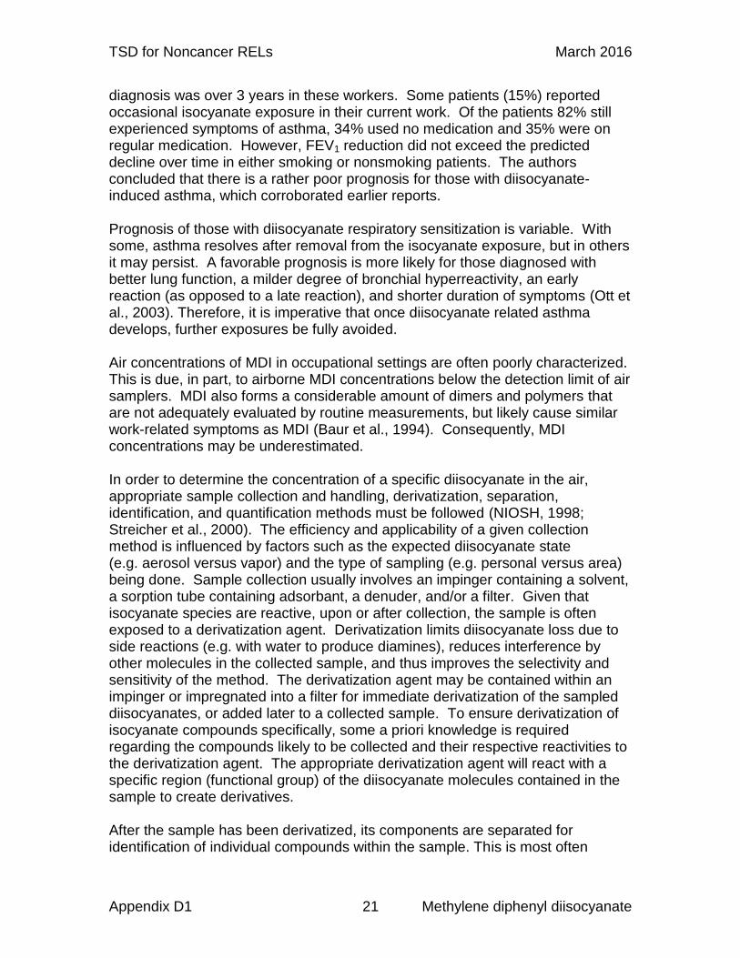
diagnosis was over 3 years in these workers. Some patients (15%) reported occasional isocyanate exposure in their current work. Of the patients 82% still experienced symptoms of asthma, 34% used no medication and 35% were on regular medication. However, FEV1 reduction did not exceed the predicted decline over time in either smoking or nonsmoking patients. The authors concluded that there is a rather poor prognosis for those with diisocyanate-induced asthma, which corroborated earlier reports. Prognosis of those with diisocyanate respiratory sensitization is variable. With some, asthma resolves after removal from the isocyanate exposure, but in others it may persist. A favorable prognosis is more likely for those diagnosed with better lung function, a milder degree of bronchial hyperreactivity, an early reaction (as opposed to a late reaction), and shorter duration of symptoms (Ott et al., 2003). Therefore, it is imperative that once diisocyanate related asthma develops, further exposures be fully avoided. Air concentrations of MDI in occupational settings are often poorly characterized. This is due, in part, to airborne MDI concentrations below the detection limit of air samplers. MDI also forms a considerable amount of dimers and polymers that are not adequately evaluated by routine measurements, but likely cause similar work-related symptoms as MDI (Baur et al., 1994). Consequently, MDI concentrations may be underestimated. In order to determine the concentration of a specific diisocyanate in the air, appropriate sample collection and handling, derivatization, separation, identification, and quantification methods must be followed (NIOSH, 1998; Streicher et al., 2000). The efficiency and applicability of a given collection method is influenced by factors such as the expected diisocyanate state (e.g. aerosol versus vapor) and the type of sampling (e.g. personal versus area) being done. Sample collection usually involves an impinger containing a solvent, a sorption tube containing adsorbant, a denuder, and/or a filter. Given that isocyanate species are reactive, upon or after collection, the sample is often exposed to a derivatization agent. Derivatization limits diisocyanate loss due to side reactions (e.g. with water to produce diamines), reduces interference by other molecules in the collected sample, and thus improves the selectivity and sensitivity of the method. The derivatization agent may be contained within an impinger or impregnated into a filter for immediate derivatization of the sampled diisocyanates, or added later to a collected sample. To ensure derivatization of isocyanate compounds specifically, some a priori knowledge is required regarding the compounds likely to be collected and their respective reactivities to the derivatization agent. The appropriate derivatization agent will react with a specific region (functional group) of the diisocyanate molecules contained in the sample to create derivatives. After the sample has been derivatized, its components are separated for identification of individual compounds within the sample. This is most often
TSD for Noncancer RELs March 2016
Appendix D1 22 Methylene diphenyl diisocyanate
accomplished by reversed-phase high-performance liquid chromatography (RP-HPLC). Quantification can then be achieved by creating a calibration curve using different standard concentrations. (Although there are only standards for pure monomeric diisocyanates, methods are available for detection and quantification of total isocyanates.). Because multiple chemicals can co-elute to produce identical/similar retention times, use of a selective detector (e.g. ultraviolet-visible or fluorescence), which responds only to specific classes of chemicals, can aid identification. Use of two different selective detectors in series can increase the selectivity and sensitivity of detection. In general, NIOSH Method 5525 may offer the most specificity, sensitivity, and applicability (NIOSH, 1998). Sample collection is achieved using a glass fiber filter impregnated with a derivatization agent, an impinger containing a derivatization agent, or a combination of the two. While the filter collects particles of all sizes, it most efficiently collects and derivatizes small particulates (≤2 µm). The impinger traps diisocyanate vapors and larger particles in the aerosol. Use of the impinger in addition to the filter improves collection of larger particles which may not disperse on the filter to allow derivatization of the collected diisocyanates. This method is appropriate for personal or area sampling, and the impinger can be used for collecting particles with short (< several minutes) or long half-lives (NIOSH, 1998). Longitudinal studies are the primary means for assessing asthma onset prevalence and changes in pulmonary function with time in diisocyanate workers. There are numerous longitudinal studies examining pulmonary changes in TDI workers. However, there have been few longitudinal studies examining the effects of MDI in workers. The following summaries represent the most comprehensive occupational studies available, most of which included limited MDI exposure data. Pham et al. (1987) Chronic exposure to mainly MDI was studied in a five-year longitudinal study of workers from two factories producing polyurethane foam. A respiratory questionnaire, measurement of vital capacity (VC) and FEV1 and a single breath CO diffusion test (DLCO) were done at the beginning of the survey, and then repeated five years later. DLCO lung diffusion testing is a measure of how well the lungs exchange gases. A total of 318 workers participated, 83 of whom were unexposed, 117 indirectly exposed and 118 directly exposed to MDI. The MDI concentration was characterized as below 20 ppb. The authors did not state what proportion of diisocyanates was MDI; only that it was mainly MDI. At the beginning of the study, both indirectly and directly exposed workers reported greater symptoms of chronic bronchitis compared to controls (p<0.05), but only directly exposed female workers reported greater symptoms of asthma (p<0.05). Pulmonary function tests of directly exposed men showed a lower

TSD for Noncancer RELs March 2016
Appendix D1 23 Methylene diphenyl diisocyanate
percent of predicted VC, FEV1 and DLCO (p<0.01). These pulmonary decrements were most pronounced in those workers with >60 months of direct exposure. Five years later, only half of the initial cohort was still active (114 males and 45 females). Asthma and chronic bronchitis had increased in both exposed groups and the unexposed group. No difference in these symptoms was observed between groups. The five year decline in VC and FEV1 was not significantly different between groups, but a significantly larger loss of DLCO (p<0.05) was found in the directly exposed workers. Petsonk et al. (2000) In a prospective study of the respiratory effects of MDI exposure, Petsonk et al. (2000) evaluated the respiratory health of workers in a new wood products manufacturing plant in which MDI was used as a binder. Health data and exposure histories were collected by questionnaire prior to the use of MDI at the plant, and semiannually for the next two years. The critical effect was asthma-like symptoms, cases of which were defined based on questionnaire responses as current or previous asthma, or current use of a bronchodilator, or current asthma attacks characterized by shortness of breath and wheezing. Cases were divided into those that met these criteria at the initial survey (IAS) before MDI exposure, those who met the definition during a follow-up survey (FAS) after exposure to MDI had begun, and a third group with new onset asthma-like symptoms (NAS), defined as workers who met the case definition for FAS, but not for IAS. Measurements of serial peak flow, spirometry, methacholine challenge, and specific IgE were used in some cases for validation of case designation, but were not available for all study participants. Thus, the authors noted that it was unlikely that all participants with respiratory symptoms have occupational asthma. Of the 178 workers with initial and at least one follow-up survey, a complete occupational history was available for 144. Of these, 77 completed the initial health survey prior to first use of MDI at the plant and also reported no previous job with MDI exposure. Thus the remaining 67 may have had MDI exposure at the plant prior to their initial health assessment. Thirty-two workers (20%) met the FAS case definition, while 22 workers (12%) met the NAS case definition. In the NAS group, the duration of work prior to symptom onset ranged from 3 to 22 months (mean, 11 months). The prevalence of FAS and NAS cases was clearly associated with reported exposure in that those who reported working with MDI were significantly more likely to have asthma (p < 0.01) than were those with no or only occasional passing exposure. Those working in areas with high potential exposure to liquid MDI (e.g., cleanup of MDI spills and cleaning the MDI blender) had a significantly elevated prevalence of asthma (p < 0.001) compared to those where potential exposure was rated medium or low. Both FAS and NAS cases were significantly elevated among those who indicated they occasionally
TSD for Noncancer RELs March 2016
Appendix D1 24 Methylene diphenyl diisocyanate
removed protective respirators compared with those who never did (p = 0.05), and by 52% of those who reported at least once observing MDI stains on their skin (p < 0.001). These observations in conjunction with the controls engineered into the plant’s design to reduce inhalation exposure suggest that the appearance of new asthma symptoms among a third of those working in the blending and press operations, and 10-30% of the workers in adjacent areas, likely reflects both inhalation and dermal exposure to MDI. Environmental sampling for diisocyanate was not carried out during the study. Six personal breathing zone samplers (OSHA method 47) worn by employees 7 months after the last health survey did not find detectable air levels of MDI. One wipe glove sample did find 78 µg of MDI. The authors suggested the high proportion of asthma-like symptoms was more related to many participants working with or cleaning spills of liquid MDI, rather than to long-term exposure to low level airborne MDI. Johnson et al. (1985) A cross-sectional study was conducted in 78 iron and steel workers exposed to Pepset, a chemical binding agent consisting of MDI, phenol, formaldehyde and their decomposition products, and silica-containing particulates. A group of 372 railway yard workers matched for socioeconomic status and smoking habit, and “without significant exposure to air contaminants” (as determined by environmental measurements taken during the health study) were used as controls. [OEHHA notes that railway yard workers are often exposed to diesel engine exhaust, and prior history of diesel exhaust exposure in this control group was not reported.] Exposure to MDI was carried out during the health survey portion of the study by area sampling (n=319) of multiple sites in the foundry with midget impingers. A colorimetric method was used for analysis of MDI air concentrations. Sampling times were not given. Of the area samples collected, 85.6% were <5 ppb, 8.5% were >5 ppb and ≤10 ppb, 4.4% were >10 and ≤15 ppb, 0.9% were >15 and ≤20 ppb, and 0.6% were >20 ppb (2 out of 319 samples). The authors noted that levels in excess of 20 ppb were more common before a new ventilation system was installed several months before the study. For prevalence of respiratory symptoms, phlegm, breathlessness, chest tightness and chest illness were statistically significantly greater (p<0.05) in foundry workers compared to the control group. The prevalence of wet cough was also significantly higher in foundry workers compared to controls. The mean FEV1, FVC and FEF25-75% of foundry workers were all significantly lower than those of controls. As expected, current smokers had significantly worse mean lung function compared to non-smokers. The foundry workers also underwent a methacholine challenge test. A provocative concentration of ≤8 mg/ml causing a 20% or greater drop in FEV1 (PC20 ≤8 mg/ml) in a worker was chosen as an indicator of bronchial hyperreactivity. By this criterion, 19.7% (13 of 66 workers)

TSD for Noncancer RELs March 2016
Appendix D1 25 Methylene diphenyl diisocyanate
had evidence of bronchial hyperreactivity. Three workers had evidence of pneumoconiosis in chest radiographs, suggestive of silicosis. The authors noted that exposure to dusts and other chemicals could have been a contributing factor to the reduced pulmonary function of the workers. Inhalation provocation tests with MDI and formaldehyde were performed on nine of the asthmatic workers in a separate study (Zammit-Tabona et al., 1983). Six had a positive asthmatic reaction to MDI exposure, but not to formaldehyde, indicating MDI was the cause of their asthma. A seventh worker had an immediate reaction to both MDI and formaldehyde. But the transient bronchoconstriction due to exposure in this worker was likely due to irritation rather than sensitization. Bernstein et al. (1993) A cross-sectional study was conducted in 243 workers exposed to MDI in a urethane plant that had been designed to minimize MDI exposure. There were 147 workers on the urethane mold lines and 96 other workers were involved with administrative, transport, or maintenance activities. The average duration of employment in the plant was 18.2 months (range 0 to 32 months). Exposure to MDI fumes was continuously monitored via a visible spectrometric method with area MDA-7100 diisocyanate monitors. During the three years the plant was in operation, short-term exposures did not exceed 5 ppb. The absence of elevated MDI levels during occasional spills was explained as either low volatility of MDI at room temperature, or by lack of monitors in close proximity to where the spills occurred. Peak expiratory flow rate (PEFR) was performed in those workers (n=43) who reported at least one lower respiratory symptom of wheezing, cough, or shortness of breath, and in those workers with MDI-HSA specific antibodies. PEFR was performed in 23 workers free of symptoms who served as controls. Greater than 15% variability in PEFR between working and non- working conditions was found in three workers and considered a diagnosis of occupational asthma. One of the control workers also was diagnosed with occupational asthma, suggesting a false negative on the workers’ questionnaire response and potential underestimation of the true prevalence of asthma. Urticarial symptoms related to MDI sensitization were confirmed by elevated specific IgE levels and cutaneous reactivity to MDI-HSA in one worker. Lack of respiratory symptoms and known dermal exposure to MDI in this worker suggest the skin was the primary route of sensitization. Another worker had only elevated levels of serum specific IgE and IgG to MDI-HSA, but was free of respiratory symptoms.
TSD for Noncancer RELs March 2016
Appendix D1 26 Methylene diphenyl diisocyanate
The authors concluded that higher than normal exposures to MDI (i.e., >5 ppb) occurred during nonroutine activities in the workers with occupational asthma and MDI-related cutaneous anaphylaxis, and speculated that unpredictable exposure to MDI liquid and fumes occurring during maintenance or excessive heating of MDI-resin mixtures caused the observed reactions. In one of the workers, onset of asthmatic symptoms began 2 weeks after accidental exposure to a large MDI spill. Sulotto et al. (1990) End of work shift and end of work week effects on pulmonary function were examined in 27 asymptomatic polyurethane workers exposed to low levels of MDI. An equal number of clerks from the same factory with no exposure to MDI and without asthma were matched by age. The polyurethane workers were identified as having 14.0 years at work, but it is unclear if this time included isocyanate exposure for the entire duration. MDI sampling was carried out during the same time when lung function tests were performed. MDI concentrations ranged from 0.5 to 1 ppb and were analyzed using continuous tape monitoring. No significant differences in pulmonary function between the two groups were observed using a paired t-test over the course of a work day (Monday) or over the course of a work week (Monday before work to end of shift on Friday). Two-way ANOVA found reductions in FEV1 and FEF25-75 over the course of a work week, but the differences were related to smoking and not occupational exposure. The authors noted that 17 workers exhibiting isocyanate-induced asthma were removed from the facility sometime before the study began when TDI or mixtures of TDI-MDI were used. The authors concluded that short-term exposures to low levels of MDI do not result in respiratory changes. Jang et al. (2000) Jang et al. conducted a cross-sectional study of workers in Korean TDI and MDI manufacturing plants. A questionnaire was given and pulmonary function was performed on 20 workers exposed to MDI at a manufacturing plant, 44 workers exposed to TDI at a TDI manufacturing plant, and a control group consisting of 27 maintenance and field staff with no known exposure to the isocyanates. A total of 60 personal breathing zone samples were collected from the TDI and MDI workers during manufacturing processes using impingers. Sampling times were 30-60 min. The mean and maximum air concentrations of MDI were 1.3 and 6.4 µg/m3 (0.13 and 0.63 ppb), and the mean and maximum air concentrations of TDI were 17.4 and 42.9 µg/m3 (2.5 and 6.0 ppb). FEV1 was comparable among all three groups. Airway hyperresponsiveness (AHR) was considered positive when a PC20 FEV1 <16.0 mg/mL of methacholine was measured. By this criterion, AHR was greater (p<0.05) in MDI workers (20%) compared to TDI workers (4.7%). However, there was no difference when the MDI workers were compared to the control group. In both TDI and MDI workers that complained of respiratory symptoms, AHR was more prevalent (p<0.05). The authors observed

TSD for Noncancer RELs March 2016
Appendix D1 27 Methylene diphenyl diisocyanate
no clear evidence of sensitization, likely a result of the healthy worker effect (i.e., sensitized workers did not remain working at the plants). Littorin et al. (2002) Respiratory symptoms and biomarkers for isocyanate exposure were investigated in car industry workers (n=29) spraying or applying hot-melt glues containing PMDI onto flexible TDI polyurethane foam. Applying and heating of PMDI-based glues were associated with biomarkers of inflammation in nasal lavage fluid (albumin, myeloperoxidase and neutrophils) and work-related symptoms of nasal irritation, including stuffiness, runny nose or sneezing. After work, workers who had complained of nasal irritation had higher levels of nasal inflammatory biomarkers than a control group of 15 office workers with no such history of exposure. However, biomarkers of TDI metabolites (mainly 2,6-toluene metabolites) in urine showed a stronger association with biomarkers of nasal inflammation than did metabolites of MDI. In addition, MDI metabolites were found in nasal lavage fluid of only two workers, whereas TDI metabolites were found in nasal lavage fluid of four workers. Finally, the presence of serum antibodies specific for TDI and MDI was associated with increased levels of nasal inflammatory biomarkers. Littorin et al. (2002) had assumed the main exposure would be to fumes and gases emitted from the PMDI-glue. However, a separate study conducted by the researchers showed that urinary TDI metabolites rose over a work shift when hot-melt glue was used, which was probably caused by thermodegradation of TDI-based polyurethane foam. The workers selected for this study were healthy workers (atopic workers were excluded) in order to reduce the “noise” of non-specific symptoms and signs. Table 2 presents a summary of the findings from the occupational studies.

TSD for Noncancer RELs March 2016
Appendix D1 28 Methylene diphenyl diisocyanate
Table 2. Summary of occupational studies in workers exposed to MDI
Study Study type, Industry & Exposure
Results
Pham et al., 1988
Longitudinal 5-yr study Polyurethane production Mean age of workers not provided <20 ppb for all groups 118 directly exposed, 117 indirectly exposed and 83 unexposed workers
Initially, ↑ asthma in directly exposed women (p<0.05) and lower predicted VC, FEV1 and DLCO in directly exposed men with >60 mo of exposure After 5 yrs, no change in FEV1, but observed increased loss of DLCO in directly exposed workers (p<0.05)
Petsonk et al., 2000
Prospective 2-year study at new wood products plant. 178 workers Mean age range 31.1 to 32.6 yrs Limited air measurements after the study below LOD. Possible dermal exposure
15 of 56 workers with high exposure had new onset of asthma after 2 years vs. 0 of 42 workers with low exposure (p<0.001).
Johnson et al., 1985
Cross-sectional study at foundry plant (n=78) Mean age 43.7 yrs MDI Sampling: 86% <0.05 ppb 9% >5 & <10 ppb 4% >10 & <15 ppb 1.5% >15 ppb 372 railway worker controls, mean age 38.6 yrs
Increased wet cough, lower FEV1, FVC and FEF25-75% compared to controls (p<0.05). 13 of 66 workers had increased bronchial hyperreactivity by methacholine test. 6 of 9 tested by MDI provocation were positive for asthma. Cross contamination with silica dust.
Bernstein et al., 1993
Cross-sectional study in polyurethane workers 147 workers, 96 controls Ages not specified ≤5 ppb during short-term measurements >5 ppb during spills
3 workers and 1 control diagnosed with occupational asthma: Significant association between >15% variability in PEFR and respiratory symptoms (X2 p<0.002)
Sulotto et al., 1990
Cross-sectional study in polyurethane workers 27 asymptomatic workers 27 controls Mean group ages matched but not stated Exposures ranged from 0.5 to 1 ppb MDI
No end of work shift or end of work week changes in FEV1 or FEF25-75 in exposed workers. 17 workers with occupational asthma had been removed from plant prior to study

TSD for Noncancer RELs March 2016
Appendix D1 29 Methylene diphenyl diisocyanate
Study Study type, Industry & Exposure
Results
Jang et al., 2000
Cross-sectional study in MDI/TDI manufacturing plants. 20 MDI workers 44 TDI workers 27 control workers Mean ages 34.9 to 35.9 yrs MDI exposure: mean 1.3 µg/m3, maximum 6.4 µg/m3
No difference in FEV1 among the groups. Airway hyperresponsiveness (PC20 FEV1 at <16.0 mg/mL of methacholine) greater in MDI and TDI workers (p<0.05). Occupational asthma not seen, likely due to healthy worker effect.
Littorin et al., 2002
Cross-sectional study of workers applying/spraying glues with PMDI 29 non-atopic workers 15 control workers Mean ages not specified No air levels sampled Exposure estimated by MDI and TDI urinary metabolites
Increased biomarkers of inflammation in nasal lavage and increased work-related symptoms of nasal irritation in workers (p<0.05). Exposure better correlated with TDI exposure from polyurethane foam than with PMDI in glue
Possible neurological effects of workers exposed to MDI have been reported. In cases reported by Reidy and Bolter (1994), five individuals were occupationally exposed to MDI over a two-year span, and examined while exposure was ongoing (1 case), or up to 9 months after cessation of exposure (4 cases). The intensity and frequency of exposure were not reported and there was co-exposure to hydrocarbon solvent vapors. Subjective complaints included respiratory distress, headaches, forgetfulness, mood alterations, irritability, and difficulty concentrating. All subjects were diagnosed with isocyanate-induced occupational asthma and allergic rhinitis. Formal neuropsychological evaluations indicated that psychomotor, psychosensory, visuographic and language skills were largely intact. However, there was marked slowing in the rate of information processing, discrepancies in immediate recall of verbal versus nonverbal material, and deficiencies in learning ability. Complex, nonverbal abstract reasoning was impaired, and there was evidence of emotional distress in the form of depression, anxiety, and altered mentation. The authors concluded the data indicate compromised cognitive functions characteristic of CNS involvement, but do not clearly identify a single pattern of neuropsychological deficits associated with MDI exposure. Hughes et al. (2014) reviewed the study by Reidy and Bolter (1994), along with a number of other studies suggesting neurological deficits resulting from exposure to other diisocyanates. They believe that the Reidy and Bolter study was biased as a result of testing obtained by litigating attorneys, and that there was a lack of comparison with other exposed workers. They also point out that the authors say

TSD for Noncancer RELs March 2016
Appendix D1 30 Methylene diphenyl diisocyanate
that selection bias was present, as there were other workers exposed to MDI who refused to participate for various reasons. Several reports suggest that skin exposure to MDI in the workplace can increase the risk for sensitization and isocyanate-induced asthma (Bello et al., 2007). It was proposed that MDI skin exposure induces systemic sensitization, which then leads to occupational asthma following MDI inhalation exposure. Evidence largely results from settings in which known skin exposure occurs, but extremely low to non-detectable air levels of MDI are measured. In supporting evidence, MDI air concentrations were below the limit of detection in most breathing zone air samples in an occupational study, but detectable amounts of the MDI metabolites, measured as 4,4’-methylenedianiline (MDA) in acid hydrolyzed urine, were found in nearly all urine samples (Kaaria et al., 2001). MDA is formed following acid hydrolysis of MDI metabolites in urine samples and is preferred for quantitative analysis. The presence of MDA in acid-hydrolyzed urine samples was explained, in part, by the long half-life of MDI metabolites in the body, and that exposure from previous days contributed to the urinary amount of metabolite. In addition, analysis of MDA in acid-hydrolyzed urine samples was said to be a more sensitive and less arduous method than the established measurement of airborne MDI. Case reports of MDI-exposed workers with allergic contact eczema on hands arms and face, and contact allergy (delayed dermal hypersensitivity) have been reported. Several cases of sensitization occurred after a few weeks or months of exposure at work (Estlander et al., 1992). In some cases the patient had MDI-induced asthma in addition to the contact eczema.
6.2 Chronic Toxicity to Infants and Children
No studies of the chronic effects of MDI on infants and children were located. It has been postulated that early life exposure to TDI and other diisocyanates may occur through inhalation and dermal contact with polyurethane products(Krone et al., 2003b). However, emissions of detectable levels of free MDI and TDI from polyurethane consumer products and other products made with MDI (e.g., mattresses, adhesives, sealants and other products for consumer use) have not been found (Hugo et al., 2000; Boyd and Mogensen, 2007; Vangronsveld et al., 2013). Strachan and Carey (1995) found independent associations between severe wheeze and the use of non-feather bedding, especially foam pillows (odds ratio 2.78; 95% C.I. 1.89 to 4.17), among children with 12 or more wheezing attacks in the previous 12 months. The authors speculated that volatile organic compounds could be off-gassing from the foam pillows. Other researchers found that there is increased exposure to house dust-mite allergens from synthetic pillows compared to feather pillows and speculated that this may explain the increased asthma symptoms (Crane et al., 1997). Krone et al. (2003a) applied semiquantitative tests (i.e., wipe test and extraction with dimethyl sulfoxide) for isocyanate to polyurethane products manufactured

TSD for Noncancer RELs March 2016
Appendix D1 31 Methylene diphenyl diisocyanate
using TDI, including mattresses, mattress pads, sofa padding, carpet pads and pillows, and detected free isocyanate in consumer products. It was suggested by the authors that isocyanate may be available to dissolve in skin oils upon dermal contact. Hoffmann and Schupp (2009) ran tests on flexible five-day old MDI-based polyurethane foam used to upholster furniture and bed mattresses. Toluene or ethyl acetate was used to extract unreacted MDI from the surface and center of the foam. Polyurethane foam samples were also placed in a dynamic fatigue test chamber, which repeatedly compressed and released the sample to create an air exchange and release VOCs in the foam, and simulate inhalation exposures from normal mattress/furniture use. Air from the chamber was sampled using glass fiber filters attached to pumps and placed inside the chamber. Exposure by direct skin contact was simulated by testing the MDI migration from the foam. Depending on the extraction process, 1 to 14 µg MDI/g foam was extracted. Despite the extraction findings, MDI was undetectable in filters from the test chamber and contact experiments. For inhalation exposures, the limit of detection was <5.4 ng/m3 air (<6 ppt). For dermal exposures, the limit of detection was about 9 ng/cm2 per day (<44 ng/cm2 over the 5-day experiment). Adsorption of MDI on chamber walls was not measured in this study. MDI in vapor form in the chamber air was not analyzed because MDI was expected to be in the aerosol phase at ambient temperatures. The authors concluded that trace amounts of “free” MDI may remain in the polymer matrix, although it was unclear how this MDI inside the foam was bound. Diffusion of such physically bound MDI out of the matrix was not expected to occur in practice. It is unknown how the immune system in infants and children would respond to MDI exposure during critical stages of immune system and respiratory system development. These early life diisocyanate exposures may be significant since at birth, humans exhibit a dominant humoral, TH2, responsiveness (i.e., an atopic state). During the first few years of life, the TH2 response converts to a more cellular (TH1) immune response characteristic of the mature adult immune system. A delay in the transition from the predominant TH2 pattern to the more balanced TH1/ TH2 response allows an atopic TH2 type response to persist longer, thus extending the period of vulnerability to environmental stressors and allergens, and increasing the likelihood of subsequent disease expression including asthma (Prescott et al., 1999; Wills-Karp, 1999). Contrary to a TH2 pattern for childhood atopic asthma, obese children with asthma exhibit TH1 polarization and greater asthma severity, whereas lean children with asthma exhibit TH2 polarization and less asthma severity (Youssef et al., 2013). The presence of high leptin levels in the obese children is associated with an increase in IFN-у production by TH1-polarized cells. Leptin is found in higher levels in obese children and is known to promote the production of nitric oxide and pro-inflammatory cytokines in macrophages and monocytes. So, depending on body weight of the child, this research suggests either TH1- or TH2-driven pathways can be involved in childhood asthma.

TSD for Noncancer RELs March 2016
Appendix D1 32 Methylene diphenyl diisocyanate
While there is evidence that atopic asthma in children is usually TH2-based, the immunopathogenesis of diisocyanate-induced asthma is less distinct. TDI-induced asthma in workers has shown either a TH1 immune response pattern (Finotto et al., 1991; Maestrelli et al., 1994) or a mixed TH1/ TH2 immune response (Maestrelli et al., 1997; Redlich et al., 1997; Lummus et al., 1998). Regardless of the differences in T cell profiles, the clinical manifestations and pathophysiological changes observed in TDI-induced asthma are remarkably similar in some respects to those in atopic asthma including airway hyperreactivity, the presence of eosinophilic lung infiltrates, and mucus hypersecretion in airways (Del Prete et al., 1993; Herrick et al., 2003).
Similar to development of childhood allergic asthma, diisocyanate-induced asthma is multifactorial in origin and complex. The mechanism of sensitization by diisocyanates is not well understood in adults, much less children. Thus, differences in T cell profiles in childhood atopic asthma and diisocyanate-induced asthma does not inform us regarding the response of immune systems in infants and children to MDI exposure.
6.3 Chronic Toxicity to Experimental Animals
In subchronic studies carried out by Reuzel et al. (1994b) prior to conducting a chronic exposure study, 10 rats/sex were exposed to PMDI concentrations of 0.35, 1.4 or 7.2 mg/m3 in the first study, and 20 rats/sex were exposed to 4.1, 8.4 or 12.3 mg/m3 PMDI in a second study. Exposure duration for both chamber studies was 6 hrs/day, 5 days/week for 13 weeks. The respiratory tract was the main target organ. Severe respiratory distress was observed in the 12.3 mg/m3 group. In treatment groups of 7.2 mg/m3 and above, accumulation of alveolar macrophages in the lungs was observed. The investigators also observed interstitial infiltration by macrophages at the highest two levels (8.4 and 12.3 mg/m3). At all concentrations in the second study, there was also significant accumulation of yellowish pigmented macrophages in mediastinal lymph nodes. In the nasal cavity, respiratory epithelial hyperplasia and olfactory epithelial atrophy were observed primarily in the two highest levels of the second study. For these pulmonary effects, the authors reported a NOAEL of 1.4 mg/m3. The effects of chronic exposure to PMDI (approximately 50:50 monomeric:polymeric MDI) of 560 Wistar rats of both sexes were reported by Reuzel et al. (1994a). Animals were exposed to the PMDI mixture for 6 hr/day, 5 days/week for one or two years. The mean exposure concentrations were 0, 0.19, 0.98 and 6.03 mg/m3, with mass median aerodynamic diameters of 0.68, 0.70, and 0.74 µm, respectively. After both one and two years of exposure, there was significant (p < 0.01) accumulation of macrophages with yellow pigment in the lungs and the mediastinal lymph nodes at the highest dose (6.03 mg/m3), and at 0.98 mg/m3 after two years of exposure. Alveolar duct epithelialization as well as fibrosis of tissues surrounding the macrophage accumulations occurred in the 0.98 and 6.03 mg/m3 exposure

TSD for Noncancer RELs March 2016
Appendix D1 33 Methylene diphenyl diisocyanate
groups (Table 3). Localized alveolar bronchiolization was seen in the 6.03 mg/m3 group. The time sequence of the spectrum of pulmonary changes indicates that recurrent alveolar wall damage by PMDI and/or polymeric-containing alveolar macrophages leads to alveolar epithelialization. In the nasal cavity, minimal to moderate olfactory epithelial rearrangement was observed in all treatment groups, but was significant only at the highest dose. Basal cell hyperplasia and Bowman’s gland hyperplasia were significant (p < 0.05) in males at the two highest dose levels after two years (Table 3). In females, basal cell hyperplasia was significant (p < 0.01) only at the highest dose, and Bowman’s gland hyperplasia was not statistically significantly increased at any dose. The authors concluded that these data indicate a LOAEL of 0.98 mg/m3, and a NOAEL of 0.19 mg/m3 for the noncancer respiratory tract effects. Table 3. Incidences of primary microscopic findings in the lungs and nasal cavity of male and female Wistar rats exposed to PMDI for 2 years (Reuzel et al., 1994a)a
Males
Females
Exposure concentration (mg/m3) Number of lungs examined Lungs Localized fibrosis Minimal Mild Moderate Severe Alveolar duct epithelialization Local alveolar bronchiolization Nasal Cavity Basal cell hyperplasia Olfactory epithelial degeneration Bowman’s gland hyperplasia
0 60 1 0 1 0 0 1 1 14 6 0
0.19 60 0 0 0 0 0 0 1 13 9 2
0.98 60 9* 7 2 0 0 8* 2 26* 15 9**
6.03 60 44** 5 22 17 0 54** 12** 32** 25* 17**
0 59 0 0 0 0 0 0 2 4 8 2
0.19 60 0 0 0 0 0 0 3 8 16 6
0.98 60 4 3 1 0 0 8* 3 8 10 8
6.03 59 48** 8 23 14 3 57** 14** 49** 20* 8
a Values marked with asterisks differ significantly from control (*p<0.05, **p<0.01)
In a separate chronic study by Hoymann et al. (1998), female Wistar rats were subjected to whole-body exposures to monomeric MDI aerosols (0.23, 0.70, and 2.05 mg/m3; MMAD 1.03-1.06 µm) for 17 hours/day, 5 days/week for up to 24 months. Chronic exposure to MDI caused a time and dose-dependent deterioration of lung function. Lung function was assessed after 6, 12, 17 and 20 months, with histological evaluations after 12 and 24 months. At all time points, the highest exposure (2.05 mg/m3) caused a significant decrease in maximum mid-expiratory flow (MMEF), and forced expiratory flows (FEF) at 10,

TSD for Noncancer RELs March 2016
Appendix D1 34 Methylene diphenyl diisocyanate
25, and 50%, but not 75% of forced vital capacity. This indicates a significant increase in flow resistance in the small peripheral airways, but not the large airways. With the longer 12 and 17 month exposures, the decrements in these flow measures were seen at the lower doses as well. After 12 and 17 months at the high dose, the CO diffusion test showed a reduction in diffusion through the alveolar-capillary membrane. Lung weights in the high dose group were increased after 3, 12, and 20 months in the Hoymann et al. study, and correlated well with the histopathological findings of alveolar and bronchiolar hyperplasia presented in a separate report by Ernst et al. (1998) of the same study (see Table 4). In bronchoalveolar lavage fluid (BALF) obtained at these same time points, elevated hydroxyproline indicated increased collagen metabolism that correlated well with histopathological findings of interstitial fibrotic lesions in the lungs. Examination of BALF also showed an inflammatory reaction, with increased numbers of lymphocytes, at all time points at the highest dose. In the nasal cavity, MDI-related lesions included degeneration and focal squamous metaplasia of the olfactory epithelium (Ernst et al., 1998). MDI-related focal squamous metaplasia was observed in the larynx, and lymphoid hyperplasia and accumulation of particle laden macrophages were observed in the lung associated lymph nodes. These lesions were not quantified in the histopathology results. The results reported by Hoymann et al. and Ernst et al. are consistent with histopathologically determined dose-dependent interstitial and peribronchiolar fibrosis causing fibrotic thickening of walls of peripheral bronchioles and narrowing of small airways. The decline in lung function started before 6 months of exposure, increased through 12 months, and increased more slowly though 17 months. Measures of MMEF and FEF at 12 months suggest a LOAEL of 0.2 mg/m3, the lowest dose used.

TSD for Noncancer RELs March 2016
Appendix D1 35 Methylene diphenyl diisocyanate
Table 4. Incidences of MDI-related pulmonary lesions in female rats exposed to monomeric MDI for 2 years (Ernst et al. 1998)a
MDI Concentration (mg/m3)
0 0.2 0.7 2.1
Number of lungs examined Peribronchiolar and interstitial fibrosis Very slight Slight Moderate Bronchiolo-alveolar hyperplasia Very slight Slight Moderate Severe Alveolar cell hyperplasia Very slight Slight Moderate Severe
80 4 1 3 0 3 2 1 0 0 2 0 2 0 0
80 51*** 36*** 15** 0 6 2 4 0 0 8 1 4 2 1
80 73*** 29*** 43*** 1 14* 7 7 0 0 12* 0 5 5 2
80 77*** 7 45*** 25*** 41*** 10* 24*** 5 2 21*** 0 6 8** 7*
a Values marked with asterisks differ significantly from control (*p<0.05, **p<0.01,
***p<0.001)
Table 5 presents the benchmark concentration (BMC) modeling results of the respiratory system endpoints from the histopathology results presented in Tables 3 and 4. OEHHA performed BMC modeling using U.S EPA benchmark dose software, version 2.3.1 (U. S. EPA, 2012). The BMC05 represents the 5% response rate for the endpoint and the BMCL05 represents the 95% lower confidence limit of the dose producing a 5% response rate (the BMC05). Using the OEHHA BMC modeling approach outlined in the OEHHA Noncancer TSD (OEHHA, 2008), the BMCL05 is used as the point of departure for noncancer risk assessment. BMC05 and BMCL05 were derived from the model that provided the best visual and statistical fit to the data among the group of models, particularly in the low dose region where the BMC05 resides. Following U.S. EPA guidelines, we chose the model with the lowest AIC (Akaike information criterion) in instances where various acceptable model fits to the data were similar.

TSD for Noncancer RELs March 2016
Appendix D1 36 Methylene diphenyl diisocyanate
Table 5. BMCs and BMCLs for the main respiratory system lesions in the 2-year inhalation exposure studies in rats exposed to PMDI (Reuzel et al., 1994a) or MDI (Ernst et al., 1998).
Respiratory System Endpoint
Model BMC05 (mg/m3)
BMCL05 (mg/m3)
Reuzel et al. (1994a) Localized fibrosis Alveolar duct epithelialization Nasal basal cell hyperplasia Nasal olfactory epithelial degeneration
Log-probit Log-probit Weibull Log-logistic
0.721 0.705 0.313 0.854
0.554 0.569 0.253 0.549
Ernst et al. (1998) Peribronchiolar & interstitial fibrosis Bronchiolo-alveolar hyperplasia Alveolar cell hyperplasia
ND* Probit Log-logistic
ND 0.419 0.324
ND 0.351 0.213
* Not determined. An adequate fit to the data could not be found with the available BMD models. The incidence data for peribronchiolar and interstitial fibrosis from the Ernst et al. study could not be adequately modeled with any of the available BMD models (see Table 5), although this was the most sensitive endpoint of MDI exposure. This was likely due to the steep dose-response between the control and low dose groups. In consultation with the pathologists that examined the lungs in the Reuzel et al. and Hoymann et al./Ernst et al. chronic studies (from here on simply referred to as the Hoymann et al. study), Feron et al. (2001) re-examined the histopathological data of the lung sections in female rats from both studies. The nomenclature and grading schemes used to describe histopathological changes in lung tissue were harmonized in a joint effort between the pathologists involved in the original studies and an independent reviewing pathologist not involved in the original reading of the slides. In general, many similarities were found in the toxicological profiles from the review of the two studies. The differences found were ascribed as probably a consequence of the experimental protocols used rather than due to differences in intrinsic toxicity of the MDI and PMDI test materials. Specifically, Reuzel et al. employed a 6 hr/day exposure protocol, while Hoymann et al. employed an 18 hr/day (corrected upwards from 17 hrs/day as presented in the original study) exposure protocol. Major dose-related microscopic lung lesions quantified included interstitial fibrosis and bronchiolo-alveolar hyperplasia (Table 6). Subclassifications of bronchiolo-alveolar hyperplasia included alveolar- and bronchiolar-type hyperplasia, and mixed- and flat-type hyperplasia. Appearance of mixed- and flat-type hyperplasia was also recorded, but was irregular and did not show dose-related trends (data not shown).

TSD for Noncancer RELs March 2016
Appendix D1 37 Methylene diphenyl diisocyanate
Table 6. Reexamination by Feron et al. (2001) of the incidences of microscopic findings in the lungs of female rats exposed to PMDI (Reuzel et al., 1994a) and MDI (Hoymann et al., 1998)*.
Reuzel et al. Study PMDI
Hoymann et al. Study MDI
Exposure concentration (mg/m3) Number of lungs examined Bronchiolo-alveolar hyperplasia Alveolar-type hyperplasia Grade 1 Grade 2 Grade 3 Grade 4 Grade 5 Bronchiolar-type hyperplasia Grade 1 Grade 2 Grade 3 Interstitial fibrosis Grade 1 Grade 2 Grade 3 Grade 4
0 59 11 7 3 1 2 1 0 0 0 0 0 2 2 0 0 0
0.19 60 10 5 0 4 1 0 0 1 0 1 0 2 2 0 0 0
0.98 60 25 8 2 2 3 1 0 12 9 3 0 19 19 0 0 0
6.03 59 59 30 5 8 9 6 2 59 17 37 5 59 7 44 7 1
0 80 8 2 1 1 0 0 0 2 2 0 0 10 7 3 0 0
0.23 80 16 11 8 2 1 0 0 8 6 2 0 63 36 25 2 0
0.7 80 27 13 4 5 2 1 1 20 18 2 0 77 26 41 9 1
2.05 80 53 29 12 3 7 6 1 42 29 10 3 79 2 29 34 14
* Statistical analysis of lesion incidences was not conducted by the authors
Calculated as cumulative dose, the high exposure group from each study was similar (17,728 mg-hr/m3 for the Reuzel et al. (1994a) and 17,575 mg-hr/m3 for the Hoymann study). However, Table 6 shows that there was a higher incidence and greater extent of proliferative epithelial changes in rats from the Reuzel study at this dose. Feron et al. (2001) attributed this difference, in part, to the better survival of the rats in the Reuzel study. Average survival in the Reuzel rats was 700 days, whereas average survival in the Hoymann rats was 518 days. Feron et al. also surmised that there was a higher local dose and tissue deposition during exposure in the high dose rats from the Reuzel study, which received 6.03 mg/m3 PMDI versus 2.05 MDI in the high dose Hoymann rats. The incidence and/or degree of interstitial fibrosis were clearly higher in the Hoymann study than in the Reuzel study (Table 6). Feron et al. (2001) postulated that the stronger fibrotic response was probably related to the longer daily exposure period for the rats in the Hoymann study, which were exposed 18 hr/day versus 6 hr/day for the rats in the Reuzel study. The Hoymann rats experienced much longer deposition of freshly inhaled MDI in the alveolar duct region, most likely leading to greater damage of the local tissue, with a

TSD for Noncancer RELs March 2016
Appendix D1 38 Methylene diphenyl diisocyanate
subsequent shorter (overnight) recovery period. The more pronounced interstitial fibrosis in the Hoymann study cannot be related to longer survival or higher exposure concentration since, as already noted, survival was longer and exposure concentration was higher in the high-dose group of the Reuzel study (Feron et al., 2001). Low grade interstitial fibrosis was also observed in control animals, with a higher percentage showing this lesion among the Hoymann control rats (12.5%) compared to the Reuzel control rats (3.4%). The appearance of interstitial fibrosis in naturally aging rats is a frequent occurrence (Renne et al., 2003; Calabresi et al., 2007). Feron and associates do not address the greater incidence of this lesion in the Hoymann study, although they do suggest that genetic drift in the particular strain of Wistar rat used by Hoymann’s group influenced the lower survival rate. Calabresi et al. (2007) observed changes in lung collagen expression and metabolism during natural aging of rats. Collagen accumulation in the lung and progressive fibrosis was mainly due to a reduced proteolytic activity of metalloproteinases (MMP), which regulates the degradation of newly synthesized collagens. An associated change in MMP tissue inhibitors in aged rats was also observed. These data suggest reasons why the Hoymann study control rats exhibited greater incidence of interstitial fibrosis. Based on the histopathological findings of the 2-year exposure studies, Feron et al. (2001) estimated a NOAEL for MDI exposure essentially using a NOAEL/LOAEL approach. Due to the mild tissue effects at the low dose in the Hoymann et al. and Ernst et al. studies and no adverse effects at the low dose in the Reuzel study, Feron et al. suggested 6 hr exposures to 0.23 mg/m3 as a NOAEL for both MDI and PMDI. From the total incidence data presented in Table 6, dichotomous benchmark dose modeling was performed by OEHHA on the reexamination of respiratory endpoints by Feron et al. (2001). Table 7 shows the best model fit of the data and the resulting BMCs and BMCLs. Interstitial fibrosis was the most sensitive indicator of MDI exposure with the 18 hrs/day, 5 days/week exposure protocol used in the Hoymann study, and bronchiolo-alveolar hyperplasia was the most sensitive indicator of PMDI exposure with the 6 hrs/day, 5 days/week exposure protocol used in the Reuzel study. An acceptable model fit for the Hoymann fibrosis data was achieved with a BMC10 and dropping the high dose group (Figure 3). The steep dose-response from control to low dose for the endpoint could not be adequately modeled with a BMC05, likely due to the 5% response level being beyond the limit of sensitivity. US EPA (2012) generally recommends using a BMC10 for analysis unless enough data are near the observable range for the 5% response rate. In addition, a better model fit could be obtained if the high dose group was removed. The scaled residual exceeded 2 for nearly all models when the data were modeled with the high dose included. This led to a poor p-value (p<0.1) and a failure for the goodness-of-fit test. Dropping the high dose group can be done in BMD modeling if the two highest dose groups are at or

TSD for Noncancer RELs March 2016
Appendix D1 39 Methylene diphenyl diisocyanate
near 100% response, as was the case for incidence of interstitial fibrosis, resulting in the high dose group providing little useful information for estimation of the BMCL05 at the low end of the dose-response curve. Table 7. BMCs and BMCL05s for the major respiratory system endpoints in the 2-year MDI inhalation exposure studies in rats (reanalysis of Reuzel and Hoymann findings by Feron et al., 2001).
Respiratory System Endpoint
Model BMC (mg/m3)
BMCL05 (mg/m3)
Interstitial Fibrosis (Hoymann)* Log-probit 0.0326* 0.0256
Bronchiolar Hyperplasia (Hoymann) Weibull 0.144 0.116
Alveolar Hyperplasia (Hoymann) Log-Logistic 0.203 0.143
Bronchiolo-alveolar hyperplasia (Hoymann) Quantal linear 0.109 0.087
Interstitial Fibrosis (Reuzel) Logistic 0.383 0.294
Bronchiolar Hyperplasia (Reuzel) Multistage 0.416 0.233
Alveolar Hyperplasia (Reuzel) Logistic 1.154 0.931
Bronchiolo-alveolar hyperplasia (Reuzel) Multistage 0.376 0.118
* Best fit for the interstitial fibrosis data was obtained by modeling with a BMC10 and dropping the high exposure group. All other endpoints in this table were modeled with a BMC05 and with all four exposure groups.
Figure 3. Log-probit model (BMC10) fit to the 2-year MDI Hoymann study for interstitial fibrosis in rats.
BMC modeling of the microscopic findings was also run using continuous models supplied by U.S. EPA in their BMD software (U. S. EPA, 2012). A weighted average approach was used to convert the severity and incidence data in Table 6 to a mean and standard deviation for each dose group. This was achieved by

TSD for Noncancer RELs March 2016
Appendix D1 40 Methylene diphenyl diisocyanate
assigning each severity category a number: Grade 0 = zero, Grade 1 = one, Grade 2 = two, Grade 3 = 3, and Grade 4 = 4. Only limited success was attained in fitting continuous models to the data (Table 8). Table 8. BMCL1 SD and BMCL0.5 SD continuous modeling results for the major respiratory system endpoints in the 2-year MDI inhalation exposure studies in rats (reanalysis of Reuzel and Hoymann findings by Feron et al., 2001).
Respiratory Endpoint Dichotomous BMCL05
Continuous BMCL1 SD
Continuous BMCL0.5 SD
Hoymann et al. Pulmonary Fibrosis Bronchiolar Hyperplasia Alveolar Hyperplasia
0.0256 (log-probit) 0.116 (Weibull) 0.143 (log-logistic)
ND 0.270 (Exponential M4) 0.329 (Polynomial)
ND 0.130 (Exponential M4) 0.158 (Polynomial)
Reuzel et al. Pulmonary Fibrosis Bronchiolar Hyperplasia Alveolar Hyperplasia
0.294 (logistic) 0.223 (multistage) 0.931 (logistic)
1.026 (rho=0) (Exponential M4) ND 5.242 (rho=0) (Exponential M2/3)
0.524 (rho=0) (Exponential M4) ND 3.613 (rho=0) (Exponential M2/3)
ND: Not determined. An adequate fit to the data could not be found with the models in the BMD program, or not enough information was provided for an analysis with continuous models.
Models that gave acceptable values for fit and the lowest AIC are shown in parentheses in Table 8 for each respiratory system endpoint. We specified a risk of both 1 and 0.5 estimated standard deviation from the control mean as benchmarks (i.e., the BMC). The 95th lower confidence limit on the BMC in Table 8 represents the potential point of departure for a REL derivation (i.e., the BMCL1
SD and BMCL0.5 SD). Included in Table 8 are the BMCL05 based on dichotomous modeling for comparison to the continuous modeling results. Note that the results of the continuous model using 0.5 SD are generally closer to those of the dichotomous models than the continuous modeling results using 1.0 SD. Acceptable continuous model fits to the data could not be estimated for pulmonary fibrosis from the Hoymann et al. study and for bronchiolar hyperplasia from the Reuzel et al. study. In other cases, the continuous BMD modeling output recommended non-homogeneous modeling of variance (rho≠0) be used. However, non-homogeneous modeling did not provide an adequate fit to the data, so the homogeneous modeling results are presented in Table 8 for two respiratory system endpoints (i.e., pulmonary fibrosis and alveolar hyperplasia from the Reuzel et al. study). Incidence data were only supplied for bronchiolo-alveolar hyperplasia, so continuous modeling could not be performed on this endpoint. Feron et al. (2001) also compared the physical properties of MDI used in both chronic exposure studies. Chemical analysis by high pressure liquid

TSD for Noncancer RELs March 2016
Appendix D1 41 Methylene diphenyl diisocyanate
chromatography (HPLC) of the test atmospheres carried out in both the Reuzel and Hoymann studies revealed that, at the lowest exposure concentration, a certain percentage of monomeric MDI was present as vapor. The estimated saturation concentrations were 100 and 50 µg/m3 at 25ºC for monomeric and polymeric MDI, respectively. The inhaled dose of MDI aerosol was calculated by subtracting the saturation vapor concentration from the total concentration. At the lowest dose (0.19 mg/m3) in the Reuzel et al. study, 72 percent of the total inhaled dose of PMDI is expected to be in the aerosol phase. At the lowest dose (0.23 mg/m3) in the Hoymann et al. study, 56 percent of the total inhaled dose of MDI is expected to be in the aerosol phase. For regional deposition of the aerosol forms in the upper respiratory tract, Feron et al. calculated about 17 and 20 percent nasal deposition of the inhaled aerosol for MDI in the Hoymann study and PMDI in the Reuzel study, respectively. For bronchial and pulmonary regions, aerosol deposition of inhaled MDI and PMDI was approximately 10% each for both studies. Note that the observed toxicity is essentially the same for both MDI and PMDI (which includes oligomers), and the Reference Exposure Levels apply to both in either the vapor or particle phase.
6.4 Toxicogenomics
Even though diisocyanate exposure is one of the most common causes of occupational asthma, only 5-15% of exposed workers develop the disease. Thus, genetic predisposition has been implicated in the susceptibility to occupational asthma by MDI and other diisocyanates. A number of gene variants have been reported to be associated with increased sensitivity to the disease in workers, which suggests that diisocyanate-induced asthma represents a complex disease phenotype determined by multiple genes. Examples of genes include, but are not limited to, genes involved in immune regulation, inflammatory regulation, and antioxidant defense (Choi et al., 2009; Yucesoy and Johnson, 2011; Yucesoy et al., 2012). The information on associations between genes and isocyanate-induced risk is currently limited, and sometimes inconsistent results were obtained between studies. Table 9 presents the positive associations researchers have found between gene variants and increased susceptibility to diisocyanate-induced asthma. The goal of genetic association studies is to provide more accurate information on interindividual variability, thereby contributing to better protect sensitive human populations and to establish more accurate exposure limits in the workplace. No studies examined only MDI-exposed workers. The summarized toxicogenomic studies below include mixed cohorts exposed to TDI, MDI and/or HDI, or to TDI alone, to give a more complete picture of the influence of genotype on the health effects of diisocyanates. A case-control study was conducted by Yucesoy et al., (2012) to investigate whether genetic variants of antioxidant defense genes are associated with increased susceptibility to diisocyanate-induced asthma (DA). The study population consisted of 353 diisocyanate (TDI, MDI and HDI) exposed Caucasian
TSD for Noncancer RELs March 2016
Appendix D1 42 Methylene diphenyl diisocyanate
French-Canadian workers recruited from occupational clinics in Canada or, in the case of asymptomatic workers, from painters in Quebec, Canada exposed to HDI. The workers were divided into three groups: 95 workers with specific inhalation challenge confirmed DA (DA+); 116 symptomatic diisocyanate workers with a negative specific inhalation challenge (DA-); and 142 asymptomatic exposed workers (AW). Specific inhalation challenge with the work-related diisocyanate resulting in a 20% drop in FEV1 was considered positive for DA. The investigators analyzed the role of gene variants for antioxidant defense genes previously shown to modulate susceptibility to asthma and other inflammatory respiratory disease. The investigators included epoxide hydrolase, which detoxifies epoxides, because of evidence that the EPHX genotype modulates risk of asthma and chronic obstructive pulmonary disease. Genotyping of peripheral blood samples allowed examination of single nucleotide polymorphisms (SNPs) in several genes, and deletion polymorphisms in GSTT1 and GSTM1. Antioxidant defense gene variations for superoxide dismutase, glutathione-S-transferase and epoxide hydrolase and their interactions were found to contribute to DA susceptibility (Yucesoy et al., 2012). Results of regression models examining statistically significant SNPs, after adjusting for age, smoking status, and duration of exposure, are presented in Table 9 for those SNPs and interactions that increased susceptibility to diisocyanate-induced asthma. Comparisons were made for gene variants that differed between the DA+ group and the DA- group as well as the DA+ group and the AW group. Odds ratios up to 10 fold are noted for the gene variants that resulted in increased sensitivity to DA. The investigators also reported a number of gene variants that conferred protection against DA, for example, GSTM1 null and the EPHX1 rs2854450 SNP. Combinations of SNPs conferred protection or increased sensitivity, depending on the SNPs carried. These data support the hypothesis that genetic variability within antioxidant defense systems contribute to the pathogenesis of diisocyanate-induced asthma, and indicate a wide variability in susceptibility to diisocyanate-induced asthma based on genotype, including modification of susceptibility by gene-gene interactions. Piirila et al. (2001) evaluated polymorphisms in glutathione-S-transferase genes (GSTM1, GSTM3, GSTT1, and GSTP1) to look for associations with DA in workers exposed to TDI, MDI, and/or HDI in a variety of occupations. There were 109 cases of workers with DA (HDI-, MDI- and TDI-exposed) and 73 exposed non-symptomatic controls. Most (>93%) of the DA cases were diagnosed based on specific inhalation challenge tests, while the remainder were diagnosed based on lung function evaluation. Peripheral lymphocytes served as the source of DNA for genotyping. Contrary to the findings by Yucesoy et al. (2012), lack of the GSTM1 gene (null) was found to be associated with increased risk of DA by regression analysis comparing workers lacking the gene to those with the gene, after controlling for age, sex, smoking, and atopy. No other GST polymorphisms were related in this study to the risk of DA. In a later study on the

TSD for Noncancer RELs March 2016
Appendix D1 43 Methylene diphenyl diisocyanate
same worker group, Wikman et al., (2002) investigated the possible role of N-acetyltransferase (NAT) genotypes in the development of DA. Regression analysis revealed positive associations for increased DA were found with slow acetylator genotypes, especially in TDI-exposed workers, and genotype combinations with a glutathione-S-transferase (GSTM1 null) genotype, after adjusting for age, smoking, sex, and atopy (Table 9). The human leucocyte antigen (HLA) class II molecules are also thought to be involved in the development of the immune response to diisocyanates. HLA class II molecules are encoded by genes located within the major histocompatibility complex and present antigens from outside of the cell to T-lymphocytes. These particular antigens stimulate the multiplication of T-helper cells, which in turn stimulate antibody-producing B-cells to produce antibodies to that specific antigen. Mapp et al. (2000) examined the distribution of markers (DQA, DQB and DRB) for HLA class II genes in European Caucasians (67 TDI-exposed workers with DA, 27 asymptomatic TDI-exposed worker controls, and 101 normals), and also compared the results to previously generated data on 101 non-asthmatics from Northern Italy (normal subjects). The frequencies of DQA1*0104 and DQB1*0503 were significantly increased in asthmatic subjects, while DQA*0101 and DQB*0501 were significantly higher in asymptomatic exposed workers. DQB1*0503 was also more frequent among asthmatic subjects compared with normal subjects. These data suggest that genotype for HLA class II molecules influences risk of TDI-induced asthma. Kim et al. (2006) evaluated a Korean population for associations of HLA class I and II alleles with TDI-induced asthma (measured using TDI bronchoprovocation challenge). These investigators compared the HLA genotype, determined by direct DNA sequencing of genomic material from peripheral blood mononuclear cells, of workers with isocyanate-induced asthma (N=55), exposed asymptomatic workers (N=47) and unexposed healthy subjects (N=95). Single allele analysis did not reveal any statistically significant differences. However, two and three locus haplotype analysis showed several significant alleles as potential susceptibility markers for DA. The authors identified the HLA haplotype DRB1*15-DPB1*05 as the most useful marker for predicting development of TDI-induced DA in the Korean population. A more recent study by the same Korean research group expanded on the earlier study by looking for associations of HLA class I and II alleles with TDI-induced asthma using high resolution analysis (Choi et al., 2009). The Korean study population included 84 workers with DA, 47 asymptomatic controls and 127 unexposed normal controls. DNA from peripheral blood mononuclear cells was first amplified using PCR and then subjected to DNA sequencing. No significant association was found between allele frequencies and TDI-induced asthma. However, two- and three-locus haplotype frequencies were found that were associated with TDI-induced asthma compared to both asymptomatic workers and unexposed controls (DRB1*1501-DQB1*0602-DPB1*0501, DRB1*1501-
TSD for Noncancer RELs March 2016
Appendix D1 44 Methylene diphenyl diisocyanate
DQB1*0602, and DRB1*1501- DPB1*0501). The authors suggest that these genes may be involved in development of TDI-induced asthma. CTNNA3 (alpha-T catenin) is a key protein of the adherence junctional complex in epithelial cells and plays an important role in cellular adherence. The function of CTNNA3 in diisocyanate-induced asthma is not known, but it has been shown that decreased expression of CTNNA3 may lead to increased susceptibility to diisocyanate effects and contribute to development of DA (Bernstein et al., 2013). A mainly French-Canadian Caucasian study population including 132 workers (TDI-, MDI- or HDI-exposed) with DA (positive specific inhalation challenge), 131 symptomatic workers with a negative challenge for DA, and 147 asymptomatic workers was examined to determine if genetic variants of CTNNA3 genes are associated with increased susceptibility to DA. The DA+ and DA- workers were largely exposed to HDI with some exposure to TDI and MDI, while the controls were HDI-exposed painters. The frequencies of CTNNA3 SNPs 7088181 and rs10762058 were associated with the DA+ phenotype. Carriers of CTNNA3 minor allele homozygotes of rs7088181 and rs10762058 SNPs were 9 fold and almost 7-fold more likely to have DA, respectively, compared to the asymptomatic control workers, but not symptomatic workers with a negative challenge. These same CTNNA3 single-nucleotide polymorphisms (SNPs) were also significantly associated with TDI-induced asthma in a group of 84 Korean workers with DA compared to 263 normal controls (Kim et al., 2009). Sixty-two workers with DA and 75 diisocyanate workers negative for DA were analyzed for SNPs associated with the immune response genes IL4RA, IL-13, and CD14 (Bernstein et al., 2006). The TH2 cytokines IL-4 and IL-13 play key roles in airway inflammation and allergic disease and SNPs of both the IL-13 and the IL4 receptor alpha genes, as well as SNPs in the CD14 promoter region have been associated with atopy. In this study, no associations were found with individual SNPs and DA when all diisocyanate workers (TDI-, MDI- and HDI-exposed) were considered. When only HDI-exposed workers were considered (34 with DA, 62 negative for DA), associations with immune response genes and DA were found. The strongest associations were for the two-genotype variation combination IL4RA (150V) II and CD14 (C159T) CT, and the three-genotype variation combination IL4RA (150V) II, IL13 (R110Q) RR, and CD14 (C159T) CT.

TSD for Noncancer RELs March 2016
Appendix D1 45 Methylene diphenyl diisocyanate
Table 9. Variability in Observed Odds Ratio (OR) or p Value for Significant Genotype Variation Associations and Increased Susceptibility for Diisocyanate-Induced Asthma
Reference Odds Ratio and/or p value
Genetic associations for DA
Yucesoy et al., 2012
OR=2.70a (95%CI 1.38-5.27) p=0.004
SOD2 (rs4880) superoxide dismutase single-nucleotide polymorphism (SNP) Ala→Val substitution on SOD2 gene that decreases the activity of SOD2; comparing DA+ vs DA-
OR=6.10a (95%CI 1.31-28.4) p=0.021
GSTP1 (rs762803) glutathione-S-transferase SNP of unknown functional consequence; comparing DA+ vs DA-
OR=7.34a (95%CI 2.04-26.5) p=0.002
GSTM1*EPHX1 (rs2854450) copresence of glutathione-S-transferase (GSTM1) deletion and minor allele for epoxide hydrolase (EPHX1 rs2854450); comparing DA+ vs DA-
OR=8.55a (95%CI 1.05-69.9) p=0.045
EPHX1 (rs2740168)*EPHX1 (rs1051741) copresence of two EPHXs, rs2740168 variant and a variation (rs1051741) that reduces enzyme activity; comparing DA+ vs DA-
OR=10.36b (95%CI 1.47-72.96) p=0.019
EPHX1 (rs1051741) epoxide hydrolase minor allele; comparing HDI-exposed DA+ vs HDI-exposed AW
OR=6.22b (95%CI 1.95-19.82) p=0.002
EPHX1 (rs2740171) epoxide hydrolase SNP minor allele; comparing HDI-exposed DA+ vs HDI-exposed AW
Piirila et al., 2001
OR=1.89 (95%CI 1.00-3.52)
GSTM1 (null) gene lacks enzyme activity (59 cases and 29 controls with TDI, MDI or HDI exposure)
Wikman et al., 2002
OR=7.77 (95%CI 1.18-51.6)
NAT1 gene polymorphism for slow acetylation. TDI-exposed only (23 cases, 8 controls) vs fast acetylator genotype
OR=4.53 (95%CI 1.76-11.6) p=0.040
GSTM1 (null)*NAT1 (slow acetylator) copresence (43 cases and 20 controls with TDI, MDI or HDI exposure) vs fast acetylator genotype
Mapp et al., 2000
P=0.005 HLA DQA1*0104 - carried by 16 of 23 cases (23.9%) TDI-induced asthma; 0 of 10 asymptomatics (0%)
P=0.009 HLA DQB1*0503 – carried by 14 of 23 cases (20.9%) TDI-induced asthma, 0 of 10 asymptomatics (0%)
P=0.027 HLA DQB1*0503 - carried by 14 of 23 cases (20.9%) TDI-induced asthma, 9 of 30 normals (8.9%)
Kim et al., 2006
P=0.001 (cases vs. asymptomatics) P=0.003 (cases vs. normals)
HLA DRB1*15-DPB1*05 - carried by 10.6% in cases (n=110), 0% in exposed asymptomatic controls (n=94), and 2.5% in unexposed normals (n=190).

TSD for Noncancer RELs March 2016
Appendix D1 46 Methylene diphenyl diisocyanate
Reference Odds Ratio and/or p value
Genetic associations for DA
Choi et al., 2009
TDI-OA vs. AECc OR=4.43 (95%CI 1.50-13.10) p=0.007
DRB1*1501-DQB1*0602-DPB1*0501 – carried by 16 of 84 cases (19%), 1 of 47 asymptomatic workers (2.1%), and 4 of 127 normals (4%).
TDI-OA vs. AEC OR=2.024 (95%CI 1.14-3.59) p=0.016
DRB1*1501- DQB1*0602 – carried by 23 of 84 cases (27.4%), 6 of 47 asymptomatic workers (12.8%), and 15 of 127 normals (11.8%).
TDI-OA vs. AEC OR=3.127 (95%CI 1.38-7.08) p=0.006
DRB1*1501- DPB1*0501 – carried by 17 of 84 cases (20.2%), 2 of 47 asymptomatic workers (4.3%), and 4 of 127 normals (3.1%).
Bernstein et al., 2013
OR=9.05c (95%CI 1.69-48.54) p=0.01
CTNNA3 (rs7088181) – homozygous for SNP minor allele comparing DA+ vs AEC
OR=6.82 (95%CI 1.82-14.88) p=0.002
CTNNA3 (rs10762058) – homozygous for SNP minor allele comparing DA+ vs AEC
Bernstein et al., 2006
OR=5.2 (95%CI 1.65-28.24) p=0.008
IL4RA (150V) II and CD14 (C159T) CT HDI workers with DA 39% vs 11% among DA-negative workers
OR=6.4 (95%CI 1.57-26.12) p=0.01
IL4RA (150V) II, IL13 (R110Q) RR, and CD14 (C159T) CT HDI workers with DA 24% vs 5% among DA-negative workers
a DA+ vs DA-; DA-positive diisocyanate worker group compared to DA-negative
diisocyanate worker group (reported respiratory symptoms but with negative specific inhalation challenge). b HDI-exposed DA+ vs HDI-exposed AW; DA-positive worker group compared to
asymptomatic diisocyanate worker group c TDI-OA vs. AEC: workers with TDI-induced asthma vs. asymptomatic TDI-exposed
control workers. d
DA+ vs AEC; workers with diisocyanate-asthma vs asymptomatic HDI-exposed
controls
7. Developmental and Reproductive Toxicity To examine the prenatal toxic effects of monomeric MDI aerosols, Buschmann et al. (1996) exposed pregnant Wistar rats to 0, 1, 3, and 9 mg/m3 for 6 hours per day on gestational days 6 to 15. At sacrifice on gestational day 20, lung weights were significantly increased in the high dose group (p < 0.01), as were the number of litters with fetuses displaying asymmetric sternebra (p < 0.05) (Table 10). Treatment reportedly had no effect on maternal weight gain, number of corpora lutea, implantation sites, pre- and post-implantation loss, fetal and placental weight, gross and visceral anomalies, and degree of ossification. In the mid-dose range, slight deviations were observed in numbers of fetuses with dilated ureters, accessory lumbar ribs and incomplete ossification of sacral vertebral centers. Maternal food consumption decreased at 1 and 3 mg/m3 at

TSD for Noncancer RELs March 2016
Appendix D1 47 Methylene diphenyl diisocyanate
some time points, but did not affect weight gain. Maternal lung weights were increased at 9 mg/m3. A slight but significant increase in litters with fetuses displaying asymmetric sternebra(e) was observed in the 9 mg/m3 group (Table 10). Table 10. Litters with Asymmetric Sternebra (Buschmann et al., 1996)
MDI (mg/m3)
Litters (total)
Assymmetric Sternebra No. of litters % of litters % of fetuses
% Litters with skeletal anomalous
fetuses
0 25 2 8 2.3 60
1 26 7 27 4.6 61.5
3 25 5 20 2.8 64
9 23 10* 43 5.5 69.6
* p < 0.05 The authors noted asymmetric sternebrae is a minor variation and is common in the strain of rat used and in rats generally. The authors also reported that the observed incidence of this variation was within the normal historical range. The percent of fetuses with skeletal anomalies and percent of litters with skeletal anomalous fetuses were unaffected by MDI. Buschmann et al. concluded that a substance-induced effect on the sternebra cannot be ruled out at 9 mg/m3 and suggest 3 mg/m3 as a NOAEL for embryotoxicity. The prenatal effects of aerosols of the polymeric form of MDI were also examined in Wistar rats exposed on gestational days 6 through 15 to 0, 1, 4, and 12 mg /m3 for 6 hours per day (Gamer et al., 2000). Maternal toxicity was clearly evident at the highest dose with significantly reduced body weight gain during pregnancy (p < 0.01) and, at sacrifice on day 20, significantly reduced organ and carcass weights. Fetal body weight per litter and placental weights per litter were also reduced at this dose (p ≤ 0.01 and p ≤ 0.05, respectively). Significant fetal toxicity manifested primarily at the highest dose and as skeletal malformations (p < 0.01). These included irregularly shaped sternebrae, bipartite sternebrae, and incomplete ossified vertebral bodies. The number of affected fetuses per litter for skeletal variations and skeletal malformations was increased at the highest concentration of 12 mg/m3. All of these findings were above the historical control range for this strain of rat at 12 mg/m3 only. Measured in terms of affected fetuses per litter, the incidences of total fetal variations were significantly increased in all exposed groups (Table 11). However, only at the high concentration was the incidence above the historical control range. The authors attribute the total incidence of fetal variations to an unexpectedly low incidence in the control group, and suggest a NOAEL of 4 mg/m3 for maternal and developmental toxicity of PMDI.

TSD for Noncancer RELs March 2016
Appendix D1 48 Methylene diphenyl diisocyanate
Table 11. Incidence of total fetal variations (Gamer et al., 2000)
Total Variations
PMDI (mg/m3)
0.0 1.0 4.0 12.0
Historical Control
Range (%)
Fetuses: no. (%) 84 (25.0) 118 (35.0) 116 (33.6) 109 (39.1) 21.0-44.0
Litters: no. (%) 23 (92.0) 24 (100.0) 23 (95.8) 21 (100.0) 88.0-100.0
Affected fetuses/litter
24.6±15.7
34.7±15.6*
33.4±16.7*
40.0±12.1**
20.7-35.2
* p < 0.05; ** p < 0.01
8. Derivation of Reference Exposure Levels The RELs derived are relevant for both MDI and PMDI exposure. This is based primarily on the findings of two independent chronic exposure studies, one exposing rats to PMDI (Reuzel et al., 1994a) and the other exposing rats to MDI (Hoymann et al., 1998). Feron et al. (2001) re-examined the histopathological findings of both studies and found remarkable similarities in the toxicological response despite differences in experimental conditions. The major effects were seen consistently in both studies, indicating a similar qualitative response of the lung. Quantitatively, lung responses were clearly dose-related in each study, and a reasonable overall dose-response relationship was apparent for the majority of the major lung lesions when the two studies were reviewed as a whole. Exposure to MDI or PMDI could result in several adverse health effects depending on the level and duration of exposure. These effects include 1) acute sensory irritation and respiratory inflammation, 2) asthmatic episodes in acutely-exposed non-sensitized asthmatic subjects, 3) sensitization and induction of asthma in susceptible individuals with frequent repeated exposure, and 4) an accelerated decline in lung function without evidence of sensitization with long-term, repeated exposures. The RELs derived below take into consideration these possible health effects resulting from exposure to PMDI/MDI emissions. In addition, the RELs also consider potential exposure of those individuals previously sensitized to PMDI/MDI through occupational exposure or some other source, but taking into account that the RELs cannot unequivocally protect every sensitized person in the general population (See discussion below).
Strong supporting data for PMDI in animal models, with some limited data for TDI in humans, show that prevention of an inhalation dose that causes pulmonary irritation and inflammation will also deter the initiation of pulmonary sensitization (Vandenplas et al., 1993a; Pauluhn and Poole, 2011). The scrubbing ability of peptides (i.e., GSH) and proteins in epithelial lining fluid of the upper airways will prevent MDI/PMDI from reaching susceptible regions in the lower respiratory tract, provided the inhalation dose is low enough. Thus, the threshold for pulmonary irritation and sensitization are interrelated and based on the C × t relationship where the total dose is the best predictor of the threshold for penetration of PMDI to the susceptible regions in the intrapulmonary region. The accelerated decrease in lung function (i.e., FEV1) over time without evidence of

TSD for Noncancer RELs March 2016
Appendix D1 49 Methylene diphenyl diisocyanate
sensitization is thought to be related to chronic inflammatory response in lung airways. Thus, staying below the irritation/sensitization threshold dose should also be sufficient to avoid this adverse health effect. The pulmonary irritation-sensitization threshold observed with acute PMDI exposure has been shown to also hold for intermittent subacute and subchronic exposures in animal models. (Reuzel et al., 1994b; Pauluhn et al., 1999; Kilgour et al., 2002; Pauluhn, 2004). Presumably as long as the peptides and proteins in the epithelial lining fluid of the upper airways are able to sufficiently regenerate between the intermittent exposures, inhaled PMDI will be scrubbed before reaching the lower airways. This C × t relationship also appears to hold for another important diisocyanate, TDI, for acute to intermittent subchronic exposures. Furthermore, the majority of the animal exposure studies found NOAELs for respiratory sensitization and respiratory irritation were in the range of 5-30 ppb and 5-260 ppb, respectively (Schupp and Collins, 2012). The TDI LOAELs for respiratory sensitization and respiratory irritation were in the range of 20-400 ppb and 10-3100 ppb, respectively. One of the most difficult issues to contend with concerns individuals previously sensitized to MDI or PMDI through occupational exposure or some other source. Once sensitization has occurred, exposure to even exceedingly low concentrations of MDI below threshold limit values set by OSHA and other governmental agencies may precipitate symptoms (Redlich and Karol, 2002; Redlich et al., 2007). Challenge studies in MDI-sensitized workers have found exposures as low as 1 ppb to MDI can cause an asthmatic response in some workers (Burge, 1982; Lemiere et al., 2002). The lowest level of exposure in a published report resulting in an asthmatic reaction is 0.51 µg/m3 (equivalent to 0.05 ppb) for a worker with MDI-induced asthma (Suojalehto et al., 2011). The question then becomes, “Should the RELs also consider protecting sensitized individuals from adverse health effects resulting from exposure to MDI/PMDI emissions?”
This issue can be addressed, in part, as a risk estimate by estimating the number of individuals in a population that are sensitized to MDI, PMDI and other diisocyanates. If the number is exceedingly small, the risk of a sensitized person being exposed to MDI emissions under a Hot Spots scenario could be largely discounted. Very little information could be found to estimate the number of diisocyanate-sensitized individuals in a population. A review of 609 workers’ compensation claims in Ontario, Canada, between 1984 and 1988 revealed that diisocyanates were the cause of 57% (135/235) of all accepted occupational asthma claims (Tarlo et al., 1995; Ribeiro et al., 2014). Irritant-induced asthma (i.e., RADS) made up approximately 5% (12/235) of these claims (Chatkin et al., 1999). Extension of the claims review period showed that introduction of a medical surveillance program for diisocyanate workers correlated to a drop in the rate of diisocyanate irritant-induced occupational asthma claims, out of the total accepted occupational asthma claims, from a high of 64% in 1988 to 37% between 1998 and 2002. Aside from the surveillance program, other possible

TSD for Noncancer RELs March 2016
Appendix D1 50 Methylene diphenyl diisocyanate
contributing factors to this decrease could include reduced exposure and increased awareness of diisocyanate-induced asthma by workers and physicians. A similar review from 2003-2007 showed that 12 irritant- and 112 sensitizer-induced occupational asthma claims were accepted (Ribeiro et al., 2014). With respect to the latter, 26.8% (30/112) were associated with diisocyanates. Of the 30 diisocyanate claims, the specified agent was TDI (10/30), MDI (10/30), HDI (8/30), or unnamed (2/30). Given that the population of Ontario from 2001-2006 was 11,410,046-12,160,282 (http://www.citypopulation.de/Canada-Ontario.html), the estimated frequency of individuals in the general population who are diisocyanate sensitized due to occupational exposure is about 12 individuals per million [((135-12) + 30)/12 million].
Although similar population estimates have not been conducted in the United States, Verschoor and Verschoor (2014) reported that in the U.S. alone, there are approximately 280,000 workers exposed to TDI, MDI, and/or polyurethanes used in rigid foam, flexible foam, coating, adhesive, sealants and elastomer applications. Given that California accounts for approximately 12% of the U.S. population (http://quickfacts.census.gov/qfd/states/06000.html) and that no less than 5% of those potentially exposed to diisocyanates could become sensitized at some point during their work history (Redlich et al., 2007), the frequency of sensitization due to occupational diisocyanate exposure would be approximately 43 individuals per million (1680/38.8 million). This calculation assumes an equal distribution of diisocyanate workers in California compared to the U.S. as a whole.
The limited data suggest that the number of potentially sensitized individuals in a population (i.e., 12 to 43 per million) is likely very low. This population estimate is taken into account in deriving the RELs below. Not included in this estimate is the potential for exposure and sensitization to thermal degradation products of MDI and PMDI. MDI and other related compounds generated from thermal degradation of polyurethane represent an unrecognized and often unanticipated hazard (Lockey et al., 2015).

TSD for Noncancer RELs March 2016
Appendix D1 51 Methylene diphenyl diisocyanate
8.1 MDI/PMDI Acute Reference Exposure Level
Study Pauluhn, 2002 Study population Female Wistar rats Exposure method Whole body inhalation to PMDI
0, 0.7, 2.3, 8 and 20 mg/m3 Continuity Single exposure Duration 6 hr Critical effects Increased total protein in BALF three
hours post-exposure LOAEL 0.7 mg/m3 (0.068 ppm) NOAEL Not observed BMCL05 No acceptable model fit Time-adjusted exposure 4.20 mg/m3 Human equivalent concentration 7.18 mg/m3 RGDR = 1.71 for
pulmonary region (4.20 × 1.71) LOAEL uncertainty factor √10 Interspecies uncertainty factor Toxicokinetic (UFa-k) 2 Toxicodynamic (UFa-d) √10 Intraspecies uncertainty factor Toxicokinetic (UFh-k) √10 Toxicodynamic (UFh-d) 10 Cumulative uncertainty factor 600 Reference Exposure Level 12 µg/m3 (1.2 ppb) Reference Exposure Levels (RELs) are based on the most sensitive and relevant health effects reported in the medical and toxicological literature. Acute RELs are levels at which infrequent one-hour exposures are not expected to result in adverse health effects (OEHHA, 2008). The acute REL for MDI and PMDI is intended to protect 1) individuals from acute sensory irritation and respiratory inflammation, 2) non-sensitized asthmatics from asthmatic episodes, 3) and to some extent, those individuals that are already sensitized to MDI or PMDI. The acute REL is based on increased total protein in BALF of exposed rats, which is a sensitive indicator of pulmonary epithelial injury and/or compromised function of pulmonary epithelium. This outcome occurred at the lowest exposure concentration (0.7 mg/m3) three hours post-exposure. BMC continuous modeling resulted in an acceptable fit to the data for increased total protein at one day post-exposure, but not at three hours post-exposure when the effect was most pronounced. Although the acute REL is based on cellular epithelial effects of PMDI in the pulmonary region of the lung, sensitization resulting in asthma is likely a greater concern for human exposure to MDI or PMDI. However, Pauluhn and Poole

TSD for Noncancer RELs March 2016
Appendix D1 52 Methylene diphenyl diisocyanate
(2011) observed in rats that allergic responses resulting from inhalation of PMDI appear to be linked with pulmonary irritation of the lower airways. Their C × t study showed that the dose that triggers an elicitation response in the rat asthma model is slightly below that causing acute pulmonary irritation in naïve rats. This finding suggests that avoidance of MDI/PMDI exposures that result in cellular pulmonary effects may avoid triggering asthmatic responses in those individuals that are already sensitized to these diisocyanates. By extension, acute exposures below the threshold for pulmonary irritation may also avoid triggering asthmatic responses in non-sensitized asthmatic individuals. A time extrapolation from the 6 hr exposure to 1 hr was used applying Haber’s Law Cn × t = K with an “n” = 1 based on the C × t study by Pauluhn (2002). Haber’s Law states that the product of the concentration (C) and time of exposure (t) required to produce a specific physiologic effect are equal to a constant level or severity of response (K). The C × t study showed that the magnitude of BALF protein matched the exposure intensity over the range of 3.4 to 58.1 mg PMDI/m3 and exposure durations of 6 hr to 23 min, indicating equal dependence on changes in concentration and duration of exposure. Thus, n=1 in Haber’s Law equation. OEHHA notes that an assumption is made in that extrapolation of the C × t paradigm is relevant at lower concentrations in the region of the LOAEL of 0.7 mg/m3. Based on work by Feron et al. (2001), it can be expected that a significant percentage of PMDI will be in the vapor phase at concentrations near the REL value. Thus, both the regional deposited dose ratio (RDDR) for the aerosol form and the regional gas deposition ratio (RGDR) for the vapor form were calculated using the U.S. EPA Human Equivalent Concentration (HEC) method (OEHHA, 2008). To calculate these ratios, the rat body weight is used to determine the minute volume of the rats. Pauluhn (2002) described the female Wistar rats in the study as approximately two months of age. The female Wistar rat at this age is about 200 g (NLAC, 2014). Minute volume of adult humans was based on the standard 20 m3/day inhalation rate. For estimating the RDDR, the mass median aerodynamic diameter (MMAD) of the test material in the study was 1.5 µm (geometric standard deviation= 1.6) (Pauluhn, 2000). Based on these inputs, both the RDDR and the RGDR were 1.71. A default LOAEL-to-NOAEL uncertainty factor (UF) of √10 was applied based on the transient increase in total protein in BALF without evidence of cytotoxicity. Total protein was increased at the lowest concentration of 0.7 mg/m3 three hr post-exposure, but had returned to control levels one day post-exposure. Total protein was still elevated in rats exposed to higher PMDI concentrations. A dose-dependent effect on lactate dehydrogenase (LDH) levels, indicative of cytotoxicity, was not found in BALF and thus cytotoxicity was not the cause of elevated protein in BALF. LDH was elevated (p<0.01) only at the highest PMDI concentration of 20 mg/m3. Pauluhn (2000) states that MDI interferes with the pulmonary surfactant system leading to surfactant dysfunction and increased
TSD for Noncancer RELs March 2016
Appendix D1 53 Methylene diphenyl diisocyanate
alveolar surface tension. This surface tension in turn enhances transudation of fluid and solutes from the capillaries. Double-logarithmic analysis by Pauluhn (2002) of the concentration-effect relationship for total protein and ACE in BALF estimated an acute irritant benchmark no-effect threshold concentration of 0.5 mg/m3 in the rats. The no-effect threshold was described as a relative change of 100 percent from control values. Applying a LOAEL-to-NOAEL UF=√10 to the LOAEL of 0.7 mg/m3 reduces the exposure level below this estimated no-effect threshold. For potential differences between rats and humans, the interspecies toxicokinetic UF=2 is applied to account for residual toxicokinetic differences when using the HEC approach. A default interspecies toxicodynamic UF=√10 is applied to account for use of key studies employing non-primate species and the lack of data for toxicodynamic interspecies differences. For the intraspecies toxicokinetic UFh-k, the most sensitive effect occurs in the epithelial tissues of the pulmonary region of the lung where the relative pulmonary minute volume to surface area ratio is 3-fold greater in infants compared to adults (OEHHA, 2008). Therefore, the pulmonary effects are predicted to be greater in infants and children, necessitating a UF=√10 to account for intra-individual variation. The toxicogenomics data for diisocyanates show gene variants associated with increased sensitivity up to 10-fold greater in workers developing diisocyanate-induced asthma. However, these findings address long-term exposures resulting in diisocyanate-induced asthma and are relevant to the 8-hour and chronic REL derivations below. An intraspecies toxicodynamic uncertainty factor, UFh-d, is applied to address the toxicodynamic diversity in the human population, including sensitive populations. In the case of asthmagens such as MDI, OEHHA applies a UFh-d = 10 to protect children with asthma. A cumulative UF=600 results in an acute REL of 12 µg/m3 (1.2 ppb). A BMCL1SD of 0.4014 mg/m3 was calculated for total protein in BALF one day post-exposure (2nd degree polynomial model). Total protein in rats exposed to 0.7 mg/m3 had returned to control levels at one day post-exposure, although total protein was still elevated in rats exposed to higher PMDI concentrations. The continuous models available in the U.S. EPA BMD suite (U.S. EPA, 2013) could not model the data for 3 hours post-exposure, the most sensitive time point for this effect. Applying the same acute REL derivation procedure for the one day post-exposure findings, but using the BMCL=0.4014 mg/m3 as the point of departure (POD), and omitting the LOAEL-to-NOAEL UF=√10, a comparison REL of 28 µg/m3 is calculated. A comparison REL can also be derived based on developmental toxicity reported in the developmental studies by Buschmann et al. (1996) and Gamer et al. (2000). The NOAEL and LOAEL for the Buschmann study were 3 and 9 mg/m3, respectively. The NOAEL and LOAEL for the Gamer study were 4 and 12 mg/m3, respectively. OEHHA concluded that running benchmark dose modeling

TSD for Noncancer RELs March 2016
Appendix D1 54 Methylene diphenyl diisocyanate
on the data is not ideal due to all fetal effects below 12 mg/m3 being within the historical control range for the strains, lack of a good dose-response curve, and control fetal incidences for effects that were generally lower than expected. Using a NOAEL of 3 mg/m3 as a POD, no time adjustment is made and the 6-hr exposure is treated as one hour. Since the effects are systemic in nature, the default RGDR for the human equivalent concentration (HEC) adjustment is 1.
To accommodate possible differences between rats and humans, the interspecies toxicokinetic and toxicodynamic UFs are assigned 2 and √10, respectively. The intraspecies toxicokinetic and toxicodynamic UFs are assigned 10 and √10, respectively, to account for intra-individual variation when using a sensitive animal model. Since the study examined a highly sensitive life-stage, fetuses, higher intraspecies UFs are not required. The cumulative UF is thus 200 and the comparison acute REL is 15 µg/m3. Thus, an acute REL based on pulmonary changes is adequately protective for developmental toxicity.
Some individuals in a population may have been previously sensitized to MDI, PMDI or other diisocyanates from some other source, including thermal degradation of polyurethane products containing polymerized MDI. As discussed above in Krone and associates, it is conceivable that infants could be sensitized with dermal exposure to diisocyanate-containing polyurethane products. Subsequent exposure to low-level airborne MDI could then result in asthmatic symptoms. However, definitive evidence that dermal sensitization results from exposure to these consumer products is lacking. Once primary sensitization occurs it is probably not possible to identify a no effect level to protect all individuals that acquired specific hypersensitivity to diisocyanates (Redlich and Karol, 2002; Redlich et al., 2007). The same conclusion was presented in an International Consensus Report on Isocyanates (ICRI, 2002).
As described above, the number of potentially sensitized individuals to any diisocyanate, polyisocyanate or prepolymer in the California population is likely very low, perhaps on the order of 12 to 43 per million. Studies have shown measured MDI concentrations at which some sensitized individuals responded was as low as 1 ppb (10 µg/m3). This level is near the acute REL of 1.2 ppb (12 µg/m3). However, the lowest reported concentration at which an MDI-sensitized individual responded was 0.05 ppb. Keeping in mind that the RELs cannot be designed to protect all hypersensitive individuals in a population, and the likelihood that the risk of a sensitized individual being exposed to MDI emissions from a facility is very low, the acute REL is acceptable for the purposes of the Hot Spots program
In view of the concern for sensitization due to repeated exposures to MDI (which is discussed further in the derivation of the 8-hour and chronic RELs), it is appropriate to also consider whether repeated acute exposures at the acute REL level could cause sensitization. Repeated exposure to MDI generally on the order of months to years, but sometimes weeks, is observed to result in sensitization in a small percentage of workers and subsequent induction of an

TSD for Noncancer RELs March 2016
Appendix D1 55 Methylene diphenyl diisocyanate
asthmatic state. The acute REL is designed for infrequent 1-hour exposures. There is no evidence that infrequent exposures as low as 12 µg/m3 (1.2 ppb) will result in sensitization, and it is unknown if this pattern of infrequent exposure can initiate and promote sensitization. The data in animal models that shows an acute threshold dose that protects against pulmonary irritation is also sufficient to protect against sensitization would indicate that occasional exposure to the acute REL is adequate to prevent this adverse effect. Thus, the acute REL is expected to be reasonably protective against sensitization under a scenario of infrequent exposures.
8.2 MDI/PMDI 8-hour Reference Exposure Level
Study Reuzel et al., 1994a; Feron et al., 2001
Study population Adult female Wistar rats (59 or 60/group)
Exposure method Discontinuous whole-body inhalation exposure to 0, 0.19, 0.98 and 6.0 mg/m3 PMDI
Continuity 6 hours per day, 5 days/week Duration 104 weeks Critical effects Bronchiolo-alveolar hyperplasia LOAEL 0.98 mg/m3 NOAEL 0.19 mg/m3 BMCL05 0.118 mg/m3 (0.0115 ppm) Time-adjusted exposure 0.0421 mg/m3
(0.118×6/24×5/7×20/10) Human equivalent concentration 0.0951 mg/m3 (RDDR: 2.26 ×
0.0421) LOAEL uncertainty factor Not applied Subchronic uncertainty factor 1 Interspecies uncertainty factor Toxicokinetic (UFa-k) 2 Toxicodynamic (UFa-d) √10 Intraspecies uncertainty factor Toxicokinetic (UFh-k) 10 Toxicodynamic (UFh-d) 10 Cumulative uncertainty factor 600 Reference Exposure Level 0.16 µg/m3 (0.015 ppb)
The 8-hour Reference Exposure Level is a concentration at or below which adverse noncancer health effects would not be anticipated for repeated daily 8-hour exposures, up to 7 days per week. The 8-hour REL for PMDI and MDI is intended to protect individuals from, 1) accelerated lung function decrements not related to MDI-induced asthma, and 2) sensitization and induction of asthma. In addition, the REL also takes into account the potential exposure of those

TSD for Noncancer RELs March 2016
Appendix D1 56 Methylene diphenyl diisocyanate
individuals previously sensitized to MDI through occupational exposure or some other source.
Eight-hour and chronic RELs based on MDI sensitization or pulmonary function decrements could not be derived from any of the occupational studies presented in this summary due to lack of adequate dose-response data. Thus, the 8-hour and chronic REL derivations relied on animal studies. Two chronic studies have been conducted in rats: one by Hoymann et al. (1998) in which rats were exposed to MDI for 18 hrs/day, 5 days/week, and a study by Reuzel et al. (1994a) in which rats were exposed to PMDI for 6 hrs/day, 5 days/week. Interstitial fibrosis in rats resulting from 18 hr/day exposure in the Hoymann study is the most sensitive endpoint in the two studies and strongly suggests a dose-dependent effect (see Table 6). However, for an 8-hr REL, it is more appropriate to consider the most sensitive endpoint from the Reuzel study, since these rats were exposed closer to the time duration of the 8-hr REL (6 hrs/day, 5 days/week). The high incidence of fibrotic lesions at all dose levels in the 18 hr/day study were probably related to the longer daily exposures and the resulting reduction in recovery time between exposures (Feron et al., 2001; Pauluhn, 2011).
BMD modeling by OEHHA of the reanalyzed histopathology data in Feron et al. (2001) revealed that pulmonary bronchiolo-alveolar hyperplasia is the most sensitive endpoint (see Table 7) to use for REL derivation, with pulmonary fibrosis also considered a critical endpoint. The BMCL05 for pulmonary fibrosis is about 2.5-fold higher than the BMCL05 for bronchiolo-alveolar hyperplasia. Feron et al. (2001) suggested that the greater incidence of bronchiolo-alveolar hyperplasia when comparing the high dose groups from each study was due to the higher concentration used by Reuzel et al. (6.03 mg/m3 vs. 2.05 mg/m3 used by Hoymann et al.) resulting in a higher local tissue dose, and longer survival at the top dose in the Reuzel animals (average survival for the Reuzel study rats was 700 days, whereas average survival in the Hoymann rats was 518 days). A time-adjusted exposure of 6 hrs/24 hrs x 5 days/7 days x 20 m3/10 m3 was used for the 8-hr REL derivation, which accounts for extrapolation from the lab exposure paradigm to a continuous exposure and includes the assumption that half the daily volume of air intake in humans occurs during an active 8-hr period in accordance with our guidelines. Based on work by Feron et al. (2001), it can be expected that a significant percentage of PMDI will be in the vapor phase at concentrations near the REL value. Thus, both the regional deposited dose ratio (RDDR) for the aerosol form and the regional gas deposition ratio (RGDR) for the vapor form were calculated using the U.S. EPA Human Equivalent Concentration (HEC) method (OEHHA, 2008). The average body weight of the female Wistar rats used to calculate minute volume was 281 g (Feron et al., 2001). Minute volume of adult humans was based on the standard 20 m3/day inhalation rate. In addition, the MMAD

TSD for Noncancer RELs March 2016
Appendix D1 57 Methylene diphenyl diisocyanate
(0.68 to 0.74 µm) presented in Feron et al. (2001) is used to calculate the RDDR. The calculated RDDR and RGDR (2.26) were the same. For reactive chemicals such as MDI where lesions are formed in the pulmonary tract at points of tissue contact, toxicokinetic differences between rats and humans are not expected to be large and are partially accounted for with a HEC adjustment. Thus, the interspecies UFa-k was a 2, in accordance with OEHHA guidelines. An intraspecies toxicokinetic (UFh-k) uncertainty factor of 10 was used to account for the up to 3-fold greater pulmonary minute volume-to-surface area ratio in infants and children compared to adults, which is not accounted for in the rat-to-human interspecies HEC adjustment (OEHHA, 2008), and to account for differences in risk of diisocyanate-induced asthma, as observed in workers, based on genotype for a number of enzymes including GST, NAT, and epoxide hydrolase. A default interspecies toxicodynamic UF of √10 was used. However, due to MDI’s sensitizing potential and the greater susceptibility of children to the asthma-exacerbating effects of substances such as MDI (described in Section 5.2), an intraspecies toxicodynamic UF of 10 was applied. The toxicogenomic data indicating associations between specific genotype and diisocyanate-induced asthma (ORs between 2 and 9) for enzymes and factors related to toxicodynamic properties, including immune and inflammatory regulation, also support a UF of 10. Dividing by a total UF of 600 gives an 8-hr REL of 0.16 µg/m3 (0.015 ppb). Contrary to the animal data that suggests otherwise, there is currently no known threshold level of exposure to MDI or other isocyanates in humans below which DA-asthma can be avoided (Tarlo and Liss, 2002). Issues that make it difficult to define a threshold for sensitization include the potential for systemic sensitization via dermal exposure in workers, the role of occasional short-term high exposures, and the large variation in the toxicogenomic response of sensitized vs. non-sensitized diisocyanate workers. Some studies suggest that dermal exposure is a component in sensitization of workers handling MDI, in part because air levels of MDI were very low or not measurable. Animal models support this theory, as systemic sensitization via dermal exposure to MDI has been demonstrated with subsequent asthma-like symptoms resulting from inhalation exposure to MDI. However, dermal exposure that may augment systemic sensitization in workers is not expected to be an issue for community exposure in the Hot Spots program.
The supporting evidence for 8-hour and chronic RELs also protecting the general public from TDI-induced sensitization, as well as those that may have already become sensitized to MDI by some other source, is discussed in the chronic REL derivation below.
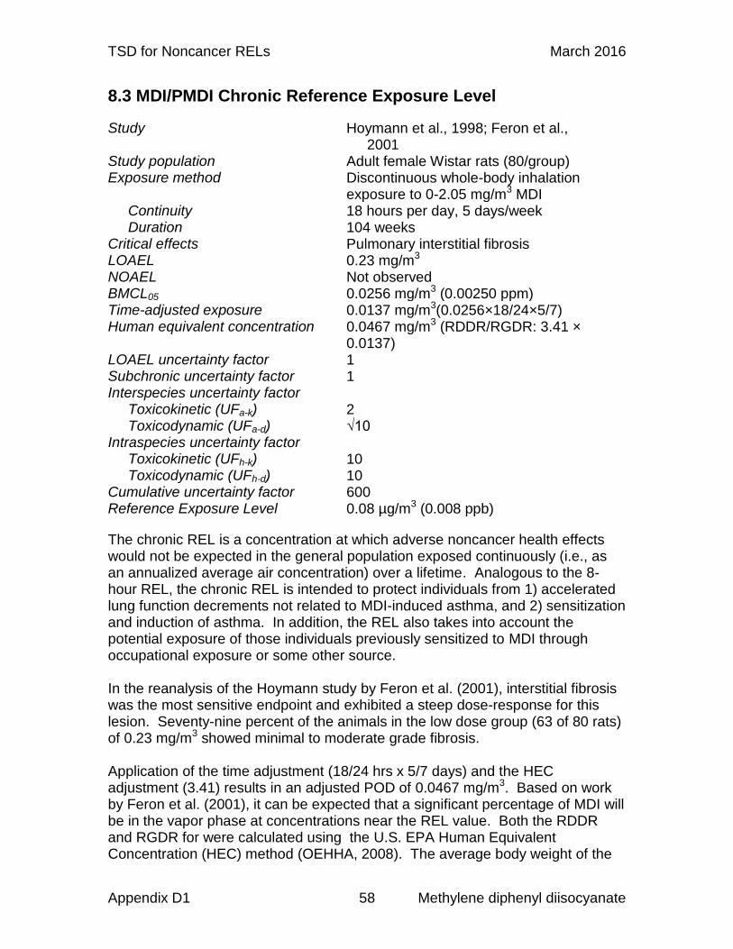
TSD for Noncancer RELs March 2016
Appendix D1 58 Methylene diphenyl diisocyanate
8.3 MDI/PMDI Chronic Reference Exposure Level
Study Hoymann et al., 1998; Feron et al., 2001
Study population Adult female Wistar rats (80/group) Exposure method Discontinuous whole-body inhalation
exposure to 0-2.05 mg/m3 MDI Continuity 18 hours per day, 5 days/week Duration 104 weeks Critical effects Pulmonary interstitial fibrosis LOAEL 0.23 mg/m3 NOAEL Not observed BMCL05 0.0256 mg/m3 (0.00250 ppm) Time-adjusted exposure 0.0137 mg/m3(0.0256×18/24×5/7) Human equivalent concentration 0.0467 mg/m3 (RDDR/RGDR: 3.41 ×
0.0137) LOAEL uncertainty factor 1 Subchronic uncertainty factor 1 Interspecies uncertainty factor Toxicokinetic (UFa-k) 2 Toxicodynamic (UFa-d) √10 Intraspecies uncertainty factor Toxicokinetic (UFh-k) 10 Toxicodynamic (UFh-d) 10 Cumulative uncertainty factor 600 Reference Exposure Level 0.08 µg/m3 (0.008 ppb)
The chronic REL is a concentration at which adverse noncancer health effects would not be expected in the general population exposed continuously (i.e., as an annualized average air concentration) over a lifetime. Analogous to the 8-hour REL, the chronic REL is intended to protect individuals from 1) accelerated lung function decrements not related to MDI-induced asthma, and 2) sensitization and induction of asthma. In addition, the REL also takes into account the potential exposure of those individuals previously sensitized to MDI through occupational exposure or some other source. In the reanalysis of the Hoymann study by Feron et al. (2001), interstitial fibrosis was the most sensitive endpoint and exhibited a steep dose-response for this lesion. Seventy-nine percent of the animals in the low dose group (63 of 80 rats) of 0.23 mg/m3 showed minimal to moderate grade fibrosis. Application of the time adjustment (18/24 hrs x 5/7 days) and the HEC adjustment (3.41) results in an adjusted POD of 0.0467 mg/m3. Based on work by Feron et al. (2001), it can be expected that a significant percentage of MDI will be in the vapor phase at concentrations near the REL value. Both the RDDR and RGDR for were calculated using the U.S. EPA Human Equivalent Concentration (HEC) method (OEHHA, 2008). The average body weight of the

TSD for Noncancer RELs March 2016
Appendix D1 59 Methylene diphenyl diisocyanate
female Wistar rats used to calculate minute volume was 449 g (Feron et al., 2001). Minute volume of adult humans was based on the standard 20 m3/day inhalation rate. In addition, the MMAD=1.03 µm presented in Feron et al. (2001) is used to calculate the RDDR. The calculated RDDR was 3.49 and the RGDR was 3.33. The average value for the two ratios (3.41) was used for the HEC adjustment. For reactive chemicals such as MDI where lesions are formed in the pulmonary tract at points of tissue contact, interspecies toxicokinetic differences are not expected to be large. Thus, following our guidance when applying a Human Equivalent Concentration adjustment, the interspecies UFa-k was assigned a value of 2. We assigned a value of 10 to the intraspecies UFa-k. The intraspecies UFa-k accounts for the up to 3-fold greater pulmonary minute volume-to-surface area ratio in infants and children compared to adults (OEHHA, 2008), as well as gene variants associated with increased sensitivity in workers that were diagnosed with diisocyanate-induced asthma that suggest a wide variation (up to 10-fold) in response among the human population. A default interspecies toxicodynamic UF of √10 was applied. The intraspecies toxicodynamic UF of 10 was used to address MDI’s sensitizing potential and the greater susceptibility of children to the asthma-exacerbating effects of substances such as MDI described in Section 5.2. The toxicogenomic data indicating associations between specific genotype and diisocyanate-induced asthma (ORs between 2 and 9) for enzymes and factors related to toxicodynamic properties, including immune and inflammatory regulation, also support a UF of 10. This gives a cumulative UF of 600, and a chronic REL of 0.08 µg/m3 (0.008 ppb).
The 100-fold intraspecies UF accounts for the uncertainty in establishing a minimum level of MDI/PMDI exposure that will not lead to sensitization in susceptible individuals. What is known is that the proportion of exposed workers who become sensitized is reduced when exposure to MDI, PMDI or other diisocyanates are reduced in the workplace (Tarlo et al., 1997; Tarlo and Liss, 2002; Redlich et al., 2007). In addition, animal models that have been rendered hypersensitive via dermal exposure to diisocyanates have indicated a threshold air level for induction of an asthmatic-like response. Using standard OEHHA risk assessment methodology, a comparison REL based on the sensitized animal model threshold for an asthmatic response is higher than the 8-hour and chronic RELs (see below). Given these findings and the consideration that the RELs are not designed to protect every hypersensitive individual in a population, public health is sufficiently protected with the OEHHA 8-hour and chronic RELs.
Some individuals in a population may have been previously sensitized to MDI, PMDI, or other diisocyanates from some other source(s). Once primary sensitization occurs it is probably not possible to identify a no-effect level to protect all individuals that acquired specific hypersensitivity to diisocyanates (Redlich and Karol, 2002; Redlich et al., 2007). The same conclusion was

TSD for Noncancer RELs March 2016
Appendix D1 60 Methylene diphenyl diisocyanate
presented in an International Consensus Report on Isocyanates (ICRI, 2002). As described above, the number of potentially sensitized individuals in the California population is likely very low (e.g., 12 to 43 per million). Two studies found that the lowest measured MDI concentration at which some sensitized individuals responded was 1 ppb (10 µg/m3) (Burge, 1982; Lemiere et al., 2002). One report exists of an MDI-sensitized worker responding at 0.05 ppb (Suojalehto et al., 2011). The 8-hour and chronic RELs of 0.015 and 0.008 ppb (0.16 and 0.08 µg/m3), respectively, are lower than the lowest level resulting in sensitized individuals responding. Keeping in mind that the RELs are not designed to protect every sensitized individual in a population, and the likelihood that the risk of a sensitized individual being exposed to MDI or PMDI emissions from a facility is very low, the 8-hour and chronic RELs are appropriate for the purposes of the Hot Spots program For comparison, the US EPA based its RfC (similar to a chronic REL) of 0.6 µg/m3 on a benchmark dose analysis of Reuzel et al. (1994a) using basal cell hyperplasia of the olfactory epithelium as the critical effect (U. S. EPA, 1998a). Whereas the RfC was based on data for males only, our analysis utilized the data for female rats in the reexamination of the Hoymann data by Feron et al. (2001). OEHHA chose interstitial fibrosis as the critical effect because this was the most sensitive endpoint for an exposure duration (18 hrs/ day, 5 days/week) that came closest to a continuous chronic exposure. In addition, OEHHA used a larger toxicodynamic UFh-d than USEPA specifically to protect against the onset of asthma symptoms in children, and a toxicokinetic UFh-k of 10 to account for differences in risk of diisocyanate-induced asthma, as observed in workers, based on genotype for a number of metabolic and protective enzymes. Pauluhn and Poole (2011) determined a threshold level of 5 mg/m3 × 30 min for prevention of an asthmatic-like response (increase PMNs in the lung) in a sensitized rat model, in which induction occurred by repeated inhalation exposure to PMDI. This rat model was also used to derive an 8-hour TWA worker exposure level for TDI (Pauluhn, 2014). A summary of this study is presented in Section 5.3. OEHHA used the rat “asthma” threshold to derive 8-hour and chronic RELs and compare it with the RELs derived above based on chronic exposure studies in rats.
To derive an 8-hour REL, 5 mg/m3 × 30 min is divided by 480 min for a concentration of 0.31 mg/m3 for the equivalent 8-hour exposure. OEHHA could justifiably apply a toxicokinetic adjustment developed by Pauluhn (2014) of √10 for obligate vs. oronasal breathing. Since PMDI and MDI are primarily pulmonary irritants, no toxicokinetic adjustment is made for depression of respiration rate and minute volume by the rats (e.g., as done for upper respiratory irritants such as TDI). To the interspecies toxicokinetic adjustment, OEHHA would also include a default interspecies toxicodynamic uncertainty factor of √10. For intraspecies uncertainty, OEHHA would use a 100-fold factor (10 for toxicokinetic and 10 for toxicodynamic) based mainly on gene variants associated with increased sensitivity in workers that were diagnosed with diisocyanate-induced

TSD for Noncancer RELs March 2016
Appendix D1 61 Methylene diphenyl diisocyanate
asthma. The total uncertainty adjustment factor would then = 1000 (√10 × √10 × 10 × 10). The OEHHA-derived comparison 8-hour REL is 0.0003 mg/m3 (0.3 µg/m3 or 0.03 ppb). Use of chronic rat exposure data to derive the 8-hour REL (i.e., 0.16 µg/m3, or 0.015 ppb) is roughly 2-fold lower than the comparison REL, a more health-protective level for the REL. To derive a chronic REL, a time adjustment of 1440 min would be used. For a chronic REL, applying a 24-hour time adjustment to the point of departure (5 mg/m3 × 30 min / 1440 min), and the same dosimetric adjustments and uncertainty factors as used in the 8-hour REL derivation, a comparison chronic REL of 0.1 µg/m3 (0.01 ppb) is calculated. The chronic REL based on a chronic exposure study derived above of 0.008 ppb is about 10-fold lower than the comparison REL and, thus is the more health protective level.
8.4 MDI/PMDI as a Toxic Air Contaminant Especially Affecting Infants and Children
Under Health and Safety Code Section 39669.5, OEHHA establishes and maintains a list of Toxic Air Contaminants (TACs) that may disproportionately impact infants and children. OEHHA evaluates TACs for addition to this list as we develop Reference Exposure Levels for TACs. MDI was identified by the ARB as a toxic air contaminant (TAC) in accordance with section 39657(b) of the California Health and Safety Code (Title 17, California Code of Regulations, section 93001) (CCR, 2007). MDI has been shown to cause asthmatic reactions in sensitized asthmatic adults in controlled exposure studies, and possibly in non-sensitized children with asthma as well as asthma-like effects in normal children exposed acutely in an accidental exposure (Jan et al., 2008). OEHHA considers asthma a disease that disproportionately impacts children, and thus chemicals that induce or exacerbate asthma are considered more impactful for children (OEHHA, 2001). In addition, an animal study has shown that younger rats are more sensitive to the acute effects of MDI than young adult rats (Reuzel et al., 1994b). In view of the potential of MDI to exacerbate asthma and the differential impacts of asthma on children including higher prevalence rates, OEHHA recommends that MDI be identified as a TAC that may disproportionally impact children pursuant to Health and Safety Code, Section 39669.5(c).

TSD for Noncancer RELs March 2016
Appendix D1 62 Methylene diphenyl diisocyanate
9.0 References
Baur X (1983). Immunologic cross-reactivity between different albumin-bound isocyanates. J Allergy Clin Immunol 71(2): 197-205.
Baur X (1995). Hypersensitivity pneumonitis (extrinsic allergic alveolitis) induced by isocyanates. J Allergy Clin Immunol 95(5 Pt 1): 1004-10.
Baur X, Dewair M and Rommelt H (1984). Acute airway obstruction followed by hypersensitivity pneumonitis in an isocyanate (MDI) worker. J Occup Med 26(4): 285-7.
Baur X, Marek W, Ammon J, Czuppon AB, Marczynski B, Raulf-Heimsoth M, Roemmelt H and Fruhmann G (1994). Respiratory and other hazards of isocyanates. Int Arch Occup Environ health 66(3): 141-52.
Bello D, Herrick CA, Smith TJ, Woskie SR, Streicher RP, Cullen MR, Liu Y and Redlich CA (2007). Skin exposure to isocyanates: reasons for concern. Environ Health Perspect 115(3): 328-35.
Bello D, Woskie SR, Streicher RP, Liu Y, Stowe MH, Eisen EA, Ellenbecker MJ, Sparer J, Youngs F, Cullen MR and Redlich CA (2004). Polyisocyanates in occupational environments: a critical review of exposure limits and metrics. American journal of industrial medicine 46(5): 480-91.
Bernstein DI, Kashon M, Lummus ZL, Johnson VJ, Fluharty K, Gautrin D, Malo JL, Cartier A, Boulet LP, Sastre J, Quirce S, Germolec D, Tarlo SM, Cruz MJ, Munoz X, Luster MI and Yucesoy B (2013). CTNNA3 (alpha-catenin) gene variants are associated with diisocyanate asthma: a replication study in a Caucasian worker population. Toxicol Sci 131(1): 242-6.
Bernstein DI, Korbee L, Stauder T, Bernstein JA, Scinto J, Herd ZL and Bernstein IL (1993). The low prevalence of occupational asthma and antibody-dependent sensitization to diphenylmethane diisocyanate in a plant engineered for minimal exposure to diisocyanates. J Allergy Clin Immunol 92(3): 387-96.
Bernstein DI, Wang N, Campo P, Chakraborty R, Smith A, Cartier A, Boulet LP, Malo JL, Yucesoy B, Luster M, Tarlo SM and Hershey GK (2006). Diisocyanate asthma and gene-environment interactions with IL4RA, CD-14, and IL-13 genes. Ann Allergy Asthma Immunol 97(6): 800-6.
Blomqvist P, McNamee MS, Stec AA, Gylestam D and Karlsson D (2014). Detailed study of distribution patterns of polycyclic aromatic hydrocarbons and isocyanates under different fire conditions. Fire Mater 38: 125-44.

TSD for Noncancer RELs March 2016
Appendix D1 63 Methylene diphenyl diisocyanate
Bolognesi C, Baur X, Marczynski B, Norppa H, Sepai O and Sabbioni G (2001). Carcinogenic risk of toluene diisocyanate and 4,4'-methylenediphenyl diisocyanate: epidemiological and experimental evidence. Crit Rev Toxicol 31(6): 737-72.
Bonauto DK, Sumner AD, Curwick CC, Whittaker SG and Lofgren DJ (2005). Work-related asthma in the spray-on truck bed lining industry. J Occup Environ Med 47(5): 514-7.
Booth K, Cummings B, Karoly WJ, Mullins S, Robert WP, Spence M, Lichtenberg FW and Banta J (2009). Measurements of airborne methylene diphenyl diisocyanate (MDI) concentration in the U.S. workplace. J Occup Environ Hyg 6(4): 228-38.
Boyd HB and Mogensen BB (2007). Survey and health assessment of selected respiratory sensitizers in consumer products. Survey of Chemical Substances in Consumer Products, No. 82, Danish Ministry of the Environment. Online at: http://www2.mst.dk/Udgiv/publications/2007/978-87-7052-506-0/pdf/978-87-7052-507-7.pdf.
Buchdahl R, Willems CD, Vander M and Babiker A (2000). Associations between ambient ozone, hydrocarbons, and childhood wheezy episodes: a prospective observational study in south east London. Occup Environ Med 57(2): 86-93.
Budnik LT, Nowak D, Merget R, Lemiere C and Baur X (2011). Elimination kinetics of diisocyanates after specific inhalative challenges in humans: mass spectrometry analysis, as a basis for biomonitoring strategies. J Occup Med Toxicol 6(1): 9.
Burge PS (1982). Non-specific bronchial hyper-reactivity in workers exposed to toluene di-isocyanate, diphenyl methane di-isocyanate and colophony. Eur J Resp Dis (Suppl) 123: 91-6.
Buschmann J, Koch W, Fuhst R and Heinrich U (1996). Embryotoxicity study of monomeric 4,4'-methylenediphenyl diisocyanate (MDI) aerosol after inhalation exposure in Wistar rats. Fundam Appl Toxicol 32(1): 96-101.
Calabresi C, Arosio B, Galimberti L, Scanziani E, Bergottini R, Annoni G and Vergani C (2007). Natural aging, expression of fibrosis-related genes and collagen deposition in rat lung. Exp Gerontol 42(10): 1003-11.
CARB. (2013). California Toxics Inventory. California Air Resources Board. Online at: http://www.arb.ca.gov/toxics/cti/cti.htm.
Carpenter CP, Kinkead ER, Geary DL, Jr., Sullivan LJ and King JM (1975). Petroleum hydrocarbon toxicity studies. V. Animal and human response to vapors of mixed xylenes. Toxicol Appl Pharmacol 33(3): 543-58.

TSD for Noncancer RELs March 2016
Appendix D1 64 Methylene diphenyl diisocyanate
Cartier A, Grammer L, Malo JL, Lagier F, Ghezzo H, Harris K and Patterson R (1989). Specific serum antibodies against isocyanates: association with occupational asthma. J Allergy Clin Immunol 84(4 Pt 1): 507-14.
CCR (2007). California Code of Regulations Section 93001 Hazardous Air Pollutants Identified as Toxic Air Contaminants. Sacramento, CA: California Office of Administrative Law. 8-20-07. http://ccr.oal.ca.gov/linkedslice/default.asp?SP=CCR-1000&Action=Welcome.
Chatkin JM, Tarlo SM, Liss G, Banks D and Broder I (1999). The outcome of asthma related to workplace irritant exposures: a comparison of irritant-induced asthma and irritant aggravation of asthma. Chest 116(6): 1780-5.
Choi JH, Lee KW, Kim CW, Park CS, Lee HY, Hur GY, Kim SH, Hong CS, Jang AS and Park HS (2009). The HLA DRB1*1501-DQB1*0602-DPB1*0501 haplotype is a risk factor for toluene diisocyanate-induced occupational asthma. Int Arch Allergy Immunol 150(2): 156-63.
Crane J, Kemp T, Siebers R, Rains N, Fishwick D and Fitzharris P (1997). Increased house dust mite allergen in synthetic pillows may explain increased wheezing. BMJ 314(7096): 1763-4.
Crespo J and Galan J (1999). Exposure to MDI during the process of insulating buildings with sprayed polyurethane foam. Ann Occup Hyg 43(6): 415-9.
Del Prete GF, De Carli M, D'Elios MM, Maestrelli P, Ricci M, Fabbri L and Romagnani S (1993). Allergen exposure induces the activation of allergen-specific Th2 cells in the airway mucosa of patients with allergic respiratory disorders. Eur J Immunol 23(7): 1445-9.
Delfino RJ, Gong H, Linn WS, Hu Y and Pellizzari ED (2003). Respiratory symptoms and peak expiratory flow in children with asthma in relation to volatile organic compounds in exhaled breath and ambient air. J Expo Anal Environ Epidemiol 13(5): 348-63.
Ernst H, Hoymann HG, Rittinghausen S, Mohr U and Heinrich U (1998). Pulmonary lesions in Wistar rats associated with chronic inhalation of 4,4'methylene-diphenyl diisocyanate (MDI). In: Relationships Between Respiratory Disease and Exposure to Air Pollution. Mohr, U, Dungworth DL, Brain JD, Driscoll KE, Grafstrom RC, Harris CC (eds). ILSI Press, Washington, pp 320-24.
Estlander T, Keskinen H, Jolanki R and Kanerva L (1992). Occupational dermatitis from exposure to polyurethane chemicals. Contact Dermatitis 27(3): 161-5.

TSD for Noncancer RELs March 2016
Appendix D1 65 Methylene diphenyl diisocyanate
Feron VJ, Kittel B, Kuper CF, Ernst H, Rittinghausen S, Muhle H, Koch W, Gamer A, Mallett AK and Hoffmann HD (2001). Chronic pulmonary effects of respirable methylene diphenyl diisocyanate (MDI) aerosol in rats: combination of findings from two bioassays. Arch Toxicol 75(3): 159-75.
Finotto S, Fabbri LM, Rado V, Mapp CE and Maestrelli P (1991). Increase in numbers of CD8 positive lymphocytes and eosinophils in peripheral blood of subjects with late asthmatic reactions induced by toluene diisocyanate. Br J Ind Med 48(2): 116-21.
Gamer AO, Hellwig J, Doe JE and Tyl RW (2000). Prenatal toxicity of inhaled polymeric methylenediphenyl diisocyanate (MDI) aerosols in pregnant wistar rats. Toxicol Sci 54(2): 431-40.
Gledhill A, Wake A, Hext P, Leibold E and Shiotsuka R (2005). Absorption, distribution, metabolism and excretion of an inhalation dose of [14C] 4,4'-methylenediphenyl diisocyanate in the male rat. Xenobiotica 35(3): 273-92.
Herrick CA, Das J, Xu L, Wisnewski AV, Redlich CA and Bottomly K (2003). Differential roles for CD4 and CD8 T cells after diisocyanate sensitization: genetic control of TH2-induced lung inflammation. J Allergy Clin Immunol 111(5): 1087-94.
Hertzberg T, Blomqvist P, Dalene M and Skarping G. (2003). Particles and isocyanates from fires. Brandforsk Project 324-021, SP Swedish National Testing and Research Institute, Sweden, pp. 50.
Hoffmann HD and Schupp T (2009). Evaluation of consumer risk resulting from exposure against diphenylmethane-4,4'-diisocyanate (MDI) from polyurethane foam. EXCLI J 8: 58-65.
Hoymann HG, Creutzenberg O, Ernst H, Hohlfeld J and Mohr U (1998). Lung function changes in rats after chronic inhalation exposure to 4,4'-methylene diphenyl diisocyanate (MDI). In: Relationships Between Respiratory Disease and Exposure to Air Pollution. Mohr, U, Dungworth DL, Brain JD, Driscoll KE, Grafstrom RC, Harris CC (eds). ILSI Press, Washington, pp 353-56.
HSDB (2015). Hazardous Substances Data Bank. Chemical/Physical Properties; Emergency Medical Treatment. National Library of Medicine, Bethesda, MD. Available at: http://toxnet.nlm.nih.gov.
Hughes MA, Carson M, Collins MA, Jolly AT, Molenaar DM, Steffens W and Swaen GM (2014). Does diisocyanate exposure result in neurotoxicity? Clin Toxicol (Phila) 52(4): 242-57.
Hugo JM, Spence MW and Lickly TD (2000). The determination of the ability of polyurethane foam to release toluene diisocyanate into air. Appl Occup Environ Hyg 15(6): 512-9.

TSD for Noncancer RELs March 2016
Appendix D1 66 Methylene diphenyl diisocyanate
ICRI (2002). International Consensus Report on Isocyanates: Risk Assessment and Management. Ed, Jan Vilhelm Bakke, held November 22-22, 2001 at Hotel Norge Hoesbjoer, N-2320, Furnes, Norway.
Innocenti A, Cirla AM, Pisati G and Mariano A (1988). Cross-reaction between aromatic isocyanates (TDI and MDI): a specific bronchial provocation test study. Clin Allergy 18(4): 323-9.
Jan RL, Chen SH, Chang HY, Yeh HJ, Shieh CC and Wang JY (2008). Asthma-like syndrome in school children after accidental exposure to xylene and methylene diphenyl diisocyanate. J Microbiol Immunol Infect 41(4): 337-41.
Jang AS, Choi IS, Koh YI, Moon JD and Lee KJ (2000). Increase in airway hyperresponsiveness among workers exposed to methylene diphenyldiisocyanate compared to workers exposed to toluene diisocyanate at a petrochemical plant in Korea. Am J Ind Med 37(6): 663-7.
Johnson A, Moira CY, MacLean L, Atkins E, Dybuncio A, Cheng F and Enarson D (1985). Respiratory abnormalities among workers in an iron and steel foundry. Br J Ind Med 42(2): 94-100.
Kaaria K, Hirvonen A, Norppa H, Piirila P, Vainio H and Rosenberg C (2001). Exposure to 4,4'-methylenediphenyl diisocyanate (MDI) during moulding of rigid polyurethane foam: determination of airborne MDI and urinary 4,4'-methylenedianiline (MDA). Analyst 126(4): 476-9.
Kilgour JD, Rattray NJ, Foster J, Soames A and Hext PM (2002). Pulmonary responses and recovery following single and repeated inhalation exposure of rats to polymeric methylene diphenyl diisocyanate aerosols. J Appl Toxicol 22(6): 371-85.
Kim SH, Cho BY, Park CS, Shin ES, Cho EY, Yang EM, Kim CW, Hong CS, Lee JE and Park HS (2009). Alpha-T-catenin (CTNNA3) gene was identified as a risk variant for toluene diisocyanate-induced asthma by genome-wide association analysis. Clin Exp Allergy 39(2): 203-12.
Kim SH, Choi GS, Ye YM, Jou I, Park HS and Park SM (2010). Toluene diisocyanate (TDI) regulates haem oxygenase-1/ferritin expression: implications for toluene diisocyanate-induced asthma. Clin Exp Immunol 160(3): 489-97.
Kim SH, Oh HB, Lee KW, Shin ES, Kim CW, Hong CS, Nahm DH and Park HS (2006). HLA DRB1*15-DPB1*05 haplotype: a susceptible gene marker for isocyanate-induced occupational asthma? Allergy 61(7): 891-4.
Krone CA, Ely JT, Klingner T and Rando RJ (2003a). Isocyanates in flexible polyurethane foams. Bull Environ Contam Toxicol 70(2): 328-35.

TSD for Noncancer RELs March 2016
Appendix D1 67 Methylene diphenyl diisocyanate
Krone CA and Klingner TD (2005). Isocyanates, polyurethane and childhood asthma. Pediatr Allergy Immunol 16(5): 368-79.
Krone CA, Klingner TD and Ely JT (2003b). Polyurethanes and childhood asthma. Med Sci Monit 9(12): HY39-43.
Kullman G, Knutti E, Buono G and Allen E. (1998). Health Hazard Evaluation Report 94-0265-2703. J.L. Long Middle School, Dallas Texas. NIOSH, Cincinatti, OH.
Lastbom L, Colmsjo A, Johansson R, Karlsson D, Melin J, Nordqvist Y and Skarping G (2003). Effects of thermal degradation products from polyurethane foams based on toluene diisocyanate and diphenylmethane diisocyanate on isolated, perfused lung of guinea pig. Scand J Work Environ Health 29(2): 152-8.
Latza U, Baur X and Malo JL (2002). Isocyanate-induced health effects. In: Bakke JV, Noren JO, Thorud S, Aasen TB, editors. International Consensus Report on Isocyanates: Risk Assessment and Management, Appendices 10.2.1. Gjovik: Norwegian Labour Inspection Authority; 37-51.
Lemiere C, Romeo P, Chaboillez S, Tremblay C and Malo JL (2002). Airway inflammation and functional changes after exposure to different concentrations of isocyanates. J Allergy Clin Immunol 110(4): 641-6.
Leroyer C, Perfetti L, Cartier A and Malo JL (1998). Can reactive airways dysfunction syndrome (RADS) transform into occupational asthma due to "sensitisation" to isocyanates? Thorax 53(2): 152-3.
Lesage J, Stanley J, Karoly WJ and Lichtenberg FW (2007). Airborne methylene diphenyl diisocyanate (MDI) concentrations associated with the application of polyurethane spray foam in residential construction. J Occup Environ Hyg 4(2): 145-55.
Liljelind I, Norberg C, Egelrud L, Westberg H, Eriksson K and Nylander-French LA (2010). Dermal and inhalation exposure to methylene bisphenyl isocyanate (MDI) in iron foundry workers. Ann Occup Hyg 54(1): 31-40.
Liss GM, Bernstein DI, Moller DR, Gallagher JS, Stephenson RL and Bernstein IL (1988). Pulmonary and immunologic evaluation of foundry workers exposed to methylene diphenyldiisocyanate (MDI). J Allergy Clin Immunol 82(1): 55-61.
Littorin M, Hou S, Broberg K, Bjork J, Falt S, Abdoulaye G, Kalemba M, Ryk C and Skerfving S (2008). Influence of polymorphic metabolic enzymes on biotransformation and effects of diphenylmethane diisocyanate. Int Arch Occup Environ Health 81(4): 429-41.

TSD for Noncancer RELs March 2016
Appendix D1 68 Methylene diphenyl diisocyanate
Littorin M, Welinder H, Skarping G, Dalene M and Skerfving S (2002). Exposure and nasal inflammation in workers heating polyurethane. Int Arch Occup Environ Health 75(7): 468-74.
Lockey JE, Redlich CA, Streicher R, Pfahles-Hutchens A, Hakkinen PB, Ellison GL, Harber P, Utell M, Holland J, Comai A and White M (2015). Isocyanates and human health: multistakeholder information needs and research priorities. J Occup Environ Med 57(1): 44-51.
Lofgren DJ, Walley TL, Peters PM and Weis ML (2003). MDI exposure for spray-on truck bed lining. Appl Occup Environ Hyg 18(10): 772-9.
Lummus ZL, Alam R, Bernstein JA and Bernstein DI (1998). Diisocyanate antigen-enhanced production of monocyte chemoattractant protein-1, IL-8, and tumor necrosis factor-alpha by peripheral mononuclear cells of workers with occupational asthma. J Allergy Clin Immunol 102(2): 265-74.
Macpherson LJ, Dubin AE, Evans MJ, Marr F, Schultz PG, Cravatt BF and Patapoutian A (2007). Noxious compounds activate TRPA1 ion channels through covalent modification of cysteines. Nature 445(7127): 541-5.
Maestrelli P, Del Prete GF, De Carli M, D'Elios MM, Saetta M, Di Stefano A, Mapp CE, Romagnani S and Fabbri LM (1994). CD8 T-cell clones producing interleukin-5 and interferon-gamma in bronchial mucosa of patients with asthma induced by toluene diisocyanate. Scand J Work Environ Health 20(5): 376-81.
Maestrelli P, Occari P, Turato G, Papiris SA, Di Stefano A, Mapp CE, Milani GF, Fabbri LM and Saetta M (1997). Expression of interleukin (IL)-4 and IL-5 proteins in asthma induced by toluene diisocyanate (TDI). Clin Exp Allergy 27(11): 1292-8.
Mapp CE, Beghe B, Balboni A, Zamorani G, Padoan M, Jovine L, Baricordi OR and Fabbri LM (2000). Association between HLA genes and susceptibility to toluene diisocyanate-induced asthma. Clin Exp Allergy 30(5): 651-6.
Marand A, Dahlin J, Karlsson D, Skarping G and Dalene M (2004). Determination of technical grade isocyanates used in the production of polyurethane plastics. J Environ Monit 6(7): 606-14.
NIOSH. (1998). NIOSH Manual of Analytical Methods. . K: CDC. http://www.cdc.gov/niosh/docs/2003-154/pdfs/chapter-k.pdf.
NLAC (2014). National Laboratory Animal Center. Online at: http://www.nlac.mahidol.ac.th/nlacmuEN/p_animal_Rat.htm.
O'Brien IM, Harries MG, Burge PS and Pepys J (1979). Toluene di-isocyanate-induced asthma. I. Reactions to TDI, MDI, HDI and histamine. Clin Allergy 9: 1-6.

TSD for Noncancer RELs March 2016
Appendix D1 69 Methylene diphenyl diisocyanate
OEHHA (2001). Prioritization of Toxic Air Contaminants Under the Children's Environmental Health Protection Act. California Environmental Protection Agency, Office of Environmental Health Hazard Assessment, Oakland, CA. Online at: http://oehha.ca.gov/air/toxic_contaminants/SB25finalreport.html.
OEHHA. (2008). Air Toxics Hot Spots Program Risk Assessment Guidelines. Technical Support Document for the Derivation of Noncancer Reference Exposure Levels California Environmental Protection Agency, Office of Environmental Health Hazard Assessment, Oakland, CA. Online at: http://www.oehha.ca.gov/air/hot_spots/rels_dec2008.html.
Ott MG, Diller WF and Jolly AT (2003). Respiratory effects of toluene diisocyanate in the workplace: a discussion of exposure-response relationships. Crit Rev Toxicol 33(1): 1-59.
Pauluhn J (2000). Acute inhalation toxicity of polymeric diphenyl-methane 4,4'-diisocyanate in rats: time course of changes in bronchoalveolar lavage. Arch Toxicol 74(4-5): 257-69.
Pauluhn J (2002). Short-term inhalation toxicity of polyisocyanate aerosols in rats: comparative assessment of irritant-threshold concentrations by bronchoalveolar lavage. Inhal Toxicol 14(3): 287-301.
Pauluhn J (2011). Interrelating the acute and chronic mode of action of inhaled methylenediphenyl diisocyanate (MDI) in rats assisted by computational toxicology. Regul Toxicol Pharmacol 61(3): 351-64.
Pauluhn J (2014). Development of a respiratory sensitization/elicitation protocol of toluene diisocyanate (TDI) in Brown Norway rats to derive an elicitation-based occupational exposure level. Toxicology 319: 10-22.
Pauluhn J and Poole A (2011). Brown Norway rat asthma model of diphenylmethane-4,4'-diisocyanate (MDI): determination of the elicitation threshold concentration of after inhalation sensitization. Toxicology 281(1-3): 15-24.
Pauluhn J, Thiel A, Emura M and Mohr U (2000). Respiratory sensitization to diphenyl-methane-4,4'-diisocyanate (MDI) in guinea pigs: impact of particle size on induction and elicitation of response. Toxicol Sci 56(1): 105-13.
Petsonk EL, Wang ML, Lewis DM, Siegel PD and Husberg BJ (2000). Asthma-like symptoms in wood product plant workers exposed to methylene diphenyl diisocyanate. Chest 118(4): 1183-93.
Pham QT, Teculescu D, Meyer-Bisch C and Mur JM (1987). Effects of chronic exposure to diisocyanates. Bull Eur Physiopathol Respir 23(6): 561-4.

TSD for Noncancer RELs March 2016
Appendix D1 70 Methylene diphenyl diisocyanate
Piirila P, Wikman H, Luukkonen R, Kaaria K, Rosenberg C, Nordman H, Norppa H, Vainio H and Hirvonen A (2001). Glutathione S-transferase genotypes and allergic responses to diisocyanate exposure. Pharmacogenetics 11(5): 437-45.
Piirila PL, Nordman H, Keskinen HM, Luukkonen R, Salo SP, Tuomi TO and Tuppurainen M (2000). Long-term follow-up of hexamethylene diisocyanate-, diphenylmethane diisocyanate-, and toluene diisocyanate-induced asthma. Am J Respir Crit Care Med 162(2 Pt 1): 516-22.
Prescott SL, Macaubas C, Smallacombe T, Holt BJ, Sly PD and Holt PG (1999). Development of allergen-specific T-cell memory in atopic and normal children. Lancet 353(9148): 196-200.
Rattray NJ, Botham PA, Hext PM, Woodcock DR, Fielding I, Dearman RJ and Kimber I (1994). Induction of respiratory hypersensitivity to diphenylmethane-4,4'-diisocyanate (MDI) in guinea pigs. Influence of route of exposure. Toxicology 88(1-3): 15-30.
Raulf-Heimsoth M and Baur X (1998). Pathomechanisms and pathophysiology of isocyanate-induced diseases--summary of present knowledge. Am J Ind Med 34(2): 137-43.
Redlich CA, Bello D and Wisnewski AV (2007). Isocyanate exposures and health effects. In Rom WM, editor: Environmental and Occupational Medicine. 4th Edn. Philadelphia: Lippincott Williams & Wilkins, pp. 502-16. In.
Redlich CA and Karol MH (2002). Diisocyanate asthma: clinical aspects and immunopathogenesis. Int Immunopharmacol 2(2-3): 213-24.
Redlich CA, Karol MH, Graham C, Homer RJ, Holm CT, Wirth JA and Cullen MR (1997). Airway isocyanate-adducts in asthma induced by exposure to hexamethylene diisocyanate. Scand J Work Environ Health 23(3): 227-31.
Reidy TJ and Bolter JF (1994). Neuropsychological toxicology of methylene diphenyl diisocyanate: a report of five cases. Brain Inj 8(3): 285-94.
Renne RA, Dungworth DL, Keenan CM, Morgan KT, Hahn FF and Schwartz LW (2003). Non-proliferative lesions of the respiratory tract in rats. R-1. In: Guides for Toxicologic Pathology. STP/ARP/AFIP, Washington, DC.
Reuzel PG, Arts JH, Lomax LG, Kuijpers MH, Kuper CF, Gembardt C, Feron VJ and Loser E (1994a). Chronic inhalation toxicity and carcinogenicity study of respirable polymeric methylene diphenyl diisocyanate (polymeric MDI) aerosol in rats. Fundam Appl Toxicol 22(2): 195-210.

TSD for Noncancer RELs March 2016
Appendix D1 71 Methylene diphenyl diisocyanate
Reuzel PG, Kuper CF, Feron VJ, Appelman LM and Loser E (1994b). Acute, subacute, and subchronic inhalation toxicity studies of respirable polymeric methylene diphenyl diisocyanate (polymeric MDI) aerosol in rats. Fundam Appl Toxicol 22(2): 186-94.
Ribeiro M, Tarlo SM, Czyrka A, Vernich L, Luce CE and Liss GM (2014). Diisocyanate and non-diisocyanate sensitizer-induced occupational asthma frequency during 2003 to 2007 in Ontario, Canada. J Occup Environ Med 56(9): 1001-7.
Schupp T and Collins MA (2012). Toluene diisocyanate (TDI) airway effects and dose-responses in different animal models. EXLCI J 11: 416-35.
Skarping G, Dalene M, Svensson BG, Littorin M, Akesson B, Welinder H and Skerfving S (1996). Biomarkers of exposure, antibodies, and respiratory symptoms in workers heating polyurethane glue. Occup Environ Med 53(3): 180-7.
Strachan DP and Carey IM (1995). Home environment and severe asthma in adolescence: a population based case-control study. BMJ 311(7012): 1053-6.
Streicher RP, Reh CM, Key-Schwartz RJ, Schlecht PC, Cassinelli ME and O'Connor PF (2000). Determination of airborne isocyanate exposure: considerations in method selection. AIHAJ 61(4): 544-56.
Sulotto F, Romano C, Piolatto G, Coggiola M, Polizzi S, Ciacco C and Berra A (1990). Short-term respiratory changes in polyurethane foam workers exposed to low MDI concentration. Int Arch Occup Environ Health 62(7): 521-4.
Suojalehto H, Linstrom I, Henriks-Eckerman ML, Jungewelter S and Suuronen K (2011). Occupational asthma related to low levels of airborne methylene diphenyl diisocyanate (MDI) in orthopedic casting work. Am J Ind Med 54(12): 906-10.
Tarlo SM, Liss G, Corey P and Broder I (1995). A workers' compensation claim population for occupational asthma. Comparison of subgroups. Chest 107(3): 634-41.
Tarlo SM and Liss GM (2002). Diisocyanate-induced asthma: diagnosis, prognosis, and effects of medical surveillance measures. Applied occupational and environmental hygiene 17(12): 902-8.
Tarlo SM, Liss GM, Dias C and Banks DE (1997). Assessment of the relationship between isocyanate exposure levels and occupational asthma. American journal of industrial medicine 32(5): 517-21.
Taylor-Clark TE, Kiros F, Carr MJ and McAlexander MA (2009). Transient receptor potential ankyrin 1 mediates toluene diisocyanate-evoked respiratory irritation. Am J Respir Cell Mol Biol 40(6): 756-62.

TSD for Noncancer RELs March 2016
Appendix D1 72 Methylene diphenyl diisocyanate
Tury B, Pemberton D and Bailey RE (2003). Fate and potential environmental effects of methylenediphenyl diisocyanate and toluene diisocyanate released into the atmosphere. J Air & Waste Manage Assoc 53(1): 61-6.
U. S. EPA. (1998a). Methylene Diphenyl Diisocyanate (monomeric MDI) and polymeric MDI (PMDI) (CASRN 101-68-8, 9016-87-9). Integrated Risk Information System. U.S. Environmental Protection Agency, Washington DC. Online at: http://www.epa.gov/iris/subst/0529.htm.
U. S. EPA. (1998c). Toxicological Review of Methylene Diphenyl Diisocyanate (MDI). CAS No. 101-68-8 and 9016-87-9. U.S. Environmental Protection Agency, Washington DC.
U. S. EPA. (2012). Benchmark Dose Software, Version 2.3.1. National Center for Environmental Assessment. Online at: http://www.epa.gov/ncea/bmds.
U. S. OSHA (2015). OSHA Annotated Table Z-1, Occupational Safety & Health Administration, U.S. Department of Labor. Online at: www.osha.gov/dsg/annotated-pels/tablez-1.html.
Ulvestad B, Melbostad E and Fuglerud P (1999). Asthma in tunnel workers exposed to synthetic resins. Scand J Work Environ Health 25(4): 335-41.
Vandenplas O, Cartier A, Ghezzo H, Cloutier Y and Malo JL (1993a). Response to isocyanates: effect of concentration, duration of exposure, and dose. Am Rev Respir Dis 147(5): 1287-90.
Vandenplas O, Cartier A, Lesage J, Perrault G, Grammer LC and Malo JL (1992). Occupational asthma caused by a prepolymer but not the monomer of toluene diisocyanate (TDI). J Allergy Clin Immunol 89(6): 1183-8.
Vandenplas O, Malo JL, Dugas M, Cartier A, Desjardins A, Levesque J, Shaughnessy MA and Grammer LC (1993b). Hypersensitivity pneumonitis-like reaction among workers exposed to diphenylmethane [correction to piphenylmethane] diisocyanate (MDI). Am Rev Respir Dis 147(2): 338-46.
Vangronsveld E, Berckmans S and Spence M (2013). Toluene diisocyanate emission to air and migration to a surface from a flexible polyurethane foam. Ann Occup Hyg 57(5): 650-61.
Verschoor L and Verschoor AH (2014). Nonoccupational and occupational exposure to isocyanates. Curr Opin Pulm Med 20(2): 199-204.
Vock EH, Cantoreggi S, Gupta RC and Lutz WK (1995). 32P-postlabeling analysis of DNA adducts formed in vitro and in rat skin by methylenediphenyl-4,4'-diisocyanate (MDI). Toxicol Lett 76(1): 17-26.

TSD for Noncancer RELs March 2016
Appendix D1 73 Methylene diphenyl diisocyanate
Vock EH, Hoymann HG, Heinrich U and Lutz WK (1996). 32P-postlabeling of a DNA adduct derived from 4,4'-methylenedianiline, in the olfactory epithelium of rats exposed by inhalation to 4,4'-methylenediphenyl diisocyanate. Carcinogenesis 17(5): 1069-73.
Weyel DA and Schaffer RB (1985). Pulmonary and sensory irritation of diphenylmethane-4,4'- and dicyclohexylmethane-4,4'-diisocyanate. Toxicol Appl Pharmacol 77(3): 427-33.
Wikman H, Piirila P, Rosenberg C, Luukkonen R, Kaaria K, Nordman H, Norppa H, Vainio H and Hirvonen A (2002). N-Acetyltransferase genotypes as modifiers of diisocyanate exposure-associated asthma risk. Pharmacogenetics 12(3): 227-33.
Wills-Karp M (1999). Immunologic basis of antigen-induced airway hyperresponsiveness. Annu Rev Immunol 17: 255-81.
Wisnewski AV and Liu J (2013). Molecular determinants of humoral immune specificity for the occupational allergen, methylene diphenyl diisocyanate. Mol Immunol 54(2): 233-7.
Wisnewski AV, Liu J and Colangelo CM (2015). Glutathione Reaction Products with a Chemical Allergen, Methylene-diphenyl Diisocyanate, Stimulate Alternative Macrophage Activation and Eosinophilic Airway Inflammation. Chem Res Toxicol 28(4): 729-37.
Wisnewski AV, Liu J and Redlich CA (2013). Connecting glutathione with immune responses to occupational methylene diphenyl diisocyanate exposure. Chem Biol Interact 205(1): 38-45.
Wisnewski AV, Xu L, Robinson E, Liu J, Redlich CA and Herrick CA (2011). Immune sensitization to methylene diphenyl diisocyanate (MDI) resulting from skin exposure: albumin as a carrier protein connecting skin exposure to subsequent respiratory responses. J Occup Med Toxicol 6: 6.
Woellner RC, Hall S, Greaves I and Schoenwetter WF (1997). Epidemic of asthma in a wood products plant using methylene diphenyl diisocyanate. Am J Ind Med 31(1): 56-63.
Yakabe Y, Henderson KM, Thompson WC, Pemberton D, Tury B and Bailey RE (1999). Fate of methylenediphenyl diisocyanate and toluene diisocyanate in the aquatic environment. Environ Sci Technol 33: 2579-83.
Youssef DM, Elbehidy RM, Shokry DM and Elbehidy EM (2013). The influence of leptin on Th1/Th2 balance in obese children with asthma. J Bras Pneumol 39(5): 562-8.

TSD for Noncancer RELs March 2016
Appendix D1 74 Methylene diphenyl diisocyanate
Yucesoy B and Johnson VJ (2011). Genetic variability in susceptibility to occupational respiratory sensitization. J Allergy 2011: 346719.
Yucesoy B, Johnson VJ, Lummus ZL, Kissling GE, Fluharty K, Gautrin D, Malo JL, Cartier A, Boulet LP, Sastre J, Quirce S, Germolec DR, Tarlo SM, Cruz MJ, Munoz X, Luster MI and Bernstein DI (2012). Genetic variants in antioxidant genes are associated with diisocyanate-induced asthma. Toxicol Sci 129(1): 166-73.
Zammit-Tabona M, Sherkin M, Kijek K, Chan H and Chan-Yeung M (1983). Asthma caused by diphenylmethane diisocyanate in foundry workers. Clinical, bronchial provocation, and immunologic studies. Am Rev Respir Dis 128(2): 226-30.