Embed Size (px)
Citation preview

Preorder‘Infections of the musculoskeletal system – Basic principles, prevention, diagnosis and treatment’, published by Swiss Orthopaedics and the Swiss Society for Infectious Diseases, is expected to be available from autumn 2014.
Take the opportunity to preorder your personal edition now:
herae.us/infections_musculoskeletal

Published by Swiss Orthopaedics and the Swiss Society for Infectious Diseases expert group “Infections of the musculoskeletal system”
Infections of the musculoskeletal systemBasic principles, prevention, diagnosis and treatment
1st English edition 2014
Excerpt

In a fast growing discipline such as orthopaedic surgery, continuous specialisation is a given situation and is essential to maintain expertise. However, the narrowing area of specialisation that results entails the risk of losing the general overview, which can only be compensated by specialists joining forces. Such networking does not always come naturally and can be difficult since it depends on the communication skills of human beings.
It is therefore with great pleasure and respect that the board of the Swiss Society of Orthopaedics and Traumatology (Swiss Orthopaedics) presents here the result of an exceptional joint effort by the Swiss Society of Infectious Diseases and Swiss Orthopaedics. They have united in a common task to provide a summary of the basic principles, diagnosis, prevention and treatment of infections of the musculoskeletal system.
Special thanks go to Peter Ochsner and Werner Zimmerli, both honorary members of Swiss Orthopaedics, who developed a sophisticated approach for periprosthetic joint infections in Switzerland and acted as catalysts for this code of practice.
Infections of the musculoskeletal system, especially when associated with an implant such as a joint prosthesis, are still a serious complication that can entail long-term treatment. Infection is often associated with loss of the implant and success for a particular patient is sometimes uncertain. Infections present a serious challenge to the confidence a patient has in his or her doctor because the path to success is not wholly predictable for either party. As complications are fortunately rather rare, knowledge of the diagnostic steps involved and the therapy required is limited. This compendium is intended to help infectious disease specialists and orthopaedic surgeons determine the correct treatment for patients with infections of the musculoskeletal system, particularly for implant-associated infections. The manual emphasises the importance of pooling the specific knowledge and experience of infectious disease specialists and orthopaedic surgeons to increase the success rate of treatments and thus the well-being of the patient.
The German edition of this handbook sold out quickly and we hope that the English version will follow suit. It would be an even greater success if it inspires doctors specialising in infections to join forces to make ongoing improvements and updates to the textbook.
Bern, April 2014 Bernhard Christen President of Swiss Orthopaedics
Preface by the President of Swiss Orthopaedics

Infections of the musculoskeletal system occur either spontaneously (e.g., arthritis of the native joint, spondylodiscitis, etc.) or as a postoperative complication (e.g., implant-associated infection). Orthopaedic surgeons rarely deal with infections on a routine basis. If these infections develop as nosocomial complications, affected patients often feel threatened and anxious. A frank discussion and a thorough explanation of the diagnostic and therapeutic measures can restore the patient’s trust in the treating doctors. Close collaboration between orthopaedic surgeons and infectious disease specialists is critical in bone infection and patients should experience this collaboration at their bedsides.
These guidelines are intended to provide recommendations for the procedures to follow in individual situations. They are aimed primarily at those orthopaedic surgeons and infectious disease specialists who do not regularly treat these sorts of patients. They explain both the diagnostic steps as well as the rational therapies. An additional objective is to introduce the orthopaedic surgeon to the approach of the infectious disease specialist and vice versa.
The ‘Infections of the musculoskeletal system’ expert group was commissioned by Swiss Orthopaedics to produce guidelines to promote greater understanding of these diseases among orthopaedic surgeons. Approximately half of the expert group is composed of orthopaedic surgeons and the other half of infectious diseases specialists from Swiss Orthopaedics and the Swiss Society for Infectious Diseases, respectively.
Drafting these guidelines was considerably more difficult than originally thought. Our aim was to select the most important ones. Concise presentation took priority over exhaustive treatment of the issues. Brand names were omitted as a matter of principle. Many meetings, emails and a working weekend were necessary to discuss the sections prepared by individual members and to reach a consensus.
This booklet would not have been possible without the active support of our sponsor. We would like to thank Heraeus Medical GmbH for the creative design of the text and for printing the guidelines.
We welcome your reviews, indications of errors and any other feedback. Kindly send them to the email addresses of the undersigned.
On behalf of the expert group,Frenkendorf and Lausanne, April 2014 Peter E. Ochsner and Olivier Borens
Foreword by the expert group

The appreciation that musculoskeletal infection involves interplay between the patient physiology and the pathological Infection in the bone provides an ideal condition for multi-disciplinary working between surgeons, physicians and laboratory scientists. This book is an excellent example of that essential team-working. Swiss Orthopaedics and the Swiss Society for Infectious Diseases are to be congratulated in overcoming the difficulties of communication between different professional groups in producing these helpful, concise and evidence-based guidelines.
Our understanding of bone and joint infections has changed dramatically in the last 30 years. But also, the disease is changing with a reduction in haematogenous osteomyelitis, both acute and chronic and a move towards more implant-related and contiguous focus infection. The rise of resistant strains of bacteria, the problems of small colony variants and intra-celluar persistence of micro-organisms present new challenges for treatment.
It is a pleasure to read a textbook which emphasizes clinical assessment, good sampling techniques, accurate diagnosis and general principles of treatment along-side the more usual descriptions of surgery and antimicrobial therapy. The group of experts and guest authors draws together a wealth of experience, scientific knowl-edge and published work which gives real authority to the contents of each section.
The organisation of infection services in Switzerland and other parts of Europe has been very influential in promoting modern management of infection. The pro-duction of this new edition of the book in English should help to disseminate this knowledge more widely. The format of a small book will allow it to be carried into many clinical settings,
In the past, there has been a rather negative view of the treatment of musculoskel-etal infection, particularly among those who see chronic infection rarely. This book makes clear what is possible in the prevention, diagnosis and treatment today and should encourage clinicians to engage with bone infection patients and specialists to achieve the best outcome.
Oxford, April 2014 Martin McNally Translation Editor
Preface to the 1st Edition in English

Group of expertsInfections of the musculoskeletal system
Prof. Dr med. Peter E. OchsnerEmeritus Extraordinarius in Orthopaedics at the University of BaselRuttigasse 7, 4402 Frenkendorf, CH, [email protected]
Dr med Olivier Borens PD MER (President) Head of the Centre for Septic Surgery, Clinic for Orthopaedics and Trauma Surgery University Hospital of Lausanne, CHUVRue Bugnon 46, 1001 Lausanne, CH, [email protected]
Dr med. Paul-Michael BodlerOrthopädie am Rosenberg [group of private practices]Rorschacherstr. 150, 9006 St. Gallen, CH, [email protected]
Dr med. Ivan Broger Joint Chief Consultant/Acting Head of Orthopaedics, Graubunden Cantonal HospitalLoestr. 99, 7000 Chur, CH, [email protected]
Dr med. Thomas Maurer Head of the Clinic for Orthopaedic and Traumatology, Langenthal HospitalSt. Urbanstr. 67, 4900 Langenthal, CH, [email protected]
Prof. Dr med. Hubert NötzliOrthopädie Sonnenhof [private practice]Buchserstr. 30, 3006 Berne, CH, [email protected]
Dr med. Stefan SeilerPraxis bei der Klinik Linde [private clinic]Blumenrain 91, 2503 Biel, CH, [email protected]
PD Dr med. Andrej Trampuz Charite – University Medicine, Free University and Humboldt UniversityCharitéplatz 1, 10117 Berlin, D, [email protected]
PD Dr med. Ilker Uçkay Infectious Diseases, Clinic for Orthopaedics, University Hospital of Geneva 4, rue Gabrielle Perret-Gentil, 1211 Geneva 14, CH, ilker.uckay @ hcuge.ch
Prof. Dr med. Markus Vogt Chief Consultant of the Medical Clinic, Zug Cantonal HospitalLandhausstr. 11, 6340 Baar, CH, [email protected]
Prof. Dr med. Werner ZimmerliEmeritus Ordinarius for Internal Medicine at the University of BaselOchsengasse 38, 4123 Allschwil, CH, [email protected]

6
Group of experts – Infections of the musculoskeletal system
Guest authors:
Dr med., dipl. chem. ETH Gerhard Eich, med. Mikrobiologie FAMH Head of Infectious Diseases, Hospital Hygiene, Occupational Medicine, Triemli Hospital Birmensdorferstr. 497, 8063 Zurich, CH, [email protected]
Prof. Dr med. Fritz HeftiEmeritus Ordinarius for Paediatric Orthopaedics at the University of BaselConsultant at the University Children’s Hospital Basel (UKBB)Im Bertschenacker 5, 4103 Bottmingen, CH, [email protected]
PD Dr med. Domizio Suvà Orthopaedic Surgery and Traumatology of the Musculoskeletal System, University Hospital of Geneva 4, rue Gabrielle Perret-Gentil, 1211 Geneva 14, CH, [email protected]
Translation Editor:
Mr. Martin A McNally MD FRCSEd FRCS(Orth)Consultant In Limb Reconstruction Surgery, The Bone Infection Unit, Nuffield Orthopaedic Centre, Oxford University Hospitals, Windmill Road, Headington, Oxford, OX3 7LD, UK, [email protected]

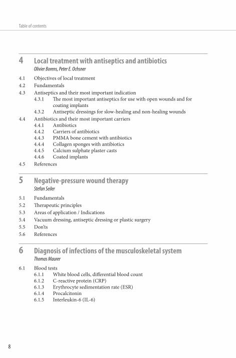
Table of contents
Fundamentals
1 Implant-associated biofilmAndrej Trampuz
1.1 Definition1.2 Biofilm development and maturation1.3 Implant/biofilm interactions1.4 Development of microorganism variants1.5 Pathogenesis of implant-associated infections1.6 References
2 Prevention of perioperative infectionsMarkus Vogt, Ilker Uçkay, Paul Bodler
2.1 General2.2 Preoperative measures2.3 Intraoperative measures2.4 Postoperative measures2.5 Perioperative management of patients with infections/colonisation with
multi-resistant bacteria2.6 Antibiotic prophylaxis in orthopaedic surgery
2.6.1 Fundamentals of perioperative antibiotic prophylaxis2.6.2 Practical approach2.6.3 Procedures with no evidence for the efficacy of antibiotic prophylaxis2.6.4 When is prophylaxis recommended for possible haematogenous
prosthetic joint infections?2.7 References
3 Systemic antibiotic therapyWerner Zimmerli
3.1 Fundamentals3.2 Definitions concerning the use of antibiotics3.3 Procedures in the case of therapeutic failure3.4 Antibiotics3.5 References
8
4 Local treatment with antiseptics and antibioticsOlivier Borens, Peter E. Ochsner
4.1 Objectives of local treatment4.2 Fundamentals4.3 Antiseptics and their most important indication
4.3.1 The most important antiseptics for use with open wounds and for coating implants
4.3.2 Antiseptic dressings for slow-healing and non-healing wounds4.4 Antibiotics and their most important carriers
4.4.1 Antibiotics4.4.2 Carriers of antibiotics4.4.3 PMMA bone cement with antibiotics4.4.4 Collagen sponges with antibiotics4.4.5 Calcium sulphate plaster casts4.4.6 Coated implants
4.5 References
5 Negative-pressure wound therapyStefan Seiler
5.1 Fundamentals5.2 Therapeutic principles5.3 Areas of application / Indications5.4 Vacuum dressing, antiseptic dressing or plastic surgery5.5 Don’ts5.6 References
6 Diagnosis of infections of the musculoskeletal systemThomas Maurer
6.1 Blood tests6.1.1 White blood cells, differential blood count6.1.2 C-reactive protein (CRP)6.1.3 Erythrocyte sedimentation rate (ESR)6.1.4 Procalcitonin6.1.5 Interleukin-6 (IL-6)
Table of contents
6.2 Arthrocentesis6.2.1 Technique of arthrocentesis6.2.2 Cell count, differentiation, Gram stain test6.2.3 Microbiological results6.2.4 Testing the synovial fluid with biomarkers6.2.5 Contrast-enhanced arthrography
6.3 Imaging diagnostics6.3.1 Conventional x-ray6.3.2 Computed tomography with contrast agent6.3.3 Magnetic resonance imaging6.3.4 Contrast-enhanced arthrography6.3.5 Sonography6.3.6 Nuclear imaging6.3.7 Summary of imaging diagnostics for infections of the musculoskeletal
system6.4 Biopsies
6.4.1 Discontinuation of antimicrobial therapy – Pretreatment before diagnostic sampling
6.4.2 Biopsy technique6.4.3 Transport to the lab6.4.4 Bacteriological tests6.4.5 Polymerase chain reaction (PCR) testing6.4.6 Histological tests
6.5 Testing of explanted foreign bodies using sonication6.6 References
Special infections
7 Infected prosthetic jointsPeter E. Ochsner, Werner Zimmerli, Hubert Nötzli
7.1 Fundamentals7.1.1 Preliminary remarks7.1.2 Aetiology7.1.3 Localisation7.1.4 Incidence7.1.5 Risk factors7.1.6 Classification

7.2 Clinical symptoms and diagnostic procedures7.2.1 Medical history and findings7.2.2 Laboratory tests7.2.3 Imaging
7.3 Treatment selection, algorithm7.4 Surgical treatment
7.4.1 General building blocks for surgical treatment7.4.2 Specific building blocks for surgical treatment
7.5 Antibiotic therapy7.6 Don’ts7.7 Expected clinical results7.8 References
8 Infected osteosynthesis – infected pseudarthrosis – chronic osteomyelitisPeter Ochsner, Andrej Trampuz, Werner Zimmerli
8.1 Fundamentals8.1.1 Aetiology8.1.2 Incidence8.1.3 Classification options for estimating the severity of post-traumatic
infections and/or the spread of bone necrosis8.2 Bone development in the fracture area in the case of infections8.3 Proof of infection8.4 Post-traumatic arthritis8.5 Antibiotic therapy
8.5.1 Indications8.5.2 Duration of therapy, problems
8.6 Early initial manifestation – infected osteosynthesis8.6.1 Clinical symptoms and diagnostic procedures8.6.2 Treatment indications8.6.3 Surgical treatment8.6.4 Prognosis and complications
8.7 Delayed initial manifestation – infected pseudarthrosis8.7.1 Clinical symptoms and diagnostic procedures8.7.2 Treatment indications8.7.3 Surgical treatment8.7.4 Specific treatment options8.7.5 Prognosis and complications
Table of contents

8.8 Late initial manifestation – chronic post-traumatic osteomyelitis8.8.1 Clinical symptoms and diagnostic procedures8.8.2 Treatment indications8.8.3 Surgical treatment8.8.4 Specific treatment options8.8.5 Prognosis and complications
8.9 Differential diagnosis: Chronic haematogenous osteomyelitis8.9.1 Clinical symptoms and diagnostic procedures8.9.2 Treatment indications8.9.3 Surgical treatment8.9.4 Prognosis and complications
8.10 Don’ts8.11 References
9 Infectious arthritisWerner Zimmerli, Olivier Borens
9.1 Fundamentals9.1.1 Aetiology9.1.2 Incidence9.1.3 Risk factors9.1.4 Affected joints
9.2 Clinical symptoms and diagnostic procedures9.2.1 Medical history9.2.2 Clinical findings9.2.3 Laboratory tests9.2.4 Imaging9.2.5 Differential diagnosis
9.3 Therapeutic principles9.3.1 Arthrocentesis9.3.2 Arthroscopy9.3.3 Arthrotomy9.3.4 Synovectomy9.3.5 Antibiotic treatment9.3.6 General information about physiotherapy
9.4 Don’ts9.5 References

10 SpondylodiscitisIvan Broger, Stefan Seiler
10.1 Fundamentals10.1.1 Definition10.1.2 Pathogenesis10.1.3 Epidemiology
10.2 Clinical symptoms and diagnostic procedures10.2.1 Clinical observations10.2.2 Laboratory tests10.2.3 Radiological diagnostics / Imaging techniques10.2.4 Microbiology
10.3 Therapeutic principles10.3.1 Conservative treatment – Antibiotics and rest10.3.2 Surgical treatment – Surgery plus antibiotics
10.4 Don’ts10.5 Prognosis and complications10.6 References
11 Soft tissue infectionsDomizio Suvà, Olivier Borens, Ilker Uçkay
11.1 Classification11.2 General diagnosis11.3 Some important infections
11.3.1 Furunculosis and localised skin abscesses11.3.2 Erysipelas11.3.3 Cellulitis11.3.4 Septic bursitis11.3.5 Necrotising fasciitis
11.4 References
12 Open woundsStefan Seiler
12.1 Fundamentals12.1.1 Aetiology12.1.2 Wound types
Table of contents

12.2 Diagnostics and clinical observations12.2.1 Medical history12.2.2 Clinical assessment12.2.3 Laboratory tests12.2.4 Microbiology12.2.5 Radiological diagnostics / Imaging techniques12.2.6 Prerequisites for wound healing
12.3 Therapy12.3.1 Treatment of acute wounds (soft tissue defect, grade II/III open fracture)12.3.2 Treatment of subacute and chronic wounds12.3.3 Systemic measures
12.4 Don’ts12.5 References
13 Diabetic footOlivier Borens
13.1 Fundamentals13.1.1 Incidence13.1.2 Pathogenesis13.1.3 Classification
13.2 Clinical observations13.3 Diagnostics13.4 Therapeutic principles
13.4.1 Treatment steps for the open diabetic foot13.4.2 Prophylactic measures
13.5 Prognosis and complications13.6 References
14 Osteomyelitis and purulent arthritis in children and adolescentsFritz Hefti
14.1 Classification14.2 Acute haematogenous osteomyelitis
14.2.1 Aetiology and pathology14.2.2 Incidence and localisation14.2.3 Clinical observations, diagnostics14.2.4 Therapy14.2.5 Follow-up examinations and prognosis

14.3 Special forms of acute osteomyelitis14.3.1 Acute multifocal haematogenous osteomyelitis14.3.2 Neonatal osteomyelitis14.3.3 Spondylodiscitis
14.4 (Primary) chronic osteomyelitis14.4.1 Aetiology14.4.2 Clinical observations, diagnostics14.4.3 Therapy14.4.4 Follow-up treatment14.4.5 Follow-up examinations, post-infection deformities14.4.6 Growth prognosis
14.5 Special forms of chronic osteomyelitis14.5.1 Garré’s sclerosing osteomyelitis14.5.2 Chronic recurrent multifocal osteomyelitis (CRMO)14.5.3 Specific osteomyelitis (tuberculosis)14.5.4 BCG osteomyelitis14.5.5 Exogenous osteomyelitis
14.6 Infectious (purulent) arthritis14.6.1 Aetiology, localisation14.6.2 Growth prognosis14.6.3 Clinical observations14.6.4 Diagnosis and treatment14.6.5 Post-infection deformities
14.7 References
Appendix
15 A microbiological guideGerhard Eich
15.1 Introduction15.2 Fundamentals
15.2.1 Virulence and pathogenicity15.2.2 Endogenous and exogenous infections15.2.3 Bacterial lifeforms15.2.4 Diagnosis15.2.5 Resistance testing
Table of contents

15.3 Specific bacteria15.3.1 Gram-positive bacteria15.3.2 Gram-negative bacteria15.3.3 Anaerobic bacteria15.3.4 Other microorganisms
15.4 Fungi15.5 Nomenclature and notation guidelines for microorganisms15.6 References
16 DefinitionsPaul Bodler
17 Common errors in the treatment of infections of the musculoskeletal systemIlker Uçkay, Markus Vogt
17.1 Diagnostics17.2 Antibiotic therapy17.3 Miscellaneous
18 Infection Therapy PassportThomas Maurer
18.1 Infection Therapy Passport18.2 Use of the Infection Therapy Passport
19 Documentation of samples collected for bacteriology and histologyPeter E. Ochsner
19.1 Issues19.2 Purpose of a special form19.3 Design of the form
Index of terms
Index of figures

16
Thomas Maurer
6 Diagnosis of infections of the musculoskeletal system
This chapter describes the technical options for diagnosing infections of the musculoskeletal system. The focus is on diagnosing periprosthetic joint infections (PJI) as well as the early stages of post-traumatic infection. This chapter does not deal with the important aspects of recording medical histories and clinical diagnostics. These details are provided in the individual sections for the medical conditions.
6.1 Blood tests
6.1.1 White blood cells, differential blood count
Determining the white blood cell count (normal range 4–10 x 106/L) is part of a routine examination. An elevated white blood cell count can have a number of causes. A supplementary differential blood count is also important.
Bacterial infections are one of the possible causes of a high neutrophil count with left shift. In the case of infectious arthritis, the white blood cell count sensitivity is only 75% while the specificity is 55% (n=156).
6.1.2 C-reactive protein (CRP)
CRP is an acute-phase protein. The normal level depends on the method used and is usually 0.5 mg/dL or 5 mg/L. The CRP level is independent of age, sex, blood loss and anaesthesia. The extent of the surgical procedure, the administration of steroids or other immunosuppressants and/or postoperative haematoma influence the CRP level. It increases within 6–24 hours in response to inflammatory processes and has a half life of approximately one day. As a result, the CRP level is an important postoperative clinical parameter. It usually peaks on postoperative day 2–3 and then continues to fall steadily over a postoperative course free of complications. A persistently elevated CRP level or a postoperative increase may indicate an infection at the site of the operation. The sensitivity and specificity values reported in the case of periprosthetic hip and knee joint infections are: sensitivity 91–96%, specificity 74–92% (n=105–296).
6.1.3 Erythrocyte sedimentation rate (ESR)
The ESR (normal range: women = 6–20 mm/h; men = 3–15 mm/h) is a non- specific haematology test whose diagnostic specificity is usually low. It is worth noting that significant deviations have been reported, depending on the publication.

17
While a diagnostic sensitivity of 75% and a specificity of only 11% are reported for infectious arthritis (n=107), better results have been published for periprosthetic hip and knee infections (sensitivity 82–93%, specificity 66–85%, n=105–296). A number of authors have reported an increase in sensitivity using ESR together with CRP for the diagnosis of PJI. In many countries, ESR is considered obsolete for diagnosing an infection.
6.1.4 Procalcitonin
Procalcitonin is a good parameter for distinguishing between bacterial and viral respiratory infections. The place of procalcitonin in the diagnosis of bacterial and nonbacterial joint infections is under debate. A recent study cast doubt on its value in the diagnosis of periprosthetic infections.
6.1.5 Interleukin 6 (IL-6)
The cytokine interleukin 6 may be considered a useful parameter because it returns to baseline levels as early as 3 days postoperatively during normal healing processes. This enables more rapid detection of postoperative infections. IL-6 has also achieved high specificity as a diagnostic marker in some studies. The authors recommend combining IL-6 and CRP. Because the costs of determining IL-6 levels in the lab are considerably higher than CRP, it is too expensive for routine clinical use.
A recent study compared the diagnostic value of CRP, procalcitonin and interleukin-6 in prosthetic joint infections. Despite the valuable information provided by the latter, CRP remains the most important test.
With any blood test it must be remembered that an infection may be present even if the values are normal! Additional diagnostic tools must be used.
6.2 ArthrocentesisNative jointsIn cases of suspected joint infection, an arthrocentesis should be done as soon as possible for diagnosis. Because the cell count alone does not provide sufficient evidence of an infection in the case of a native joint, it is necessary to wait for the bacteriology results. This usually takes too long. Therefore, if there is a strong suspicion of a native joint infection, it is recommended to combine diagnostics and

18
Diagnosis of infections of the musculoskeletal system
treatment, e.g., using arthroscopic lavage. This approach presumes that the patient is suitably stable and the procedure can be performed without delay. The objective is to protect the cartilage against bacterial destruction through rapid , early intervention.
Prosthetic joints ■ Newly suspected infection: The joint should be punctured as soon as possible
because timely diagnosis enables the option of simpler treatment and implant retention (see also Chapter 2.1). The decision to intervene and then initiate empirical antibiotic therapy is based on the number of white blood cells and/or granulocytes found in the puncture specimen.
■ Chronic symptoms: In cases of suspected low-grade infection with no signs of systemic infection, the antibiotic regimen should be discontinued for at least two weeks prior to the arthrocentesis.
6.2.1 Technique of arthrocentesis
An arthrocentesis must be performed under the most sterile conditions possible. There are a number of recommendations regarding the technique:
■ From an orthopaedic and infectious-disease standpoint, effective disinfection (alcohol-based agent), sterile draping and the wearing of sterile gloves and a face mask are recommended.
■ Important: aspiration through skin lesions (infections, psoriasis) must be avoided! ■ Arthrocentesis of prosthetic joints should never be performed in the office
or patient examination room but rather in a separate room and preferably in the operating theatre where the risk of infection is significantly lower (at least 100,000 times in the case of PJI compared to native joints).
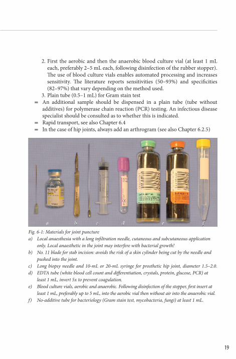
■ Sample containers (plain tube, EDTA tube, blood culture vial, aerobic and anaerobic) are placed at the ready (Fig. 6-1).
■ Apply local anaesthetic to the subcutaneous level only (local anaesthetic in the joint distorts the cell count and causes false-negative bacteriology results due to its bacteriostatic effect). A subsequent stab incision avoids the biopsy needle to cut a skin cylinder eventually later pushed in the joint with the risk of contaminating/infecting the joint.
■ The puncture volume obtained is then dispensed according to the following priorities. In the case of small puncture specimens it is not possible to fill all of the tubes:1. EDTA tube, 1 mL (must be inverted several times immediately following
transfer of the synovial fluid to be completely mixed with the EDTA to prevent coagulation, otherwise an automated cell count will not be possible).
19
Fig. 6-1: Materials for joint puncturea) Local anaesthesia with a long infiltration needle, cutaneous and subcutaneous application
only. Local anaesthetic in the joint may interfere with bacterial growth!b) No. 11 blade for stab incision: avoids the risk of a skin cylinder being cut by the needle and
pushed into the joint.c) Long biopsy needle and 10-mL or 20-mL syringe for prosthetic hip joint, diameter 1.5–2.0.d) EDTA tube (white blood cell count and differentiation, crystals, protein, glucose, PCR) at
least 1 mL, invert 5x to prevent coagulation.e) Blood culture vials, aerobic and anaerobic. Following disinfection of the stopper, first insert at
least 1 mL, preferably up to 5 mL, into the aerobic vial then without air into the anaerobic vial.f) No-additive tube for bacteriology (Gram stain test, mycobacteria, fungi) at least 1 mL.
2. First the aerobic and then the anaerobic blood culture vial (at least 1 mL each, preferably 2–5 mL each, following disinfection of the rubber stopper). The use of blood culture vials enables automated processing and increases sensitivity. The literature reports sensitivities (50–93%) and specificities (82–97%) that vary depending on the method used.
3. Plain tube (0.5–1 mL) for Gram stain test ■ An additional sample should be dispensed in a plain tube (tube without
additives) for polymerase chain reaction (PCR) testing. An infectious disease specialist should be consulted as to whether this is indicated.
■ Rapid transport, see also Chapter 6.4 ■ In the case of hip joints, always add an arthrogram (see also Chapter 6.2.5)
a b c d e f

20
Diagnosis of infections of the musculoskeletal system
6.2.2 Cell count, differentiation, Gram stain test
Cell countSynovial cell counts provide reliable results for the diagnosis of infection, particularly in patients with prosthetic joints.. In the case of periprosthetic knee and hip joint infections, there are defined thresholds for the leucocyte counts and granulocyte fraction in the puncture specimen. These counts can be distorted in patients with inflammatory rheumatic diseases and neutropenia.
■ Total knee arthroplasty: An infection is present with a cell count > 1.7 x 10³/µL (sensitivity 94%, specificity 88%) or with granulocyte ratio > 65% (sensitivity 97%, specificity 98%).
■ Total hip arthroplasty: An infection is present with a cell count > 4.2 x 10³/µL (sensitivity 84%, specificity 93%) or with granulocyte ratio > 80% (sensitivity 84%, specificity 82%).
■ Native joints: In contrast to prosthetic joints, there are no precisely defined thresholds for native joints. An infection is considered likely with a cell count > 50 x 103/µL (sensitivity 62%, specificity 92%) or with a neutrophil granu-locyte level > 90% (sensitivity 73%, specificity 79%) (see also Chapter 9.2.3).
Cell count determinations are useful in verifying a suspected infection. Even if the cause is not yet known, therapeutic measures can be initiated immediately.
CrystalsCrystal analysis provides information about crystal-induced synovitis but it does not rule out an infection.
ViscosityA highly viscous puncture specimen decreases the likelihood for infection.
Gram stain testThe Gram stain test is only useful for a rapid diagnosis if it is positive. Its low sensitivity of < 26% (specificity > 97%) shows that this is seldom the case.A punctio sicca (‘dry tap’ in which no fluid is aspirated) can never rule out an infection. For example, hip joint pressure due to periarticular fistulous tract can be reduced to such an extent that no material can be aspirated. Joint lavage with sterile fluid and subsequent aspiration have proven successful in such cases. However, the quantitative cell count determined using this technique can no longer be used to establish the diagnosis.

21
6.2.3 Microbiological results
The interpretation of cultures is easy, when the growth is early and the synovial cell count is typical for a periprosthetic infection. If contamination is suspected arthrocentesis may be repeated to clarify the situation with a second culture.
6.2.4 Testing the synovial fluid with biomarkers
Five different biomarkers seem to support the diagnosis of periprosthetic joint infections: α-defensin, ELA2, BPI, NGAL, and lactoferrin. While tests with these biomarkers are justifiably considered promising, there are still no reliable data concerning their sensitivity and specificity, to date.
6.2.5 Contrast-enhanced arthrography
Imaging using contrast agent administered once the puncture is complete via the in situ needle is especially useful in the case of prosthetic hip joints. It reveals protrusions from the joint cavity, abscess cavities, and fistulous tracts, even if no external fistula is visible (Fig. 6-2). These signs are often criteria for two-stage revision. Improved resolution through digital subtraction technique is possible.
Fig. 6-2: Contrast-enhanced arthrography. In the image on the left the contrast agent extends into the subcutaneous tissue in two fistulous tracts; in the image on the right there is an intraosseous abscess cavity.

22
Diagnosis of infections of the musculoskeletal system
6.3 Imaging diagnosticsIf radiology is used to diagnose infection, the pathophysiology of the infection must be understood in addition to imaging. In pathophysiological terms, we distinguish between:
■ Acute infection: vasodilatation, increased neutrophil and monocyte penetra-tion, exudation and migration
■ Chronic infection: proliferation of lymphocytes and macrophages in the tissue, vascularisation, scarring
Methods of imaging diagnostics ■ Conventional x-ray examination ■ Computed tomography (CT) ■ Magnetic resonance imaging (MRI) ■ Scintigraphy ■ Single-photon emission computed tomography (SPECT/CT) ■ Positron emission tomography (PET) ■ PET/CT

23
Fig. 6-3: Radiological signs of periprosthetic joint infection: Osteolysis along the implants, irregular thickening of the diaphyseal femur
6.3.1 Conventional x-ray
Conventional x-ray scans only show the bone remodelling in the case of osteomyelitis and prosthesis-associated infections. However, osteolysis and loosening are also often associated with aseptic processes. This reduces the specificity of these radiological signs. Serial x-rays over a certain period can measure changes to the cortical bone and migration of a prosthesis, for example (Fig. 8-3). Rapidly progressive and/or irregular periprosthetic osteolysis suggests an infection (see cover image).
Exposures: AP and lateral views and possibly additional oblique views can greatly increase the predictive value (Fig. 8-7).

24
Diagnosis of infections of the musculoskeletal system
Advantages Drawbacks
– Good imaging of the bone structure– Development of changes can be detected by
comparing with previous X-rays– Fewer metallic artefacts
– Visible changes often less specific for infections– Poor imaging of soft tissue– Sequestra, fistulas, abscesses only partially
detectable, improved detection by contrast filling of the fistula
Indication: Quick overview examination, simplest monitoring of progression by comparing to all earlier x-rays
What should you look for? ■ Bones: density, sclerosis, sequestra, osteolysis ■ loosening of prothetic joint or internal fixation device ■ Surrounding tissue reaction
Tab. 6-1: Role of conventional x-ray for the diagnosis of infections
6.3.2 Computed tomography with contrast agent
(Contrast agent concentration: 10-3 mol/kg body weight)Computed tomography (CT) with contrast agent enables the diagnosis of joint effusion, fistulas, soft tissue abscesses, sequestra (Fig. 6-4), bone erosion and periprosthetic rarefaction of the bone. Metallic artefacts have a major negative impact on image quality. Artefacts can be reduced by using special techniques which enable detection of prosthetic loosening and signs of soft tissue infection.

25
Fig. 6-4: Osteomyelitis 2 years after internal fixation, 1 year following plate removal. Numerous sequestra could only be localised using CT. Arrow: 1 peripheral sequestrum, 2 central sequestra quite deeply embedded in the newly formed bone (see also Fig. 8-12).
1
2
2
Advantages Drawbacks
– Short examination time– Good imaging of the bone structure – Best imaging of sequestra– Reconstructions possible in all planes– No claustrophobia (unlike with MRI)
– Relatively poor imaging of soft tissue– Metal artefacts– Radiation exposure
Indication: Preoperative search for sequestra and necrosis in the bones combined with filling of fistulas, distribution of fistula system, air bubbles
Tab. 6-2: Role of CT scan for the diagnosis of infections

26
Diagnosis of infections of the musculoskeletal system
6.3.3 Magnetic resonance imaging
(Contrast agent concentration: 10-5 mol/kg body weight)Magnetic resonance imaging (MRI) can be performed on patients with and without non-ferromagnetic implants. MRI can resolve soft tissue changes better than CT or conventional x-rays. It also visualizes anatomic details more precisely than scintigraphy. The method is especially important in the spinal region (Fig. 10-1, 10-2). As with CT scans, the main disadvantage of MRI involves imaging interferences close to metallic implants.
Functional magnetic resonance imaging in the case of infectionsThere have been experimental studies conducted using iron nanoparticles which accumulate in macrophages during inflammation. This enables acute and chronic infections to be very easily distinguished and imaged.
This method cannot be used in humans at this stage because the iron nanoparticles are too toxic at the concentrations administered.
Advantages Drawbacks
– Shows acute inflammations with high sensitivity and specificity
– Shows complications such as abscesses and fistulas
– Specificity is reduced with chronic infections (only 60% with chronic postoperative osteomyelitis)
– Bone oedema is overestimated– Susceptibility artefacts*
* Susceptibility artefacts: No differentiation between chronic inflammation and scarred repair. Differentiation tips: The bone repair process should be completed approximately 1 year after surgery; if not, then the presence of a chronic infection is likely. Do not disregard scarring (medullary cavity).
Tab. 6-3: Role MRI for the diagnosis of infections
6.3.4 Contrast-enhanced arthrography
See Chapter 6.2.5 and Fig. 6-2
6.3.5 Sonography
Ultrasound examinations can be used to locate a joint effusion or for controlled puncture and drainage of such an effusion. Sonography is especially useful in the case of prosthetic hip joint-associated infections where effusions often cannot be diagnosed clinically.

27
6.3.6 Nuclear imaging
Using molecular imaging, scintigraphic scans show the physiological processes that precede radiologically visible, anatomical changes. The concentrations of radioactive substances that are required are much lower than the concentrations required for MRI and CT (concentration of radiopharmaceuticals: 10-9 to 10-12 mol/kg). Another bene-fit is that a general, functional diagnosis of the bone metabolism can be established in addition to the principle issue in question. Drawbacks include the limited spatial res-olution associated with conventional systems and the relatively long time required for the scanning procedure. We will limit ourselves to the following:
■ Bone scintigraphy with 99mTechnetium phosphate (with or without SPECT/CT) ■ 99mTc antigranulocyte scintigraphy and 99mTc-HMPAO leukocyte scintigraphy
(with or without SPECT/CT) ■ 18F-fluorodeoxyglucose positron-emission tomography/computed tomo-
graphy (FDG-PET/CT)
Bone scintigraphy99mTechnetium (99mTc)-labelled phosphonates are used in bone scintigraphy. The substance is distributed via the circulatory system following injection. This enables zones that are well supplied with blood (zones of infection) to be imaged immediately following injection during the tissue perfusion and blood-pool phase. The substance then accumulates in the bones within approximately 3–4 hours (late phase). Sites of increased bone metabolism, e.g., inflammations and infections, are especially well imaged (increased uptake), which makes the scan very sensitive.
Due to its poor specificity, bone scintigraphy with 99mTc does not usually provide enough information when used as the sole imaging technique to confirm diagnosis of an implant-associated joint infection. Depending on the type of prosthesis, increased accumulation is a normal physiological process during the first (1–2) postoperative years and corresponds to remodelling in periprosthetic bones. In addition, bone scintigraphy is often unable to distinguish aseptic loosening from a prosthesis-associated infection. As a result, bone scintigraphy can be combined with inflammation scintigraphy.
99mTc-antigranulocyte scintigraphy and 99mTc-HMPAO leukocyte scintigraphy99mTc-labelled monoclonal antigranulocyte antibodies have an accuracy of 81% in the diagnosis of a prosthesis-associated infection (Fig. 6-5). This technique is widespread in Europe. There are physiological accumulations in the liver, spleen and colon. Antigranulocyte scintigraphy does not work on the axial skeleton because the promyelocytes in the blood-forming bone marrow are also labelled. Ectopic blood-forming bone marrow also accumulates antibodies. It is potentially difficult to distinguish between a low-grade infection and aseptic inflammation. Follow-up

28
Diagnosis of infections of the musculoskeletal system
examinations may trigger immune reactions (in 4% of cases formation of human anti-mouse antibodies [HAMA]). For the very complex method involving labelled autologous leukocytes, the values for sensitivity, specificity and accuracy are 94%, 83% and 89%, respectively.
Fig. 6-5: Persistent fistula despite 3 months of negative-pressure wound therapy following total knee arthroplasty and early revision due to staphylococcal infection. Antigranulocyte scintigraphy with massive periprosthetic accumulation. Tuberosity of the tibia has not healed following oste-otomy. Rifampicin-resistant coagulase-negative staphylococci identified upon explantation.
Single-photon emission computed tomography (SPECT/CT)In the case of SPECT/CT, the scintigraphic methods described above are carried out using a next-generation hybrid device; this involves coupling the gamma camera used in nuclear medicine with CT to produce and then merge anatomical and functional scans using the same device. The highly active sites detected using the scintigraphic method are integrated into the CT. This makes localization of scintigraphically active sites easier, thus increasing the specificity of the scans. It also means additional scans are often unnecessary.
18F-fluorodeoxyglucose positron-emission tomography/computed tomography (FDG-PET/CT or PET/CT)
■ Colour imaging of regional glucose metabolism. Phagocytes consume approximately 50 times more glucose than the surrounding environment, meaning that accumulation of phagocytes during an infection is accompanied by significantly increased glucose uptake in the scintigram.
■ High sensitivity, satisfactory local resolution ■ 3D data

29
■ Relatively quick scanning procedure (2 hours) ■ Normal: major accumulation in the brain, individually variable levels in the
heart, liver and gastrointestinal tract ■ PET implements an approach using specific radioactively labelled molecules
to image cell and organ functions. By comparison, MRI uses a different approach based on the imaging of protons and Brownian motion and cannot always be applied due to certain contraindications (e.g., pacemaker, toxicity of the contrast agents, etc.).
■ Therapy monitoring possible ■ Diagnosis of spondylodiscitis possible (unlike antigranulocyte scintigraphy) ■ Indications: can possibly be used to establish status following internal fixation
(depending on the time since the procedure), abdominal infections, vascular implants, autoimmune diseases, fever of unclear origin in neutropenic patients.
■ Administrative problem: health insurance funds are not yet required to cover this procedure in outpatient settings. Health insurances must be consulted.
In combination with computed tomography (FDG-PET/CT) the diagnostic value is significantly improved:
■ Excellent local resolution of CT combined with functional PET scan ■ High sensitivity and specificity for chronic inflammations ■ Can also be performed with metal implants ■ Difficult to differentiate tumour/inflammation. In such cases, however,
FDG-PET can identify the ideal site of a possible biopsy. Depending on the region of the body/organ, tumour-specific PET tracers (e.g., amino acids/ oligopeptides, choline) can be used for further differentiation.
■ Administrative issues: – Availability: Complex nuclear imaging with PET scanning is not univer-
sally available across Europe, but is increasingly used in University-based hospitals and some private institutions.
– Cost effectiveness: Simple nuclear imaging is not expensive and is covered in most public and insurance-based systems. Combined scanning (particu-larly with FDG) may require specific funding arrangements.
The role of FDG-PET in differentiating between an infection and aseptic loosening of a prosthetic hip joint is still considered controversial because of the lack of standardised interpretation criteria. While Love et al. reported a poor specificity of just 9% compared to a high sensitivity of 100%, Stumpe et al. of the Zurich group reported a high specificity of 83% with a low sensitivity of 28%. Obviously, more studies are needed before the procedure can be routinely used in the diagnosis of implant-associated infections.

30
Diagnosis of infections of the musculoskeletal system
6.3.7 Summary of imaging diagnostics for infections of the musculoskeletal system
■ A conventional x-ray is usually sufficient as the first imaging procedure (issue: signs of loosening, radiolucent lines, etc.)
■ In the case of suspected low-grade infections or complex situations, further imaging (MRI, scintigraphy with SPECT/CT, etc.) must be considered.
■ Extremities: Antigranulocyte scintigraphy possible, ideally combined with SPECT/CT
■ CT in the case of osteomyelitis in order to plan a procedure to investigate sequestrum formation
■ FDG-PET/CT is a modern, highly sensitive option for chronic infections. Limitations: reduced availability, costly, currently not everywhere covered by health insurance funds in outpatient settings, depending on localisation, reli-able differentiation between tumour/inflammation not possible
6.4 Biopsies
6.4.1 Discontinuation of antimicrobial therapy – Pretreatment before diagnostic sampling
Prior to collecting microbiological samples, any antibiotic regimen should be discontinued for 2 weeks, provided the progression of the disease allows this. Prophylactic antibiotics should not be administered until after the samples have been collected. If samples are collected with the use of a tourniquet (constriction of blood flow), then prophylactic and/or therapeutic antibiotics should be introduced into the blood stream at the full dosage just prior to removal of the tourniquet.
6.4.2 Biopsy technique
Sample type Swabs should not be used. Biofilm bacteria cannot be extracted from biofilms using swabs. Swabs may contain contaminating microbes. Their sensitivity is significantly less than that of tissue samples. Foreign bodies such as plates, screws, prosthetic joints can be sonicated. Pus ensures rapid diagnosis of acute infections. It is recommended, in some centres, to use separate instruments for each sample. We think, that contamination is better ruled out by the parallel analysis of each tissue biopsy by bacteriology and histology.

31
Fig. 6-6: Two samples each from the same region are taken for bacteriology and histology (left), both assigned the same number. Larger numbers of samples are collected if a difficult to diagnose low-grade infection is expected.
Technical procedureA large piece is collected from each region to be tested and divided into 2 pieces for bacteriology and histology with each edge being approximately 0.5 cm long (Fig. 6-6). Contamination of the collected samples is prevented by immediately transferring them to transport containers. Individual samples can be collected for PCR (for economical reasons, PCR examinations should be limited to cases without microbial growth).
Sample collection siteSamples should always be collected from a zone in which the tissue structure is visibly inflamed (e.g., abnormally soft, degraded). Tissue in the vicinity of sequestra is informative. If an implant-associated infection is suspected, then the samples must be collected from the immediate vicinity of the foreign body. In these cases, infection tends to be localised around the implant, while more remote regions will not be affected in low-grade infections. Samples collected directly from the skin or superficially from a fistula canal are unsuitable because these samples are often contaminated with skin microbes and lead to false results.
It is important to precisely document the collection site so that the region in which the microbes are growing can be isolated. Microbiology and histology samples must be assigned the same number so that the bacteriology results can be compared with the corresponding histology results. Intraoperative biopsies have a sensitivity of 65–94% and a specificity of 69–87%.
Number of samplesA minimum of 3 tissue samples must be collected. One study found that sensitivity was 50% with 2–3 samples and 72.7% with more than 5 samples. More samples reduce the risk of an incorrect assessment due to contamination. Bacterial growth in
32
Diagnosis of infections of the musculoskeletal system
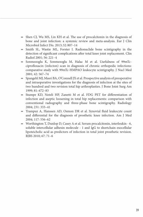
one of 2 samples does not indicate whether infection or contamination is involved. Contamination can be assumed if there is growth in 1 of 7 samples. Simultaneously collecting tissue for bacteriology and histology enables more reliable differentiation between contamination and infection.
Documentation of samplesA standard form that is acceptable to both the microbiology and histology labs and that ensures uniform collection of patient data and identification of samples is suitable for documenting the various samples (see also Chapter 19).
Note: The more acute and obvious the infection is, the fewer the samples required (at least 3). If a low-grade infection is suspected, then a large number of samples (at least 6) should be collected for bacteriology and histology.
Joint puncture specimen Biopsies
Volume Approx. 1–20 mL (see also Chapter 6.2 Arthrocentesis)
Approx. 0.5 x 0.5 x 0.5 cm per biopsy
Number 1 At least 3; if low-grade infection is suspected, at least 6
Transport container
Sterile container with no additives (Gram stain test) EDTA tube (automated cell count and one aerobic and one anaerobic blood culture vial, see also Chapter 6.2 Arthrocentesis)
Sterile container with no additivesFor longer shipping times, it is recommended to consult the lab to determine if a special transport container should be used.
Lab form Precise description of the material, collection date and time, and note as to whether periprosthetic joint infection is suspected
Gram stain test Low sensitivity Not recommended due to very low sensitivity
Culture Aerobic and anaerobic cultures are performed. The cultures are prepared using non-selective media such as sheep blood agar and chocolate agar. Liquid media must also be used to detect growth. The incubation period must be of sufficient length (10–14 days) so that slow-growing microbes (Propionibacterium spp. or SCV) can also be detected.
PCR Eubacterial (broad-range) PCR has low sensitivity. Its routine use is not recom-mended for all patients. An additional biopsy is sent to the lab as a reserve. This is used for PCR if bacteriology remains negative.
Tab. 6-4: Summary of bacteriology sample processing

33
6.4.3 Transport to the lab
Samples should be received by the laboratory as soon as possible, optimally within 4 hours of collection. Samples sent by post must always be sent by express post. The lab should be notified by phone about the samples to ensure they are processed immediately. If samples cannot be sent immediately, for instance, if samples are collected at night, then they should be stored at room temperature.
6.4.4 Bacteriological tests
If an implant-associated infection is suspected, then extended incubation of the biopsies in fluid enrichment broth for 10 days will improve the sensitivity of pathogen detection in the samples. This period is followed by subcultivation on solid media with resistance testing. The extended incubation period of 14 days is important because otherwise slow-growing microbes will not be detected, e.g., Propionibacterium spp. or small colony variants (SCV) (Table 6-4).
6.4.5 Polymerase chain reaction (PCR) testing
The polymerase chain reaction (PCR) is a procedure used to detect bacterial DNA. It is a supplementary molecular biology test used for suspected infections if there is no growth in conventional bacteriology tests. It is recommended to use a no-additive tube or EDTA tube for sample collection during joint puncture; tissue samples do not require any special medium. In many cases, only single samples are analysed if conventional bacteriology results were negative. The diagnostic value of PCR is considerably reduced with colonisation by multiple microbes. Resistance testing is also not possible with the exception of methicillin- and rifampicin-resistance.
6.4.6 Histological tests (Fig. 6-7)
A single positive tissue sample is already evidence of an infection, even without positive bacteriology results. It is critically important for the reliability of the diagnosis that the tissue for the test is collected from the region in which an infection is suspected. In the case of suspected implant-associated infections, samples must always be collected in the immediate vicinity of the implant (membranes surrounding the implant or prosthesis, joint neocapsule). The histology results can also be helpful for distinguishing between infection and contamination. If, for

34
Diagnosis of infections of the musculoskeletal system
example, coagulase-negative staphylococci are found in only one sample and positive histology is present we conclude that there is an infection (see also Chapter 6.4.2).
Neutrophil granulocytes are indicators of bacterial infections. They are found in greater numbers in the case of acute infections (Fig. 6-7 a). Bacteria are more rarely found. In the case of low-grade infections in connection with implants, a search for granulocytes must be performed (Fig. 6-7 c). The granulocytes are counted in at least 10 high-power fields (400x magnification) and must exceed a total of 20–25 (Morawietz et al. 2009). Neutrophil granulocytes can be specifically imaged using immunohistochemical staining (CD15) (Fig. 6-7 d). This makes detection of granulocytes considerably easier, enabling a meaningful diagnosis even if the number of neutrophil granulocytes is low. Typical accumulations of granulocytes are not found in the case of all infections. Granulocyte accumulation is not present in every type of implant-associated infection. As an example, tissue granulocytes may be very scarce in Propionibacterium acnes infection.
Prosthetic loosening is often simultaneously present in the case of delayed or late infections. The analysis of periprosthetic membranes is often complicated due to the
a
c
b
d
Fig. 6-7: High-grade infection (top): a) Synovial membrane with numerous granulocytes, high-grade infection, foreign body giant cells; b) Large numbers of granulocytes with simultaneous presence of giant cells with phagocytised material.Low-grade infection (below): c) Isolated granulocytes (arrow), d) Granulocytes much more visible with immunohistochemical staining (CD15)

35
presence of foreign body reactions to wear particles. Four types of periprosthetic membranes (Plural) have been defined (Morawietz et al. 2006), including:
■ Type I: Wear particle induced type = with signs of wear only ■ Type II: Infectious type = with signs of infection only ■ Type III: Combined type = simultaneous signs of wear and infection (Fig. 6-7 b) ■ Type IV: Indeterminate type = does not fulfil criteria for type I or type II
6.5 Testing of explanted foreign bodies using sonicationSonication is used to identify the bacteria in the biofilm on explanted implants and prosthetic joints. The procedure involves first vortexing the implants in a liquid bath. The ultrasonic waves generate a rapid change in pressure on the surface of the implant which dislodges the biofilm (Fig. 6-8). Vortexing is repeated and the liquid is then used to diagnose bacterial infection with conventional culture.
In a study of 79 patients with periprosthetic joint infection and 252 patients with aseptic failure it was found that sonication was significantly more sensitive than tissue biopsy (tissue biopsy 60.8%, sonication 78.5%) with almost identical specificity (tissue biopsy 99.2%, sonication 98.8%).
In patients whose antibiotic therapy was discontinued less than 14 days prior to surgery, sonication fluid culture was significantly more sensitive than conventional bacteriology.
Sonication
0.5 mL aerobic and anaerobic culture dishes
Addition of 400 mLRinger’s (in the lab)
Implant in sterile container
Sonication in the ultrasonic bath
Vortex Vortex
s s
CC CR R
Fig. 6-8: Diagram of the sonication work flow. Ultrasonic penetration takes place through alternating phases of compression (C) and rarefaction (R).

36
Diagnosis of infections of the musculoskeletal system
The pathogen is also identified more quickly with sonication fluid culture than conventional bacteriology, making it possible to switch more rapidly to a suitable postoperative antibiotic if resistance has developed. Sonication is not targeted for identifying mycobacteria or fungi; special cultures should still be used.
Explantation and shipmentIn order to prevent inadvertent contamination, implants must be shipped in sterile, stable containers. Laboratories usually provide their own transport containers that have been evaluated for sonication (Fig. 6-9).
Because gloves represent the highest risk for microbial contamination and sonication is very sensitive, the implant should only be handled with suitable instruments or fresh gloves and placed directly into the special transport container.
Fig. 6-9: Example of a sterile transport container
When explanting a prosthesis that has been anchored with antibiotic cement, it is recommended to remove the cement from the prosthesis and ship the prosthetic parts without cement. Newly fractured cement may release antibiotics that are still active and this could have a negative impact on the identification of microbes. For shipment, see also Chapter 6.4.3.

37
6.6 References
Further reading
■ Gemmel F, Van den Wyngaert H, Love C et al. Prosthetic joint infections: radionucleide state-of-the-art imaging. Review article. Eur J Med Mol Imaging 2012; 39: 892–909
■ Morawietz L, Classen R-A, Schröder JH et al. Proposal for a histopathological consensus classification of the periprosthetic interface membrane. J Clin Pathol 2006; 59: 591–7
■ Morawietz L, Tiddens O, Mueller M et al. Twenty-three neutrophil granulocytes in 10 high-power fields is the best histopathological threshold to differentiate between aseptic and septic endoprosthesis loosening. Histopathology 54; 2009: 847–53
■ Trampuz A, Piper KE, Jacobson MJ et al. Sonication of removed hip and knee prostheses for diagnosis of infection. N Engl J Med 2007; 357: 654–63
■ Trampuz A, Zimmerli W. Diagnosis and treatment of implant-associated septic arthritis and osteomyelitis. Curr Infect Dis Rep 2008; 10: 394–403
Additional articles
■ Atkins BL et al. Prospective evaluation of criteria for microbiological diagnosis of prosthetic-joint infection at revision arthroplasty. The OSIRIS Collaborative Study Group. J Clin Microbiol 1998; 36: 2932–9
■ Austin MS, Ghanem E, Joshi A et al. A simple, cost-effective screening protocol to rule out periprosthetic infection. J Arthroplasty 2008; 23: 65–8
■ Bori G et al. Usefulness of histological analysis for predicting the presence of microorganisms at the time of reimplantation after hip resection arthroplasty for the treatment of infection. J Bone Joint Surg Am 2007; 89: 1232–7
■ Bottner F et al. Interleukin-6, procalcitonin and TNF-alpha: markers of peri-prosthetic infection following total joint replacement. J Bone Joint Surg Br 2007; 89: 94–9
■ Bühler M, Engelhardt M, Schmidt H. Septische postoperative Komplikationen, Springer, Wien, 2003
■ Cheung A, Lachieziwcz PF, Renner JB. The role of aspiration and contrast-enhanced arthrography in evaluating the uncemented hip arthroplasty. Am J Roentgenol 1997; 168: 1305–9

38
Diagnosis of infections of the musculoskeletal system
■ Deirmengian C, Kardos K, Kilmartin P et al. Diagnosing periprosthetic joint infection. Has the era of the biomarkers arrived? Clin Orthop Relat Res 2014; under print
■ Della Valle CJ, Sporer SM, Jacobs JJ et al. Preoperative testing for sepsis before revision total knee arthroplasty. J Arthroplasty 2007; 22 (Suppl 2): 90–3
■ De Man FH, Graber P, Lüem M et al. Broad-range PCR in selected episodes of prosthetic joint infection. Infection 2009; 37: 292–4
■ Glehr M, Friesenbichler J, Hofmann G et al. Novel biomarkers to detect infection in revision hip and knee arthroplasties. Clin Orthop Relat Res 471; 2013: 2621–8
■ Greidanus NV, Masri BA, Garbuz DS et al. Use of erythrocyte sedimentation rate and C-reactive protein level to diagnose infection before revision total knee arthroplasty. A prospective evaluation. J Bone Joint Surg Am 2007; 89: 1409–16
■ Ivancevic V, Perka C, Hasart O et al. Imaging of low-grade bone infection with a technetium-99m labelled monoclonal anti-NCA-90 Fab’ fragment in patients with previous joint surgery. Eur J Nucl Med Mol Imaging 2002; 29: 547–51
■ Levine BR, Evans BG. Use of blood culture vial specimens in intraoperative detection of infection. Clin Orthop Relat Res 2001; 382: 222–31
■ Li SF, Cassidy C, Chang C et al. Diagnostic utility of laboratory tests in septic arthritis. Emerg Med J 2007; 24: 75–7
■ Love C, Marwin SE, Tomas MB et al. Diagnosing infection in the failed joint replacement: a comparison of coincidence detection 18F-FDG and 111In-labeled leukocyte/99mTc-sulfur colloid marrow imaging. J Nucl Med 2004; 45: 1864–71
■ Mackowiak PA, Jones SR, Smith JW. Diagnostic value of sinus-tract cultures in chronic osteomyelitis. JAMA 1978; 239: 2772–5
■ Ochs BG, Kommerell M, Geiss HK et al. [Improving microbiological diagnostics in septic orthopaedic surgery. Comparative study of patients receiving systemic antibiotic therapy]. Orthopäde 2005; 34: 345–51
■ Ochsner PE. (Hrsg.) Die Hüfttotalprothese, Springer, Berlin, 2003 ■ Pandey P, Drakoulakis, Athanasou NA. An assessment of the histological
criteria used to diagnose infection in hip revision arthroplasty tissues. J Clin Pathol 1999; 52: 118–23
■ Schäfer P, Fink B, Sandow D et al. Prolonged bacterial culture to identify late periprosthetic joint infection: a promising strategy. Clin Infect Dis 2008; 47: 1403–9
■ Schinsky MF et al. Perioperative testing for joint Infection in patients undergoing revision total hip arthroplasty. J Bone Joint Surg Am 2008; 90: 1869–75
39
■ Shen CJ, Wu MS, Lin KH et al. The use of procalcitonin in the diagnosis of bone and joint infection: a systemic review and meta-analysis. Eur J Clin Microbiol Infect Dis. 2013; 32: 807–14
■ Smith SL, Wastie ML, Forster I. Radionuclide bone scintigraphy in the detection of significant complications after total knee joint replacement. Clin Radiol 2001; 56: 221–4
■ Sonmezoglu K, Sonmezoglu M, Halac M et al. Usefulness of 99mTc- ciprofloxacin (infecton) scan in diagnosis of chronic orthopedic infections: comparative study with 99mTc-HMPAO leukocyte scintigraphy. J Nucl Med 2001; 42: 567–74
■ Spangehl MJ, Masri BA, O’Connell JX et al. Prospective analysis of preoperative and intraoperative investigations for the diagnosis of infection at the sites of two hundred and two revision total hip arthroplasties. J Bone Joint Surg Am 1999; 81: 672–83
■ Stumpe KD, Notzli HP, Zanetti M et al. FDG PET for differentiation of infection and aseptic loosening in total hip replacements: comparison with conventional radiography and three-phase bone scintigraphy. Radiology 2004; 231: 333–41
■ Trampuz A, Hanssen AD, Osmon DR et al. Synovial fluid leukocyte count and differential for the diagnosis of prosthetic knee infection. Am J Med 2004; 117: 556–62
■ Worthington T, Dunlop D, Casey A et al. Serum procalcitonin, interleukin - 6, soluble intercellular adhesin molecule - 1 and IgG to shortchain exocellular lipoteicholic acid as predictors of infection in total joint prosthetic revision. BJBS 2010; 67: 71–6