Embed Size (px)
DESCRIPTION
prabhat
Citation preview
ANATOMY1. In the advancement of surgery of shoulder joint, one muscle was not given much importance earlier but now came in picture, this forgotten muscle of rotator cuff is a. Subscapularisb. Supraspinatus c. Infraspinatusd. Teres minorAnswer: (a) SubscapularisReference:Explanation:Musculotendinous cuff of shoulder is a fibrous sheath formed by the four flattened tendons which blend with the capsule of shoulder joint and strengthen it. Muscles which form the cuff arise from scapula and are inserted into the lesser and greater tubercles of humerus. They are subscapularis, supaspin-taous, infraspinatus and teres minor. There tendons while crossing the shoulder joint become flattened and blend with each other on one end and with capsule of the joint on the other end, before reaching their points of insertion.Cuff gives strength to the capsule of shoulder joint all around except inferiorly. That is why dis-location of humerusoccur most commonly in a down ward insertion.2. Stuctures at anorectal junction all excepta. External sphincterb. Internal sphincterc. Anococcygeal raphed. PuborectalisAnswer (c)AnococcygealraphaeReference: BDC 2nd vol,5/e, pg.414Explanation: Anorectal ring is a muscular ring present at the anorectal junction. It is formed by the fusion of the puborectalis, uppermost fibers of external sphincter and internal sphincter. Surgical division of anorectal ring results in rectal incontinence. Anococcygeal ligament or anococcygeal raphe is a multilayer musculotendinous structure between anal ca-nal & hip of coccyx. It is attached anteriorly to the superficial part of external anal sphincter i.e. middle part of external anal sphincter which lies below to deep part of external anal sphincter at the anorectal junction present deep part of external anal sphincter. 3. Structure not present at floor of third ventricle- a. Optic stalkb. Third nervec. Infundibulumd. Mamillary bodyAnswer: (b) Third nerve (Occulo motor N.)Reference: BDC 3rdvol, 5/e, pg. 428Explanation: Third ventricle is a median cleft b/w two thalami. Embryologically it represents the cavity of diencephalon. Communication :Antero superiorly, on each side it communicates with the lateral ventricle through the interventriclar foramen (or Foramen of Monro). Postero inferiorly it communicates with the fourth ventricle through the Cerebral Aqueduct.Boundaries:Anterior wall – i. Lamina terminalisii. Anterior commissureiii. Anterior columns of fornixPosterior wall – i. Pineal bodyii. Post commissureiii. Cerebral aqueductRoof -i. Ependymal liningof the under surface of telachoroidea of third ventricle.
www.examrace.com

Floor – i. Optic chiasmaii. Tubercineriumiii. Infndibulum (Pituitary stalk)iv. Mamillary bodiesv. Posterior perforated substancevi. Tegmentum of the mid rain
Lateral wall –i. Medial surface of thalamusii. Hypothalamusiii. Hypothalamic sulcus
Occulomotor N arises at the medial end of crus cerebri of mid brain.So it is not present at the floor of IIIrd ventricle
4. Which among the following forma a complete cartilaginous ring around tracheobronchial tree- a. Cricoidcartilageb. Epiglottisc. Cuneiformcartilaged. Thyroid cartilage Answer (a) Cricoids cartilage Ref: BDC 3rdvol, 5/e pg. 238Explanation: Cricoid cartilage is shaped like a ring. It encircles the larynx below the thyroid cartilage. The ring has a narrow anterior part called the arch and a broad posterior part called the lamina. Lamina projects upwards behind the thyroid cartilage and articulates superiorly with the arytenoids cartilages. Larynx contains nine cartilages out of which 3 are paired & 3 are unpaired.Unpaired cartilagesi. Thyroid – V shapedii. Cricoids – Ring shapediii. Epiglottic – Leaf shapedPaired cartilages i. Arytenoids – Pyramid shaped ii. Corniculate – Small conical shapeiii. Cuneiform – Small rod shape5. Father of neuro-otology wasa. William Houseb. John sheac. Joseph lampertd. Not recalledAnswer: (a) William F. HouseInMemorian: Reference: Wiliam F. House, D.D.S. M.D. the “Father of Neurotology” 1923 – 2012Explanation: Berlinger, Karen Otology and Neurotology 34 (3): 386 – 387 April 2013 Otology &Neurotology is the name of journal. In which this article has been published available on internet.
6. Vaginal endothelium is derived from: a. Endoderm of urogenital sinus b. Mesoderm of urogenital sinus c. Endoderm of genital ridge d. Mesoderm of genital ridge Ans: A, Endoderm of urogenital sinus Ref: BDC 2ndvol, 5/e pg. 397Exp: As the fused Müllerian or paramesonephric ducts which form the uterovaginal canal open into the
www.examrace.com
definitive urogenital sinus, the endoderm bulges to form the Müllerian tubercle. Uterovaginal canal forms upper third of vagina.Endoderm on either side of Müllerian tubercle proliferates to form two sinovaginal bulbs which fuse to form vaginal plate. The vaginal plate surrounds the caudal end of the uterovaginal canal. Soon there is a canaliza-tion of the vaginal plate to form lower 2/3rd of vagina and vaginal fornices. It opens through an endodermal partial septum- the hymen in definitive urogenital sinus.
7. Floor of orbit is not formed by all excepta. Ethmoidb. Maxillac. Zygomaticd. Palatine Ans: A,EthmoidRef: [REF TEXTBOOK OF HUMAN OSTEOLOGY INDERBIR SING 2/E P-128-130; BDC 4/E VOL III P. 27]Exp: MEDIAL WALL OF ORBIT IS FORMED BY MAXILLA, SPHENOID, ETHMOID & LACRIMAL BONE
Orbital wall Formed by Medial wall Sphenoid (body) , Maxilla (frontal process), Lacrimal bone, Ethmoid (orbital plate) Lateral wall Zygomatic (frontal process)Greater wing of Sphenoid Roof Frontal Lesser wing of sphenoid Floor Palatine (Orbital process), maxilla, Zygomatic
8. Sentinel node biopsy of the breast. Which nerve damage is most likely to be seen? a. Nerve to lattismusdorsiib. Nerve toserratus anterior c. Intercostobrachial nerve d. Interthorasic nerveAns: C, Intercostobrachial nerveRef: Schwartz 8 Ed. Pg. 482 Exp: In sentinel node biopsy the most common affected nerve is Intercostobrachial
9. Urethral crest is due toa. Ridge of mucosa b. Puboprostatic spread c. Insertion of detrusor d. Insertion of trigoneAnswer: (d) Insertion of trigoneRef: reference-- A.K.Datta page 291 6th ed essentials of human anatomy vol 1Explanation: it is a median longitudinal mucus fold which gradullay increase inheight as it descends.the height reaches maximumof about 3mm at themiddle of crest.it fades away below by dividing into branches.crest isproduced by insertion of trigonal muscles of ureter.Gray,s anatomy Ed. 39th pg.1295the pre prostatic urethra is 1-1.5 cm long.extending vertically fromthe bladder neck to the superior aspect of urethral crest.the smoothmuscle bundles surrounds the bladder neck and preprostatic urethra arearranged as distanct circular collar.
www.examrace.com
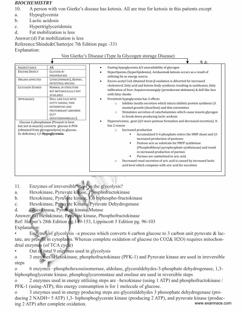
BIOCHEMISTRY10. A person with von Gierke’s disease has ketosis. All are true for ketosis in this patients except a. Hypoglycemiab. Lactic acidosisc. Hypertriglyceridemiad. Fat mobilization is less Answer:(d) Fat mobilization is lessReference:Shinde&Chatterjee 7th Edition page -331Explanation: Von Gierke’s Disease (Type Ia Glycogen storage Disease)
INHERTITANCE AR ENZYME DEFECT GLUCOSE-6-
PHOSPHATASE ORGANS AFFECTED LIVER (PRIMARY), KIDNEY,
INTESTINAL MUCOSA GLYCOGEN STORED NORMAL IN STRUCTURE
BUT METABOLICALLY NOT AVAILABLE
APPEARANCE DOLL LIKE FACE WITH FATTY CHEEKS, THIN EXTREMITIES AND PROTUBERANT ABDOMEN (D/T HEPATORENOMEGALY)
Glucose 6 phosphatase (Present in liver but not in muscle) converts glucose 6-PO4 (obtained from glycogenolysis) to glucose. So deficiency 1/t Hypoglycemia.
• Fasting hypoglycemia d/t unavailability of glycogen • Hyperlipemia (hyperlipidemia). Acidaemia& ketosis occurs as a result of
utilizing fat as energy source. • Excess acetyl CoA obtained from β-oxidation is dilverted for increased
cholesterol, fatty acid and ketone body synthesis resulting in xanthomas, fatty infiltration of liver, hepatorenomegaly (protuberant abdomen) & doll like face with fatty cheeks
• Persistent hypoglycemia has 2 effects o Inhibits insulin secretion which inturn inhibits protein synthesis l/t
stunted growth (dwarfism) and thin extremities o Stimulates secretion of catecholamines which cause muscle glycogen
to break down producing lactic acidosis • Hyperuricemia, gout (d/t more pentose formation and decreased excretion). It
has 2 reason o Increased production
Accumulated G-6 phosphate enters the HMP shunt and l/t increased production of pentoses
Pentose acts as substrate for PRPP synthetase (PhosphoRibosyl pyrophosphate synthetasae) and result in increased production of purines
Purines are catebolized to uric acid. o Decreased renal excretion of uric acid is caused by increased lactic
acid level which competes with uric acid for excretion
11. Enzymes of irreversible steps in the glycolysis? a. Hexokinase, Pyruvate kinase, Phosphofructokinaseb. Hexokinase, Pyruvate kinase, 1,6 biphospho-fructokinasec. Hexokinase, Pyruvate Kinase, Pyruvate Dehydrogenased. Glucokinase, Pyruvate kinase,MutaseAnswer: (a) Hexokinase, Pyruvate kinase, PhosphofructokinaseRef: Harper’s 28th Edition pg.149-153, Lippincott 5 Edition pg. 96-103Explanation:• Enzymes of glycolysis –a process which converts 6 carbon glucose to 3 carbon unit pyruvate & lac-tate, are present in cytoplasm. Whereas complete oxidation of glucose (to CO2& H2O) requires mitochon-drial enzymes (of TCA cycle) • Out of total 9 enzymes used in glycolysis o 3 enzymes –Hexokinase, phosphofructokinase (PFK-1) and Pyruvate kinase are used in irreversible steps o 6 enzymes –phosphohexoseisomerase, aldolase, glyceraldehydes-3-phosphate dehydrogenase, 1,3- biphosphoglycerate kinase, phosphoglyceromutase and enolase are used in reversible stepso 2 enzymes used in energy utilizing steps are –hexokinase (using 1 ATP) and phosphofructokinase /PFK-1 (using-ATP), this energy consumption is for 1 molecule of glucose. o 3 enzymes used in energy producing steps are-glyceraldehydes 3 phosophate dehydrogenase (pro-ducing 2 NADH= 5 ATP) 1,3- biphosphoglycerate kinase (producing 2 ATP), and pyruvate kinase (produc-ing 2 ATP) after complete oxidation. www.examrace.com
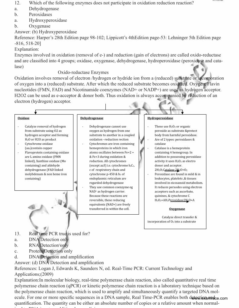
12. Which of the following enzymes does not participate in oxidation reduction reaction?a. Dehydrogenaseb. Peroxidasesa. Hydroxyperoxidaseb. OxygenaseAnswer: (b) HydroxyperoxidaseReference: Harper’s 28th Edition page 98-102; Lippicott’s 4thEdition page-53: Lehninger 5th Edition page -816, 518-20]Explanation:Enzymes involved in oxidation (removal of e-) and reduction (gain of electrons) are called oxido-reductase and are classified into 4 groups; oxidase, oxygenase, dehydrogenase, hydroperoxidase (peroxidase and cata-lase) Oxido-reductase Enzymes Oxidation involves removal of electron /hydrogen or hydride ion from a (reduced) substrate or incorporation of oxygen into a (reduced) substrate. After which the reduced substrate becomes oxidized. Oxygen, Flavin nucleotides (FMN, FAD) and Nicotinamide coenzymes (NAD+ or NADP+) are used as hydrogen acceptor. H2O2 can be used as e-acceptor & donor both. Thus oxidation is always accompanied by reduction of an electron (hydrogen) acceptor.
Oxidase
- Catalyze removal of hydrogen from substrate using O2 as hydrogen acceptor and forming H2O or H2O as product
- Cytochrome oxidase (aa3)contain copper
- Flavoprotein containing oxidase are L amino oxidase (FMN linked), Xanthine oxidase (Mo containing) and aldehyde dehydrogenase (FAD linked molybdenum & non heme iron containing)
Dehydrogenase
- Dehydrogenase cannot use oxygen as hydrogen from one substrate to another in a coupled oxidation –reduction rection
- Cytochromes are iron containing hemoproteins in which iron atoms oscillates between Fe+2 + & Fe+3 during oxidation & reduction. All cytochromes (except aa3) i.e. cytochrome b,C1, c of respiratory chain and cytochrome p-450 & b5 of endoplasmic reticulum are regarded dehydrogenase
- They use common coenzyme eg NAD+ as hydrogen carrier. Because these reactions are reversible, these reducing equivalents (NAD+) are freely transferred in within the cell.
Hydroperoxidase
- These use H2O2 or organic peroxide as substrate &protect body from harmful peroxidase.
- Are of 2 types: peroxidases & catalase
- Catalase is a hemeprotein containing 4 hemegroup. In addition to possessing peroxidase activity it uses H2O2 as electro donor and acceptor. 2H2O2Catalase 2H2O+O2
- Peroxidase are found in mild & in leukocytes, platelets ,& tissues involved in eicosanoid metabolism. It reduces perxodes using electron acceptors such as ascorbate, quinines, & cytochrome C H2O2+AH2Peroxidase2H2O+A
Oxygenase
Catalyze direct transfer & incorporation of O2 into a substrate
13. Real time PCR true is used for?a. DNA Detection onlyb. RNA Detection only c. Protein Detection onlyd. DNA Detection and amplification Answer: (d) DNA Detection and amplificationReferences: Logan J, Edwards K, Saunders N, ed. Real-Time PCR: Current Technology and Applications;(2009)Explanation:In molecular biology, real-time polymerase chain reaction, also called quantitative real time polymerase chain reaction (qPCR) or kinetic polymerase chain reaction is a laboratory technique based on the polymerase chain reaction, which is used to amplify and simultaneously quantify a targeted DNA mol-ecule. For one or more specific sequences in a DNA sample, Real Time-PCR enables both detection and quantification. The quantity can be either an absolute number of copies or a relative amount when normal-i d t DNA i t dditi l li i
www.examrace.com
The procedure follows the general principle of polymerase chain reaction; its key feature is that the amplified DNA is detected as the reaction progresses in real time. This is a new approach compared to stan-dard PCR, where the product of the reaction is detected at its end. Two common methods for the detection of products in real-time PCR are: (1) non-specific fluorescent dyes that intercalate with any double-stranded DNA, and (2) sequence-specific DNA probes consisting of oligonucleotides that are labelled with a fluores-cent reporter which permits detection only after hybridization of the probe with its complementary sequence to quantify messenger RNA (mRNA) and non-coding RNA in cells or tissues. qPCR is the abbreviation used for real-time PCR. Real-time reverse-transcription PCR is often denoted as: qRT-PCR. The acronym “RT-PCR” commonly denotes reverse transcription polymerase chain reaction and not real-time PCR, but not all authors adhere to this convention. A DNA-binding dye binds to all double-stranded (ds) DNA in PCR, causing fluorescence of the dye. An increase in DNA product during PCR therefore leads to an increase in fluorescence intensity and is measured at each cycle, thus allowing DNA concentrations to be quantified. However, dsDNA dyes such as SYBR Green will bind to all dsDNA PCR products, including nonspecific PCR products (such as Primer di-mer). This can potentially interfere with, or prevent, accurate quantification of the intended target sequence. The SYBR Green is excited using a blue light (λmax = 488 nm) and it emits a green light (λmax = 522 nm). The reaction is prepared as usual, with the addition of fluorescent dsDNA dye.The reaction is run in a real-time PCR instrument, and after each cycle, the levels of fluorescence are measured with a detector; the dye only fluoresces when bound to the ds DNA (i.e. the PCR product). With reference to a standard dilution, the dsDNA concentration in the PCR can be determined. This method has the advantage of only needing a pair of primers to carry out the amplification, which keeps costs down; however it is only possible to amplify a product using a chain reaction. Like other real-time PCR methods, the values obtained do not have absolute units associated with them (i.e., mRNA copies/cell). As described above, a comparison of a measured DNA/RNA sample to a standard dilution will only give a fraction or ratio of the sample relative to the standard, allowing only rela-tive comparisons between different tissues or experimental conditions. To ensure accuracy in the quantifica-tion, it is usually necessary to normalize expression of a target gene to a stably expressed gene (see below). This can correct possible differences in RNA quantity or quality across experimental samples.Fluorescent reporter probe method (1) An intact probes, reporter fluorescence is quenched. (2) Probes and the complementary DNA strand are hybridized and reporter fluorescence is still quenched. (3) During PCR, the probe is degraded by the Taq polymerase and the fluorescent reporter released. Fluorescent reporter probes detect only the DNA containing the probe sequence; therefore, use of the reporter probe significantly increases specificity, and enables quantification even in the presence of non-spe-cific DNA amplification. Fluorescent probes can be used in multiplex assays—for detection of several genes in the same reaction—based on specific probes with different-colour labels, provided that all targeted genes are amplified with similar efficiency. The specificity of fluorescent reporter probes also prevents interference of measurements caused by primer dimers, which are undesirable potential by-products in PCR. However, fluorescent reporter probes do not prevent the inhibitory effect of the primer dimers, which may depress ac-cumulation of the desired products in the reaction. The method relies on a DNA-based probe with a fluorescent reporter at one end and a quencher of fluorescence at the opposite end of the probe. The close proximity of the reporter to the quencher prevents detection of its fluorescence; breakdown of the probe by the 5’ to 3’ exonuclease activity of the Taq poly-merase breaks the reporter-quencher proximity and thus allows unquenched emission of fluorescence, which can be detected after excitation with a laser. An increase in the product targeted by the reporter probe at each PCR cycle therefore causes a proportional increase in fluorescence due to the breakdown of the probe and release of the reporter.The PCR is prepared as usual (and the reporter probe is added. As the reaction commences, during the annealing stage of the PCR both probe and primers anneal to the DNA target.Polymerization of a new DNA strand is initiated from the primers, and once the polymerase reaches the probe, its 5’-3’-exonuclease degrades the probe, physically separating the fluorescent reporter from the quencher, resulting in an increase in fluorescence.
www.examrace.com

Fluorescence is detected and measured in a real-time PCR machine, and its geometric increase corre-sponding to exponential increase of the product is used to determine the quantification cycle (Cq) in each reaction.
14. Ketone Bodies are not utilized by a. Brainb. RBCc. Renal cortex -d. Skeltal muscle Answer (b) RBCReference: Harper’s Illustrated Biochemistry 27th Edition page 190-191; 26th Edition page 184:Explanation:Liver is the only organs which produces ketone bodies and add to the blood, but it lacks the enzyme respon-sible for their degradation and utilization. This is the reason why liver does not utilize ketone bodiesKETOSIS• Under certain metabolic conditions associated with high rate of fatty acid oxidation, liver produces ketone bodies, which is used as respiratory substrate (Fuel) by extra-hepatic tissue ketone bodies is collec-tive name given to 3 Compounds: 1. Acetone 2. Acetoacetate 3. Beta hydroxybutyrate• In a well fed state concentration of ketone bodies in blood does not exceed 1mg /100ml
PHARMACOLOGY15. Rho kinase inhibitor is seen in :a. Fasudilb. Nicorandil c. Amiodarone d. Ranolazine Answer: (a) FasudilReference: Katjung 12th edition page 206Explanation: Fasudil Hydrochloride is a potent Rho-kinase inhibitor and vasodilator. Since it was discov-ered, it has been used for the treatment of cerebral vasospasm, which is often due to subarachnoid hemor-rhage, as well as to improve the congnitive decline seen instroke victims. It has been found to be effective for the treatment of pulmonary hypertension. It was demonstrated in February 2009 that Fasudil could also be used to enhance memory and improve the prognosis of Alzheimer’s patients 16. Orphan drugs area. Drugs used in Orphansb. Drugs used for rare diseasesc. Rarely using drugsd. Drugs of common dis used rarelyAns: B, Drugs used for rare diseases Ref: Katjung 12th edition , page 77Exp: An orphan drug is a pharmaceutical agent that has been developed specifically to treat a rare medical condition, the condition itself being referred to as an orphan disease.
17. Suicide enzyme is :a. COXb. Lipo-oxygenase c. Nucleosidased. Thromboxane SynthaseAns: (a) COX
www.examrace.com
Ref:Harper 29th Ed. Pg. 225, Exp: Cyclooxygenase is a “Suicide Enzyme” “Switching off” of prostaglandin activity is partly achieved by a remarkable property of cyclooxygenase-that of self –catalyzed destruction; i.e. it is a :Suicide enzyme.”18. Prolonged post antibiotic effect and concentration dependent killing seen in a. flouroquinolonesb. Vancomycin c. clindamycind. erythromycinAns: (a) flouroquinolonesRef: Katjung 12th edition page 823Exp: The minimum inhibitory concentration and minimum bactericidal concentration are used to measure in vitro activity antimicrobial and is an excellent indicator of antimicrobial potency. They don’t give any infor-mation relating to time-dependent antimicrobial killing the so called post antibiotic effectPost Antibiotic EffectConcentration dependent (time independent) means that the rate and extent of microorganism killing are a function of the antimicrobial concentration (increase as the concentration increases). The pharmacodynamic parameter that is most often predictive of outcome for concentration dependent drugs is peak/MIC, although the AUC/MIC can be used because the AUC takes both the antimicrobial concentration and time into ac-count. Examples of concentration dependent antimicrobials include: fluoroquinolones, aminoglycosides, and amphotericin BThe post antibiotic effect (PAE) is defined as persistent suppression of bacterial growth after a brief exposure (1 or 2 h) of bacteria to an antibiotic even in the absence of host defense mechanisms.[3] Factors that affect the duration of the post antibiotic effect include duration of antibiotic exposure, bacterial species, culture medium and class of antibiotic. It has been suggested that an alteration of DNA function is possibly respon-sible for post antibiotic effect following the observation that most inhibitors of protein and nucleic acid syn-thesis (aminoglycosides, fluoroquinolones, tetracyclines, clindamycin, certain newer macrolides/ketolides, and rifampicin and rifabutin) induce long-term PAE against susceptible bacteria. [4][3] Theoretically, the ability of an antibiotic to induce a PAE is an attractive property of an antibiotic since antibiotic concentra-tions could fall below the MIC for the bacterium yet retain their effectiveness in their ability to suppress the growth
19. Dysphoric effects of opioid receptors are mediated bya. Kappa b. Lamba c. Mu d. Sigma Ans: A, kappa Ref: Katjung 12th edition 546-559Exp: psychomimetic effects of opioid are due to kappa receptors.20. Design study which aims to asses the maximum tolerable dose of new drug a. Case control b. Phase 1 trial c. Phase 2RCTd. Phase 4 RCTAns: B, Phase 1 trial Ref: Katjung 12th edition page 58-59Exp: Phase I trials are the first to take place and are primarily concerned with the saftey of the treatment. In drug development trials the main objective is to estimate the maximum tolerated dose (MTD) and investi-gate drug toxicity.Phase I trials for cytostatic drugsThe aims of such trials are typically to:Estimate the maximum tolerated dose (MTD)
www.examrace.com
Determine the extent, duration and reversibility of toxicityObserve any anti-tumour activity21. Ritonovir inhibits all except? a. Amiodarone b. Cisapride c. Phenytoin d. Midazolam Ans: C, Phenytoin Ref: katjung 12th edition pages 880 and 889Exp: ritonavir induces CYP 1A2 and inhibits the major P450 isoforms (3A4 and 2D6).amiodarone – decreased metabolism, possible toxicitymidazolam and triazolam – contraindicatedcarbamazepine – decreased metabolism, possible toxicitycisapride – decreased metabolism, possible prolongation of Q-T interval and life-threatening arrythmiasdisulfiram (with ritonavir oral preparation) – decreased metabolism of ritonavirvoriconazole – ritonavir increases metabolism of voriconazoleBecause of potential toxicities, ritonavir should not be used concurrently with various antiarrhythmics (amiodarone, encainide, flecainide, quinidine) and highly metabolized sedative/hypnotics (i.e., alprazolam, diazepam, flurazepam, midazolam, and triazolam).
22. The site of action of the vasopressin receptor antagonists is a. Proximal convoluted tubule b. Distal convoluted lobulec. Cortical collecting tubuled. Medullary collecting duct Ans: (c) Cortical collecting tubuleRef: katjung 12th edition page no. 303Exp: The collecting ducts, in particular, the outer medullary and cortical collecting ducts, are largely imper-meable to water without the presence of antidiuretic hormone (ADH, or vasopressin).A vasopressin receptor antagonist (VRA) is an agent which interferes with action at the vasopressin recep-tors. Most commonly VRAs are used in the treatment of hyponatremia, especially in patients with conges-tive heart failure, liver cirrhosis or SIADHTetracyclines [edit]Demeclocycline, a tetracycline antibiotic, is sometimes used to block the action of vasopressin in the kidney in hyponatremia due to inappropriately high secretion of vasopressin (SIADH), when fluid restriction has failed.[2]Vaptans [edit]A new class of medication, the “vaptan” drugs, act by inhibiting the action ofvasopressin on its receptors (V1A, V1B and V2).V2 receptor [edit]V2 receptor (V2R) differs from V1R primarily in the number of sites susceptible to N-linked glycosylation; the V1R has sites at both the amino-terminus and at the extracellular loop, whereas the V2R has a single site at the extracellular amino-terminuThe well known antidiuretic effect of vasopressin occurs via activation of V2R.[1] Vasopressin regulates wa-ter excretion from the kidney by increasing the osmotic water permeability of the renal collecting duct – an effect that is explained by coupling of the V2R with the Gs signaling pathway, which activates cAMP. The increased intracellular cAMP in the kidney in turn triggers fusion of aquaporin-2-bearing vesicles with the apical plasma membrane of the collecting duct principal cells, increasing water reabsorption.
23. Iodine use in thyroid disorder not true?a. Cuases Iodism b. Contraindicated in Hyperthyroidism c. Inhibit Formation of Iodo Thyronine
www.examrace.com
d. Thyroxine ReleaseAns: (b) Contraindicated in HyperthyroidismRef: Katzung 12th Edition pg. 689Exp: Iodine has several effects on thyroid function. In hyperthyroid patients, iodine acutely inhibits hor-monal secretion within hours , but the responsible mechanisms are uncertain. This is the most acute effect of iodine on thyroid status, occurring within one to two days of the start of therapy.A second effect involves inhibition of thyroid hormone synthesis. In normal subjects, the administration of pharmacologic amounts of iodine leads to temporary inhibition of iodine organification in the thyroid gland, thereby diminishing thyroid hormone biosynthesis, a phenomenon called the Wolff-Chaikoff effect [2]. However, within two to four weeks of continued exposure to excess iodine, organification and thyroid hormone biosynthesis resume in a normal fashion. This is called escape from the Wolff-Chaikoff effect.
24. Which of the following drugs can be given in subcutaneous form?a. Terbutalineb. Almeterol c. Fenoterol d. Metaproterenol Ans: (a) TerbutalineRef: Katzung 12th Edition pg. 344Exp: --After subcutaneous administration of 0.25 mg of Terbutaline sulfate injection, a measurable change in expiratory flow rate usually occurs within 5 minutes
25. Drug induced SLE is seen with all except:a. Hydralazineb. penicillinc. Sulfonamided. Isoniazide Ans (b) PenicillinRef: Harrison 16th Ed. Pg. 1960-61, CMDT 2006 pg. 834Exp: EXPL…High risk:Procainamide (antiarrhythmic)Hydralazine (antihypertensive)Moderate to low risk:Infliximab anti (TNF-alpha)Etanercept anti (TNF-alpha)Isoniazid (antibiotic)Minocycline (antibiotic)Pyrazinamide (antibiotic)Quinidine (antiarrhythmic)D-Penicillamine (anti-inflammatory)Carbamazepine (anticonvulsant)Oxcarbazepine (anticonvulsant)Phenytoin (anticonvulsant)Propafenone (antiarrhythmic)
26. 16 yr old girl complains of severe abdominal pain after receiving sulphur drugs. She was incoherent of suffered a seizure. The probable diagnosis of the patient is a. Acute intermittent porphyriab. Congenital erythropoeitic porphyriac. Erythropoietic protoporphyria (EEP)d. Hepatic porphyriasAns: A, Acute intermittent porphyriaRef: Harrison’s 17th Ed. Pg. 2434-40: Fitzpatrick 7ed. Pg. 1228-56
www.examrace.com
Exp: Acute intermittent porphyria (AIP), • Characterised by abdominal pain (most common-90%), neurological & psychiatric symptoms.• Abdominal pain is often intermittent, steady, poorly localized & spasmodic. • Vomiting, nausea, constipation (ileus), decreased bowel sound are common • Abdominal tenderness, fever, leukocytopsis are usually absent or mild because symptoms are neu-rological rather than inflammatory • Mental (psychiatric) symptoms include anxiety, insomnia, depression, disorientation, hallucination & paronia• Peripheral neuropathy d/t aomnal degernation primarily affects motor neuroms of proximal muscles (shoulder & arms) initially > focal cranial nerve> sensory (L/t paresthesia, pain in limb, head –neck & sen-sory loss) > respiratory & bulbar paralysis (l/t death)• Seizures can be d/t hyponatremia (resulting from vomiting & inappropriate fluid therapy) or neu-rological effect. Treatment of seizure is difficult because most anti seizure drugs can exacerbate AIP (clon-azepam may be safer than phenytoin or barbiturates) • Sympathetic overactivity may l/t tachycardia, hypertension, restlessness, tremors, excessive sweat-ing & cardiac arrhythmias causing sudden death 27. Comparison of efficacy of a drug with placebo is confirmed in which phase of clinical trials? a. Phase Ib. Phase 2c. Phase 3d. Phase 4Ans: (c)Phase 3 Ref: Katzung 12th Edition pg. 59 & 74Exp: Efficacy of a new chemical entity is first known in phase 2 clinical trials. Phase 2 studies are indica-tive studies. On the other hand, the finding of phase 2 are confirmed in phase 3 clinical trials. Phase 3 trials are also know as Therapeutic confirmatory studies
PATHOLOGY28. If a chromosome divides in an axis perpendicular to usual axis of division it is going to form:a. Ring chromosome b. Isochromosome c. Acrocentric d. SubtelocentricAnswer: b. Isochromosome Reference: Emery’s Elements of Medical Genetics, 14th editionExplanation: • An isochromosome is formed when the centromere divides transversely rather than longitudinally. • An isochromosome shows loss of one arm with duplication of the other. • The most commonly encountered isochromosome is that which consists of 2 long arms of X chro-mosome. • Isochromosome accounts for upto 15% of all cases of Turner Syndrome
29. Marker for rhabdomyosarcomaa. Desminb. Synaptophysinc. Keratind. SynaptophysinAnswer: (a) DesminReference: Robbin’s 8TH edition pg 1253Explanation: Rhabdomyosarcoma, the most common soft tissue sarcoma of childhood and adolescence, usually appears before age 20. They may arise in any anatomic location, but most occur in the head and neck or
www.examrace.com
genitourinary tract, where there is little, if any, skeletal muscle as a normal constituent. Only in the extremi-ties do they appear in relation to skeletal muscle Rhabdomyosarcoma is histologically subclassified into the embryonal, alveolar, and pleomorphic variants. The rhabdomyoblast—the diagnostic cell in all types—contains eccentric eosinophilic granular cyto-plasm rich in thick and thin filaments. The rhabdomyoblasts may be round or elongate; the latter are known as tadpole or strap cells and may contain cross-striations visible by light. Ultrastructurally, rhabdomyoblasts contain sarcomeres, and immunohistochemically they stain with antibodies to the myogenic markers desmin, MYOD1, and myogenin30. Due to popularity of refrigeration reducing the need to preserve food, which cancer’s incidence has dramatically declined:a. Esophagusb. Stomachc. Colond. Nasopharynx Answer: (b) Stomach Reference: Robbons 7th ed: Robbin’s 8th ed:pg 784Explanation: Factors Associated with Increased Incidence of Gastric CarcinomaThe overall reduction in the incidence of Gastric cancer is unknown. One possible explanation is decresed consumtion of diatary carcinogens,such as N-nitroso compounds and benzo-pyrene, because of reduced use of salt and smokink in food preservation and widespread use of refrigeration.
Environmental FactorsInfection by H. pylori• Present in most cases of intestinal-type carcinomaDiet• Nitrites derived from nitrates (water, preserved food)• Smoked and salted foods, pickled vegetables, chili peppers• Lack of fresh fruit and vegetablesLow socioeconomic statusCigarette smokingHost FactorsChronic gastritis• Hypochlorhydria: favors colonization with H. pylori• Intestinal metaplasia is a precursor lesionPartial gastrectomy• Favors reflux of bilious, alkaline intestinal fluidGastric adenomas• 40% harbor cancer at time of diagnosis• 30% have adjacent cancer at time of diagnosisBarrett esophagus• Increased risk of gastroesophageal junction tumorsGenetic FactorsSlightly increased risk with blood group AFamily history of gastric cancerHereditary nonpolyposis colon cancer syndromeFamilial gastric carcinoma syndrome (E-cadherin mutation
www.examrace.com

Environmental FactorsInfection by H. Pylori• Present in most cases of intestinal-type carcinomaDiet• Nitrites derived from nitrates (water, preserved food)• Smoked and salted foods, pickled vegetables, chili peppers• Lack of fresh fruit and vegetablesLow socioeconomic statusCigarette smokingHost FactorsChronic gastritis• Hypochlorhydria: favors colonization with H. pylori• Intestinal metaplasia is a precursor lesionPartial gastrectomy• Favors reflux of bilious, alkaline intestinal fluidGastric adenomas• 40% harbor cancer at time of diagnosis• 30% have adjacent cancer at time of diagnosisBarrett esophagus• Increased risk of gastroesophageal junction tumorsGenetic FactorsSlightly increased risk with blood group AFamily history of gastric cancerHereditary nonpolyposis colon cancer syndromeFamilial gastric carcinoma syndrome (E-cadherin mutation
31. What is used for diagnosis of fragment of chromosomesa. FISHb. CGHc. PCRd. RFLPAnswer: (a) FISHReference: Robbins and Cotran, pathologic Basis of Disease 8th Ed. Pg. 179Explanation: FISH is used to detect and localize the presence or absence of specific DNA sequences on chro-mosomes. FISH uses fluorescent probes that bind to only those parts of the chromosome with which they show a high degree of sequence complementarity. Fluorescence microscopy can be used to find out where the fluorescent probe is bound to the chro-mosomes. FISH is often used for finding specific features in DNA for use in genetic counseling, medicine, and species identification. FISH can also be used to detect and localize specific RNA targets (mRNA, ln-cRNA and miRNA) in cells, circulating tumor cells, and tissue samples. In this context, it can help define the spatial-temporal patterns of gene expression within cells and tissues.
32. Embryonic haemoglobin is composed up of :a. Alpha and betab. Epsilon and gammac. Gamma and betad. Zeta and epsilonAnswer: (d) Zeta and epsilon Reference: Nelson ped 19th ed;pg 1663
www.examrace.com
Explanation: Hemoglobin is a tetramer consisting of 2 pairs of globin chains. Abnormalities in these pro-teins are referred to as hemoglobinopathies. There are ~800 variant hemoglobins. The most common and useful clinical classification of hemoglobinopathies is based on nomenclature associated with alteration of the involved globin chain. Two hemoglobin gene clusters are involved in the produc-tion of hemoglobin and are located at the end of the short arms of chromosomes 16 and 11. Their control is complex, including an upstream locus control region on each respective chromosome and an X-linked control site. On chromosome 16, there are 3 genes within the αgene cluster, namely zeta (ζ), alpha 1 (α1), and alpha 2 (α2). On chromosome 11, there are 5 genes within the beta gene cluster, namely epsilon (ε), 2 gamma genes (γ), a delta gene (δ), and a beta gene (β). The order of the gene expression within each cluster roughly follows the order of expression during the embryonic period, fetal period, and eventually childhood. After 8 wk of fetal life the embryonic hemoglobins, Gower-1 (ζ2ε2), Gower-2 (α2ε2), and Portland (ζ2γ2), are formed. At 9 wk of fetal life, the major hemo-globin is Hb F (α2γ2). Hb A (α2β2) appears at ~1 mo of fetal life but does not become the dominant hemoglobin until after birth, when Hb F levels start to de-cline. Hb A2(α2δ2) is a minor hemo-globin that appears shortly before birth and remains at a low level after birth. The final hemoglobin distribution pattern that occurs in childhood is not achieved until at least 6 mo of age and sometimes later. The normal hemoglobin pattern is ≥95% Hb A ≤3.5 Hb A2, and <2.5% Hb F33. Which is not seen with apoptosisa. Cell shrinkageb. Nuclear condensationc. Inflammationd. All of the aboveAnswer: (c) Inflammation Reference: Robbins 8th ed; pg 27Explanation: Apoptosis is a pathway of cell death that is induced by a tightly regulated intracellular program in which cells destined to die activate enzymes that degrade the cells’ own nuclear DNA and nuclear and cytoplasmic proteins. The cell’s plasma membrane remains intact, but its structure is altered in such a way that the apoptotic cell becomes an avid target for phagocytosis. The dead cell is rapidly cleared, before its contents have leaked out, and therefore cell death by this pathway does not elicit an inflammatory reaction in the host. Thus, apoptosis is fundamentally different from necrosis, which is characterized by loss of mem-brane integrity, enzymatic digestion of cells, and frequently a host reaction34. True about MHC isa. Antigen presentingb. Regulation of T cell mediated immune response c. Class II Gene present on T cells d. Class I Gene present on B cells Answer: Antigen presentingReference: Robbins 8 th ed; pg 190 Explanation: On the basis of their chemical structure, tissue distribution, and function, the MHC gene prod-ucts are classified into three categories. Class I and class II genes encode cell surface glycoproteins involved in antigen presentation.Class I MHC molecules are expressed on all nucleated cells and platelets. They are encoded by three closely linked loci, designated HLA-A, HLA-B, and HLA-C Each of these molecules is a heterodimer, consisting of a polymorphic α, or heavy, chain (44-kD) linked noncovalently to a smaller (12-kD) nonpolymorphic peptide called β2-microglobulin, which is not encoded within the MHC. The extracellular region of the heavy chain is divided into three domains: α1, α2, and α3 Crystal structure of class I molecules has revealed that the α1 and α2 domains form a cleft, or groove, where peptides bind to the MHC molecule.Biochemical analyses of several different class I alleles have revealed that almost all polymorphic residues line the sides or the base of the peptide-binding groove. As a result, different class I alleles bind and display different peptide fragments. In general, class
www.examrace.com
I MHC molecules bind and display peptides that are derived from proteins, such as viral antigens, synthe-sized within the cell. The generation of peptide fragments within the cells, and their association with MHC molecules and transport to the cell surface, is a complex process. Involved in this sequence are proteolytic complexes (proteasomes), which digest antigenic proteins in the cytoplasm into short peptides, and trans-port proteins, which ferry peptide fragments from the cytoplasm to the endoplasmic reticulum. Within the endoplasmic reticulum, peptides bind to the antigen-binding cleft of newly synthesized class I heavy chains, which then associate with β2-microglobulin to form a stable trimer that is transported to the cell surface for presentation to CD8+ cytotoxic T lymphocytes
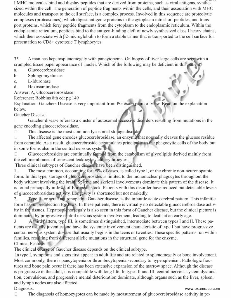
35. A man has hepatosplenomegaly with pancytopenia. On biopsy of liver large cells are seen with a crumpled tissue paper appearance of nuclei. Which of the following may be deficient in this patient?a. Glucocerebrosidaseb. Sphingomyelinasec. L-Iduronased. HexosaminidaseAnswer: A, GlucocerebrosidaseReference: Robbins 8th ed: pg 149Explanation: Gauchers Disease is very important from PG exam point of view. Read on the explanation below.Gaucher Disease Gaucher disease refers to a cluster of autosomal recessive disorders resulting from mutations in the gene encoding glucocerebrosidase. This disease is the most common lysosomal storage disorder. The affected gene encodes glucocerebrosidase, an enzyme that normally cleaves the glucose residue from ceramide. As a result, glucocerebroside accumulates principally in the phagocytic cells of the body but in some forms also in the central nervous system. Glucocerebrosides are continually formed from the catabolism of glycolipids derived mainly from the cell membranes of senescent leukocytes and erythrocytes. Three clinical subtypes of Gaucher disease have been distinguished. 1. The most common, accounting for 99% of cases, is called type I, or the chronic non-neuronopathic form. In this type, storage of glucocerebrosides is limited to the mononuclear phagocytes throughout the body without involving the brain. Splenic and skeletal involvements dominate this pattern of the disease. It is found principally in Jews of European stock. Patients with this disorder have reduced but detectable levels of glucocerebrosidase activity. Longevity is shortened but not markedly. 2. Type II, or acute neuronopathic Gaucher disease, is the infantile acute cerebral pattern. This infantile form has no predilection for Jews. In these patients, there is virtually no detectable glucocerebrosidase activ-ity in the tissues. Hepatosplenomegaly is also seen in this form of Gaucher disease, but the clinical picture is dominated by progressive central nervous system involvement, leading to death at an early age.3. A third pattern, type III, is sometimes distinguished, intermediate between types I and II. These pa-tients are usually juveniles and have the systemic involvement characteristic of type I but have progressive central nervous system disease that usually begins in the teens or twenties. These specific patterns run within families, resulting from different allelic mutations in the structural gene for the enzyme.Clinical Feature:The clinical course of Gaucher disease depends on the clinical subtype. In type I, symptoms and signs first appear in adult life and are related to splenomegaly or bone involvement. Most commonly, there is pancytopenia or thrombocytopenia secondary to hypersplenism. Pathologic frac-tures and bone pain occur if there has been extensive expansion of the marrow space. Although the disease is progressive in the adult, it is compatible with long life. In types II and III, central nervous system dysfunc-tion, convulsions, and progressive mental deterioration dominate, although organs such as the liver, spleen, and lymph nodes are also affected.Diagnosis: The diagnosis of homozygotes can be made by measurement of glucocerebrosidase activity in pe-
www.examrace.com
ripheral blood leukocytes or in extracts of cultured skin fibroblasts. Because there is substantial overlap between the enzyme levels in normal individuals and heterozy-gotes, such assays are not reliable for carrier detection. In principle, detection of specific mutations can be used for detecting heterozygotes. Because more than 150 allelic mutations can cause Gaucher disease, however, it is not possible to use a single genetic test. Chitotriosidase, an enzyme synthesized by macrophages, is markedly elevated in patients with Gau-cher disease. It is a reasonably specific biomarker for Gaucher disease because levels are only slightly elevated in other disorders affecting macrophages.Diagnosis on biopsy: The glucocerebrosides accumulate in massive amounts within phagocytic cells throughout the body in all forms of Gaucher disease. The distended phagocytic cells, known as Gaucher cells, are found in the spleen, liver, bone marrow, lymph nodes, tonsils, thymus, and Peyer patches. Similar cells may be found in both the alveolar septa and the air spaces in the lung. Gaucher cells rarely appear vacuolated but instead have a fibrillary type of cytoplasm likened to crumpled tissue paper. Gaucher cells are often enlarged, sometimes up to 100 µm in diameter, and have one or more dark, eccentri-cally placed nuclei. Periodic acid-Schiff (PAS) staining is usually intensely positive. With the electron mi-croscope, the fibrillary cytoplasm can be resolved as elongated, distended lysosomes, containing the stored lipid in stacks of bilayers.Treatment:As with all lysosomal storage diseases, the treatment of Gaucher disease is difficult. Replacement therapy with recombinant enzymes is effective, and those with type I disease can expect normal life expectancy with enzyme replacement therapy. However, such therapy is extremely expensive. Because the fundamental de-fect resides in mononuclear phagocytic cells originating from marrow stem cells, bone marrow transplanta-tion has been attempted. Attempts are also directed toward correction of the enzyme defect by transfer of the normal glucocerebrosidase gene into the patient’s cells.
MICROBIOLOGY36. A woman is having sore throat with high grade fever with headache nausea and vomiting. On exami-nation, she is having RR of 36/min, temperature of 39 degrees and BP of 80/50 mm Hg. On her arm some red spots are seen distal to BP cuff. The probable diagnosis is :a. N. Meningitidisb. Brucellasuisc. Brucella abortusd. Staph. Aureus.Answer ( a) N. Meningitidis Reference- Harrison 18th /p1166, 1211-13, Ananthnarayan 8th /p224-227.o Features like .....Female with Fever, headache, hypotension, tachycardia and petechial rashes..............Points towards ............. a case of meningococcal speticemia........o St aureus TSS may present with similar features, but can be ruled out because... there is no h/o use of vaginal tampons.Meningococcal septicaemia-o Meningococcal septicemia alone accounts for up to 20% of cases of meningococcal disease. The condition may progress from early nonspecific symptoms to death within hours.o Early symptoms are nonspecific and presented as-o influenza-like illness with fever, o headache, and myalgia accompanied byo vomiting and abdominal pain• A nonblanching rash (petechial or purpuric) develops in >80% of cases of meningococcal disease;• Shock is manifested by tachycardia, poor peripheral perfusion, tachypnea, and oliguria.• Decreased cerebral perfusion leads to confusion, agitation, or decreased level of consciousness.
www.examrace.com
• With progressive shock, multiorgan failure ensues; hypotension is a late sign in children, who more commonly present with compensated shock.• Poor outcome is associated with an absence of meningismus, hypotension, young age, coma, rela-tively low temperature (< 38oC), leukopenia, and thrombocytopenia. • Spontaneous hemorrhage (pulmonary, gastric, or cerebral) may result from consumption of coagula-tion factors and thrombocytopenia.Also read this lines....o The most common form of infection with N. meningitidis is asymptomatic carriage of the organism in the nasopharynx. o Despite the location of infection in the upper airway, meningococcal pharyngitis is rarely reported; however, upper respiratory tract symptoms are common prior to presentation with invasive disease.
37. Rapidly frowing Atypical organism involved in lung infection ?a. m.chelonaeb. m.fortuitumc. m.abscessusd. M.kansasiAnswer (c ) M.abscessusReference- Harrison 18th /p1369, Koneman’s Diagnostic Microbiology 6th/p1105• Any of the rapidly growing Mycobacteria such as M chelonei, M fortuitum & M. Abscessus can cause pulmonary infection but infection with M. Abscessus may be particularly dangerous ..Koneman’s Diagnostic Microbiology 6th/p1105• M. kansasii ....... Belongs to photochromogen. o Can cause a clinical syndrome that strongly resembles tuberculosis, consisting of hemoptysis, chest pain, and cavitary lung disease. • MAC organisms are the most common causes of pulmonary nontuberculous mycobacterial infection in North America.• The rapidly growing NTM, M chelonei, M fortuitum & M. abscessus, acquired via skin contami-nation from surgical instruments (especially in cosmetic surgery), injections, and other procedures. These infections are typically accompanied by painful, erythematous, draining subcutaneous nodules, usually with-out associated fever or systemic symptoms.Atypical MycobacteriumRunyon classifcation Definition SPECIESI Photo chromogen Pigmentation only in light M kansasii, M marinum, M simiae,
M.asciaticum (MASK)II Scoto chromogen Pigmentations only in light &
darkSSG-M. scrofulaceum, M szulgai , S.gordonae
III Non-chromogen No Pigmentation MAC, M xenopi M. ulceransIV Rapid growers Grows within a week M chelonei, M fortuitum M.abscessus
Diseases caused by Atypical Mycobacterium• Post trauma abscess - M chelonei , M fortuitum• Swimming pool granuloma - M marinum • Buruli ulcer - M. ulcerans • Mycobacteria Causing Johnes Disease -M. paratuberculosis• Lymphadenopathy - M. scrofulaceum • Pulmonary disease - M kansasii• Disseminated disease- M avium intracellulare
38. 15 Year old complains of loose motion, intermittent abdominal pain of 1year wet mount of stool shows multiple ova> 100 microns in length.Which of the following agent is responsible a. Fosciola Giganticab Gastrodiscoides Hominis
www.examrace.com
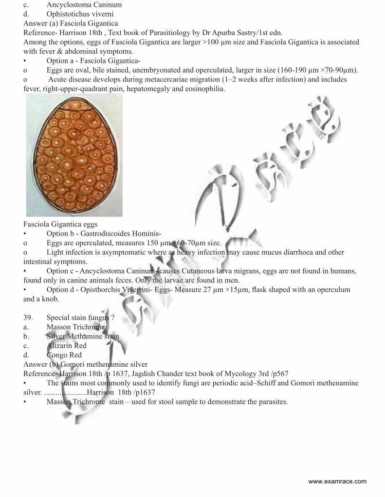
c. Ancyclostoma Caninumd. Ophistotichus viverniAnswer (a) Fasciola GiganticaReference- Harrison 18th , Text book of Parasitiology by Dr Apurba Sastry/1st edn.Among the options, eggs of Fasciola Gigantica are larger >100 µm size and Fasciola Gigantica is associated with fever & abdominal symptoms.• Option a - Fasciola Gigantica-o Eggs are oval, bile stained, unembryonated and operculated, larger in size (160-190 µm ×70-90µm).o Acute disease develops during metacercariae migration (1–2 weeks after infection) and includes fever, right-upper-quadrant pain, hepatomegaly and eosinophilia.
Fasciola Gigantica eggs• Option b - Gastrodiscoides Hominis- o Eggs are operculated, measures 150 µm ×60-70µm size. o Light infection is asymptomatic where as heavy infection may cause mucus diarrhoea and other intestinal symptoms.• Option c - Ancyclostoma Caninum- causes Cutaneous larva migrans, eggs are not found in humans, found only in canine animals feces. Only the larvae are found in men.• Option d - Opisthorchis Viverrini- Eggs- Measure 27 µm ×15µm, flask shaped with an operculum and a knob.
39. Special stain fungus ? a. Masson Trichrome b. Silver Methamine stainc. Alizarin Red d. Congo RedAnswer (b) Gomori methenamine silverReference- Harrison 18th /p 1637, Jagdish Chander text book of Mycology 3rd /p567• The stains most commonly used to identify fungi are periodic acid–Schiff and Gomori methenamine silver. ......................Harrison 18th /p1637• Masson Trichrome stain – used for stool sample to demonstrate the parasites.
www.examrace.com
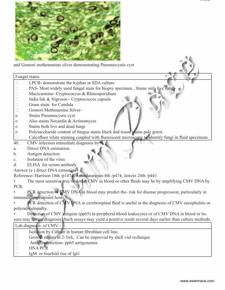
and Gomori methenamine silver demonstrating Pneumocystis cyst
Fungal stains LPCB- demonstrate the hyphae in SDA culture PAS- Most widely used fungal stain for biopsy specimen , Stains only live fungi Mucicarmine- Cryptococcus & Rhinosporidium India Ink & Nigrosin-- Cryptococcus capsule Gram stain- for Candida Gomori Methinamine Silver- o Stains Pneumocystis cysto Also stains Nocardia & Actinomyceso Stains both live and dead fungio Polysaccharide content of fungus stains black and tissue stains pale green Calcofluor white staining coupled with fluorescent microscopy to identify fungi in fluid specimens
40. CMV infection immediate diagnosis by?a. Direct DNA estimation b. Antigen detectionc. Isolation of the virusd. ELISA for serum antibodyAnswer (a ) direct DNA estmationReference- Harrison 18th /p1474, Ananthnarayan 8th /p474, Jawetz 24th /p441• The most sensitive way to detect CMV in blood or other fluids may be by amplifying CMV DNA by PCR. • PCR detection of CMV DNA in blood may predict the- risk for disease progression, particularly in immunocompromised hosts • PCR detection of CMV DNA in cerebrospinal fluid is useful in the diagnosis of CMV encephalitis or polyradiculopathy.• Detection of CMV antigens (pp65) in peripheral-blood leukocytes or of CMV DNA in blood or tis-sues may hasten diagnosis. Such assays may yield a positive result several days earlier than culture methods.Lab diagnosis of CMV– Isolation by Culture in human fibroblast cell line, Growth occurs in 2-3wk, Can be improved by shell vial technique Antigen detection- pp65 antigenemia DNA PCR IgM or fourfold rise of IgG
www.examrace.com
Transplacental CMV infection Postnatal CMV infectionBaby’s IgM Antibody titre-• At birth- just appearing• At 1 month- peak• At 3 month- falls
Baby’s IgM Antibody titre-• At birth-• At 1 month-- just appearing• At 3 month- peak
41. Legionella pneumophila spreads bya. Person to person b. A.C Aerosolc. Infected Meat d. Water Drinking which is contaminatedAnswer (b) A.C AerosolReference- Harrison 18th /p1236, Ananthnarayan 8th /p400-401• Multiple modes of transmission of Legionella to humans exist, including-o Aerosolization, o Aspiration,o Direct instillation into the lungs during respiratory tract manipulations. • Aspiration is now known to be the predominant mode of transmission• Aerosolization of Legionella by devices filled with tap water, including whirlpools, nebulizers, and humidifiers, cooling tower, air-conditioners, has been implicated. • Reservoir –o Natural water source- Rivers/streams/amoebae; o Artificial Aquatic source- AC, water cooling tanks• No human – to human transmission, • No carriers, No animal reservoirLegionnaires’ disease (“Atypical Pneumonia”) –• CAP + Diarrhea + encephalitis• MC extrapulmonary site – heart• Numerous PMN, but no organism in sputumPontiac Fever –mild flu likeDiagnosis: Fastidious requiring iron and cysteine -(BCYE medium)Treatment- Azithromycin or Clarithromycin is DOC. Alternate is quinolones.B lactamase & AMG- not affective
PHYSIOLOGY Urinary metabolite of progesterone: Pregnanolone Progestriol 17 Hydroxypregnanolone PregnanediolAnswer: (d) PregnanediolReference:Ganong’s Review of Medical Physiology, 22nd edition, pg. 443Explanation: Pregnanediol is most abundant plasma as well as urinary metabolite of progesterone. In plas-ma 80 % is bound to albumin and 18 % to corticosteroid binding globulin. Progesterone has short half-life and in liver it is converted to Pregnanediol and conjugated to glucoronic acid and excreted in urine.
In healthy female ovulation occurs when: Before LH surge After biphasic rise in basal body temp. After follicular ripening by FSH After cervical mucus breaks down
www.examrace.com
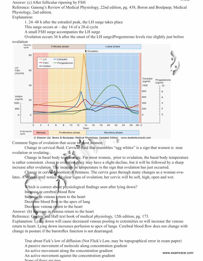
Answer: (c) After follicular ripening by FSHReference: Ganong’s Review of Medical Physiology, 22nd edition, pg. 438, Boron and Boulpaep, Medical Physiology, 2nd edition.Explanation: 1. 24–48 h after the estradiol peak, the LH surge takes place This surge occurs at ~ day 14 of a 28-d cycle A small FSH surge accompanies the LH surge Ovulation occurs 36 h after the onset of the LH surge)Progesterone levels rise slightly just before ovulation
Common Signs of ovulation that occur in most women: Change in cervical fluid. Cervical fluid that resembles “egg whites” is a sign that women is near ovulation or ovulating.. Change in basal body temperature. For most women, prior to ovulation, the basal body temperature is rather consistent. closer to ovulation, they may have a slight decline, but it will be followed by a sharp increase after ovulation. The increase in temperature is the sign that ovulation has just occurred. Change in cervical position or firmness. The cervix goes through many changes as a woman ovu-lates. A woman may notice the clear signs of ovulation; her cervix will be soft, high, open and wet.
Which is correct about physiological findings seen after lying down? Increase in cerebral blood flow Increase in venous return to the heart Decrease blood flow to the apex of lung Decrease venous return to the heartAnswer: (b) Increase in venous return to the heartReference: Guyton and Hall text book of medical physiology, 12th edition, pg. 173.Explanation: Lying down will cause decreased venous pooling to extremities so will increase the venous return to heart. Lying down increases perfusion to apex of lungs. Cerebral blood flow does not change with change in posture if the baroreflex function is not dearranged.
True about Fick’s low of diffusion (Not Flick’s Law, may be topographical error in exam paper) A passive movement of molecule along concentration gradient An active movement along the concentration gradient An active movement against the concentration gradient None of these are true
www.examrace.com

Answer: (a)A passive movement of molecule along concentration gradientReference: Ganong 24th e/ pg 7, 22nde/pg 4Fick’s low Diffusion cJ=-DA xWhere J is the net rate of diffusion, D is the diffusion coefficient, A is the area, and c/ x is the concentra-tion gradient. The minus sign indicates the direction of diffusion
FORENSIC MEDICINE46. Priapism is seen in: a. Cobra bite b. Spanish fly poisoning c. Scorpion bite d. Viper biteAnswer (B) Spanish fly poisoning Reference:: Reddy 27th edition page 490Explanation: It is persistent abnormal erection of penis with pain and tenderness.Spanish fly (Blister Beetle) can cause priapism.47. Difficulty in identifying entry and exit wounds due to surgical alteration is also known as: a. Mac naughten’s rule b. Kennedy’s phenomenonc. Alecjeffrey’s rule d. Edmond locard exchange principleAnswer (B) Kennedy’s phenomenonRef: Reddy 27th edition page 203Explanation: Kennedy Phenomenon is surgical intervention of firearm wound resulting in an artifact and hence rendering the wound difficult to interpret during autopsy as happend in Kennedy’s case
48. Which of the following is a signature fracturea. Depressed fractureb. Ring fracturec. Separation of suturesd. Pond #Answer (A), Depressed fractureRef: Reddy 27th edition page 216Explanation: The outer table is driven into diploe, the inner table is fractured irregularly and to greater extent and may be comminuted.Localized depressed fracture are caused by blows from heavy weapon with a small striking surface49. Methanol poisoning .false is ??a. Snow field visionb. Fomepizole is a comp inhibitor of aldehyde dehydrogenasec. Formic acid is responsbile for toxicity . d. Min lethal dose is 1.25mg/mlAnswer (b) fomepizole is a comp inhibitor of aldehyde dehydrogenaseRefernce: Reddy 27th edition page 513Explanation: Fomepizole is used as an antidote in methanol or ethylene glycol poisoning. Fomepizole is a competitive inhibitor of alcohol dehydrogenase, the enzyme that catalyzes the initial steps in the metabolism of ethylene glycol and methanol to their toxic metabolites. So this eliminates option C and marks B as the correct answer..50. A child is burnt by hot water, the best method to calculate the surface area of the burnt areas is: fo-rensica. Palm methodb. Lund and browder method
www.examrace.com
c. Rule of 9d. Brocas methodAnswer (B) Lund and browder methodReference:: Reddy 27th edition page 284Explanation : Estimation of percentage of body surface area Lund and Browder.51. A girl coming with kerosene like odour on breath, lacrimation, pin point pupil, rhinorrhea, weakness. Which statement is false:a. Atropine is the antidote b. Plasma AChesterase has no prognostic value c. Atropine reverses neuromuscular weakness d. Activated charwal has no proven benefitsAnswer (c) Atropine reverses neuromuscular weakness Reference: Reddy 27th edition page 458Explanation :Atropine is a muscarinic receptor antagonist and it is antidote used for organophosphorus poi-soning. Activated charchol is not used in organophosphorus poisoning.
P.S.M
52. Number of Vision centers under Vision 2020, National Program for Control of Blindness area. 20b. 200c. 2000d. 20000
Ans. (d) 20000 Reference: Review of PSM including Biostatistics by Vivek Jain, 5th Ed., Figure 5.3, Pg 298/ K Park, 22nd Ed., Pg 408EXPLANATIONProposed Structure for Vision 2020, (NPCB)o Vision centers 20,000 (Primary level)o Service centers 2,000 (Secondary level)o Training centers 200 (Tertiary level)o Centers for Excellence 20 (Tertiary level)
53. All can be used to reduce the risk of transmission of HIV to neonate in a pregnanct-woman excepta. Vaginal Delivery b. Vitamin A supplementationc. Stopping Breast feedingd. Zidovudine to mother antenatal and newborn after delivery
Ans. (a) Vaginal Delivery Reference: Dutta 7th Edition page 301-302, Williams Obs 23 Edition page 1248 onwards COGDT 10 Edi-tion page 692-693EXPLANATION: Vertical Transmission (mother to child transmission of HVI)Vertical transmission can occur• In utero • During delivery • After birth by breast feeding• M/C time= peripartum (7-49%) >labour Risk factors for Increased transmission • High maternal viral load • Low CD 4T cell count
www.examrace.com
• Chorioamnionitis • Vitamin A deficiency Methods to decrease vertical transmission- I. Antiretroviral prophylaxis- Vertical transmission can be prevented by giving antireto viral therapy to mother and early prophylaxis to newborn. Zidovudine treatment is started from beginning of second trimes-ter and continued till delivery. The newborn babies of HIV positive mothers are given prophylactic zidovu-dine for 6 weeks. Such treatment reduces the rate of HIV transmission by 22.5 to 25% II. Cesarean section- Elective cesarean section reduces the risk of transmission by 87% in women on ART can by 50% in patients without zidovudine treatment. The efficacy of elective cesarean section among women with low viral loads is unknown, therefore ACOG recommends the use of cesarean delivery for women with viral load of 1000 copies/ml III. Breast feeding –Vertical transmission is increased by breast feeding, and it is generally not recom-mended in HIV positive women.In all developed countries –breast feeding is contraindicated in HIV
WHO (2008) has recommended continuing breast feeding promotion with early wearing by 6 months for women living in developing countries in which infectious diseases and malnutrition are the primary causes of infant deaths.
IV. Adjuvant therapies- which are being further studies for decreasing vertical transmission are a. Vitamin A supplementation b. Antiseptic washes Micronutrients and Vertical transmission of HIV_ many prospective and Cohort studies have examined the relation of nutrition al status and vertical transmission of HIV, both in developing and developed countries. Evidence show that low serum Vitamin A concentration among HIV infected pregnant women are associated with an increased risk of vertical transmission. In the light of these observations, placebo controlled trials were conducted to assess the efficacy of vitamin A supplementation in reducing the risk of vertical transmis-sion –but these trials have not given any conclusive evidences. So this option can be kept in +/- status As fas as vaginal delivery is concerned, it has no role in decreasing vertical transmission.
54. A 3 year graduate MBBS programme was suggested by which committee?a. Sundar Committeeb. Srivastava Committeec. Expert Level Committee on Universal Health Coveraged. Krishnan Committee
Ans. (c) Expert Level Committee on Universal Health Coverage Reference: Universal Health Coverage in India, Planning Commission, Government of India, 2010EXPLANATIONHLEG Recommendationso High Level Expert Group (HLEG, Planning Commission, GOI) on Universal health Coverage has suggested 3½ year MBBS course for serving rural populationo HLEG was developed for XII Five Year Plano Rural doctors will be called as ‘Community Health Officers’o 3½ Degree given: B.Sc. Community Health
55. ASHA gets remuneration on all excepta. Institutional deliveryb. Zero doses of OPV and BCGc. Recording birth weightd. Birth registration
www.examrace.com
Ans (b) Zero dose of OPV and BCGReference: Operational Guidelines for ASHA, NHRSCEXPLANATIONASHA payment mechanisms under JSY: ON 45th DAYo 6 visits in Institutional deliveries (Day 3,7,14,21,28,42)o 7 visits in home deliveries (Day 1,3,7,14,21,28,42)o Birth weight recordo Immunized with BCG, First dose of OPV & DPTo Birth registrationo Mother and child are safe
Other ASHA paymentso Institutional deliverieso Arrange transport of AN mothero Escort AN mother to facilityo Completed immunization upto 1 year and 2 years ageo Pulse Polio immunizationo Family planning serviceso Sanitary napkins to adolescent girlso Promote use of sanitary toiletso DOTS providero Leprosy treatmento P/S for Malariao Malaria treatment
56. 50% population having disease with estimated prevalence to be 45-55% with 95% of probability of identifying them minimum sample size required is a. 100b. 200c. 300d. 400
Ans. (d) 400 Reference: Applied Statistics in Health Sciences by Rao & Murthy, 1st Ed., Pg 105EXPLANATIONMinimum sample size for prevalence calculation in Cross-sectional studies (Field surveys) is calculated by formula,Sample size = 4pq/L2Where, p= prevalence; q=1-p; l=error in estimation of prevalenceIn the given question, p = 50% (50/100); q = 1-p = 1-0.50 = 0.50 (50/100); L=5% (5/100 as range permis-sible is 45-50% i.e. +5%)So, Sample size = [4*50/100*50/100]/ [5/100]2= 400
57. Social pathology is a. Change in disease pattern due to change in lifestyleb. Study of social problems which cause disease in populationc. Conflicts arising from new opportunities in transitional societiesd. Study of human relationships and behaviour
Ans. (b) Study of social problems which cause disease in population Reference: Review of PSM including Biostatistics by Vivek Jain, 5th Ed., Pg 644/ K Park, 22nd Ed., Pg 624
www.examrace.com
EXPLANATIONSocial Pathologyo Social Pathology: Is the study of social problems which undermine the social, psy¬chological or eco-nomical health of the populations; it is used to describe relation¬ship between disease and social conditions o Social pathology is uncovered by ‘Social Surveys’ o Social Problems studied under social pathology: o Social constraints: Poverty and destitution Illiteracy and ignorance Migration and environmental crisis Industrialization and Urbanization o Social evils: Smoking and drinking Caste and casteism Gender bias and gender discrimination Child neglect and child abuse Child labour and child abandonment Stress and stress behaviour Crime and corruption Prostitution and STDs o Social deviance: Drug abuse Juvenile delinquency Suicide
58. Number of health related goals in millennium development goals? a. 1b. 2c. 3d. 4
Ans(c) 3 Reference: Review of PSM including Biostatistics by Vivek Jain, 5th Ed., Annexure 11 & Pg 788/ K Park, 22nd Ed., Pg 11EXPLANATIONMillennium Development Goals (MDGs)o Description: In September 2000, 189 countries adopted UN Millennium Declara¬tion. Millennium Development Goals (MDGs) Goals place health at the heart of development and represent commitments by governments o Baseline Year for MDGs: 1990 o Deadline year for MDGs: 2015o There are 8 MDGs: Goal 1: Eradicate extreme poverty and hunger Goal 2: Universalise primary education Goal 3: Gender equality and women empowerment Goal 4: Reduce child mortality Goal 5: Improve maternal health Goal 6: Combat HIV/AIDS, malaria and other disease (Tuberculosis)Q Goal 7: Ensure environmental sustainability Goal 8: Develop global partnerships for development o 3 out of 8 goals, 8 out of 18 targets required to achieve them and 18 out of 48 indica¬tors of progress are ‘directly health related’ Goal 4, 5 and 6 are ‘directly health related’
www.examrace.com
Goal 2 and 3 ‘do not pertain to health’
59. Which of the following diseases is not under surveillance in Integrated Disease Surveillance Project? a. Snake biteb. Acute Respiratory Tract Infectionsc. Tuberculosisd. Leptospirosis
Ans (a) Snake bite Reference: IDSP. Internet (idsp.nic.in)EXPLANATIONDiseases covered under IDSP (P-FORM)COMMUNICABLE DISEASES UNDER IDSPFOR THE PURPOSE OF SURVEILLANCE UNDER IDSP, THE PARAMEDICAL STAFF NEEDS TO BE FAMILIAR WITHTHE DISEASES THAT ARE TO BE REPORTED UNDER THE P FORM. THE LIST IS AS GIVEN BE-LOW:1. ACUTE DIARRHOEAL DISEASE (INCLUDING ACUTE GASTROENTERITIS)2. BACILLARY DYSENTERY3. VIRAL HEPATITIS4. ENTERIC FEVER5. MALARIA6. DENGUE/DENGUE HEMORRHAGIC FEVER(DHF)/ DENGUE SHOCK SYNDROME(DSS)7. CHIKUNGUNYA8. ACUTE ENCEPHALITIS SYNDROME( AES)9. MENINGITIS10. MEASLES11. DIPHTHERIA12. PERTUSSIS13. CHICKEN POX14. FEVER OF UNKNOWN ORIGIN(PUO)15. ACUTE RESPIRATORY INFECTION (ARI)/INFLUENZA LIKE ILLNESS(ILI)16. PNEUMONIA17. LEPTOSPIROSIS18. ACUTE FLACCID PARALYSIS < 15 YEARS OF AGE19. DOG BITE20. SNAKE BITE21.ANY OTHER STATE SPECIFIC DISEASE (CHECK WITH YOUR DISTRICT SURVEILLANCE OF-FICER FOR ANYADDITIONAL LIST OF DISEASES)22. UNUSUAL SYNDROME (NOT BEING CAPTURED BY ANY OF THE ABOVE)
60. Not included in the human poverty index isa. % of population not surviving up to 40 yrs ageb. Underweight for age c. Occupationd. % population not using safe water supply
Ans(c) Occupation Reference: Review of PSM including Biostatistics by Vivek Jain, 5th Ed., Pg 58EXPLANATIONHuman Poverty Index (HPI) o HPI measures: Deprivation in basic dimensions of human development
www.examrace.com
o HPI is complimentary to: Human Development Index (HDI) o Components of HPI – I (Used for developing countries): Probability at birth of not surviving to age 40 Adult Illiteracy Rate Un-weighted average of two indicators: % population not using an improved water source + % chil-dren underweight-for-age o Components of HPI – II (Used for developed countries): Probability at birth of not surviving to age 60 % adults (aged 16-65 years) lacking functional literacy skills % people living below poverty line (BPL) Rate of long term employment (12 months or more)
61. National Program for Prevention and Control of Cancer, Diabetes, Cardiovascular diseases and Stroke (NPCDCS), true isa. Separate centre for stroke, DM, cancer b. Implementation in some 5 states over 10 districts c. District hospital has specialised facilitiesd. Subcentre has facility for diagnosis and treatment
Ans(c) District hospital has specialised facilities Reference: K Park, 22nd Ed., Pg 424-25EXPLANATIONNational Program for Prevention and Control of Cancer, Diabetes, Cardiovascular diseases and Stroke (NPCDCS)o Introduction: Single centre for Cancer, Diabetes, Cardiovascular disease, Stroke 100 districts in 21 states being covered in 11th Five year plan 20,000 Subcentres and 700 Community health centres (CHCs) coveredo Activities at Sub-centres: Health promotion for behaviour and lifestyle change Opportunistic screening of BP, Blood glucose (Strip method) in age >30 years Referral to CHC of cases of DM, HT o Activities at CHCs: Diagnosis and management at NCD clinic Home visits by nurse for bedridden cases Referral to District hospital for complicated caseso Activities at District hospital: Health promotion Screening of population >30 years Diagnosis and management of cardiovascular diseases Home-based palliative care for chronic, debilitating, progressive patientso Urban health check-up scheme for Diabetes and High BP: Screen urban slum population Screen population >30 years and pregnant femaleso Cancer control in NPCDCS: Regional cancer control scheme: Regional cancer centres to act as Referrral centres for complicated cases Oncology wing development scheme Decentralized NGO scheme: IEC activities and early cancer detection IEC at Central level Research and training
www.examrace.com
62. Ridley Jopling Leprosy classification is a type of a. Clinical, bacteriological, Immunological, epidemiological classificationb. Clinical, bacteriological, Immunological, therapeutic classification c. Clinical, bacteriological, Immunological, histological classificationd. Operational classification
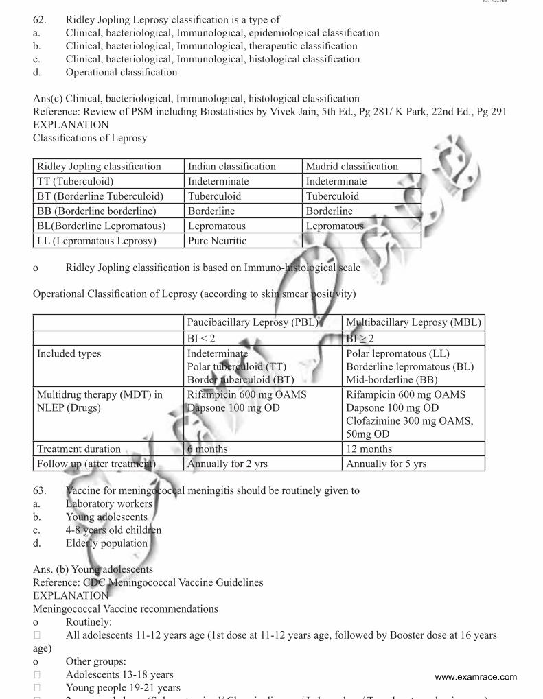
Ans(c) Clinical, bacteriological, Immunological, histological classificationReference: Review of PSM including Biostatistics by Vivek Jain, 5th Ed., Pg 281/ K Park, 22nd Ed., Pg 291EXPLANATIONClassifications of Leprosy
Ridley Jopling classification Indian classification Madrid classification TT (Tuberculoid) Indeterminate Indeterminate BT (Borderline Tuberculoid) Tuberculoid Tuberculoid BB (Borderline borderline) Borderline Borderline BL(Borderline Lepromatous) Lepromatous Lepromatous LL (Lepromatous Leprosy) Pure Neuritic
o Ridley Jopling classification is based on Immuno-histological scale
Operational Classification of Leprosy (according to skin smear positivity)
Paucibacillary Leprosy (PBL) Multibacillary Leprosy (MBL)BI < 2 BI ≥ 2
Included types Indeterminate Polar tuberculoid (TT) Border tuberculoid (BT)
Polar lepromatous (LL) Borderline lepromatous (BL) Mid-borderline (BB)
Multidrug therapy (MDT) in NLEP (Drugs)
Rifampicin 600 mg OAMS Dapsone 100 mg OD
Rifampicin 600 mg OAMS Dapsone 100 mg OD Clofazimine 300 mg OAMS, 50mg OD
Treatment duration 6 months 12 monthsFollow up (after treatment) Annually for 2 yrs Annually for 5 yrs
63. Vaccine for meningococcal meningitis should be routinely given to a. Laboratory workersb. Young adolescentsc. 4-8 years old childrend. Elderly population
Ans. (b) Young adolescents Reference: CDC Meningococcal Vaccine GuidelinesEXPLANATIONMeningococcal Vaccine recommendationso Routinely: All adolescents 11-12 years age (1st dose at 11-12 years age, followed by Booster dose at 16 years age)o Other groups: Adolescents 13-18 years Young people 19-21 years 2 d b (S l t i d/ Ch i di / L b k / T l t d i )
www.examrace.com

64. WHO criteria for High endemicity for Meningococcal disease includea. 0.1%b. 0.01%c. 0.001%d. 1.0%
Ans(b) 0.01% Reference: K Park, 22nd Ed., Pg 156EXPLANATIONWHO Classification of Meningococcal areaso Low endemicity: < 2 cases per 100,000 population per yearo Moderate endemicity: 2-10 cases per 100,000 population per yearo High endemicity: > 10 cases per 100,000 population per year (0.01%)o Epidemic: > 100 cases per 100,000 population per year (0.1%)
65. If Blindness is surveyed using Schools as compared to Population Surveys, then estimation of preva-lence of blindness will have?a. Overestimationb. Underestimationc. Remains samed. None of them is used for evaluation
Ans(b) Underestimation Reference: K. Park, 22nd Ed., Pg 371EXPLANATIONBlindness situation in Indiao Estimated prevalence of Blindness in India (Total): 11.2 per 1000 populationo Estimated prevalence of Blindness in India (0-14 years): 0.1 per 1000 populationo Estimated prevalence of Blindness in India (15-49 years): 0.6 per 1000 populationo Estimated prevalence of Blindness in India (50+ years): 77.3 per 1000 population
So if Schools are used where only refractive errors generally constitute blindness (that too very few are actu-ally blind i.e. <6/60) AS COMPARED TO POPULATION (where age-related cataract constitute as major cause of blindness), it would lead to underestimation of prevalence of blindness in the country
66. Type of Growth Charts used by Anganwadi workers (ICDS) for growth monitoringa. NCHSb. IAPc. MRGSd. CDC
Ans(c) MRGS Reference: K Park, 22nd Ed., Pg 504-506EXPLANATIONICDS Growth Charto In NRHM and ICDS, Government of India has adopted WHO Child Growth Standards 2006 (also known as MGRS ‘Multicentre Growth Reference Study’ Standards) Normal zone Below -2 SD: Malnutrition Below -3 SD: Severe Malnutrition
67. Maximum tolerated dose of a new drug is evaluated inwww.examrace.com
a. Phase 1b. Phase 2c. Phase 3d. Phase 4
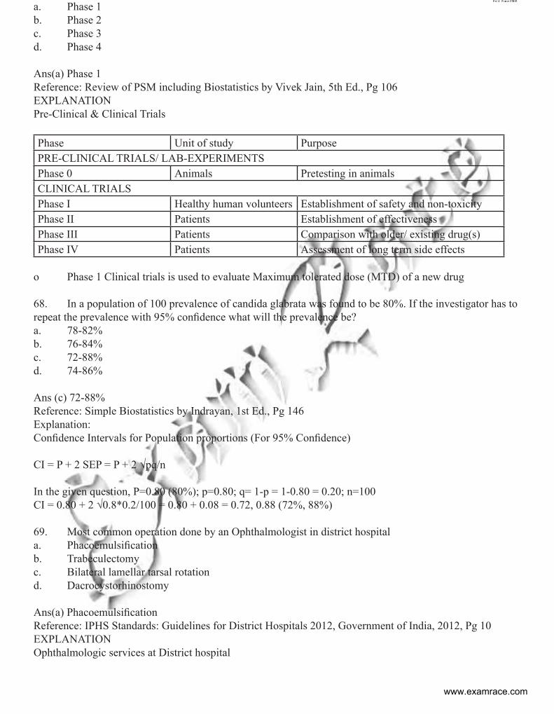
Ans(a) Phase 1 Reference: Review of PSM including Biostatistics by Vivek Jain, 5th Ed., Pg 106EXPLANATIONPre-Clinical & Clinical Trials
Phase Unit of study Purpose PRE-CLINICAL TRIALS/ LAB-EXPERIMENTSPhase 0 Animals Pretesting in animals CLINICAL TRIALSPhase I Healthy human volunteers Establishment of safety and non-toxicity Phase II Patients Establishment of effectiveness Phase III Patients Comparison with older/ existing drug(s) Phase IV Patients Assessment of long term side effects
o Phase 1 Clinical trials is used to evaluate Maximum tolerated dose (MTD) of a new drug
68. In a population of 100 prevalence of candida glabrata was found to be 80%. If the investigator has to repeat the prevalence with 95% confidence what will the prevalence be? a. 78-82%b. 76-84%c. 72-88%d. 74-86%
Ans (c) 72-88% Reference: Simple Biostatistics by Indrayan, 1st Ed., Pg 146Explanation:Confidence Intervals for Population proportions (For 95% Confidence)
CI = P + 2 SEP = P + 2 √pq/n
In the given question, P=0.80 (80%); p=0.80; q= 1-p = 1-0.80 = 0.20; n=100CI = 0.80 + 2 √0.8*0.2/100 = 0.80 + 0.08 = 0.72, 0.88 (72%, 88%)
69. Most common operation done by an Ophthalmologist in district hospitala. Phacoemulsificationb. Trabeculectomyc. Bilateral lamellar tarsal rotationd. Dacrocystorhinostomy
Ans(a) Phacoemulsification Reference: IPHS Standards: Guidelines for District Hospitals 2012, Government of India, 2012, Pg 10EXPLANATIONOphthalmologic services at District hospital
www.examrace.com
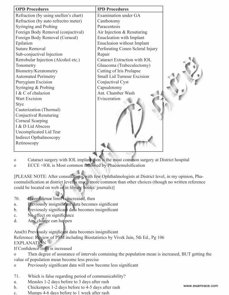
OPD Procedures IPD ProceduresRefraction (by using snellen’s chart)Refraction (by auto refrectro meter)Syringing and ProbingForeign Body Removal (conjuctival)Foreign Body Removal (Corneal)EpilationSuture RemovalSub-conjuctival InjectionRetrobular Injection (Alcohol etc.)TonometryBiometry/KeratometryAutomated PerimetryPterygium ExcisionSyringing & ProbingI & C of chalazionWart ExcisionStyeCauterization (Thermal)Conjuctival ResuturingCorneal ScarpingI & D Lid AbscessUncomplicated Lid TearIndirect OpthalmoscopyRetinoscopy
Examination under GACanthotomyParacentesisAir Injection & ResuturingEnucleation with ImplantEnucleaion without ImplantPerforating Coneo Scleral Injury RepairCataract Extraction with IOLGlaucoma (Trabeculectomy)Cutting of Iris ProlapseSmall Lid Turnour ExcisionConjuctival CystCapsulotomyAnt. Chamber WashEvisceration
o Cataract surgery with IOL implantation is the most common surgery at District hospitalo ECCE +IOL is Most common followed by Phacoemulsification
[PLEASE NOTE: After consultations with few Ophthalmologists at District level, in my opinion, Pha-coemulsification at district level is much more common than other choices (though no written reference could be located on web or in library books/ journals)]
70. If confidence limit is increased, thena. Previously insignificant data becomes significant b. Previously significant data becomes insignificant c. No effect on significanced. Any change can happen
Ans(b) Previously significant data becomes insignificant Reference: Review of PSM including Biostatistics by Vivek Jain, 5th Ed., Pg 106EXPLANATIONIf Confidence limit is increasedo Then degree of assurance of intervals containing the population mean is increased, BUT getting the value of population mean become less preciseo Previously significant data will now become less significant
71. Which is false regarding period of communicability?a. Measles 1-2 days before to 3 days after rashb. Chickenpox 1-2 days before to 4-5 days after rash c. Mumps 4-6 days before to 1 week after rash
www.examrace.com
d. German measles 7 days before and after rash
Ans(a) Measles 1-2 days before to 3 days after rash Reference: Review of PSM including Biostatistics by Vivek Jain, 5th Ed., Pg 245EXPLANATIONPeriod of Communicability o Chicken pox: 1 – 2 days before to 4 – 5 days after appearance of rasho Measles: 4 days before to 5 days after appearance of rasho Rubella: 7 days before symptoms to 7 days after appearance of rash o Mumps: 4 – 6 days before symptoms to 7 days thereafter o Influenza: 1 – 2 days before to 1 – 2 days after onset of symptoms o Diphtheria: 14 – 28 days from disease onseto Pertussis: 7 days after exposure to 3 weeks after paroxysmal stage o Meningococccus: Until absent from nasal and throat dischargeso Tuberculosis: As long as not treated o Poliomyelitis: 7 – 10 days before and after onset of symptomso Hepatitis A: 2 weeks before to 1 week after onset of jaundice o Hepatitis B: Till disappearance of HBsAg & appearance of anti-HBso Tetanus: None
72. Due to use of preservative for refrigerated foods following cancer can be reduced a. Stomach Cancerb. Esophagus Cancerc. Colon Cancerd. Oropharyngeal Cancer
Ans(a) Stomach Cancer Reference: K Park, 22nd Ed., Pg 361/ Cancer & Nutrition by CB Simone, 3rd Ed., Pg 179EXPLANATIONDecline of Stomach cancer in Industrialized nations is due too Improved food preservation practices (esp refrigerated foods)o Better nutrition (esp vitamins from fresh fruits/ vegetables)o Lesser consumption of preserved, cured, salted foodso Reduced use of salt picklingo Lesser consumption of smoked foods
73. Dose of vitamin A given to post partum woman a. 50000 IUb. 1 lakh IUc. 2 lakh IUd. 3 lakh IU
Ans(c) 2 lakh IU Reference: WHO Vitamin A Supplementation Guidelines]EXPLANATIONPotential target groups and immunization contacts in countries with vitamin A deficiency WHO GUIDE-LINES
www.examrace.com
Target group Immunization contact Vitamin A doseAll mothers irrespective of their mode of infant feeding up to six weeks postpartum if they have not received vitamin A supplementa-tion after delivery
BCG, OPV-0 or DTP-1 up to 6 weeks
200 000 IU
Infants aged 9–11 monthsChildren aged 12 months and older
Measles vaccine contact 100 000 IU200 000 IU
Children aged 1–4 years Booster doses*Special campaigns*Delayed primary immunization doses*
200 000 IU
* The optimal interval between doses is four to six months. A dose should not be given too soon after a previous dose of vitamin A supplement: the minimum recommended interval between doses for the preven-tion of vitamin A deficiency is one month (the interval can be reduced in order to treat clinical vitamin A deficiency and measles cases).
[PLEASE NOTE: Currently WHO DOESNOT recommend Vitamin A supplementation to Postpartum fe-males]
E.N.T74. BAHA is used for which of these a. Cochlear malformation b. SNHL c. Bl acoustic neuromad. Boy with microtionea and auditory canal atresia Answer: (d), Boy with microtionea and auditory canal atresia Reference: Dhingra 5th Ed. Pg. 136-137Explanation: Boy with microtia and auditory canal atresia baha stands for “bone anchoring hearing aid”. This is surgically fitted onto the mastoid bone . it amplifies sound and transmits the sound into the ear through bony conduction. It has two main indicationsi. When air conduction pathways cannot be used like in case of microtia and auditory canal atresia ii. Small child requiring hearing aid.75. Facial recess not surrounded by- a. Short proces f incusb. Chorda tympanic. Facial nerved. Stapedius tendonAnswer: (d) Stapedius tendonReference: Dhingra 5th Ed. Pg. 7Explanation: Posterior wall of middle ear has four recess where cholesteotoma may hide. These arei. Facial recessii. Sinus tympaniiii. Posterior tympanic sinusiv. Lateral tympanic sinus76. A patient with h/o right ear discharge undergoes mastoidectomy but his symptoms r not relieved nd he presents with purulent discharge, vertigo nd deafness.. diagnosis: a Labyrinthitis
www.examrace.com

b. Thrombophlebitisc. Meningitisd. PetrositisAnswer: (a) Labyrinthitis. Reference: Dhingra 4th Ed. Pg. 22,23,41Explanation: A pt of ear discharge undergoing mastoidectomy is probably suffering from unsafe csom. If the symptoms are not relieved after surgery and also has vertigo nd deafness will probably has some complica-tion of csom. among the common complications of csom only two can cause vertigo(a symptom in the above patient). These arei. Labyrinthine fistula andii. Labyrinthitis77. A 55 yr man presented with CHL, fullness sensation in ear , no discharge, T.M. Normal, Tympanom-etry shows B type graph. What is the Next Step of managementa. Myringotomy with grommet b. Evaluation of Nasopharyngeal Massc. Antibioticsd. ObservationAnswer (b) Evaluation of Nasopharyngeal MassReference: Dhingra 261-266Explanation: This is a case of unilateral glue ear in an adult. One of the most serious primary disease that may cause glue ear in adult is nasopharyngeal carcinoma. Nasopharyngeal ca is most commonly seen in fossa of rosenmuller which is just behind the eustachean tube. This tube gets blocked by this ca to cause glue ear. The best investigation for ca nasopharynx are ct scan of nasopharynx and nasopharyngoscopy.78. 50 year old male Unilateral SOM B type tympanogram intact TM.... MANAGEMENT?a. Myringotomy with grommet insertionb. Endoscopy of nesophyraxc. Examination under microscoped. Adenoidectomy and grommet insertion.Answer (a) Myringotomy with grommet insertionReference: Dhingra 5th Ed. Pg. 71 Explanation:This is a case of unilateral glue ear in an adult. One of the most serious primary disease that may cause glue ear in adult is nasopharyngeal carcinoma. Nasopharyngeal ca is most commonly seen in fossa of rosenmuller which is just behind the eustachean tube. This tube gets blocked by this cancer to cause glue ear. The best investigation for ca nasopharynx are ct scan of nasopharynx and nasopharyngoscopy79. Patient presents with 4×5 cm lymph node ,mobile firm , not fixed in left lateral neck. Clinical exami-nation and PET scan cannot identify primary. Examination of nose oral cavity and pharynx is normal What is the stage?a. T0N2Mxb. T1 N2aM0c. T0N2aMxd. T1N2aMxAnswer: (d) T1N2aMxRef: Dhingra 5th Pg. 266 Explanation:
www.examrace.com
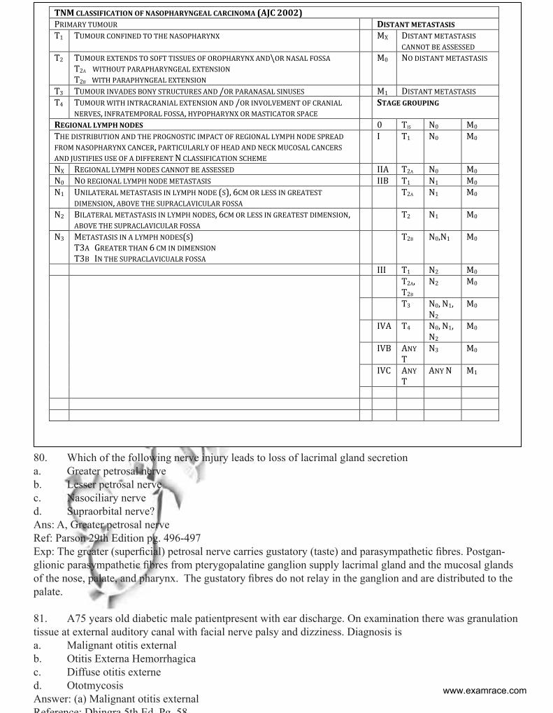
TNM CLASSIFICATION OF NASOPHARYNGEAL CARCINOMA (AJC 2002) PRIMARY TUMOUR DISTANT METASTASIS T1 TUMOUR CONFINED TO THE NASOPHARYNX MX DISTANT METASTASIS
CANNOT BE ASSESSED T2 TUMOUR EXTENDS TO SOFT TISSUES OF OROPHARYNX AND\OR NASAL FOSSA
T2A WITHOUT PARAPHARYNGEAL EXTENSION T2B WITH PARAPHYNGEAL EXTENSION
M0 NO DISTANT METASTASIS
T3 TUMOUR INVADES BONY STRUCTURES AND /OR PARANASAL SINUSES M1 DISTANT METASTASIS T4 TUMOUR WITH INTRACRANIAL EXTENSION AND /OR INVOLVEMENT OF CRANIAL
NERVES, INFRATEMPORAL FOSSA, HYPOPHARYNX OR MASTICATOR SPACE STAGE GROUPING
REGIONAL LYMPH NODES 0 TIS N0 M0 THE DISTRIBUTION AND THE PROGNOSTIC IMPACT OF REGIONAL LYMPH NODE SPREAD FROM NASOPHARYNX CANCER, PARTICULARLY OF HEAD AND NECK MUCOSAL CANCERS AND JUSTIFIES USE OF A DIFFERENT N CLASSIFICATION SCHEME
I T1 N0 M0
NX REGIONAL LYMPH NODES CANNOT BE ASSESSED IIA T2A N0 M0 N0 NO REGIONAL LYMPH NODE METASTASIS IIB T1 N1 M0 N1 UNILATERAL METASTASIS IN LYMPH NODE (S), 6CM OR LESS IN GREATEST
DIMENSION, ABOVE THE SUPRACLAVICULAR FOSSA T2A N1 M0
N2 BILATERAL METASTASIS IN LYMPH NODES, 6CM OR LESS IN GREATEST DIMENSION, ABOVE THE SUPRACLAVICULAR FOSSA
T2 N1 M0
N3 METASTASIS IN A LYMPH NODES(S) T3A GREATER THAN 6 CM IN DIMENSION T3B IN THE SUPRACLAVICUALR FOSSA
T2B N0,N1 M0
III T1 N2 M0 T2A,
T2B N2 M0
T3 N0, N1, N2
M0
IVA T4 N0, N1, N2
M0
IVB ANY T
N3 M0
IVC ANY T
ANY N M1
80. Which of the following nerve injury leads to loss of lacrimal gland secretiona. Greater petrosal nerveb. Lesser petrosal nervec. Nasociliary nerve d. Supraorbital nerve?Ans: A, Greater petrosal nerveRef: Parson 29th Edition pg. 496-497Exp: The greater (superficial) petrosal nerve carries gustatory (taste) and parasympathetic fibres. Postgan-glionic parasympathetic fibres from pterygopalatine ganglion supply lacrimal gland and the mucosal glands of the nose, palate, and pharynx. The gustatory fibres do not relay in the ganglion and are distributed to the palate.
81. A75 years old diabetic male patientpresent with ear discharge. On examination there was granulation tissue at external auditory canal with facial nerve palsy and dizziness. Diagnosis isa. Malignant otitis externalb. Otitis Externa Hemorrhagicac. Diffuse otitis externe d. Ototmycosis Answer: (a) Malignant otitis externalReference: Dhingra 5th Ed Pg 58
www.examrace.com
Explanation: Malignant (necrotizing) otits externa it is an inflammatory condition caused by pseudomonas infection usually in the elderly diabetics, or in those on immunosuppressive drugs. Its early manifestation resemble diffuse otitis externa but there is excruciating pain and appearance of granulations in the meatus. Facial paralysis is common. Infection may spread to the skull base and jugular foramen causing multiple cranial nerve palsies. Anteriorly, infection spreads to temporomandibular fossa, posteriorly to the mastoid and medially into the middle ear and petrous bone. CT scan is useful, to know the extent of disease. Treatment consist of high doses of i.v antibiotics directed against pseudomonas (tobramycin, antibi-otics directed against pseudomonas (tobramycin, ticarcillin or third generation cephralosporins). Antibiotics are given for 6-8 weeks or longer. Diabetes should be controlled. Surgical debridement of devitalized tissue and bone should done judiciously. Radical resenctions have been abandoned in favour of prolonged inten-sive medical therapy.
OPHTHALOMOLOGY
82. False about acute conjunctivitis: a. Vision not affectedb. Cornea infiltrates c. Pupil remains unaffected d. Topical antibiotics is the treatment Answer: (b) Cornea infiltrates Reference: Parsons 21st Edition Page 166-167 Explanation: “Neisseria gonorrhoeae is a bun-shaped Gram-negative diplococcus, found within both leuco-cytes and epithelial cells. N. Catarrhalis and N. Meningitides, both gram-negative in the conjunctival sac. They may be differentiated from the gonococcus by agglutination tests. N. Catarrhalis is rarely found in acute conjunctivitis, but may be seen in chronic forms. Clinical features: The disease is acute, occurring usually in adult males, often developing first in the right eye and is due to direct infection from the genitals. Apart from severe purulent conjunctivitis there is a marked tendency to involvement of the cornea. Constitutional disturbance, including a rise of temperature and mental depression, may also occur. The incubation period is a few hours to 3 days. There after, the upper lid becomes swollen and tense, overhanging the lower lid and edged with pus. Eversion, which is difficult, shows that the palpebral conjunc-tiva is deep red and velvety; rarely there is a membrane. There is intense pain and the preauricualr lymph node is enlarged and tender and may suppurate. After 2 or 3 weeks the purulent discharge diminishes, but subacute conjunctivitis with papillary thickening of the conjunctive persists for several weeks longer. The gonococcus is still present, a point of great importance, both in terms of contagion and treatment. No immu-nity is conferred by the attack. The most important point in diagnosis is the coincidence of urethritis. The most important point in prognosis is the involvement of the cornea. The gonococcus is capable of invading the normal cornea through an intact epithelium. Corneal complications are the rule, and constitute the cause of blindness. There may be diffuse haziness of the whole cornea, with grey or yellow spots near the centre. Ulcers may occur at any part, and are due to necrosis of the epithelium through direct invasion by the organisms. Marginal ulceration, which may extend completely round the cornea, may be due to retention of pus in the angle formed by the chemotic conjunctiva. When ulceration has commenced, it progresses rapidly and deeply and perforation is common with all its attendant dangers. The greatest care should therefore be taken to prevent injury to the cornea during diagnosis and treatment. Treatment: Specific antigonococcal therapy is initiated based on the findings of intracellular Gram-negative diplococcic on conjunctival scaraping and smear examination or on clinical suspicion. 83. Most common presentation of retinoblastoma: e. Leucokoria with pseudohypopyonf. Leucokoria with hyphemag. Leucokoria with heterochromiairidish. Leucokoria with strabismus
www.examrace.com
Answer: (d) Leucokoria with strabismusReference: Abramson DH et all 1998Explanation:A retrospective chart review was performed on the charts of 1265 patients with retinoblastoma who were on file at New York Hospital and whose conditions had been diagnosed between the years 1960 and 1990. The mean follow-up was 90 months, ranging from 0 to 409 months. Thirty-two distinct presenting signs of retinoblastoma were identified, the most common of which were leukocoria (56.2%), strabismus (23.6%), poor vision (7.7%), and family history (6.8%). No correlation was found between any of the presenting signs and laterality, sex, race, or survival. Leukocoria correlated to Reese-Ellsworth Group Va or Vb, whereas strabismus was invariably associated with either macular tumors or macular retinal detachments. Eighty-three (96.5%) patients presenting with a family history did so before the age of 24 months. The presenting sign leading to a diagnosis of retinoblastoma was correlated with degree of advancement and tumor loca-tion. Leukocoria and strabismus are the most common presenting signs, with leukocoria correlated to more advanced disease and strabismus always associated with macular involvement. Vitreous hemorrhage (1 case, 0.1%), microphthalmos (4 cases, 0.3%), and orbital cellulitis (3 cases, 0.2%) are extremely rare presenting findings in retinoblastoma.
84. Salt n pepper retinopathy is not seen with: i. Phenothiazine toxicity j. Congenital rubella k. Old retinal detachment l. Fundus flavimaculatusAnswer: (d) Fundus flavimaculatus Reference: Parsons 21st Edition Page 303-312Explanation: Salt and pepper retinopathy• Congenital Rubella• Leber’s congenital amaurosis• Congenital Syphilis• Cystinosis• Albinism (carrier state)• Retinitis pigmentosa (carrier state)• Choroideremia (carrier state)• Old retinal detachment• Phenothiazine toxicity85. Rhegmatogenous RD is not seen with: m. Pseudophakian. Hyperopia o. Lattice degenerationp. Trauma Answer: (b) hyperopia Reference: Parsons 21st Edition page 318Explanation: Myopic Choriretinal degeneration
www.examrace.com
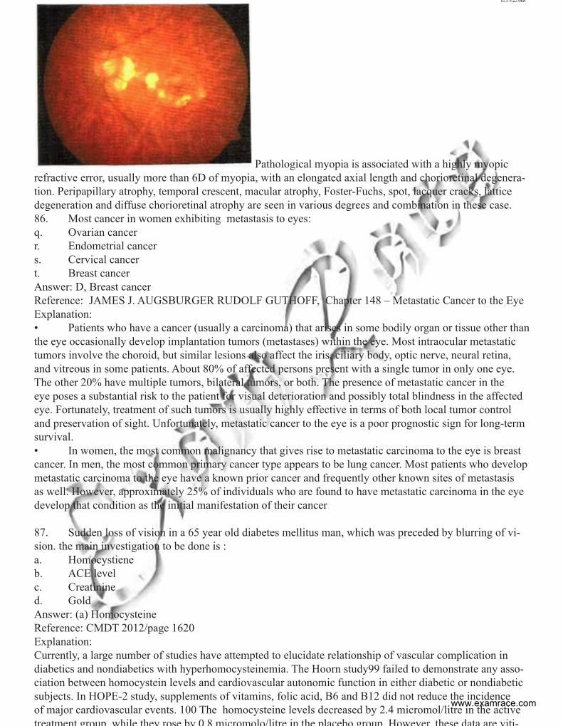
Pathological myopia is associated with a highly myopic refractive error, usually more than 6D of myopia, with an elongated axial length and chorioretinal degenera-tion. Peripapillary atrophy, temporal crescent, macular atrophy, Foster-Fuchs, spot, lacquer cracks, lattice degeneration and diffuse chorioretinal atrophy are seen in various degrees and combination in these case. 86. Most cancer in women exhibiting metastasis to eyes:q. Ovarian cancerr. Endometrial cancers. Cervical cancert. Breast cancerAnswer: D, Breast cancer Reference: JAMES J. AUGSBURGER RUDOLF GUTHOFF, Chapter 148 – Metastatic Cancer to the EyeExplanation:• Patients who have a cancer (usually a carcinoma) that arises in some bodily organ or tissue other than the eye occasionally develop implantation tumors (metastases) within the eye. Most intraocular metastatic tumors involve the choroid, but similar lesions also affect the iris, ciliary body, optic nerve, neural retina, and vitreous in some patients. About 80% of affected persons present with a single tumor in only one eye. The other 20% have multiple tumors, bilateral tumors, or both. The presence of metastatic cancer in the eye poses a substantial risk to the patient for visual deterioration and possibly total blindness in the affected eye. Fortunately, treatment of such tumors is usually highly effective in terms of both local tumor control and preservation of sight. Unfortunately, metastatic cancer to the eye is a poor prognostic sign for long-term survival.• In women, the most common malignancy that gives rise to metastatic carcinoma to the eye is breast cancer. In men, the most common primary cancer type appears to be lung cancer. Most patients who develop metastatic carcinoma to the eye have a known prior cancer and frequently other known sites of metastasis as well. However, approximately 25% of individuals who are found to have metastatic carcinoma in the eye develop that condition as the initial manifestation of their cancer
87. Sudden loss of vision in a 65 year old diabetes mellitus man, which was preceded by blurring of vi-sion. the main investigation to be done is :a. Homocystiene b. ACE level c. Creatinine d. GoldAnswer: (a) Homocysteine Reference: CMDT 2012/page 1620Explanation: Currently, a large number of studies have attempted to elucidate relationship of vascular complication in diabetics and nondiabetics with hyperhomocysteinemia. The Hoorn study99 failed to demonstrate any asso-ciation between homocystein levels and cardiovascular autonomic function in either diabetic or nondiabetic subjects. In HOPE-2 study, supplements of vitamins, folic acid, B6 and B12 did not reduce the incidence of major cardiovascular events. 100 The homocysteine levels decreased by 2.4 micromol/litre in the active treatment group while they rose by 0 8 micromolo/litre in the placebo group However these data are viti-
www.examrace.com
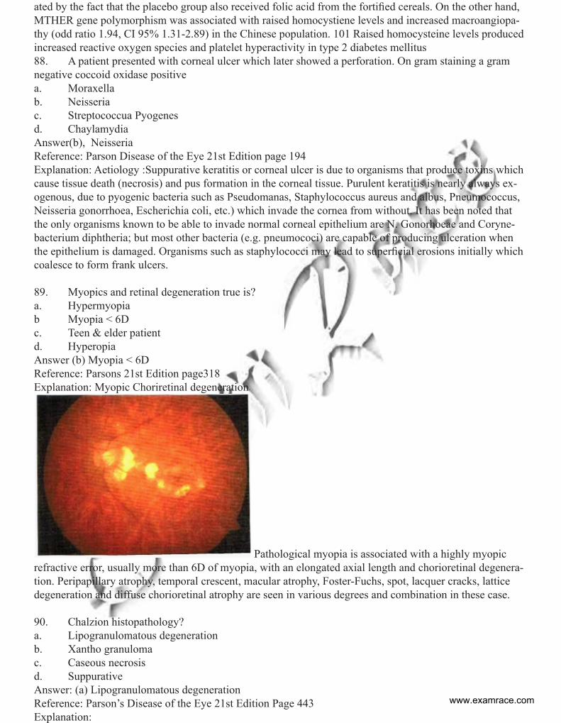
ated by the fact that the placebo group also received folic acid from the fortified cereals. On the other hand, MTHER gene polymorphism was associated with raised homocystiene levels and increased macroangiopa-thy (odd ratio 1.94, CI 95% 1.31-2.89) in the Chinese population. 101 Raised homocysteine levels produced increased reactive oxygen species and platelet hyperactivity in type 2 diabetes mellitus 88. A patient presented with corneal ulcer which later showed a perforation. On gram staining a gram negative coccoid oxidase positive a. Moraxella b. Neisseriac. Streptococcua Pyogenesd. Chaylamydia Answer(b), Neisseria Reference: Parson Disease of the Eye 21st Edition page 194Explanation: Aetiology :Suppurative keratitis or corneal ulcer is due to organisms that produce toxins which cause tissue death (necrosis) and pus formation in the corneal tissue. Purulent keratitis is nearly always ex-ogenous, due to pyogenic bacteria such as Pseudomanas, Staphylococcus aureus and albus, Pneumococcus, Neisseria gonorrhoea, Escherichia coli, etc.) which invade the cornea from without. It has been noted that the only organisms known to be able to invade normal corneal epithelium are N. Gonorhoeae and Coryne-bacterium diphtheria; but most other bacteria (e.g. pneumococi) are capable of producing ulceration when the epithelium is damaged. Organisms such as staphylococci may lead to superficial erosions initially which coalesce to form frank ulcers.
89. Myopics and retinal degeneration true is? a. Hypermyopia b Myopia < 6Dc. Teen & elder patient d. Hyperopia Answer (b) Myopia < 6DReference: Parsons 21st Edition page318Explanation: Myopic Choriretinal degeneration
Pathological myopia is associated with a highly myopic refractive error, usually more than 6D of myopia, with an elongated axial length and chorioretinal degenera-tion. Peripapillary atrophy, temporal crescent, macular atrophy, Foster-Fuchs, spot, lacquer cracks, lattice degeneration and diffuse chorioretinal atrophy are seen in various degrees and combination in these case.
90. Chalzion histopathology? a. Lipogranulomatous degenerationb. Xantho granulomac. Caseous necrosis d. Suppurative Answer: (a) Lipogranulomatous degeneration Reference: Parson’s Disease of the Eye 21st Edition Page 443Explanation:
www.examrace.com
Also known as tarsal or meibomian cystIt is not a cyst. It results from obstruction of a sebaceous gland either meibomian or Zeis. Extravasated lipid material produces a surrounding chronic lipogranulomatous inflammation (Yanoff & Duker Ophthal-mology)Acute lesions are treated with hot compresses while chronic chalazia are treated using intralesional corticos-teroid (e.g. Triamcinolone acetonide) or by surgical drainage.91. In which of the following calcification is not seena. Retinoblastomab. Primary hyperplastic persistent vitreousc. Choroid osteomad. Optic nerve drusenzAnswer: (b) Primary hyperplastic persistent vitreousReference: Parson’s Disease of the Eye 21st Edition Explanation:Causes of Intraocular Calcificationsi. Retinoblastoma (>50% of all cases)ii. Astrocytichamartomaiii. Choroidalosteomaiv. Optic drusenv. Scleral calcifications in systemic hypercalcemic statesvi. Retrolental fibroplasiasvii. Phthisis bulbi
92. Most common Non hogdkin lymphoma of orbit is a. B cellb. T cellc. NK Celld. Plasma cell Answer: B cellReference:Paron’s diseases of the Eye page 365-369Explanation: Orbital lymphoma is common type of non-Hodgkin lymphoma that occurs near or on the eye. Common symptoms include decreased vision and uveitis. Orbital lymphoma can be diagnosed via a biopsy of the eye and is usually treated with Radiotherapy or with combination with chemotherapy.SymptomsPrimary visible symptoms of ocular lymphoma include proptosis and a visible mass in the eye. Other symp-toms are due to mass effect.PathophysiologyRecent studies have detected the presence of viral DNA in ocular lymphoma cells. This implies that patho-gens play a role in ocular lymphoma. Other studies have found that the aging population, the increasing number of immunosuppressive drugs, and theAIDS epidemic have also contributed to the increased inci-dence of Non-Hodgkin lymphomas.Ocular MALT lymphomas may also be associated with Chlamydia psittaci, although whether or not this is the case is still debated. Follicular lymphoma, diffuse large B cell lymphoma, mantle cell lymphoma, B-cell chronic lymphocytic leukemia, peripheral T-cell lymphoma, and natural killer cell lymphoma have also been reported to affect the orbitEpidemiology55% of all lymphoma cases in adults and 10% in older patients is a form of ocular lymphoma. In 2008, a prediction by the National Cancer Institute Surveillance, estimated that in 1,340 men and 1,050 women would be diagnosed with eye cancer and 240 people would die of the disease that year Orbital lymphoma is more prevalent in Asia and Europe than in the United States.Although intraocular lymphoma is rare, the number of cases per year is rising, affecting mainly people in their seventies and immunocompromised patients A recent study has shown that ocular lymphoma is more
www.examrace.com
prevalent in women than men.The survival rate is approximately 60% after 5 years.TypesThere are two types of ocular lymphomas: intraocular lymphomas and adnexal lymphomas. An intraocular lymphoma occurs within the eye, while an adnexal lymphoma occurs outside, but adjoined to the eye.TreatmentRadiotherapy is the most effective treatment for local disease either as the sole treatment for low-grade lym-phoma or in combination with chemotherapy for intermediate- and high-grade lymphoma Radiotherapy dose in range of 30-45 Gy administered in fractions are advised in treating the local orbital lymphomas93. OCREOPLASMIN is a recombinant protease & it is used to treat a. Retinal break b. Diabetic mellitus macular edemac. Uveovitrealmembranbe adhesion d. Submacular bleedingAnswer: (c) Uveovitrealmembranbe adhesion Reference: Current and Potential Uses of Ocriplasmin Jeffrey S 05/05/2013Explanation: Many attempts have been made to develop a pharmacologic agent that could promote enzy-matic vitreolysis. Until now, none had been successful in at inducing both synchesis and syneresis without adversely affecting the retina. Phase III data for ocriplasmin has been published and its Food and Drug Administration approval has confirmed that there is finally a viable pharmacologic option for symptomatic vitreomacular adhesion. There are two primary indications for ocriplasmin. The first is for patients who have mild to moder-ate symptomatic VMA, and also have good visual acuity. Patients may be experiencing metamorphopsia, but test 20/40 or better on a Snellen visual acuity chart. Metamorphopsia can be tremendously disabling to functional capabilities, especially for those whose work requires them to read frequently or perform detailed visual tasks. However, vitrectomy surgery would not be a viable option for this group, because their vision is too good to risk the complications associated with surgery, including cataract formation, retinal tear, retinal detachment, bleeding and end-ophthalmitis.6 Historically, disease management for these patients has been careful observation, hoping the VMA would spontaneously resolve. However, the literature shows that many do progress and lose vision.7 The FDA approval of ocriplasmin provided surgeons with a minimally inva-sive means of treating these patients who previously had no viable option. The second set of patients are those with more moderate VMA whose visual acuity has deteriorated to 20/80 or worse, sufficient to justify surgery. Ocriplasmin is the ideal first choice in these patients. I have found that it does not require drops before or after, nor any eye patch, and the patient need not be in the prone position. Administering the injection is very straightforward and patients go home shortly thereafter. It is important to note that the large majority of responders demonstrated resolution of VMA by day seven, and all did so by day 28 of the study.94. Correct match of drug and action is :a. Brimonidine decrease aqueous production b. Latanoprost carbonic anhydrase inhbtorc. Pilocarpine increases uvoescleralpoutflowd. Betaxolol increases trabecular outflowAnswer: (a) Brimonidine decrease aqueous production Reference: Drug Facts and Comperisons 2006/2257Explanation: The relationship between ciliary perfusion and aqueous production is poorly understood. It was recently reported that aqueous production decreases when ciliary blood flow is reduced by lowering the ocular perfusion pressure, and hypothesized that drug-induced reduction of ciliary blood flow would also decrease aqueous production. alpha2 adrenergic agonist (brimonidine) formulated for topical application. When used acutely, brimonidine decreases intraocular pressure (IOP) by suppressing aqueous production, although its mechanism of action is unclear
OBSTETRICS AND GYNAECOLOGY www.examrace.com
95. A girl with short stature widely spaced nipples and primary amennorhea. Karyotypea. 47XXXb. 47XYYc. 45XOd. 46XXAnswer: (c) 45XO Reference: D.C Dutta’s Text Book of Gynaecology 6th Edition page 22-23 Explanation: Turner’s Syndrome In Turner ‘s syndrome the gonads are not properly (streak gonads) o The ovary is not properly developed and it cannot secrete adequate amount of estrogen o This leads to lack of secondary sexual characteristic o Breast is not developed properly o Uterus is not developed properly in most of the patients who have not received estrogen therapy Turner;s syndrome• Underdeveloped uterus• Undeveloped breast • Primary amenorrhoea
96. A woman after normal labour suddenly goes into shock. Diagnosis: a. PPHb. inversion of uterus c. Amniotic fluid embolismd. Eclampsia Answer: (b) inversion of uterusReference: Munroken’s operative obstetric 10th Edition page 69, 476: I m DonaldExplanation: The patient went into unexplained shock after postpartum Both unterine inversion and amniotic fluid embolism can cause unexplained shock Uterine inversion is more common and important cause of unexplained shock than amniotic fluid embolism Munrokerr Further adds Uterine inversion is the most important Three degrees of inversion are generally described i. Where the inverted fundus reaches the internal os ii. Where the whole body of the uterus is inverted upto the internal os. iii. Where uterus, cervix and vagina are completely inverted Symptoms of uterine Inversion• Symptoms of uterine inversion are not always well defined • If however the inversion occurs quickly or is of severe degree there is feelling of something soming down and pain followed by collapse and hemorrhage of variable severity • When the inversion is of lesser degree and is contained within the vagina or when the fundus does not come beyond the os externum, only pain and hemorrhage may be present • Inversion is also the most important cause of unexplained shock o In these cause the shock is not primarily due to loss of blood or trauma but is really of neurogenic origin. o In this connection it has been suggested by several authors that as one or both invagination, pressure on them may be responsible for shock. o The traction on the infundibulopelvic ligaments, peritoneum and broad ligaments stimulate the “ parasympathetic system” producing shock.
97. Female with normal pubic and axiillary hair, with primary amenorrhea. Diagnosis: a. Androgen insensitvity syndrome
www.examrace.com
b. Mullerian agenesis c. 47XYY d. 45XOAnswer: (b) Mullerian agenesisReference: Shows Text book of Gynaecology 14th Edition page 252Explanation; The laboratory evaluation of a woman with primary amenorrhea depends upon the findings on physical examination, in particular, whether mullerian structures are present or absent Thus, the single most important step in the evaluation is to determine by physical examination or ultrasonography whether there are any anatomic abnormalities of the vagina, cervix, or uterus If a normal vagina or uterus is not abviously present on physical examination, pelvic ultrasonography should be performed to confirm the presence or absence of ovaries, uterus, and cervix, in addition, ultra-sonography can be useful to look for vaginal or cervical outlet obstruction in patients with cyclic pain Uterus absent For those with absence of the uterus, further evaluation should include a karyotype and measurement of serum testosterone. These tests should then allow the clinician to distinguish between:-o Abnormal mulerian development (mullerian agenesis) (46, XX, karyotype with normal female serum testosterone concentrations). )Meyer Rokitansky Kuster Hauser syndrome) o Andtrogen insensitivity syndrome (46, XY karyotype and normal male serum testosterone concentra-tions)Uterus present For patients with normal mullerian structures and no evidence of an imperforate hymen, vagina sep-tum, or congenital absence of the vagina, an endocrine evaluation should be performed This includes measurement of serum beta human chorionic gonadotropin to axclude pregnancy and of serum FSH, and perphas other hormones A high serum FSH concentration is indicative of primary ovarian failure. A karyotype is then re-quired and may demonstrate complete or partial deletion of the X chromosome (Turner syndrome) or the presence of Y chromatin. The presence of a Y chromosome (eg, vanishing tests syndrome, absent testis determining factor) is associated with a higher risk of gonadal tumors and makes gonadectomy mandatory A low or normal serum FSH concentration suggests functional hypothalamic amenorrhea, congenital GnRH deficiency, or other disorders of the hypothalamic-pituitary axis. o Cranial MR imaging is indicated in most cases of hypogonadotropic hypogonadism to evaluate for hypothalamic or pituitary disease, cranial imaging should be recommended in all women with primary hypogonadotropic hypoganadism. Cranial imanging should be recommended in all women with primary hy-pogonadotropic hypogonadism, visual field defects, headaches, or any other signs of hypothalmaci –pituitary dysfunction Serum prolactin and thyrotropin should be measured is FSH is low or normal, especially if galactor-rhea is present. If there are sings or symptoms of hyperandrogenism, serum testosterone and dehydroepiandrosterone sulfate (DHEA-S) should be measured to assess for an androgen –secreting tumor. Among women who are also hypertensive, blood tests should be drawn for evaluation for CYP17 deficiency.o The characteristic findings are elevations in serum progester- on (>3 ng/mL [9.5 nmol/L] ) and de-oxycorticosterone and low values for serum 17-alpha-hydroxyprogesterone (<0.2 ng/ml [0.6 nmol/L]98. Most accurate and safest way to diagnose viable pregnancy at 6 weeks: a. Beta HCG levels b. pv examination to check uterus size of 6 weeks c. Trans vaginal scan for fetal cardiac activity d. Doppler for fetal cardiac activityAnswer: (b) Trans vaginal scan for fetal cardiac activityReference: D.C Dutta Text book of Obstetrics 7th Edition page 64-68Explanation: An intrauterine GS should be seen by TVS when the maternal serum β hCH level is 1000-1200 mIU /ml. and by TAS with the level of β hCH 6000 mIU/ml. Yolk sac is seen at the level of 7000 mIU/ml
www.examrace.com
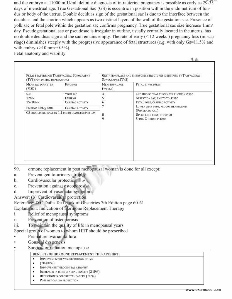
and the embryo at 11000 mIU/ml. definite diagnosis of intrauterine pregnancy is possible as early as 29-35 days of menstrual age. True Gestational Sac (GS) is eccentric in position within the endometrium of fun-dus or body of the uterus. Double deciduas sign of the gestational sac is due to the interface between the deciduas and the chorion which appears as two distinct layers of the wall of the gestation sac. Presence of yolk sac or fetal pole within the gestation sac confirms pregnancy. True gestational sac size increase 1mm/day. Pseudogestational sac or pseudosac is irregular in outline, usually centrally located in the uterus, has no double deciduas sign and the sac remains empty. The rate of early (< 12 weeks ) pregnancy loss (miscar-riage) diminishes steeply with the progressive appearance of fetal structures (e.g. with only Gs=11.5% and with embryo >10 mm=0-5%). Fetal anatomy and viability
FETAL FEATURES ON TRANSVAGINAL SONOGRAPHY (TVS) FOR DATING IN PREGNANCY
GESTATIONAL AGE AND EMBRYONIC STRUCTURES IDENTIFIED BY TRASVAGINAL SONOGRAPHY (TVS)
MEAN SAC DIAMETER (MSD)
FINDINGS MENSTRUAL AGE (WEEKS)
FETAL STRUCTURES
5-8 12MM 15-18MM
YOLK SAC EMBRYO CARDIAC ACTIVITY
4 5 6 7 8 9
CHORIODECIDUAL THICKNESS, CHORIONIC SAC GESTATION SAC, EMBYO YOLK SAC FETAL POLE, CARDIAC ACTIVITY LOWER LIMB BUDS, MIDGUT HERNIATION (PHYSIOLOGICAL) UPPER LIMB BUDS, STOMACH SPINE. CHOROID PLEXUS
EMBRYO CRL > 4MM CARDIAC ACTIVITY GS SHOULD INCREASE BY 1.1 MM IN DIAMETER PER DAY
99. ormone replacement in post menopausal woman is done for all except:a. Prevent genito-urinary atrophyb. Cardiovascular protectionc. Prevention against osteoporosisd. Imprevent of vasomatar symptoms Answer: (b) Cardiovascular protectionReference: D.C Dutta Text book of Obstetrics 7th Edition page 60-61Explanation: Indication of Hormone Replacement Therapy i. Relief of menopausal symptoms ii. Prevention of osteoporosis iii. To maintain the quality of life in menopausal years Special group of women to whom HRT should be prescribed• Premature ovarian failure• Gonadal dysgenesis • Surgical or radiation menopause BENEFITS OF HORMONE REPLACEMENT THERAPY (HRT)
• IMPROVEMENT OF VASOMOTOR SYMPTOMS • (70-80%) • IMPROVEMENT UROGENITAL ATROPHY • INCREASED IN BONE MINERAL DENSITY (2-5%) • REDUCTION IN COLORECTAL CANCER (20%) • POSSIBLY CARDIO PROTECTION
www.examrace.com
RISKS OF HORMONE REPLACEMENT THERPAY • Endometrial cancer: When oestrogen is given alone to a woman with intact uterus, it cause endome-trial proliferation, hyperplasia and carcinoma. It is therefore advised that a progestogen should be added to ERT to counter balance such risks • Breast cancer: Combined oestrogen and progestin replacement therapy increased the risks breast can-cer slightly (R.R. 1.26). adverse effects of hormone therapy are related to the dose and duration of therapy • Venous Thromboembolic (VTE) disease has been found to be increased with the use of combined oral oestrogen and progestin. Transdermal oestrogen use does not have the some risk compared to oral oe-strogen • Coronary Heart Disease (CHD) : Combined HRT therapy shows a relative hazard (R.R 1.29) of CHD. Hypertension has not been observed to be a complication of HRT • Lipid metabolism: An increased incidence of gallbladder disease has been observed following ERT due to rise in cholesterol (in bile) • Dementia, Alzheimer: disease are increased 100. A female from hilly region, of poor socioeconomic status, coming with amenorrhea, weight loss, low grade fever. no pain abdomen. diagnosis: a. Pelvic inflammatory disease b. Urogenital tuberculosis c. Bacterial cystitis d. Bacterial vaginitis Answer: (b) Urogenital tuberculosisReference: D.C Dutta Text book of Obstetrics 7th Edition page 132-135Explanation: the incidence of genital tuberculosis varies widely with the social status of the patient and her environment. The incidence is about 1 % amongst the gynaecological patients attending the out-patient department in the developing counties. Incidence is high (5-10%) amongst the patients with infertility. With the prevalence of HIV infection incidence of gential tuberculosis is rising. About 10% of women with pelvic tuberculosis, have urinary tract tuberculosis. 101. Maternal blood, fetal blood difference test? a. APT testb. Kleihouer-Betka Test c. Bubblin Test d. Osmotic of Fragility test Answer: (a) ATP TestReference: Williams Obs 23th Edition 617-618:Explanation: Both Apt test and Kleihaou-betke test can be used to presence of fetal blood within a sample APT Test o Is used to detect the pressure or absence of fetal blood (qualitative) in a vaginal discharge to rule out vasa previa late in pregnancy or to detect the origin of a neonatal blood vomiting whether it’s a genuine upper GI hemorrhage/hemoptysis or simply a swallowed maternal blood during delivery or from cracked nipple. Kleihauer_Betke Testo The sample is maternal peripheral smear and is used to see how much of fetal blood (quantitative) has been transfused into the maternal serum in order to assess the risk of isoimmunization and then the risk of hemolytic disease of the newborn.
102. Breathing movements in a fetus lead to all excepta. Increase towards end of termb. Can lead to RDSc. Develops the Respiratory Muslcesd. Prevents lung hypoplasiaAnswer: (a) Can lead to RDSReference: Dutta, 5thEdn, page 469
www.examrace.com

Explanation: Fetal breathing movements start at 11 weeks of gestationThe breathing movements are regular in the early weeks, as the gestational age advances, it becomes irregu-lar and rapid.Fetal breathing increase with hyperglycemia and an increase in fetal oxygen tension.Breathing movements also help in the development of the THORAX(respiratory muscles).The etiology of RDS is deficiency of SURFACTANT but not fetal breathing movements, hence the answer of exclusion.
103. Female present with single elevated painless ulcer with 3cm on labia majora with everted margins a. Chlamydiab. Herpes c. Papilloma virus d. Syphilis Answer: (d) Syphilis.Reference: Dutta, 5thedn, page; 153Explanation: TABLE 90-1 CLINICAL FEATURES OF GENITAL ULCERSFEATURE SYPHILIS HERPES CHANCROID LYMPHOGRANULOMAVENERE-
UMDONOVANOSIS
INCUBATION PERIOD
9–90 DAYS 2–7 DAYS 1–14 DAYS 3 DAYS–6 WEEKS 1–4 WEEKS (UP TO 6 MONTHS)
EARLY PRI-MARY 1LE-SIONS
PAPULE VESICLE PUSTULE PAPULE, PUSTULE, OR VESICLE PAPULE
NO. OF LE-SIONS
USUALLY ONE
MULTIPLE USUALLY MUL-TIPLE, MAYCOALESCE
USUALLY ONE; OFTEN NOTDETECTED, DESPITELYMPHADENOPATHY
VARIABLE
DIAMETER 5-15 MM 1-2 MM VARIABLE 2-10 MM VARIABLEEDGES SHARPLY
DEMAR-CATED, ELEVATED,ROUND, OR OVAL
ERYTHEMA-TOUS
UNDERMINED, RAGGED,IRREGULAR
ELEVATED, ROUND, OR OVAL ELEVATED, IRREGU-LAR
DEPTH SUPERFI-CIAL OR DEEP
SUPERFI-CIAL
EXCAVATED SUPERFICIAL OR DEEP ELEVATED
BASE SMOOTH, NONPURU-LENT,RELATIVELY NONVASCU-LAR
SEROUS, ERYTHEMA-TOUS,NONVASCU-LAR
PURULENT, BLEEDS EASILY
VARIABLE, NONVASCULAR RED AND VELVETY,BLEEDS READILY
INDURATION FIRM NONE SOFT OCCASIONALLY FIRM FIRMPAIN UNCOMMON FREQUENT-
LY TENDERUSUALLY VERY TENDER
VARIABLE UNCOMMON
LYMPHADE-NOPATHY
FIRM, NON-TENDER, BILATERAL
FIRM, TEN-DER, OFTENBILATERAL WITH INI-TIALEPISODE
TENDER, MAY SUPPURATE,LOCULATED,USUALLY UNILAT-ERAL
TENDER, MAY SUPPURATE,LOCULATED, USUALLY UNI-LATERAL
NONE; PSEUDOBU-BOES
104. Which is not included in 3rd stage of labor to prevent PPH?a. Oxytocin injection with shoulder injury
www.examrace.com
b. Early cutting with cord clampc. Prophylactic Misoprostold. Controlled and sustained cord tractionAnswer: (b) Early cutting with cord clamp Reference: Williams Obstetrics, 22 edn, table 302-5, practical obstretrics problems, 5thEditionb , page-748Explanation: Active management of the third stage of labor is highly effective at preventing postapartum hemorrhage (PPH). In a systematic review of randomized controlled trials, active management of the third of labor was more effective than physiological management in preventing blood loss. Active management of the third stage of labor (AMTSL) includes 3 steps I. Administrate of a uterotonic drug (oxytocin, 10 IU injection, is the drug of choice) II. Controlled cord tractionIII. Uterine massage after delivery of placenta, followed by palpation of the uterus every 15 minutes for 2 hours to assess the continued need for massage. Oxytocin (10 IU), administered intramuscularly, is the preferred medication and route for the prevention of PPH is low-risk vaginal deliveries. Care providers should administer this medication after delivery of the anterior shoulder. Intravenous infusion of oxytocin (20-40 IU in 1000mL, 150 mL/h) is an acceptable alter-native for AMTSL. Ergometrine (Methergine) can be used for prevention of PPH but may be considered second choice to oxytocin owing to the greater risk of maternal adverse effects and of the need for manual removal of a re-tained placenta. Ergometrine 0.2 mg IM and misoprostol 600-800 mg given by the oral, sublingual, or rectal route may be offered as alternatives in vaginal deliveries when oxytocin in not available. Timing of cord clamping (early or late) is controversial at present. There are no clear guidelines available at present. But as mentioned in earlier MCQ, cord clampling is a part of second stage of labor.
The Active Management of third stage of labour includes:1. Controlled traction of cord.2. Injection IV oxytocin(preferable over methergin) immediately after the delivery of the anterior shoul-der.3. And delayed clamping of the cord.Note: Early cord clamping is employed in Infant of a diabetic mother, Rh negative pregnancy, and asphyxia neonatorum.According to various clinical trials advocated for prophylactic use of misoprostol in the active management of third stage of labor, it was found that, prophylactic misoprostol is not superior to administration of IV oxytocin or methergin (ueterotonic). Hence the anwer of exclusion105. Duncan method of separation of placenta, all are true except: a. Commonest mode of separation of placentab. Maternal surface of placenta presents at vulvac. Peripheral separation of placenta occursd. Blood accumulates between placenta and membranes and escapes through vagina.Answer: a. Commonest mode of separation of placentaReference: WHO reference physiology of labour for midwifery page 25.Methods of separation of placenta are:i. CENTRAL SEPARATION(SCHULTZ)Detachment from uterus starts at the centre.The Schultze method is said to be the more common.Accumulation of blood behind the placenta results in retroplacentalhemotoma.Factors that help in further sepaprationare, weight of the placenta and retroplacental hematoma.The placenta detaches from a central point and slips down into the vagina through the hole in the amniotic sac; the fetal surface appears at the vulva, with the membranes trailing behind like an inverted umbrella as they are peeled off the uterine wall. The maternal surface of the placenta is not seen, and any blood clot is inside the inverted sac.ii. MARGINAL SEPARATION(DUNCAN’S METHOD):
www.examrace.com

Separation starts at the periphery.In this method, the placenta slides down sideways and comes through the vulva with the lateral border first, like a button through a buttonhole. The maternal surface is seen, and the blood escapes as it is not inside the sac. It is more likely that parts of the membranes will be left behind with the Matthews Duncan method, as they may not be peeled off as completely as in the Schultze method.The Matthews Duncan method may be associated with a placenta lying lower in the uterus. The process of separation takes longer and blood loss is greater (because there are fewer oblique fibres in the lower segment).
SURGERY
106. A guy driving a car fast suddenly slams on the brakes. He was wearing a seat belt. The most likely organ affected is :a. Liver b. Spleenc. Mesenteryd. KidneyAnswer: (c) MesenteryReference: Bailey & Love 25th Edition page 1003Explanation:107. A child is having few drops of blood with act of defecation. The probable diagnosis isa. Juvenile rectal polypb. Adenomatous Polyposis c. Rectal ulcerd. SxAnswer: (a) Juvenile rectal polypReference: Bailey & Love 25th Edition page 83Explanation: The most common cause of bleeding per rectum in children is juvenile rectal polyp up to ado-lescence age. Though in infants and young children (up to 1 yrs) the cause is anal fissure.108. Damage control surgery include a. Used in triage b. Surgery avoided to controlled major damagec. Minor disability rectified first then posted for surgeryd. Controlled major hemorrhage than disabilityAns: D, Controlled major hemorrhage than disabilityRef: (TRAUMA Emergency Resuscitation, Perioperative, Anesthesia and Surgical Management Volume 1, chapter 21, page 405)Explanation: STAGES OF DAMAGE CONTROL Stage I: Limited Operation (Control of Hemorrhage and Contamination)In patients with high-grade spleen injuries - grades III, IV, and V, splenectomy is the procedure of choice for damage control.Stage II: Resuscitation (Restoration of Physiologic Homeostasis)Stage III: RE-OPERATION: REMOVAL OF PACKS,WASHOUT, DEFINITIVE REPAIR
109. A child is having circumferential burns over both his thighs , buttocks , face and scalp . Calculate the surface area of the burnt area. a. 27%b. 10%c. 37%d. 45%Answer: (a) 27%Reference: CSDT 11th Edition page 269
www.examrace.com
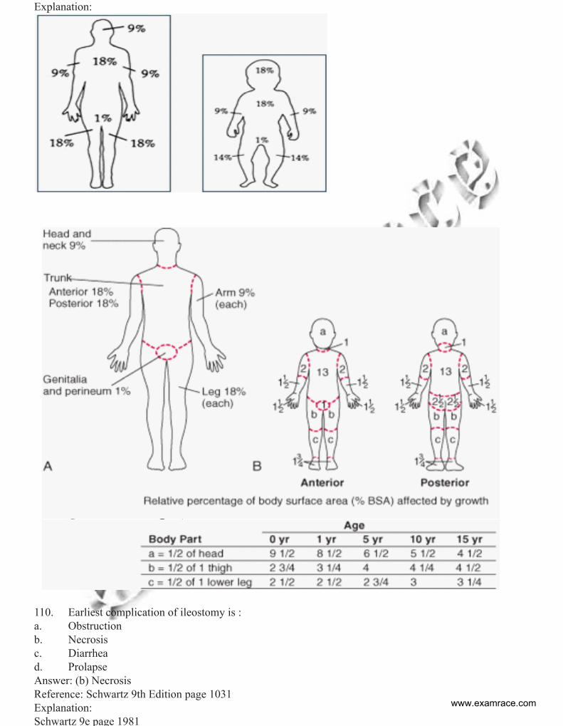
Explanation:
110. Earliest complication of ileostomy is : a. Obstructionb. Necrosisc. Diarrhea d. Prolapse Answer: (b) NecrosisReference: Schwartz 9th Edition page 1031Explanation:Schwartz 9e page 1981
www.examrace.com
Complications of IleostomyStoma necrosis may occur in the early postoperative period and usually is caused by skeletonizing the distal small bowel and/or creating an overly tight fascial defect.
111. A motor cyclist after multiple trauma is having hypoventilation. The cause is ss. damage to respiratory centrett. damage to respiratory apparatusuu. bothvv. None of the Above Ans: (a) damage to respiratory centreRef: CSDT 13th Ed. Pg. 817Explanation: The Cushings triad is the constellation of bradycardia, hypertension, and respiratory irregular-ity that often occupies a herniation event clinically. It is likely due to brainstem compression. The hyperten-sion can be conceptualized as an attempt to protect the brain’s perfusion pressure from the high intracranial pressure. Intubation and mechanical ventilation often obscure the respiratory part of the triad, but the other two are observed regularly. i. HTNii.Bradycardiaiii. Bradypnoea
112. A smoker 30 packets year present with, microscopic hematuria and painless left scrotal mass, LDH negative AFP negative, DD?a. Epididymitides b. Seminoma c. RCC d. Lung CancerAns: C, RCCRef: Bailey and Love 25th page 1311Exp:Clinical features Haematuria is usually the presenting symptom, sometimes with clot colic. There may be a dragging discom-fort in the loin or the patient may detect a mass. In men, arapidly developing varicocele is a rare but impressive sign, occurring most often on the left side because the left gonadal vein is obstructed where it joins the left renal vein.113. A new born boy is having lumbosacral meningomyelocele n awaiting the surgical repair. The sac is best protected with sterile guaze piece soaked with a. Mercurochrome b. Tincture benzoin c. Methylene blue d. Normal saline Answer: D, Normal saline Reference: Explanation:The first step in the management of a newborn with myelomeningocele is a careful clinical assessment with particular emphasis on motor, sensory, reflex, and sphincter function. Open defects should be covered with a saline-moistened nonadherent dressing to prevent injury to and desiccation of the neural placode. Neurotoxic substances are avoided, and the child should be kept prone or in a lateral recumbent position until surgery114. Mortality in emergency abdominal aorta aneurysm repair surgeryww. 10%xx. 20%yy. 40%zz. >50%
www.examrace.com

Ans: B, 20%Reference: Harrison 17th Edition page 1565Sabiston 17 edition page 1976/77Exp:Results after open repair of ruptured aneurysm vary. For patients in stable condition with a contained rup-ture, the mortality rate is less than 50%. For patients with free intraperitoneal rupture who arrive in shock with possible cardiac arrest, the outlook is grim and mortality rates exceed 90%.
ORTHOPEDIC115. Unconscious man lying in right lateral position with injury to face, hand lateral aspect of right knee. Most common cause for this clinical presentation? a. Trigeminal n. b. Radial n. c. Common Peroneal nerve d. Femoral nAnswer: (c) Common Peroneal nerveRef: Apleys OrthopaedicsExp: the common peroneal nerve is around fibular neck which can be injured in fall on lateral aspect of knee.116. A child was given gallows traction with weight. He is suffering from a. Fracture of shaft of femurb. Fracture of neck humerusc. Spine injury d. Fracture ulnaAnswer (a) Fracture of shaft of femurRef:ApleysOrthopaedics, J MaheswariorthopaedicsExp: in child less than one year femur shaft is treated by gallows traction 117. Most common cause of death in pelvic #e. Hypovolumic shockf. Bladder Injuryg. Neurogenic Injuryh. Pelvic instability Answer (a) Hypovolumic shock: Ref:ApleysOrthopaedics, J MaheswariorthopaedicsExp: blood loss on average is 2 litresie 40% of blood volume118. A patient with hip dislocation with limitation of Abduction at hip and flexion and internal rotation deformity at hip and shortening. Diagnosis is i. Central dislocationj. Anterior dislocationk. Posterior dislocationl. Fracture dislocation Ans: C, Posterior dislocationRef: ApleysOrthopaedics, J MaheswariorthopaedicsExp: Typical attitude is Flexion adduction Internal rotation and shortening in Posterior hip dislocation
119. A lady with colle’s fracture. The fracture healed but after few days patient develops pain and swell-ing over wrist and forearm, red hot and shiny skin and on X Ray diffuse osteopenia. Diagnosis is?m. Sudeck’sosteodystrophyn. Causalgiao. Non union p. Nerve injury Ans: A, Sudeck’sosteodystrophy
www.examrace.com
Ref: ApleysOrthopaedics, J MaheswariorthopaedicsExp: it is same as reflex sympathetic dystrophy, the cause is unknown
120. True about supracondylar fracture q. Cubitus valgus more common than varusr. Nerve injuries are usually tranisitorys. Anterior displacement of distal segment t. Weakness in elbow flexion eventually Ans: B, Nerve injuries are usually tranisitoryRef: ApleysOrthopaedics, J MaheswariorthopaedicsExp: Cubitusvarus is more common, posterior displacement of fragment, Nerve injury is usually neuroproxia121. Patient presents with knee problem. He gives history of injury during playing hockey 3 months back. On testing knee was unstable in extension but was stable in 90 degrees of flexion probably injury involvesu. ACL anteromedial fiber v. ACL posterolateral fiberw. PCL x. Anterior portion of medial meniscus Ans: B, ACL posterolateral fiberRef: Campbell orthopaedicsExp: Anterior cruciate ligament with postero lateral corner in jury iepopliteus are common122. 70year female is on treatment with Alendronate for severe osteoporosis. Now she complains of pain in right thigh. What is the next investigation to be performed?a. DEXA scanb. X- rayc. Serum vitamin D levelsd. Serum alkaline phosphate levels|Answer (b) (X-ray)Reference: CMDT 2013/page 1118Explanation:In this question patient is complaining of pain in right hip and thigh. Thus diagnosis of the patient can be1. Fracture of neck of femur or shaft of femur.2. Chronic pain probably not bony in origin; instead, it is related to abnormal strain on muscles, liga-ments, and tendons and to secondary facet-joint arthritis associated with alterations in thoracic and/or ab-dominal shape.3. Lumbar spinal canal stenosis resulting in nerve compression4. TendinitsCMDT states that patients of osteoporosis on bisphosphonates can develop “chalk” like fractures of shaft of femur unlike the spiral fractures that are read by us in orthopedic textbooks. The clinical feature of these patients is thigh pain and X ray reveals a shocking fracture. Infact Harrison also states in osteoporosis chap-ter in volume II that improvement in bone density scores with bisphosphonates over ten years of intake are minimal. If the question asked regarding MONITORING the patient in future then the best answer should have been DEXA123. Osteoblasticsecondaries are seen in y. Prostate metastasis z. Lung metastasis aa. Bladder metastasis bb. Stomach metastasis Ans: A, Prostate metastasis Ref: ApleysOrthopaedics, J MaheswariorthopaedicsExp: most common cause of osteblasticsecondaries in males is prostrate , in females breast carcinomawww.examrace.com
PSYCHIATRY124. Anti craving agent in alcoholism is :a. Buprenorphine b. Disulfiramc. Acamprosate d. Diazepam Answer: (c) Acamprosate Reference: Keplan & Sadock’s –Synopsis of Psychiatry 9 Edition page 1286-1287Expalnation: Data support the probable modest effect of two medications in addition to the usual cognitive-behavioral approaches for treating alcohol dependence. These have been hypothesized to decrease the rewarding effects of alcohol if an individual returns to drinking, diminish the symptoms of the protracted withdrawal syndrome, or, perhaps, decrease craving. The first drug is acamprosate, which is an analog of the amino acid neurotransmitter taurine and structurally resembles GABA. Although the mechanism of action in alcoholics is unknown, acamprosate antagonizes neuronal overactivity related to the excitatory neurotrans-mitter glutamate, at least in part by acting as an antagonist to NMDA receptors. Thus, one possibly impor-tant mechanism for this drug may be in diminishing mild anxiety, mood swings, and other sleep difficulties associated with the subacute and protracted withdrawal syndrome observed after the first 4 to 5 days of alcohol abstinence. At the usual dose of about 2,000 mg per day the majority of studies report modest but significantly better outcomes with active drug than placebo. The level of improvement is often in the range of 15 to 20 percent over results seen with placebo, and in most studies, rates of side effects are similar for active drug and placebo except for GI problems such as diarrhea. At least one cost-effectiveness study indicated that the improved result produces significant cost savings above the financial obligations of prescribing the drug. Al-though there are no clear guidelines, it is reasonable to prescribe this drug for about 6 months, during which time it is hoped an alcohol-free lifestyle can be developed. The second promising medication is the long-acting, oral, opioid antagonist naltrexone. This agent has been marketed for many years for the treatment of acute opioid overdose. It may also be effective for highly motivated opioid-dependent individuals, who understand that because of the use of this blocking drug, they cannot get high from opioids. Naltrexone works by blocking opioid receptors in the brain and, thus, at least indirectly, changing levels of activity of dopamine and serotonin. In alcohol-dependent individ-uals, naltrexone and its cousin nalmefene (Revex) have been hypothesized to decrease the rewarding effects of a drink and to diminish craving. The large majority of double-blind, placebo-controlled trials support the superiority of naltrexone to placebo regarding drinking-related behaviors with about 15 to 20 percent better outcomes than for placebo. The side effect profile of the 50 to 150 mg per day usually used in these trials is relatively benign. The most frequent complaints involving GI upset, with a possible modest increase in leth-argy and, perhaps, the subjective report of a dampened level of interest in activities and life events. Naltrex-one is also available in a 380 mg injection to be given once a month as Vivitrol. This might be particularly useful for patients who find it difficult to comply with oral medications. Several additional medications are worth brief mention. First, while not yet approved for the treat-ment of alcoholism, the anticonvulsant topiramate (Topamax) has the usual GABA-boosting activity associ-ated with treatments of seizure disorders along with an additional glutamate receptor blockade and effects on dopamine systems. Perhaps related to this combination of effects, several studies using about 300 mg of topiramate per day reported improvements in drinking patterns. Second, some preliminary data are available on the serotonin 3 receptor antagonist ondansetron (Zofran), indicating a possible benefit for this medica-tion, particularly in early onset alcoholics with comorbid drug dependence and criminality. However, these studies are very preliminary and there are no indications for the use of this expensive medication with a high level of associated side effects in clinical practice. Additional medications that have been evaluated for the treatment of alcoholism include the nonbenzodiazepine antianxiety drug buspirone (BuSpar), several selec-tive serotonin reuptake inhibitors (SSRIs), the GABA-B receptor agonist baclofen (Lioresal), and antipsy-chotic medications in the treatment of alcoholism, although none of these medications have proven to be consistently better than placebo in controlled trials125. On Wechsler intelligence scale if a child has average IQ the score of child shall bea. 71
www.examrace.com
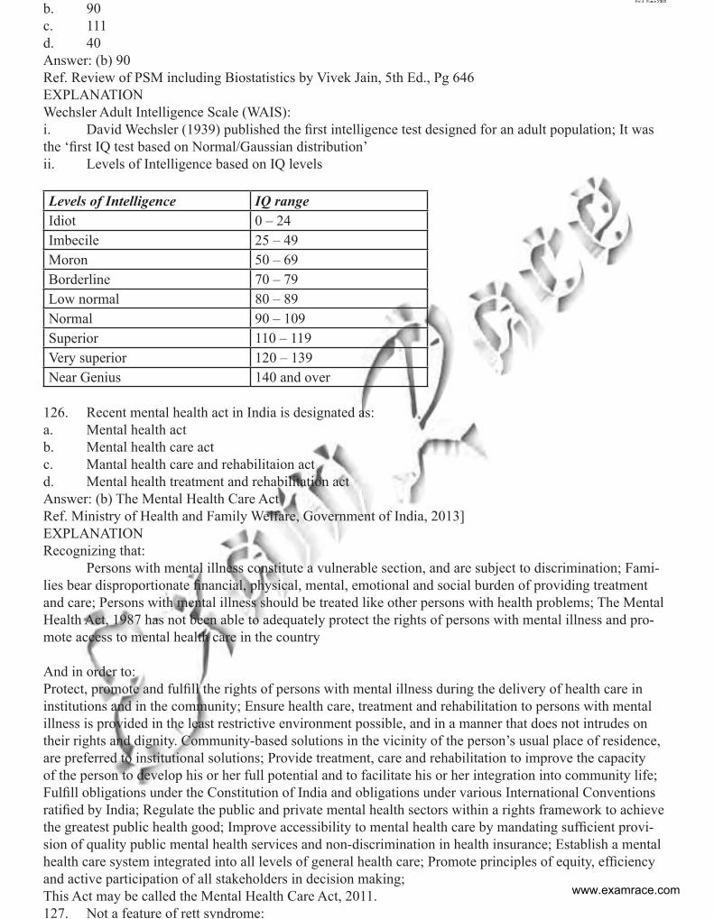
b. 90c. 111d. 40Answer: (b) 90Ref. Review of PSM including Biostatistics by Vivek Jain, 5th Ed., Pg 646EXPLANATIONWechsler Adult Intelligence Scale (WAIS): i. David Wechsler (1939) published the first intelligence test designed for an adult population; It was the ‘first IQ test based on Normal/Gaussian distribution’ ii. Levels of Intelligence based on IQ levels
Levels of Intelligence IQ range Idiot 0 – 24 Imbecile 25 – 49 Moron 50 – 69 Borderline 70 – 79 Low normal 80 – 89 Normal 90 – 109 Superior 110 – 119 Very superior 120 – 139 Near Genius 140 and over
126. Recent mental health act in India is designated as: a. Mental health act b. Mental health care actc. Mantal health care and rehabilitaion act d. Mental health treatment and rehabilitation act Answer: (b) The Mental Health Care ActRef. Ministry of Health and Family Welfare, Government of India, 2013]EXPLANATIONRecognizing that: Persons with mental illness constitute a vulnerable section, and are subject to discrimination; Fami-lies bear disproportionate financial, physical, mental, emotional and social burden of providing treatment and care; Persons with mental illness should be treated like other persons with health problems; The Mental Health Act, 1987 has not been able to adequately protect the rights of persons with mental illness and pro-mote access to mental health care in the country And in order to:Protect, promote and fulfill the rights of persons with mental illness during the delivery of health care in institutions and in the community; Ensure health care, treatment and rehabilitation to persons with mental illness is provided in the least restrictive environment possible, and in a manner that does not intrudes on their rights and dignity. Community-based solutions in the vicinity of the person’s usual place of residence, are preferred to institutional solutions; Provide treatment, care and rehabilitation to improve the capacity of the person to develop his or her full potential and to facilitate his or her integration into community life; Fulfill obligations under the Constitution of India and obligations under various International Conventions ratified by India; Regulate the public and private mental health sectors within a rights framework to achieve the greatest public health good; Improve accessibility to mental health care by mandating sufficient provi-sion of quality public mental health services and non-discrimination in health insurance; Establish a mental health care system integrated into all levels of general health care; Promote principles of equity, efficiency and active participation of all stakeholders in decision making;This Act may be called the Mental Health Care Act, 2011.127. Not a feature of rett syndrome:
www.examrace.com
a. Macrocephalyb. Seizuresc. repetitive stereotyped movementsd. DystoniaAnswer: (a) MacrocephalyReference: Nelson : chapter 29.4 rett syndromeExplanation: The DSM-IV-TR diagnostic criteria for the condition are presented in Table 41-6. Early devel-opment of the child is normal. The onset of the condition may be insidious and follow a period of develop-mental stagnation and delay recognition slightly. Over time, the developmental delay, decelerated head and body growth, and diminished interest in the environment become quite striking. Previously acquired abilities are lost, including purposeful hand movements. The lack of social interest and potential for misdiagnosis of autism is greatest in the preschool years, as, usually, by the time the child reaches school age, the autistic-like features are less prominent and development plateaus for a time. At this point, severe mental retardation, seizures, and motor problems are areas of major concern. During this plateau, or “pseudo-stationary” phase, breathing difficulties, bruxism, motor problems, and early scoliosis may be noted. Apneic episodes may alternate with hyperventilation. Most children remain ambulatory until a final period of motor deterioration. EEG is frequently abnormal and seizures are common
128. Alcoholic brought by his wife with emergency, he is alcoholic for past 15 years. 2 days he left drink-ing because of some religious case, had c/o rest;essmess, tremor insomnia, on 2nd day he started throwing convulsions, after 6 hrs he had 2nd episode, now the treatment to control seizure isa. Sodium valproateb. Diazepam c. Phenyton d. Clonidine Answer: (b) DiazepamReference: Keplan & Sadock’s –Synopsis of Psychiatry 9 Edition page 1285Explanation: For the less than 1 percent of alcoholic patients with extreme autonomic dysfunction, agitation, and confusion—that is, those with alcoholic withdrawal delirium, also called delirium tremens—there is no perfect treatment. The first key step is to ask why such a severe and relatively uncommon withdrawal syn-drome has occurred; the answer often relates to a concomitant medical problem that needs immediate treat-ment. The withdrawal symptoms can then be minimized either through the use of benzodiazepines (in which case high doses are sometimes required) or through antipsychotic agents such as haloperidol (Haldol). Once again, doses are used on the first or second day to control behavior, and the patient can be weaned off the medication by approximately the fifth day. Another 1 percent or so of patients may have a single grand mal convulsion. The rare person has multiple fits, and the peak incidence is on the second day of withdrawal. Such patients require a neurological evaluation, but in the absence of evidence of an independent seizure disorder, those with a single seizure do not benefit from anticonvulsant drugs.
129. Piagnet cognitive model question , if object out of sight out of mind and “here and now” are included in which stagea. Concrete operation stage b. Pre operational stage c. Formal operational stage d. Sensory and motor stageAnswer: (d) Sensory and motor stageReference: Kaplan & sadock’s –Synopsis of Psychiatry 9 Edition page 136-139Explanation:
www.examrace.com

Stage Age Achievements of stage 1. Sensorimotor stage
Birth to 2 years • Infants being to learn through sensory observation & gain control of their motor functions. • Development of object performance i.e. ability to un-derstand that objects have an existence • Symbolization (ability to create visual image of object)
2. Preoperation-al thought stage
2 to 7 years • Intuitive (primitive) thinking i.e. unable to think logi-cally & child learn without use of reasoning Ex-if children drop a glass that then breaks –they believe that the glass was ready to break, not that they broke the glass. • Immanent justice i.e. the belief that punishment for bad deeds is inevitable • Egocentric i.e. unable to modify their behavior for someone else. • Magical thinking (or phenomenalistic causality), in which the events that occur together are thought to cause one another eg. Thunder causes lightening • Semiotic function
3. Stage of con-crete operations
7 to 11 years • Operational thought i.e. can now see things from someone else perspective • Syllogistic (logical) reasoning • Conservation i.e. if a clay ball is rolled into sausage shape the child recognizes that both form contain equal amount of clay • Reversibility i.e. to realize that one thing can turn into another & back again as ice & water
4. Stage of for-mal thoughts
11 though end of adolescence
• Ability to think abstractly, to reason deductively& to define concepts, & emergency of skills for dealing with per-mutations & combination.
130. In Mental Status examination , which of the following is a measure of awareness response a. Perceptionb. Judgementc. Insightd. CognitionAnswer: (a) PerceptionReference: Kaplan & sadock’s –Synopsis of Psychiatry 9 Edition page 619ExplanationThe terms sensation, perception, and cognition are used to describe the three broadening tiers of human information processing: Think of sensation as the immediate result of the stimulation of sensory neurons and perception as involving the organization and conscious awareness of these sensations. Cognition refers to the set of interwoven processes, such as memory, language, and problem solving, that we bring to bear to gener-ate structures and strategies to apply to our perceptions. Although distinguishing among sensation, percep-tion, and cognition has a long academic history, our ability to separate them grows increasingly difficult as more is learned about their interdependence in the functioning nervous system.
Cognition As part of the MSE, the interviewer should get an overall sense of the patient’s cognitive function-ing The elements of cognitive functioning that should be assessed are alertness orientation concentration
www.examrace.com
memory (both short and long term), calculation, fund of knowledge, abstract reasoning, insight, and judg-ment. Note should be made of the patient’s level of alertness. The amount of detail in assessing cognitive function will depend on the purpose of the examination and also what has already been learned in the inter-view about the patient’s level of functioning, performance at work, handling daily chores, balancing one’s checkbook, etc. In addition the psychiatrist will have already elicited data concerning the patient’s memory for both remote and recent past. A general sense of intellectual level and how much schooling the patient has had can help distinguish intelligence and educational issues versus cognitive impairment that might be seen in delirium or dementia. The MMSE is a standardized screening tool commonly used to screen for cognitive impairment, and it can be used to follow the patient over time monitoring for progression or resolution of cognitive problems. The MMSE employs a 30-point scale using standardized questions and tasks and can be done in a matter of a few minutes
131. Study of brain and behavior in a normal and diseased brain called asa. Neuropsychologyb. Neuro developmental psychiatryc. Neuropsychiatryd. Criminal psychologyAnswer: (a) NeuropsychologyReference: Kaplan & Sadock’s –Synopsis of Psychiatry 9 Edition page 936Clinical neuropsychology is a specialty in psychology that examines the relationship between behavior and brain functioning in the realms of cognitive, motor, sensory, and emotional functioning. The clinical neu-ropsychologist integrates the medical and psychosocial history with the reported complaints and the pattern of performance on neuropsychological procedures in order to determine whether results are consistent with a particular area of brain damage or a particular diagnosis. Although neurological syndromes are often the focus of referrals, the neuropsychological examination has a valuable place in diagnosing and identifying behavioral symptoms that can be associated with various medical, psychological, and psychiatric conditions. Clinical neuropsychology is primarily differentiated from general clinical psychology by its focus on thorough and extensive evaluation of a broad range of cognitive and emotional factors and their potential relationship to brain damage. There is considerable overlap between the two areas in the approach to assess-ment, chiefly characterized by reliance on the psychometric foundations of reliability, validity, and norma-tive standards in order to objectively define behavioral symptoms and complaints. Emotional factors are the province of both. Both evaluate cognition, although the clinical psychologist generally focuses on issues involving general intellectual, academic, and vocational skills rather than neurological factors. Both clinical psychologists and clinical neuropsychologists are involved in treatment, which can include psychotherapy as well as cognitive retraining to remediate deficits. Psychoeducational functions are also served by both fields in discussing symptoms, assessment results, and their implications with patients, caregivers, and other health professionals.INTERNAL MEDICINE 132. LBBB is seen with all except:a. Acute MIb. Ashmann syndromec. Hypokalemiad. HyperkalemiaAnswer: B : Ashman syndrome:Reference: current diagnosis and management in cardiology, chapter19 : supraventricular tachycardiaExplanation:• Normally the septum is activated from left to right, producing small Q waves in the lateral leads.• In LBBB, the normal direction of septal depolarisation is reversed (becomes right to left), as the im-pulse spreads first to the RV via the right bundle branch and then to the LV via the septum.o This sequence of activation extends the QRS duration to > 120 ms and eliminates the normal septal Q waves in the lateral leads.o The overall direction of depolarisation (from right to left) produces tall R waves in the lateral leads
www.examrace.com

(I, V5-6) and deep S waves in the right precordial leads (V1-3), and usually leads to left axis deviation.• As the ventricles are activated sequentially (right, then left) rather than simultaneously, this produces a broad or notched (‘M’-shaped) R wave in the lateral leads.
Causes of LBBB are:• Aortic stenosis• Ischaemic heart disease• Hypertension• Dilated cardiomyopathy• Anterior MI• Primary degenerative disease (fibrosis) of the conducting system (Lenegre disease)• Hyperkalaemia• Digoxin toxicity
Ashman phenomenon is seen in atrial fibrillation. U all know atrial fibrillation has irregularly irregular pulse leading to irregular RR interval in ECG. This means there is very fast conduction occurring via bundle of his which is receiving these impulses from a supraventricular phenomenon. Now the point is that the two fascicles of bundle of his have different refractory periods. Thus if an impulse lands on the bundle of his and finds the right bundle refractory then RBBB will occur. Also remember that the refractory period of right fascicle is more than that of the left fascicle resulting in RBBB.
133. A 35 year old man with complaints of ptosis, with difficulty in chewing and occasionally swallow-ing. There is no history of diplopia and on examination there is a symmetrical ptosis, lateral arm abduction time of 60 seconds. Repetitive nerve stimulation test shows decremental response. EMG shows myopathic response. Anti –Ach receptor antibodies are negative. Probable diagnosis is:a. Ocular M.Gravisb. Generalised M.Gravisc. Since Anti-ach receptor antibodies are negative consider other diagnosisd. Chronic Progressive external opthalmoplegiaAnswer: (d) Chronic Progressive external opthalmoplegiaReference: Harrison 18th edition chapter 386 http://emedicine.medscape.com/article/1215103-overviewThis question looks intially as in favour of myaesthenia gravis but the following points are against the diag-nosis of Myasthenia gravis1. Gender given as MALE at 35 years. While MG is seen in men in 50-60 years of age.2. Symetrical Ptosis without Diplopia. In MG we have asymetrical ptosis.3. No diurnal variation in severity of weakness mentioned4. For ocular M.G. symptoms must persist for 3 years5. Anti- ach receptor antibodies are absent in 50% cases of ocular myaesthenia gravis but they would be absent in CPEO as well as.6. In CPEO- Ciliary and iris muscles are not involved and hence diplopia is absent.I have also compiled harrison and Emedicine data on these two close answers in a tabular fashion-
www.examrace.com
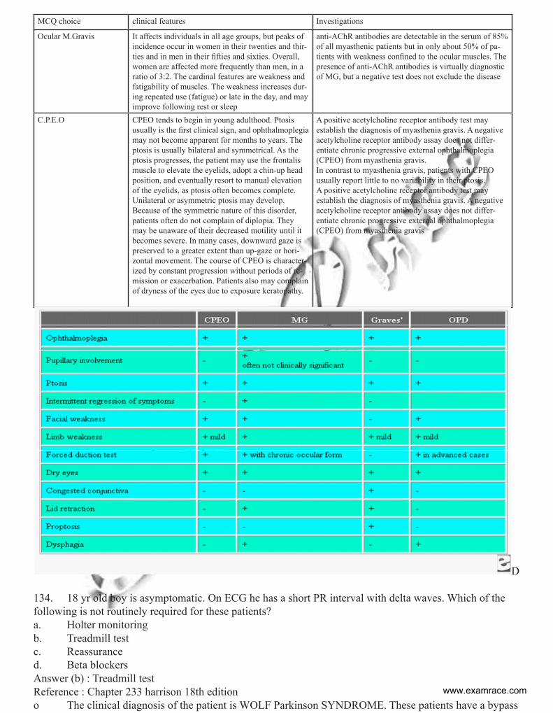
MCQ choice clinical features Investigations
Ocular M.Gravis It affects individuals in all age groups, but peaks of incidence occur in women in their twenties and thir-ties and in men in their fifties and sixties. Overall, women are affected more frequently than men, in a ratio of 3:2. The cardinal features are weakness and fatigability of muscles. The weakness increases dur-ing repeated use (fatigue) or late in the day, and may improve following rest or sleep
anti-AChR antibodies are detectable in the serum of 85% of all myasthenic patients but in only about 50% of pa-tients with weakness confined to the ocular muscles. The presence of anti-AChR antibodies is virtually diagnostic of MG, but a negative test does not exclude the disease
C.P.E.O CPEO tends to begin in young adulthood. Ptosis usually is the first clinical sign, and ophthalmoplegia may not become apparent for months to years. The ptosis is usually bilateral and symmetrical. As the ptosis progresses, the patient may use the frontalis muscle to elevate the eyelids, adopt a chin-up head position, and eventually resort to manual elevation of the eyelids, as ptosis often becomes complete. Unilateral or asymmetric ptosis may develop. Because of the symmetric nature of this disorder, patients often do not complain of diplopia. They may be unaware of their decreased motility until it becomes severe. In many cases, downward gaze is preserved to a greater extent than up-gaze or hori-zontal movement. The course of CPEO is character-ized by constant progression without periods of re-mission or exacerbation. Patients also may complain of dryness of the eyes due to exposure keratopathy.
A positive acetylcholine receptor antibody test may establish the diagnosis of myasthenia gravis. A negative acetylcholine receptor antibody assay does not differ-entiate chronic progressive external ophthalmoplegia (CPEO) from myasthenia gravis.In contrast to myasthenia gravis, patients with CPEO usually report little to no variability in their ptosis.A positive acetylcholine receptor antibody test may establish the diagnosis of myasthenia gravis. A negative acetylcholine receptor antibody assay does not differ-entiate chronic progressive external ophthalmoplegia (CPEO) from myasthenia gravis
D
134. 18 yr old boy is asymptomatic. On ECG he has a short PR interval with delta waves. Which of the following is not routinely required for these patients?a. Holter monitoringb. Treadmill testc. Reassuranced. Beta blockersAnswer (b) : Treadmill testReference : Chapter 233 harrison 18th editiono The clinical diagnosis of the patient is WOLF Parkinson SYNDROME. These patients have a bypass
www.examrace.com
tract that allows conduction of impulses from atria to ventricles without having to be subjected to the decre-mental property of AV node. o Generally, no activity restrictions are recommended in patients with ECG findings of pre-excitation in the absence of tachycardia. They should be restricted from high-risk professions (eg, airline pilot) and may be restricted from competitive sports. o Patients presenting with tachycardias and accessory pathways should avoid participating in competi-tive sports, because catecholamines can decrease the refractoriness of the bypass tract and facilitate tach-yarrhythmias. Patients with hypertrophic cardiomyopathy, or the Ebstein anomaly should also abstain from competitive sports. Walking on Treadmill can precipitate due to sympathetic stimulation an arryhtmia and would not be recommended for patient of W.P.W.o The Holter monitor records electrical signals from the heart via a series of electrodes attached to the chest. Electrodes are placed over bones to minimize artifacts from muscular activity. The number and posi-tion of electrodes varies by model, but most Holter monitors employ between three and eight. The device is responsible for keeping a log of the heart’s electrical activity throughout the recording period.More modern units record onto digital flash memory devices. The data are uploaded into a computer which then automati-cally analyzes the input, counting ECG complexes, calculating summary statistics such as average heart rate, minimum and maximum heart rate, and finding candidate areas in the recording worthy of further study by the technician. Thus Holter monitoring in these patients can identify if patient is having a life threatening event which on recording will help us know that these patients would or would not require a RADIOFRE-QUENCY ABLATION to get rid of the accessory pathway.
135. 8 yr old boy is having muscle weakness. On examination he has large muscles in legs and his CPK levels are reducing. He also has a sub normal IQ. Probable diagnosis is :a. Hereditary sensory and motor neuropathyb. Myelin deficiencyc. Dystrophin deficiencyd. Congenital myopathy
Answer (c) : dystrophin deficiencyReference: chapter 387 harrison 18th editiono Duchenne’s dystrophy is present at birth, but the disorder usually becomes apparent between ages 3 and 5 years. The boys fall frequently and have difficulty keeping up when playing. Running, jumping, and hopping are invariably abnormal.o GOWER sign is seen with PSEUDOHYPERTROPHY of the calf muscles.o Contractures of the heel cords and iliotibial bands become apparent by age 6 years, when toe walking is associated with a lordotic postureo By age 12 years, most patients are wheelchair dependent. Contractures become fixed, and a progres-sive scoliosis often develops that may be associated with pain. The chest deformity with scoliosis impairs pulmonary function, which is already diminished by muscle weakness. By age 16–18 years, patients are pre-disposed to serious, sometimes fatal pulmonary infections. Other causes of death include aspiration of food and acute gastric dilation.o A cardiac cause of death is uncommon despite the presence of a cardiomyopathy in almost all pa-tientso Intellectual impairment in Duchenne’s dystrophy is common; the average intelligence quotient (IQ) is 1 SD below the mean. Impairment of intellectual function appears to be nonprogressive and affects verbal ability more than performance.o Serum CK levels are invariably elevated to between 20 and 100 times normal. The levels are abnor-mal at birth but decline late in the disease because of inactivity and loss of muscle mass. EMG demonstrates features typical of myopathy. The muscle biopsy shows muscle fibers of varying size as well as small groups of necrotic and regenerating fibers. Connective tissue and fat replace lost muscle fibers. A definitive diagno-sis of Duchenne’s dystrophy can be established on the basis of dystrophin deficiency in a biopsy of muscle tissue or mutation analysis on peripheral blood leukocytes, as discussed below.o A diagnosis of Duchenne’s dystrophy can also be made by Western blot analysis of muscle biopsy
www.examrace.com
specimens, revealing abnormalities on the quantity and molecular weight of dystrophin protein. In addition, immunocytochemical staining of muscle with dystrophin antibodies can be used to demonstrate absence or deficiency of dystrophin localizing to the sarcolemmal membrane. Carriers of the disease may demonstrate a mosaic pattern, but dystrophin analysis of muscle biopsy specimens for carrier detection is not reliable.136. 3 yr old child is having barking cough with drooling of saliva. His RR is 36/min and Xray neck lat-eral view shows “thumb sign”. The probable diagnosis is:a. Epiglottisb. Croupc. Foreign body d. Abductor vocal cord palsy.
Answer (a) : EpiglottisReference: chapter 382.1 nelson textbook
EPIGLOTTITIS: A otherwise healthy child suddenly develops a sore throat and fever. Within a matter of hours, the patient appears toxic, swallowing is difficult, and breathing is labored. Drooling is usually present and the neck is hyperextended in an attempt to maintain the airway. The child may assume the tripod posi-tion, sitting upright and leaning forward with the chin up and mouth open while bracing on the arms. A brief period of air hunger with restlessness may be followed by rapidly increasing cyanosis and coma. Stridor is a late finding and suggests near-complete airway obstruction. Complete obstruction of the airway and death can ensue unless adequate treatment is provided. The barking cough typical of croup is rare. Usually, no oth-er family members are ill with acute respiratory symptoms.The diagnosis requires visualization of a large, “cherry red” swollen epiglottis by laryngoscopy. Occasionally, the other supraglottic structures, especially the aryepiglottic folds, are more involved than the epiglottis itself.. Anxiety-provoking interventions such as phlebotomy, intravenous line placement, placing the child supine, or direct inspection of the oral cav-ity should be avoided until the airway is secure. If epiglottitis is thought to be possible but not certain in a patient with acute upper airway obstruction, the patient can undergo lateral radiographs of the upper airway first. Classic radiographs of a child who has epiglottitis show the “thumb sign”. CROUP : Croup is a clinical diagnosis and does not require a radiograph of the neck. Radiographs of the neck may show the typical subglottic narrowing or “steeple sign” of croup on the postero-anterior view
137. 3 year old child is having sore throat with high grade fever. On examination he has a inflamed phy-ranx and exudate on tonsils with tender lymphadenopathy. Which of the following is a non suppurative complication of these patients?a. Acute rheumatic feverb. Acute glomerulonephritisc. Acute rheumatic fever and acute Glomerulonephritis bothd. Scarlet feverAnswer is C: Acute rheumatic fever and acute Glomerulonephritis bothReference: CMDT 2013 ,page no 1419
Group A β – hemolytic streptococci (Streptococcus pyogenes) are the most common bacterial cause of phar-yngitis. Transmission occurs by droplets of infected secretions. Group A streptococci producing erythrogenic toxin may cause scarlet fever in susceptible persons.Clinical FindingsA. Symptoms and Sings“Strep throat” is characterized by a sudden onset of fever, sore throat, pain on swallowing, tender cervical adenopathy, malaise, and nausea. The pharynx, soft palate, and tonsils are red and edematous. There may be a purulent exudate. The centor clinical criteria for the diagnosis of streptococcal pharyngitis are temperature > 38oC, tender anterior cervical adenopathy, lack of a cough, and pharyngotosillar exudates.The rash of scarlet fever is diffusely erythematous, resembling a sunburn, with superimposed fine red pap-ules, and is most intense in the groin and axillas. It blanches on pressure, may become petechial, and fades in
www.examrace.com
2-5 days, leaving a fine desquamation. The face is flushed, with circumoral pallor, and the tongue is coated with enlarged red papillae (strawberry tongue).B. Laboratory FindingsLeukocytosis with neutrophil predominance is common. Throat culture onto a single blood agar plate has sensitivity of 80-90%. Rapid diagnostic tests based on detection of streptococcal antigen are slightly less sensitive than culture. Clinical criteria such as the centor criteria, are useful for identifying patients in whom a rapid antigen test or throat culture is indicated. Patients who meet two or more of these criteria merit fur-ther testing. When three of the four are present, laboratory sensitivity of rapid antigen testing exceeds 90%. When only one criterion is present, streptococcal pharyngitis is unlikely. In high prevalence settings or if clinical suspicion for streptococcal pharyngitis or if clinical suspicion for streptococcal paryngitis is high, a negative antigen test or culture should be confirmed by a follow up culture.ComplicationsSuppurative complications include sinusitis, otitis media, mastoiditis, peritonsillar abscess and suppuration of cervical lymph nodes.Nonsuppurative complications are rheumatic fever and glomerulonephritis. Rheumatic fever may follow recurrent epidoses of pharyngitis beginning 1-4 weeks after the onset of symptoms. Glomerulonephritis fol-lows a single infections with a nephriotogenic strain of streptococcus group A (eg, types 4, 12, 2, 49 and 60), more commonly on the skin than in the throat, and begins 1 – 3 weeks after the onset of the infection. 138. 28 year old lady with history of recurrent abortions and repeated episodes of pain the calves is most likely to have deficiency of which of the followinga. Plasmin b. Thrombin c. Protein C d. Factor 13Answer is C: protein CReference: table 58.3 Harrison 18th Edition and NELSON chapter 478
Factor 13 deficiency and thrombin deficiency causes bleeding manifestations. Congenital plasmino-gen deficiency has been mentioned in texts but not congenital plasmin deficiency. Hence by exclusion the diagnosis is protein C deficiency. Thrombohilia should be suspected in patients has a history of recurrent venous thromboembolism, venous thrombosis in a person younger than 40 years, a familial history of venous thromboembolism, and thrombosis in unusual sites (eg, mesenteric vein, renal vein, hepatic and cerebral thrombosis). Homozygous deficiency of protein C presents with purpura fulminans in the first few hr of life. Fresh frozen plasma (FFP) is the only immediately available source of protein C. Amelioration of symptoms usu-ally requires 10–15 mL/kg of FFP every 8–12 hr. Clinical trials are in progress using a plasma protein C con-centrate, which eliminates the need for large amounts of FFP. A recombinant activated protein C concentrate (drotrecogin-α) has been approved for the treatment of adult sepsis, but it has not been approved for treating hereditary deficiency Thrombophilic disorders are usually associated with venous thrombosis. However, protein S, protein C, ATIII deficiencies, and lupus anticoagulants have been associated with arterial thrombosis. Patients with protein C and S deficiencies can develop warfarin-induced skin necrosis when placed on warfarin since protein C and S are vitamin K–dependent factors and, hence are suppressed. Remember: Purpura fulminans in infancy could suggest protein C deficiency. These antibodies occur in about 20% of patients with systemic lupus erythematosus (SLE), but they are also associated with other autoimmune diseases. Lupus anticoagulants may occur in patients taking phe-nothiazines, phenytoin, phenytoin, hydralazine, quinine, amoxicillin, and oral contraceptives. Also REMEMBER : Clinical criteria for indicating the presence of lupus anticoagulants (Sapporo criteria for the antiphospholipid syndrome) are as follows: • One or more arterial, venous, or small vessel thrombosis, affecting any organ or tissue• Pregnancy morbidity: The risk for maternal and fetal morbidity increases after the 10th week of preg-nancy. Fetal mortality in pregnancy can include spontaneous abortions, prematurity, and stillbirths. • Three or more unexplained consecutive spontaneous abortions after the 10th week of gestation
www.examrace.com

139. A lady is pregnant and is a known case of juvenile myoclonic epilepsy and is receiving sodium val-proate. Which of the following drugs is best suited after valproate for management of this patienta. phenytoinb. carbamazepinec. lamotrigine d. LacosamideAnswer is C: lamotrigineReference: Harrison 18th edition table 368-9 and 369-9In the latest issue of journal neurology in month of may 2013 he has mentioned valproate as most teratogen-ic followed by phenobarbitone being responsible for neural tube defects. Hence the older concept of pheno-barbitone as the safest anti-epileptic drug can be called as outdated. The current safest A.E.D in pregnancy is LAMOTRIGINE>>CARBAMAZEPINE.
Generalized-onset Tonic-Clonic
Focal Typical Absence Atypical Absence, Myoclonic, Atonic
First-LineValproic acidLamotrigineTopiramate
LamotrigineCarbamazepineOxcarbazepinePhenytoinLevetiracetam
Valproic acidEthosuximide
Valproic acidLamotrigineTopiramate
AlternativesZonisamideaPhenytoinCarbamazepineOxcarbazepinePhenobarbitalPrimidoneFelbamate
TopiramateZonisamideaValproic acidTiagabineaGabapentinaLacosamideaPhenobarbitalPrimidoneFelbamate
LamotrigineClonazepam
ClonazepamFelbamate
140. 60 year old patient with renal failure. He also has bone pain with X ray limb showing lytic lesions in the long bones. Serum electrophoresis shows M spike. Peripheral smear shows 35 % plasma cells. Bone marrow shows presence of immature plasma cells. Diagnosis isa. Plasma cell Leukemiab. Multiple myelomac. Smoldering myelomad. Monoclonal Gammopathy of unknown significance.Answer: A: Plasma cell leukemiaReference: http://www.medscape.com/viewarticle/731900Explanation: The question mentions Peripheral smear showing plasma cells >35% and not in the bone marrow. Hence the diagnosis goes in favour of plasma cell leukemia. The diagnostic criteria for Plasma Cell Leukemia include the Presence of a circulating clonal plasma cell count of more than 2000/µl (if the total white blood cell count is >10,000/µl) OR The presence of more than 20% CIRCULATING plasma cells.
Since in this question one of the critieria is satisfied hence diagnosis is Plasma cell leukemia
www.examrace.com
• PCL can be secondary PCL (s PCL) if arising from a known a diagnosis of plasma cell myeloma or primary PCL (p PCL) if no prior history of plasma cell myeloma can be ascertained. sPCL was felt to arise late in the course of plasma cell myeloma and those patients have been described to have a worse prognosis than those with p PCL. However, a recent report indicates that the median time to transformation to s PCL from plasma cell myeloma is approximately 21 months. The primary form accounts for 60% of all cases of PCL.• Patients with PCL may present with signs and symptoms commonly found in patients with plasma cell myeloma, such as bone pain, anemia, renal dysfunction, Hypercalcemia and lytic bone lesions; or clini-cal manifestations normally found in patients with leukemia, including leukocytosis, anemia, thrombocy-topenia, infections and hepato-splenomegaly. In addition, these patients may also have elevated levels of LDH and β-2-microglobulin. Patients with PCL are also more likely to present with advanced stage disease (stage III) and have more significant extramedullary involvement that can include the CNS• In contrast to most patients with plasma cell myeloma, the malignant cells in PCL frequently lack CD56, CD9, CD117 and human leukocyte antigen DR expression. It has been proposed that weak or absent CD56 expression could explain the propensity of these clonal plasma cells to leukemic transformation and also account for the lower osteolytic potential and higher rates of extra-medullary involvement seen in pa-tients with PCL• The use of traditional combination chemotherapy, such as vincristine, Adriamycin, dexamethasone (VAD) or high-dose cyclophosphamide-Etoposide is associated with response rates of up to 59% and median overall survival of 15–20 months. More aggressive regimens such as fractionated high-dose cyclophosph-amide and dexamethasone with infusional vincristine and Adriamycin (hyper CVAD) have been used alone or in combination with thalidomide and have revealed encouraging results including complete response (CR)
141. A patient has Pc02 of 30 mmHg, P02 of 105 mmHg and pH of 7.45. diagnosis is compensateda. respiratory acidosisb. respiratory alkalosisc. metabolic acidosisd. metabolic alkalosisAnswer: (b) respiratory alkalosis• Well if you look at values it indicates hyperventilation of the patient due to C02 washout with in-creased 02 and normal pH due to the process of compensation.• If in same question pH>7.45 we shall call it uncompensated respiratory alkalosisElse follow this table:• If direction of change of pH and pC02 is both in same direction ie like both reduced or both in-creased then problem is metabolic• If direction of change of pH and PC02 is opposite, then the primary problem is respiratory. • Else use a pen and mark arrows for increase or decrease in the table below and u shall see a pattern emerge which will solve all MCQ in books.pH pC02 HCO3- InterpretationDecrease Decrease Decrease Metabolic acidosisIncrease Increase Increase Metabolic alkalosisDecrease Increased Increased Respiratory acidosisIncrease Decrease Decrease Respiratory alkalosis
142. Bilateral Balbinski sign is seen in :a. brain- stem strokeb. thalamus and basal ganglia damagec. cerebellum damaged. mid pontine lesionsAnswer: (a) brain- stem strokeReference: Harrison 18th edition figure 370-10 point no M
www.examrace.com

Brain stem stroke Can result in development of “ locked in syndrome” where both the corticospi-nal pathways would be affected leading to bilateral balbinski sign.Harrison in figure 370-10 quotes “Basilar artery syndrome (the syndrome of the lone vertebral artery is equivalent): A combination of the various brainstem syn-dromes plus those arising in the posterior cerebral artery distribution.Bilateral long tract signs (sensory and motor; cerebellar and peripheral cranial nerve abnormalities): Bilateral long tract; cerebellar and peripheral cranial nervesParalysis or weakness of all extremities, plus all bulabar musculature.
Thalamus and basal gan-glia
Results in contralateral spasticity,,hemi-anaesthesia due to involvement of inter-nal capsule.Basal ganglia involvement would cause tremors, abulia (apathy with loss of initiative) with dystonia.
Cerebellum stroke It would cause intentional tremors with ataxia, dysmetria, dys-aidokinesia and development of Nystagmus and signs of raised ICT.
Midpontine lesions Usually results in development of ipsilateral cranial nerve palsies with contra-lateral motor manifestations due to involvement of corticospinal pathway eg millard gubler syndrome.
143. A 60 year old patient with 60 Packet per year history of smoking is having a cancer which shows small cells with high mitotic index. Which of the following is correct for this patienta. aggressive pyscho-social behaviourb. repeated blood transfusionsc. thin limbs with centripetal obesityd. increased thirst/ version 2 of this choice was gynaecomastiaAnswer: (a) aggressive pyscho-social behaviourReference: Harrison chapter 100.Aggressive psycho-social behaviour Limbic encephalitis with oat cell carcinomaRepeated BT Required for haematological malignancy.Thin limbs with centripetal obesity Not seen with ECTOPIC ACTH productionIncreased thirst Not seen with SIADH due to extra water in body.Gynaecomastia Seen with large cell carcinoma
Answer is A due to limbic encephalitis causing aggressive behaviour due to involvement of limbic system. The closest choice increased thirst due to SIADH, is negated as Harrison writes in the para-neoplastic syn-drome (chapter 100) that most patient of ectopic ACTH secretion are asymptomatic. Read on the next text for a comprehensive logic for the question.The clinical diagnosis of small cell cancer/oat cell cancer of lung which has many paraneoplastic manifesta-tions:1. Cushing syndrome is due to ECTOPIC ACTH release by lung cancer. However these patients due to this aggressive cancer CANNOT HAVE presentation of centripetal obesity but weight loss and hence choice number C is ruled out.2. SIADH due to increase secretion of ADH leading to hyponatremia , Most patients with ectopic va-sopressin secretion are asymptomatic and are identified because of the presence of hyponatremia on routine chemistry testing. Symptoms may include weakness, lethargy, nausea, confusion, depressed mental status, and seizures. The severity of symptoms reflects the rapidity of onset as well as the extent of hyponatremia. Hyponatremia usually develops slowly but may be exacerbated by the administration of IV fluids or the institution of new medications.3. Limbic encephalitis (anti –HU antibodies) due to SCLC can present with agitation and psycho-social
www.examrace.com
behaviour though the following 4 criteria must be satisfied.• Subacute onset (<12 weeks) of seizures, short-term memory loss, confusion, and psychiatric symp-toms• Neuropathologic or radiologic evidence (MRI, SPECT, PET) of involvement of the limbic system• Exclusion of other possible aetiologies of limbic dysfunction• Demonstration of a cancer within 5 years of the diagnosis of neurologic symptoms, or the develop-ment of classic symptoms of limbic dysfunction in association with a well-characterized paraneoplastic antibody (Hu, Ma2, CV2, amphiphysin, Ri)4. Paraneoplastic syndromes are common in patients with lung cancer, especially those with SCLC, and may be the presenting finding or the first sign of recurrence. Ectopic production of parathyroid hormone (PTH), or more commonly, PTH-related peptide, is the most common life-threatening metabolic compli-cation of malignancy, primarily occurring with squamous cell carcinomas of the lung. Clinical symptoms include nausea, vomiting, abdominal pain, constipation, polyuria, thirst, and altered mental status.• Hyponatremia may be caused by the syndrome of inappropriate secretion of antidiuretic hormone (SIADH) or possibly atrial natriuretic peptide (ANP). SIADH resolves within 1–4 weeks of initiating che-motherapy in the vast majority of cases. During this period, serum sodium can usually be managed and maintained above 128 meq/L via fluid restriction. Demeclocycline can be a useful adjunctive measure when fluid restriction alone is insufficient. Of note, patients with ectopic ANP may have worsening hyponatremia if sodium intake is not concomitantly increased. Accordingly, if hyponatremia fails to improve or worsens after 3–4 days of adequate fluid restriction, plasma levels of ANP should be measured to determine the caus-ative syndrome.• Ectopic secretion of ACTH by SCLC and pulmonary carcinoids usually results in additional electro-lyte disturbances, especially hypokalemia, rather than the changes in body habitus that occur in Cushing’s syndrome from a pituitary adenoma. Treatment with standard medications, such as metyrapone and ketocon-azole, is largely ineffective due to extremely high cortisol levels. The most effective strategy for manage-ment of Cushing’s syndrome is effective treatment of the underlying SCLC. Bilateral adrenalectomy may be considered in extreme cases.• Skeletal–connective tissue syndromes include clubbing in 30% of cases (usually NSCLCs) and hypertrophic primary osteoarthropathy in 1–10% of cases (usually adenocarcinomas). Patients may de-velop periostitis, causing pain, tenderness, and swelling over the affected bones and a positive bone scan. Neurologic–myopathic syndromes are seen in only 1% of patients but are dramatic and include the myas-thenic Eaton-Lambert syndrome and retinal blindness with SCLC, while peripheral neuropathies, subacute cerebellar degeneration, cortical degeneration, and polymyositis are seen with all lung cancer types. Many of these are caused by autoimmune responses such as the development of anti–voltage-gated calcium channel antibodies in Eaton-Lambert syndrome. Patients with this disorder present with proximal muscle weakness, usually in the lower extremities, occasional autonomic dysfunction, and rarely with cranial nerve symptoms or involvement of the bulbar or respiratory muscles. Depressed deep tendon reflexes are frequently pres-ent. In contrast to patients with myasthenia gravis, strength improves with serial effort. Some patients who respond to chemotherapy will have resolution of the neurologic abnormalities. Thus, chemotherapy is the initial treatment of choice. Paraneoplastic encephalomyelitis and sensory neuropathies, cerebellar degenera-tion, limbic encephalitis, and brainstem encephalitis occur in SCLC in association with a variety of antineu-ronal antibodies such as anti-Hu, anti-CRMP5, and ANNA-3. Paraneoplastic cerebellar degeneration may be associated with anti-Hu, anti-Yo, or P/Q calcium channel autoantibodies. Coagulation, thrombotic, or other hematologic manifestations occur in 1–8% of patients and include migratory venous thrombophlebi-tis (Trousseau’s syndrome), nonbacterial thrombotic (marantic) endocarditis with arterial emboli, and dis-seminated intravascular coagulation with hemorrhage, anemia, granulocytosis, and leukoerythroblastosis. Thrombotic disease complicating cancer is usually a poor prognostic sign. Cutaneous manifestations such as dermatomyositis and acanthosis nigricans are uncommon (1%), as are the renal manifestations of nephrotic syndrome and glomerulonephritis
GYNAECOMASTIA is seen with large cell carcinoma of lung. www.examrace.com
144. In HIV infection all are affected except:a. Anterior Cingulate cortexb. Caudate nucleusc. Globus pallidusd. cerebral white matterAnswer is C .globus pallidusReference: Harrison text book chapter 189/Peripheral blood HIV DNA is associated with atrophy of cerebel-lar and subcortical gray matter
Kalpana J. Kallianpur, neurology may 2013.• HIV causes subcortical dementia and therefore you can see that basal ganglia involvement is there. Cerebral white matter involvement is mentioned in Harrison. Hence by exclusion the answer is A. • AIDS related dementia causes psychomotor slowing, apathy, bradykinesia and altered posture and gait similar to those observed in advanced Parkinson’s disease. The dementia has the hallmarks attributed to subcortical dementia. The exquisite sensitivity of many of these patients to dopamine receptor blockade sug-gested a profound and, perhaps, selective abnormality of striatal dopaminergic systems.• In contrast to “cortical” dementia (such as Alzheimer’s disease), aphasia, apraxia, and agnosia are uncommon, leading some investigators to classify HIV encephalopathy as a “subcortical dementia” charac-terized by defects in short-term memory and executive function. In addition to dementia, patients with HIV encephalopathy may also have motor and behavioral abnormalities. Among the motor problems are unsteady gait, poor balance, tremor, and difficulty with rapid alternating movements. Increased tone and deep tendon reflexes may be found in patients with spinal cord involvement. Late stages may be complicated by bowel and/or bladder incontinence. Behavioral problems include apathy, irritability, and lack of initiative, with progression to a vegetative state in some instances. Some patients develop a state of agitation or mild mania. These changes usually occur without significant changes in level of alertness. This is in contrast to the find-ing of somnolence in patients with dementia due to toxic/metabolic encephalopathies.• HIV-associated dementia is the initial AIDS-defining illness in 3% of patients with HIV infection and thus only rarely precedes clinical evidence of immunodeficiency. Clinically significant encephalopathy eventually develops in 25% of untreated patients with AIDS145. A patient has developed confusion and keeps bumping into objects. He can speak fluently and on examination patient has inability to differentiate between fingers, cannot write. On MRI T2 images show cortical and sub cortical lesions.a. Gerstman syndromeb. Anton syndromec. Millard Gubler syndromed. Locked in syndromeAnswer: (b) Anton syndromeBasically since he can speak fluently so gerstman is ruled out. Since cranial nerve palsy is ruled out millard gubler is ruled out. Locked in syndrome has quadriplegia with inability to speak. Hence by exclusion the diagnosis is Anton syndrome.
www.examrace.com
GERSTAMNN syndrome Gerstmann syndrome is characterized by four primary symptoms:1. Dysgraphia/agraphia: deficiency in the ability to write. 2. Dyscalculia/acalculia: difficulty in learning or comprehending mathematics. 3. Finger agnosia: inability to distinguish the fingers on the hand. 4. Left-right disorientation.
Anton syndrome • Anton–Babinski syndrome is a rare symptom of brain damage occurring in the occipital lobe. • Those who suffer from it are “cortically blind”, but affirm, often quite adamantly and in the face of clear evidence of their blindness, that they are capable of seeing.• Failing to accept being blind gets dismissed by the sufferer through confabulation
Millard gubler syndrome Symptoms result from the functional loss of several anatomical structures of the pons, including the sixth and seventh cranial nerves and fibers of the cortico-spinal tract. 1. Paralysis of the abducens (CN VI) leads to diplopia, internal strabismus.2. Disruption of the facial nerves (CN VII) leads to symptoms including flaccid paralysis of the muscles of facial expression and loss of the corneal reflex. 3. Disruption of the cortico-spinal tract leads to contralateral hemiplegia of the extremities
Locked in syndrome • Locked-in syndrome usually results in quadriplegia and the inability to speak in otherwise cognitively intact individuals. • The site of lesion is bilateral ventral pons / brainstem leading to bilateral cortico-spinal pathway leading to quadriplegia.• The damage to cortico-bulbar fibers causes facial paralysis• The midbrain area of superior colliculus is preserved and hence vertical gaze is preserved• Clinical features of locked in syndrome1. Sustained eyes opening and preserved vertical eye movement2. Preserved higher cortical functions3. Aphonia or severe hypophonia4. Quadriplegia or quadriparesis5. Primary mode of communication that uses vertical eye movements or blinking• Individuals with the syndrome lack coordination between breathing and voice. This restricts them from producing voluntary sounds, though the vocal cords are not paralysed
146. Which can differentiate between seizures and syncopea. Unconsciousnessb. Injury due to fallc. Urinary incontinenced. Tongue biteAnswer (d) Tongue biteReference : Harrrison 18th edition table 399-7. Harrison mentions clearly that urinary incontinence which is the first thing that comes to mind is pres-ent with both of these conditions. Injury due to fall can occur in both . Unconsciousness will occur in both though the transition to unconsciousness varies and so does the recovery.
www.examrace.com
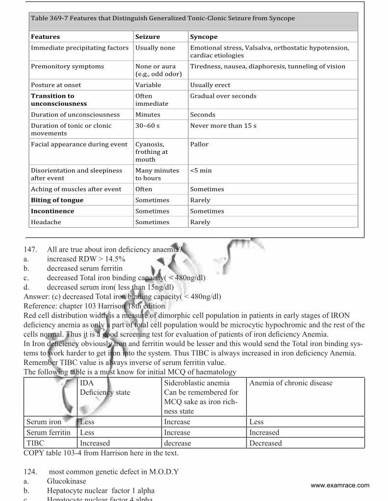
147. All are true about iron deficiency anaemia?a. increased RDW > 14.5%b. decreased serum ferritin c. decreased Total iron binding capacity( < 480ng/dl)d. decreased serum iron( less than 15ng/dl)Answer: (c) decreased Total iron binding capacity( < 480ng/dl)Reference: chapter 103 Harrison 18th editionRed cell distribution width is a measure of dimorphic cell population in patients in early stages of IRON deficiency anemia as only a part of total cell population would be microcytic hypochromic and the rest of the cells normal. Thus it is a good screening test for evaluation of patients of iron deficiency Anemia. In Iron deficiency obviously iron and ferritin would be lesser and this would send the Total iron binding sys-tems to work harder to get iron into the system. Thus TIBC is always increased in iron deficiency Anemia. Remember TIBC value is always inverse of serum ferritin value. The following table is a must know for initial MCQ of haematology
IDADeficiency state
Sideroblastic anemiaCan be remembered for MCQ sake as iron rich-ness state
Anemia of chronic disease
Serum iron Less Increase LessSerum ferritin Less Increase IncreasedTIBC Increased decrease Decreased
COPY table 103-4 from Harrison here in the text.
124. most common genetic defect in M.O.D.Ya. Glucokinaseb. Hepatocyte nuclear factor 1 alphac Hepatocyte nuclear factor 4 alpha
Table 369-7 Features that Distinguish Generalized Tonic-Clonic Seizure from Syncope
Features Seizure Syncope
Immediate precipitating factors Usually none Emotional stress, Valsalva, orthostatic hypotension, cardiac etiologies
Premonitory symptoms None or aura (e.g., odd odor)
Tiredness, nausea, diaphoresis, tunneling of vision
Posture at onset Variable Usually erect
Transition to unconsciousness
Often immediate
Gradual over seconds
Duration of unconsciousness Minutes Seconds
Duration of tonic or clonic movements
30–60 s Never more than 15 s
Facial appearance during event Cyanosis, frothing at mouth
Pallor
Disorientation and sleepiness after event
Many minutes to hours
<5 min
Aching of muscles after event Often Sometimes
Biting of tongue Sometimes Rarely
Incontinence Sometimes Sometimes
Headache Sometimes Rarely
www.examrace.com
d. Insulin promoter 1 Answer: (b) Hepatocyte nuclear factor 1 alphaReference : CMDT page no 1194Maturity onset dibetes of the young (MODY)• This subgroup is a relatively rare monogenic disorder characterized by non – insulin dependent dia-betes with autosomal dominant inheritance and an age at onset of 25 yrs or younger. • Patients are nonobese, and their hyperglycemia is due to impaired glucose induced secretion of insu-lin. Six types of mody have been described. Except for MODY 2 , in which a glucokinase gene is defective, all other types involve mutations of a nuclear transcription factor that regulates islet gene expression.• The enzyme glucokinase is a rate limiting step in glycolysis and determines the rate of adenosine triphosphate (ATP) production from glucose and the insulin secretory response in the beta cell. MODY 2, due to glucokinase mutations, is usually quite mild, associated with only slight fasting hyperglycemia and few if any microvascular diabetic complications. It generally responds well to hygienic measures or low doses of oral hypoglycemic agents. • MODY 3, due to mutations is hepatic nuclear factor 1 α is the most common form, accounting for two birds of all MODY cases. The clinical course is of progressive beta cell failure and need for insulin therapy. Mutations in both alleles of glucokinase present with more severe neonatal diabetes. Mutation in one allele of the pancreatic duodenal homeobox 1 (PDX1) causes diabetes usually at a later age (~ 35 years) than other forms of MODY; mutations in both alleles of PDX1 causes pancreatic agenesis.
125. Insulin resistance in liver disease is due to a. Steatosisb. Hepatocyte damagec. Decreased release of insulind. Decreased release of C – peptideAnswer is B: Hepatocyte damage (this is a new concept HEPATOGENOUS DIABETES)Reference: http://www.ncbi.nlm.nih.gov/pmc/articles/PMC3124882/• Increased insulin resistance is frequently associated with chronic liver disease and is a pathophysi-ological feature of hepatogenous diabetes. Distinctive factors including hepatic parenchymal cell damage, portal-systemic shunting and hepatitis C virus are responsible for the development of hepatogenous insulin resistance/diabetes• Since blood glucose is delivered to the liver through the portal vein, hyperinsulinemia in patients with liver cirrhosis may be secondary to either hepatic parenchymal cell damage or to portal-systemic shunt-ing .The rate at which insulin is degraded in the liver is reduced in patients with liver cirrhosis. Moreover, despite peripheral hyperinsulinemia, insulin levels in the portal and hepatic veins are decreased in cirrhotic patients with portal systemic shunting• Hepatogenous DM is less often associated with a positive family history, retinopathy and cardiovas-cular complications. In fact, major causes of death in cirrhotic patients with DM relate to liver disease or its complications, such as chronic liver failure, hepatocellular carcinoma (HCC) and gastrointestinal hemor-rhage. Therefore, the management of DM in patients with liver cirrhosis should aim to reduce such hepatic complications and to improve prognosis. Because the incidence of HCC has been well demonstrated to relate to DM. Therefore a major target in the management of DM should be to reduce the incidence of HCC in patients with liver cirrhosis.• Around 30% to 60% of cirrhotic patients suffer from this metabolic disorder. Insulin resistance in muscular, hepatic and adipose tissues as well as hyperinsulinemia, seem to be pathophysiologic bases for HD. An impaired response of the islet ss-cells of the pancreas and the hepatic insulin resistance are also con-tributing factors. Diabetes develops when defective oxidative and nonoxidative muscle glucose metabolism develops. Non-alcoholic fatty liver disease (NAFLD), alcoholic cirrhosis, chronic hepatitis C (CHC), and hemochromatosis are more frequently associated with HD. HD in early cirrhosis stages may be sub clinical. Only insulin resistance and glucose intolerance may be observed. As liver disease advances, diabetes be-comes clinically manifest, therefore HD may be considered as a marker for liver function deteriorationwww.examrace.com

126. Not included in armamentarium of tests for malabsorbtion syndromea. D- Xyloseb. 14C Triolein breath testc. 13 C Trioctanoin breath testd. 13 C triclosan breath testAnswer is (d) 13 C triclosan breath testReference: http://www.ncbi.nlm.nih.gov/pmc/articles/PMC1766642/pdf/v043p00S13.pdfD- xylose test Sugar malabsorbtion and tests jejunal damage or proximal enteropa-
thy14 C triolein test Fat malabsorbtion picked up by radiocarbon being tagged to the
triglycerides13 C trioactanoin Fat malabsorbtion due to pancreatic exocrine insufficiency13 C triclosan breath test Triclosan is an antiseptic and appears the best answer from the given
data.SeHCAT is the usual name for 23-seleno-25-homo-tauro-cholic acid (selenium homocholic acid taurine or tauroselcholic acid.
It is used in a clinical test to diagnose bile acid malabsorption
Hydrogen breath test For sugar malabsorbtion as unabsorbed sugar is degraded by bacteria.13C acetate breath tests For stomach emptyingUrea breath test For H. Pylori that can split urea to CO2 which is produced in breath
due the plaque build in teeth of h.Pylori can lead to erroneus results
D-xylose absorption test Xylose is a pentose that is passively absorbed by the jejunal mucosa. A standard dose of 5 g or, alternatively, 14.5 g/m2 (maximum dose of 25 g) of D-xylose is orally administered as a 10% solution in water. The test result is positive in the following instances: • Children who weigh less than 30 kg - Serum level at 1 hour after ingestion is less than 25 mg/dL.• Children who weigh more than 30 kg - The 5-hour urinary excretion is less than 15%.• A positive test result suggests malabsorption due to proximal small bowel mucosal lesion (enteropa-thy).• Beware of false-positive results (eg, from delayed gastric emptying, small bowel bacterial over-growth, accelerated transit time). The 14C-triolein breath test is used to investigate the absorption of fats from the small bowel. In principle, a 14C-labelled triglyceride is ingested, digested, absorbed and metabolised, releasing 14C-labelled carbon dioxide.The test may be performed in a day ward and requires laboratory facilities to detect the beta-emitting 14C.The test is unreliable in the following circumstances:• diabetes mellitus• gross obesity• thyroid disease• chronic respiratory insufficiency151. Not a major criteria in acute rheumatic fever- a. Polyarthralgiab. subcutaneous nodules c. choread. carditisAnswer: polyarthralagiaReference: chapter 332, Harrison textbook
www.examrace.com
Major manifestations CarditisPolyarthritisChoreaErythema marginatumSubcutaneous nodules
Minor manifestations Clinical: fever, polyarthralgiaLaboratory: elevated erythrocyte sedimentation rate or leukocyte countElectrocardiogram: prolonged P-R interval
Supporting evidence of a preceding streptococcal infection within the last 45 days
Elevated or rising anti-streptolysin O or other streptococcal antibody, orA positive throat culture, orRapid antigen test for group A streptococcus, orRecent scarlet fever
152. Most common slowly growing vascular tumor of spinal cord , cerebellum and brain is :a. Hemagioblastomab. Pilocytic astrocytomac. Meningiomad. MedulloblastomaAnswer is A ; HemangioblastomaReferences : Harrison 18th chapter 343 / http://emedicine.medscape.com/article/250670-overview#a0112The clinical presentation of hemangioblastomas usually depends on the anatomical location and growth pat-terns. 1. Cerebellar lesions may present with signs of cerebellar dysfunction, such as ataxia and discoordina-tion, or with symptoms of increased intracranial pressure due to associated hydrocephalus. In general, intrac-ranial hemangioblastomas present with a long history of minor neurological symptoms that, in most cases, are followed by a sudden exacerbation, which may necessitate immediate neurosurgical intervention. 2. Patients with spinal cord lesions most frequently present with pain, followed by signs of segmental and long-track dysfunction due to progressive compression of the spinal cord. 3. Patients with VHL disease may present with ocular or systemic symptoms due to involvement of other organs and systems.4. The polycythemia that may develop in some patients with hemangio-blastomas usually 5. is clinically asymptomatic
153. A patient is hepatitis HBsAG and HBeAg positive. Which of the following is true about this patienta. Acute hepatitis Bb. Hepatitis E infectionc. Chronic hepatitis B d. Co infection of HBV with HDVAnswer is (a) Acute hepatitis BReference : table 304-5 harrison ,chapter 304 .
www.examrace.com
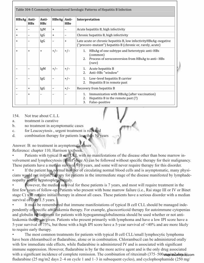
Table 304-5 Commonly Encountered Serologic Patterns of Hepatitis B Infection
HBsAg Anti-HBs
Anti-HBc
HBeAg Anti-HBe
Interpretation
+ – IgM + – Acute hepatitis B, high infectivity
+ – IgG + – Chronic hepatitis B, high infectivity
+ – IgG – + Late acute or chronic hepatitis B, low infectivityHBeAg–negative ("precore–mutant") hepatitis B (chronic or, rarely, acute)
+ + + +/– +/– 1. HBsAg of one subtype and heterotypic anti–HBs (common)
2. Process of seroconversion from HBsAg to anti– HBs (rare)
– – IgM +/– +/– 1. Acute hepatitis B 2. Anti–HBc "window"
– – IgG – +/– 1. Low–level hepatitis B carrier 2. Hepatitis B in remote past
– + IgG – +/– Recovery from hepatitis B
– + – – – 1. Immunization with HBsAg (after vaccination) 2. Hepatitis B in the remote past (?) 3. False–positive
154. Not true about C.L.La. treatment is curativeb. no treatment in asymptomatic casesc. for Leucocytosis , urgent treatment is requiredd. combination therapy for patients less than 50 years
Answer: B: no treatment in asymptomatic casesReference: chapter 110, Harrison textbook.• Patients with typical B cell CLL with no manifestations of the disease other than bone marrow in-volvement and lymphocytosis (Binet stage A) can be followed without specific therapy for their malignancy. These patients have a median survival >10 years, and some will never require therapy for this disorder. • If the patient has normal number of circulating normal blood cells and is asymptomatic, many physi-cians would not initiate therapy for patients in the intermediate stage of the disease manifested by lymphade-nopathy and/or hepatosplenomegaly.• However, the median survival for these patients is 7 years, and most will require treatment in the first few years of follow-up. Patients who present with bone marrow failure (i.e., Rai stage III or IV or Binet stage C) will require initial therapy in almost all cases. These patients have a serious disorder with a median survival of only 1.5 years. • It must be remembered that immune manifestations of typical B cell CLL should be managed inde-pendently of specific antileukemia therapy. For example, glucocorticoid therapy for autoimmune cytopenias and globulin replacement for patients with hypogammaglobulinemia should be used whether or not anti-leukemia therapy is given. Patients who present primarily with lymphoma and have a low IPI score have a 5-year survival of 75%, but those with a high IPI score have a 5-year survival of <40% and are more likely to require early therapy.• The most common treatments for patients with typical B cell CLL/small lymphocytic lymphoma have been chlorambucil or fludarabine, alone or in combination. Chlorambucil can be administered orally with few immediate side effects, while fludarabine is administered IV and is associated with significant immune suppression. However, fludarabine is by far the more active agent and is the only drug associated with a significant incidence of complete remission. The combination of rituximab (375–500 mg/m2 day 1), fludarabine (25 mg/m2 days 2–4 on cycle 1 and 1–3 in subsequent cycles), and cyclophosphamide (250 mg/
www.examrace.com
m2 with fludarabine) achieves complete responses in 69% of patients, and those responses are associated with molecular remissions in half of the cases.155. Bell’s palsy not correct is :a. Steroid is mandatoryb. Unilateral facial weaknessc. Urgent surgical decompressiond. Herpes virus is not the causeAnswer: C: urgent surgical decompressionReference: Harrison chapter 376. Bells palsy: Incidence about 1 in 60 persons in a lifetime. Causes: • Herpes simplex virus (HSV) type 1 DNA was frequently detected in endo-neural fluid and posterior auricular muscle, suggesting that a reactivation of this virus in the geniculate ganglion may be responsible for most cases. • Reactivation of varicella zoster virus is associated with Bell’s palsy in up to one-third of cases, and may represent the second most frequent cause. A variety of other viruses have also been implicated less commonly. • An increased incidence of Bell’s palsy was also reported among recipients of inactivated intranasal influenza vaccine, and it was hypothesized that this could have resulted from the Escherichia coli entero-toxin used as adjuvant or to reactivation of latent virus.Clinical Manifestations: 1. Pain behind the ear may precede the paralysis for a day or two .2. The onset of Bell’s palsy is fairly abrupt, maximal weakness being attained by 48 h as a general rule.3. Taste sensation may be lost unilaterally, and hyperacusis may be present. In some cases there is mild cerebrospinal fluid lymphocytosis. MRI may reveal swelling and uniform enhancement of the geniculate ganglion and facial nerve and, in some cases, entrapment of the swollen nerve in the temporal bone. Approximately 80% of patients recover within a few weeks or months. Electromyography may be of some prognostic value; evidence of denervation after 10 days indicates there has been axonal degeneration, that there will be a long delay (3 months as a rule) before regeneration occurs, and that it may be incomplete. The presence of incomplete paralysis in the first week is the most favorable prognostic sign.Differential Diagnosis• Lyme disease can cause unilateral or bilateral involvement• The Ramsay Hunt syndrome, caused by reactivation of herpes zoster in the geniculate ganglion, con-sists of a severe facial palsy associated with a vesicular eruption in the external auditory canal.• Sarcoidosis and in Guillain-Barré syndrome • Leprosy frequently involves the facial nerve, and facial neuropathy may also occur in diabetes mel-litus, connective tissue diseases including Sjögren’s syndrome, and amyloidosis. • Melkersson-Rosenthal syndrome consists of recurrent facial paralysis; recurrent—and eventually permanent—facial (particularly labial) edema; and, less constantly, plication of the tongue. • Acoustic neuromas frequently involve the facial nerve by local compression. Infarcts, demyelinat-ing lesions of multiple sclerosis, and tumors are the common pontine lesions that interrupt the facial nerve fibers;Treatment:1. Use of paper tape to depress the upper eyelid during sleep and prevent corneal drying, and massage of the weakened muscles. 2. A course of glucocorticoids, given as prednisone 60–80 mg daily during the first 5 days and then tapered over the next 5 days, modestly shortens the recovery period and improves the functional outcome. 3. Although two large recently published randomized trials found no added benefit of antiviral agents valacyclovir (1000 mg daily for 5–7 days) or acyclovir (400 mg five times daily for 10 days) compared to glucocorticoids alone, the overall weight of evidence suggests that the combination therapy with prednisone plus valacyclovir may be marginally better than prednisone alone, especially in patients with severe clinical presentations
www.examrace.com
4. Surgery to decompress the facial nerve is controversial. Patients with a poor prognosis, identified by facial nerve testing or persistent paralysis, appear to benefit the most from surgical intervention. Surgery may be considered in patients with complete Bell palsy that has not responded to medical therapy and with greater than 90% axonal degeneration, as shown on facial nerve EMG within 3 weeks of the onset of paraly-sis. The problem must be localized with MRI. The surgeon can then decide if the maxillary segment should be decompressed externally or if the labyrinthine segment and geniculate ganglion should be decompressed with a middle fossa craniotomy. A study in patients with greater than 90% degeneration demonstrated supe-rior results in the cohort that underwent middle fossa decompression, compared with the cohort that chose not to pursue surgical decompression. In the surgical group, 91% of cases exhibited a postoperative House-Brackmann grade of I or II. In the nonsurgical group, 58% of the patients had a poor result, with a House-Brackmann grade of III or IV at 7 months. The best surgical results were obtained when the procedure was done within 14 days after the onset of paralysis
156. 48 year old women with history of seizures has presented with gross haematuria and left flank pain. Abdominal CT scan reveals-left perinephric hematoma with 3cm angiomyolipoma along with multiple right renal angiomyolipomas measuring from 1.5-6.5 cm. The most likely diagnosis is : a. Tuberous sclerosisb. Von Hippel Landau syndromec. Familial angiolipomatosisd. ADPKD
Answer is A : tuberous sclerosisReferences: chapter 596.2 NELSON and http://emedicine.medscape.com/article/376848-overview#a20
Tuberous sclerosis Seizures/ behaviour problems with renal angio-mylipomas and they can bleed leading to the peri-nephric hematoma formation as mentioned in the question
Von Hippel Landau Von Hippel–Lindau (VHL) disease is a rare, auto-somal dominant genetic condition that predisposes individuals to benign and malignant tumours. The most common tumours found in VHL are central nervous system and retinal hemangioblastomas, clear cell renal carcinomas, pheochromocytomas, pancreatic neuroendocrine tumours, pancreatic cysts, endolymphatic sac tumors and epididymal papillary cystadenomas. VHL results from a muta-tion in the von Hippel–Lindau tumor suppressor gene on chromosome 3p.
Familial angiolipomatosis It’s a important a differential diagnosis of Neurofi-bromatosis 1. The patients have seizures and le-sion on skin which contain fat but may be wrongly ascribed to neurofibromas
Autosomal dominant polycystic kidney Presents at 30-45 yrs of age with HTN, hematuria and bilaterally enlarged kidneys showing multiple large cysts. CNS manifestation is berry aneurysm and hence is ruled out
MORE DETAILS ABOUT TUBEROUS SCLEROSIS FROM RADIOLOGICAL PERSPECTIVE
1. The most common neurologic manifestations of Tuberous Sclerosis consist of seizures, cognitive impairment, and behavioral abnormalities including autism. The characteristic brain lesion is a cortical tuber. Tubers are located in the convolutions of the cerebral hemispheres and are also present in the subependymal
www.examrace.com
region, where they undergo calcification and project into the ventricular cavity, producing a candle-dripping appearance. Generally, the greater the number of tubers, the more neurologically impaired is the patient. 2. TS may present during infancy with infantile spasms and a hypsarrhythmic EEG pattern. 3. The seizures may be difficult to control and, at a later age, they may develop into myoclonic epilep-sy, infantile spasms associated with TS are treated with vigabatrin (rather than adrenocorticotropic hormone) with good results.4. Angiomyolipomas are usually well-marginated, cortical heterogeneous tumors with predominantly fatty attenuation; rarely, higher attenuation is seen in patients who have tumors with minimal fat content. The average attenuation depends on the relative proportions of fat and other soft tissue in the angiomyolipo-ma. In small masses, fat may be averaged out with region-of-interest (ROI) circles, and pixel maps may be useful. Attenuations of less than –20 Hounsfield units (HU) are widely accepted as confirming the presence of fat; this finding virtually confirms the diagnosis of angiomyolipoma
5. Histopathology of angiomyolipomao No true capsule o Commonly bleed o Tumor composed of fat, smooth muscle, aggregates of thick-walled blood vessels
6. Types of angiomyolipomao Isolated angiomyolipoma is most common (80%) 1. Usually solitary 2. Unilateral (80% on right side) 3. Not associated with tuberous sclerosis 4. Mean age of incidence: 40s 5. Much more common in females o Angiomyolipoma associated with tuberous sclerosis (in 20%) 1. Angiomyolipoma occurs in 80% of patients with tuberous sclerosis • Commonly large • Usually bilateral • Usually multiple 2. May be only evidence of tuberous sclerosis 3. Mean age of incidence: teens 4. Equal incidence in males and females
7. Signs and symptoms o Small lesions are asymptomatic (60%) o Acute flank / abdominal pain (due to hemorrhage) most common presenting symptom o Hematuria (40%) o Palpable mass (47%) o Shock (due to massive retroperitoneal hemorrhage) 8. Imaging findings o Mostly small lesions <5 cm in diameter o Many have a large exophytic component (25%) o Calcifications not common (6%) o Plain film findings 1. Mass of fat lucency is lesion is large enough o CT findings 1. Well-marginated, cortical-based, heterogeneous tumor predominantly of fat density (<-20 HU) 2. Variable enhancement (smooth muscle, vessels)
157. Most effective management in medically intractable seizures? a. Ketotic diet
www.examrace.com
b. Vagus nerve stimulationc. Deep brain stimulationnd. SurgeryAnswer: D : surgeryReference:
Ketotic diet Useful in Pediatric seizures like rett syndrome, Lennox Gastuat syndrome. It entails consumption of 3-4 grams of fat per gram of carbohydrates. This probably works by reducing the metabo-lism of the epileptogenic focus.
Vagus nerve stimulation Vagal nerve stimulation (VNS) is a palliative device approved to treat medically refractory focal-onset epilepsy in adults. Some studies demonstrate its efficacy in focal-onset seizures and in a small number of patients with primary generalized epilepsy. Randomized studies showed modest efficacy at 3 months, but postmarketing experience shows delayed improve-ment in another group of patients
Deep brain stimulation ( emedicine) The NeuroPace RNS System, a device that is implanted into the cranium, senses and records electrocorticographic patterns and delivers short trains of current pulses to interrupt ictal dis-charges in the brain. The Neurological Devices panel of the US Food and Drug Administration (FDA) has concluded that this device is safe and effective in patients with partial-onset epi-lepsy in whom other antiepileptic treatment approaches have failed and that the benefits outweigh the risks
Surgery ( Harrison ) About 70% of patients treated with temporal lobectomy will become seizure free, and another 15–25% will have at least a 90% reduction in seizure frequency. Marked improvement is also usually seen in patients treated with hemispherectomy for catastrophic seizure disorders due to large hemispheric abnor-malities. Postoperatively, patients generally need to remain on antiepileptic drug therapy, but the marked reduction of seizures following resective surgery can have a very beneficial effect on quality of life.
Surgery ( Harrison ) About 70% of patients treated with temporal lobectomy will become seizure free, and another 15–25% will have at least a 90% reduction in seizure frequency. Marked improvement is also usually seen in patients treated with hemispherectomy for catastrophic seizure disorders due to large hemi-spheric abnormalities. Postoperatively, patients generally need to remain on antiepileptic drug therapy, but the marked reduction of seizures following resective surgery can have a very beneficial effect on quality of life.
• The most common surgical procedure for patients with temporal lobe epilepsy involves resection of the anteromedial temporal lobe (temporal lobectomy) or a more limited removal of the underlying hip-pocampus and amygdala (amygdalohippocampectomy). • Focal seizures arising from extratemporal regions may be abolished by a focal neocortical resection with precise removal of an identified lesion (lesionectomy). When the cortical region cannot be removed, multiple subpial transection, which disrupts intracortical connections, is sometimes used to prevent seizure spread. Hemispherectomy or multilobar resection is useful for some patients with severe seizures due to hemispheric abnormalities such as hemimegalencephaly or other dysplastic abnormalities, and corpus cal-losotomy has been shown to be effective for disabling tonic or atonic seizures, usually when they are part of a mixed-seizure syndrome (e.g., Lennox-Gastaut syndrome).• Presurgical evaluation is designed to identify the functional and structural basis of the patient’s sei-
www.examrace.com
zure disorder. Inpatient video-EEG monitoring is used to define the anatomic location of the seizure focus and to correlate the abnormal electrophysiologic activity with behavioral manifestations of the seizure. Routine scalp or scalp-sphenoidal recordings are usually sufficient for localization, and advances in neu-roimaging have made the use of invasive electrophysiologic monitoring such as implanted depth electrodes or subdural electrodes less common. A high-resolution MRI scan is routinely used to identify structural le-sions, and this is sometimes augmented with MEG. Functional imaging studies such as SPECT and PET are adjunctive tests that may help verify the localization of an apparent epileptogenic region. Once the presumed location of the seizure onset is identified, additional studies, including neuropsychological testing and the intracarotid amobarbital test (Wada test) may be used to assess language and memory localization and to de-termine the possible functional consequences of surgical removal of the epileptogenic region. In some cases, the exact extent of the resection to be undertaken is determined by performing cortical mapping at the time of the surgical procedure, allowing for a tailored resection. This involves electrocorticographic recordings made with electrodes on the surface of the brain to identify the extent of epileptiform disturbances. If the region to be resected is within or near brain regions suspected of having sensorimotor or language function, electrical cortical stimulation mapping is performed on the awake patient to determine the function of corti-cal regions in question in order to avoid resection of so-called eloquent cortex and thereby minimize postsur-gical deficits.• Advances in presurgical evaluation and microsurgical techniques have led to a steady increase in the success of epilepsy surgery. Clinically significant complications of surgery are <5%, and the use of function-al mapping procedures has markedly reduced the neurologic sequelae due to removal or sectioning of brain tissue.
158. Alzhiemers lobes affected?a. Frontal and temporal lobeb. Temporal and parietal lobec. Parietal and occipital lobed. Parietal and frontal lobeAnswer is (b) Temporal and parietal lobeReference: Harrison 18th edition ,chapter 371 dementiaAD most often presents with an insidious onset of memory loss followed by a slowly progressive dementia over several years. Pathologically, atrophy is distributed throughout the medial temporal lobes, as well as lateral and medial parietal lobes and lateral frontal cortex.The main purpose of imaging is to exclude other disorders, such as primary and secondary neoplasms, vascular dementia, diffuse white matter disease, and NPH; it also helps to distinguish AD from other degen-erative disorders with distinctive imaging patterns such as FTD or CJD. Functional imaging studies in AD reveal hypoperfusion or hypometabolism in the posterior temporal-parietal cortex.
159. Which of the following statements is correct about raised ESR in tuberculosis? a. Its increased due to large RBCb. Its increased due to increase in immunoglobulinsc. It is used to determine response for treatmentd. It is an incidental finding.Answer: B: It’s increased due to increase in immunoglobulinsReference:1. Wintrobe hematology 11th edition page 602. http://emedicine.medscape.com/article/2085201-overview#a30
The erythrocyte sedimentation rate (ESR) is a common but nonspecific test that is often used as an indicator of active disease. Significant specific indications for ESR testing include the following:• Diagnosis and monitoring of giant cell arteritis• Diagnosis and monitoring of polymyalgia rheumatica
www.examrace.com
• Monitoring of rheumatoid arthritis• Monitoring of systemic lupus Erythematosus
ESR reflects the tendency of red blood cells to settle more rapidly in the face of some disease states, usually because of increases in plasma fibrinogen, immunoglobulins, and other acute-phase reaction proteins. In ad-dition, changes in red cell shape or numbers may affect the ESR.
1. Sickle cells and polycythemia and spherocytosis tend to decrease the ESR, whereas anemia may increase it. 2. Factors that increase ESR• Old age• Female• Pregnancy• Anemia• Red blood cell abnormalities• • Macrocytosis• Technical factors• • Dilutional problem• • Increased temperature of specimen• • Tilted ESR tube• Elevated fibrinogen level• • Infection• • Inflammation• • Malignancy
3. ESR also increases with age in otherwise healthy people (although it tends to fall in adults older than the age of 75 and tends to be higher in women.
4. People with liver diseases, carcinomas, or other serious diseases may have a normal to low ESR because of an inability to produce the acute-phase proteins.
The ESR is measured by the Westergren or Wintrobe method or by a modification ofthese tests. Both are measured in millimeters per hour, but the normal values foreach method vary because of differences in tube length and shape. Both methods requirecorrection for patient anemia. Several technical variations to the method of ESRdetermination have been introduced, including micromethods, sedimentation at a45-degree angle, and the zeta sedimentation rate. The zeta sedimentation rate measureserythrocyte packing in four 45-second cycles of dispersion and compaction in capillarytubes. This requires a special instrument, the ZetafugeThe rate at which red blood cells settle out when anti-coagulated whole blood is allowed to stand is known as the erythrocyte sedimentation rate.
The ESR is affected by the concentrations of immunoglobulins and acute phase proteins (fibrinogen, C-reac-tive protein, alpha-1 antitrypsin, haptoglobin), and is a sensitive, but nonspecific, indicator of inflammation and tissue damageConditions that may be associated with a highly elevated ESR (>100 mm/hr) include the following: • Hypersensitivity vasculitis• Giant cell arteritis• Waldenström macroglobulinemia• Polymyalgia rheumatica• Metastatic cancer• Chronic infection• Hyperfibrogenemia www.examrace.com
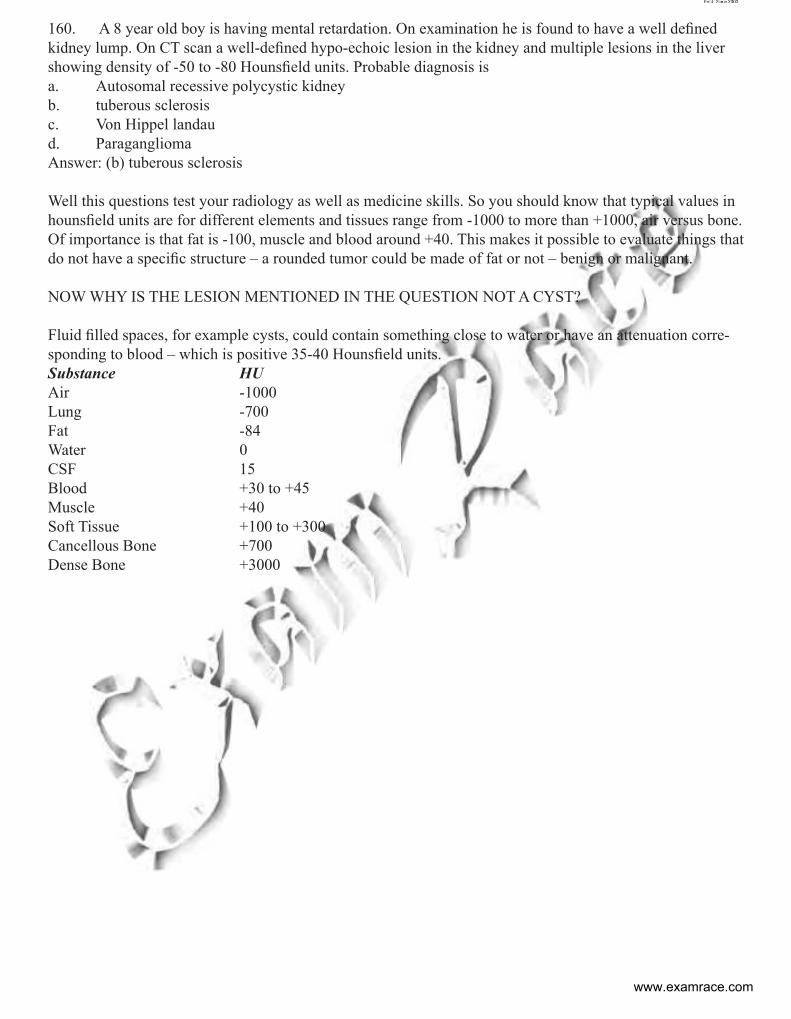
160. A 8 year old boy is having mental retardation. On examination he is found to have a well defined kidney lump. On CT scan a well-defined hypo-echoic lesion in the kidney and multiple lesions in the liver showing density of -50 to -80 Hounsfield units. Probable diagnosis isa. Autosomal recessive polycystic kidney b. tuberous sclerosisc. Von Hippel landaud. ParagangliomaAnswer: (b) tuberous sclerosis
Well this questions test your radiology as well as medicine skills. So you should know that typical values in hounsfield units are for different elements and tissues range from -1000 to more than +1000, air versus bone. Of importance is that fat is -100, muscle and blood around +40. This makes it possible to evaluate things that do not have a specific structure – a rounded tumor could be made of fat or not – benign or malignant.
NOW WHY IS THE LESION MENTIONED IN THE QUESTION NOT A CYST?
Fluid filled spaces, for example cysts, could contain something close to water or have an attenuation corre-sponding to blood – which is positive 35-40 Hounsfield units.Substance HUAir -1000Lung -700Fat -84Water 0CSF 15Blood +30 to +45Muscle +40Soft Tissue +100 to +300Cancellous Bone +700Dense Bone +3000
www.examrace.com

Now let us evaluate the choices provided individually.ARPKD • Mental retardation or seizures is not seen.
• As mentioned above the cysts have positive hounsfield units where as is the ques-tion the units are negative which are indicative of fat /angiomylipoma. • Hence ARPKD is ruled out on basis of 2 points mentioned above• Patients in ARPKD present prenatally with massively enlarged kidneys and oli-gohydramnios. In infants, Potter facies with low-set, flattened ears; short, snubbed nose; deep eye creases; and micrognathia, all secondary to oligohydramnios, can be found.. • An abdominal mass may manifest after the newborn period because of renal masses or hepato-splenomegaly. Impaired renal function is present in 70-80% of infants. Renal cysts in children may be an incidental finding. • Hepatic involvement is present in all children with autosomal recessive polycystic kidney disease but may not manifest in neonates (50-60%).
Tuberous sclerosis • Seizures or mental retardation is present dependant on number of tubers or subependymal nodules seen in these patients.• AMLs are benign mesenchymal tumors, composed of blood vessels, smooth muscle, and mature adipose tis-sue, that arise primarily in the kidneys. • AMLs may also occur, however, in extra-renal sites, such as the liver ( incidence is 25% as per emedicine 2013 update), spleen, abdominal wall, retroperitoneum, uterus, oral cavity, penis, spermatic cord, fallopian tube, va-gina, skin, and lung. Approximately 80% of renal AMLs occur sporadically, and 20% occur in patients with TSC. • AMLs may also occur in association with other phacomytoses (von Recklinghausen disease, von Hippel-Lin-dau disease( IT HAS A ADULT PRESENTATION),Sturge-Weber syndrome), autosomal dominant polycystic disease, and pulmonary lymphangio-myomatosis
VHL • The primary cause of morbidity and mortality in von Hippel-Lindau disease, as well as the most serious se-quela of the condition, involves the malignant degeneration of renal cysts. Renal cysts are seldom clinically significant; however, in von Hippel-Lindau disease they have an appreciable rate of malignant transformation. • Consequently, renal cell carcinoma is the leading cause of death in patients with von Hippel-Lindau disease, with a prevalence of as high as 75% reported in one autopsy series. • LIVER INVOLVEMENT IN VHL IS HEMANGIOMA WHICH HAS POSITIVE HOUNSFIELD UNITS.• The mean age of diagnosis of renal cell carcinoma in VHL disease is 44 years, and 70%
Paraganglioma • Paraganglioma is rare neuroendocrine neoplasm that may develop at various body sites (including the head, neck, thorax and abdomen). • About 97% are benign and cured by surgical removal; the remaining 3% are malignant because they are able to produce distant metastases. “Paraganglioma” is now the most-widely accepted term for these lesions, that have been also described as: glomus tumor, chemodectoma, perithelioma, fibroangioma, and sympathetic nevi
161. pleural tap pierces all except :a. skinb. intercostal musclec. Endo-thoracic fasciad. pulmonary pleuraAnswer: (d) pulmonary pleuraReference: This question is probably testing English of doctors. Well we have common sense and hence can solve this one. Each lung is invested by an exceedingly delicate serous membrane, the pleura,which is arranged in the form of a closed invaginated sac. A portion of the serous membrane covers the surface of the lung and dips into the fissures between its lobes; it is called the pulmonary pleura (or visceral pleura).Endothoracic fascia is layer of loose connective tissue that separates the ribs and muscles from the underlying pleural.The following layers are pierced during thoracocenteis/pleural tapping:1. skin- superficial fascia2. Serratus anterior3. external intercostal4. internal intercostal5. innermost intercoastal6. parietal pleura
www.examrace.com
7. pleural cavity is reached and fluid is accessed
162. A middle aged hypertensive male develops sudden onset unconsciousness, with nuchal rigidity. Rest of the neurological examination is within normal limits.a. SAHb. Intra-parenchymal bleedc. Extra-dural hemorrhaged. Sub dural haemorrhage.Answer: (a) SAHReference: Harrison 18 th edition chapter 275 • At the moment of aneurysmal rupture with major SAH, the ICP suddenly rises. This may account for the sudden transient loss of consciousness that occurs in nearly half of patients. Sudden loss of conscious-ness may be preceded by a brief moment of excruciating headache, but most patients first complain of head-ache upon regaining consciousness. In 10% of cases, aneurysmal bleeding is severe enough to cause loss of consciousness for several days. In 45% of cases, severe headache associated with exertion is the presenting complaint. The patient often calls the headache “the worst headache of my life”; however, the most impor-tant characteristic is sudden onset. Occasionally, these ruptures may present as headache of only moderate intensity or as a change in the patient’s usual headache pattern. The headache is usually generalized, often with neck stiffness, and vomiting is common.• Although sudden headache in the absence of focal neurologic symptoms is the hallmark of aneurys-mal rupture, focal neurologic deficits may occur. Anterior communicating artery or MCA bifurcation aneu-rysms may rupture into the adjacent brain or subdural space and form a hematoma large enough to produce mass effect. The deficits that result can include hemiparesis, aphasia, and abulia.• Occasionally, prodromal symptoms suggest the location of a progressively enlarging unruptured aneurysm. A third cranial nerve palsy, particularly when associated with pupillary dilation, loss of ipsilateral (but retained contralateral) light reflex, and focal pain above or behind the eye, may occur with an expanding aneurysm at the junction of the posterior communicating artery and the internal carotid artery. A sixth nerve palsy may indicate an aneurysm in the cavernous sinus, and visual field defects can occur with an expanding supraclinoid carotid or anterior cerebral artery aneurysm. Occipital and posterior cervical pain may signal a posterior inferior cerebellar artery or anterior inferior cerebellar artery aneurysm • Pain in or behind the eye and in the low temple can occur with an expanding MCA aneurysm. Thun-derclap headache if written in question again favours diagnosis of SAH.
163. A 30 year old patient is having high grade fever with altered sensorium. On third day the patient develops seizures and has Nuchal rigidity. The CSF examination of the patient shows presence of 300 cells/cu.mm, protein 70mg%, glucose 60mg% (BLOOD GLUCOSE= 95mg%) with polymorph 65% and the rest being lymphocytes. The most probable diagnosis of the patient is :a. Herpetic encephalitisb. Tubercular meningitisc. Pyogenic meningitisd. Cerebral malaria.
Answer is A: herpetic encephalitis Reference: chapter 381 harrison 18 th edition
www.examrace.com
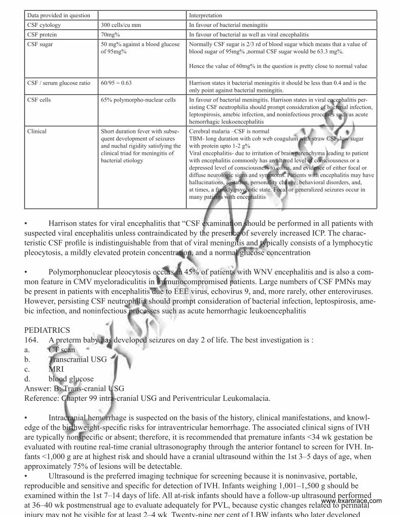
Data provided in question Interpretation CSF cytology 300 cells/cu mm In favour of bacterial meningitisCSF protein 70mg% In favour of bacterial as well as viral encephalitisCSF sugar 50 mg% against a blood glucose
of 95mg%Normally CSF sugar is 2/3 rd of blood sugar which means that a value of blood sugar of 95mg% ,normal CSF sugar would be 63.3 mg%.
Hence the value of 60mg% in the question is pretty close to normal value
CSF / serum glucose ratio 60/95 = 0.63 Harrison states it bacterial meningitis it should be less than 0.4 and is the only point against bacterial meningitis.
CSF cells 65% polymorpho-nuclear cells In favour of bacterial meningitis. Harrison states in viral encephalitis per-sisting CSF neutrophilia should prompt consideration of bacterial infection, leptospirosis, amebic infection, and noninfectious processes such as acute hemorrhagic leukoencephalitis
Clinical Short duration fever with subse-quent development of seizures and nuchal rigidity satisfying the clinical triad for meningitis of bacterial etiology
Cerebral malaria –CSF is normal TBM- long duration with cob web coagulum with straw CSF, low sugar with protein upto 1-2 g%Viral encephalitis- due to irritation of brain parenchyma leading to patient with encephalitis commonly has an altered level of consciousness or a depressed level of consciousness to coma, and evidence of either focal or diffuse neurologic signs and symptoms. Patients with encephalitis may have hallucinations, agitation, personality change, behavioral disorders, and, at times, a frankly psychotic state. Focal or generalized seizures occur in many patients with encephalitis
• Harrison states for viral encephalitis that “CSF examination should be performed in all patients with suspected viral encephalitis unless contraindicated by the presence of severely increased ICP. The charac-teristic CSF profile is indistinguishable from that of viral meningitis and typically consists of a lymphocytic pleocytosis, a mildly elevated protein concentration, and a normal glucose concentration
• Polymorphonuclear pleocytosis occurs in 45% of patients with WNV encephalitis and is also a com-mon feature in CMV myeloradiculitis in immunocompromised patients. Large numbers of CSF PMNs may be present in patients with encephalitis due to EEE virus, echovirus 9, and, more rarely, other enteroviruses. However, persisting CSF neutrophilia should prompt consideration of bacterial infection, leptospirosis, ame-bic infection, and noninfectious processes such as acute hemorrhagic leukoencephalitis
PEDIATRICS164. A preterm baby has developed seizures on day 2 of life. The best investigation is :a. CT scanb. Transcranial USGc. MRId. blood glucoseAnswer: B: Trans-cranial USGReference: Chapter 99 intra-cranial USG and Periventricular Leukomalacia.
• Intracranial hemorrhage is suspected on the basis of the history, clinical manifestations, and knowl-edge of the birthweight-specific risks for intraventricular hemorrhage. The associated clinical signs of IVH are typically nonspecific or absent; therefore, it is recommended that premature infants <34 wk gestation be evaluated with routine real-time cranial ultrasonography through the anterior fontanel to screen for IVH. In-fants <1,000 g are at highest risk and should have a cranial ultrasound within the 1st 3–5 days of age, when approximately 75% of lesions will be detectable.• Ultrasound is the preferred imaging technique for screening because it is noninvasive, portable, reproducible and sensitive and specific for detection of IVH. Infants weighing 1,001–1,500 g should be examined within the 1st 7–14 days of life. All at-risk infants should have a follow-up ultrasound performed at 36–40 wk postmenstrual age to evaluate adequately for PVL, because cystic changes related to perinatal injury may not be visible for at least 2–4 wk Twenty-nine per cent of LBW infants who later developed
www.examrace.com
cerebral palsy did not have radiographic evidence of PVL until after 28 days. Ultrasound examination also detects the precystic and cystic symmetric lesions of PVL and the asymmetric intraparenchymal echogenic lesions of cortical hemorrhagic infarction.• Furthermore, the delayed development of cortical atrophy, porencephaly, and the severity, progres-sion, or regression of posthemorrhagic hydrocephalus can be determined by serial ultrasonography.
165. a Preterm baby with PDA will have all except:a. Co2 washoutb. Pulmonary haemorrhagec. Necrotising enterocolitisd. Bounding pulsesAnswer : A carbon dioxide washoutReference: nelson pediatrics chapter 101.4Some neonates with RDS may have clinically significant shunting through a patent ductus arteriosus (PDA). Delayed closure of the PDA is associated with hypoxia, acidosis,increased pulmonary pressure secondary to vasoconstriction, systemic hypotension, immaturity, and local release of prostaglandins, which dilate the ductus. There is a relationship between early adrenal insufficiency, ductal patency, airway inflammation, and the development of BPD. Shunting through the PDA may initially be bidirectional or right to left. As RDS resolves, pulmonary vascular resistance decreases, and left-to-right shunting may occur and lead to left ven-tricular volume overload and pulmonary edema. Manifestations of PDA may include (1) apnea for unexplained reasons in an infant recovering from RDS; (2) a hyperdynamic precordium, bounding peripheral pulses, wide pulse pressure, and a continuous or systolic murmur with or without extension into diastole or an apical diastolic murmur, multiple clicks resem-bling the shaking of dice; (3) Carbon dioxide retention; (4) Increasing oxygen dependence; (5) X-ray evidence of cardiomegaly and increased pulmonary vascular markings; and (6) hepatomegaly. (7) Pulmonary hemorrhage can occur due to increased pressure in pulmonary circulation.(emedicine)The diagnosis is confirmed by echocardiographic visualization of a PDA with Doppler flow demonstrating left-to-right or bidirectional shunting. VLBW infants with PDA are at increased risk of more prolonged and more severe RDS, bronchopulmonary dysplasia, and death. Prophylactic “closure,” closure of the asymp-tomatic but clinically detected PDA, and closure of the symptomatic PDA are three strategies to manage a PDA. Interventions include fluid restriction, the use of diuretics, the use of cyclo-oxygenase inhibitors, and surgical closure
166. A preterm with 1.5 kg is given high flow oxygen, which of the following is most likely to develop in child:a. Retinopathy of prematurityb. Glaucomac. Retinal detachmentd. Brocho-pulmonary dysplasiaAnswer: A,retinopathy of prematurityReference: chapter 629 nelson.
• Retinopathy of prematurity ROP is a complex disease of the developing retinal vasculature in prema-ture infants. Exposure to the extrauterine environment including the necessarily high inspired oxygen con-centrations produces cellular damage, perhaps mediated by free radicals. Later in the course of the disease, peripheral hypoxia develops and vascular endothelial growth factors (VEGF) are produced in the nonvascu-larized retina. These growth factors stimulate abnormal vasculogenesis, and neovascularization may occur. Because of poor pulmonary function, a state of relative retinal hypoxia occurs. This causes upregulation of VEGF, which, in susceptible infants, can cause abnormal fibrovascular growth. This neovascularization may then lead to scarring and vision lossSystematic serial ophthalmologic examinations of infants at risk are
www.examrace.com
recommended. Guidelines vary but generally include infants weighing less than 1,500 g at birth and those born before 31 wk of gestational age. Infants born weighing more than 1,500 g who have an unstable clinical course and are thought to be at high risk should also be examined for ROP. The initial examination should be performed at 4–6 wk of chronological age or at 31–33 wk postconceptional age. ROP is diagnosed most often at 32–44 wk after conception.
• BPD is a disease primarily of infants <1,000 g born at less than 28 wk gestation, many of whom have little or no lung disease at birth, but develop progressive respiratory failure over the 1st few weeks of life. It causes development of oxygen dependency and is NOT DUE TO OXYGEN ADMINISTRATION.
• When compared with infants with classic BPD, most current infants with BPD do not have the prominent airway changes of squamous metaplasia and peribronchial fibrosis, severe alveolar septal fibrosis, or hypertensive vascular changesThe occurrence of BPD is inversely related to gestational age. Instead of showing improvement on the 3rd–4th day, consistent with the natural course of RDS, some infants develop an increased need for oxygen and ventilatory support. Respiratory distress persists or worsens and is char-acterized by hypoxia, hypercapnia, oxygen dependence, and, in severe cases, the development of right-sided heart failure. The chest roentgenogram may reveal pulmonary interstitial emphysema, wandering atelectasis with concomitant hyperinflation, and cyst formation. Four distinct pathologic stages of classic BPD have been identified, which are acute lung injury, exudative bronchiolitis, proliferative bronchiolitis, and oblitera-tive fibroproliferative bronchiolitis
167. A 7 year old child is having steroid DEPENDANT Nephrotic syndrome. His weight is 30 kg and eight of 106 cm. he is having truncal obesity with subcapsular bilateral cataracts. Best drug for this patient:a. Mycophenolateb. Levamisolec. Cyclophosphamided. AzathioprineAnswer is C : cyclophosphamideReference: nelson text book of paediatrics ,chapter 527Steroid-dependent patients, frequent relapsers, and steroid-resistant patients may be candidates for alterna-tive agents, particularly if the child suffers severe corticosteroid toxicity (cushingoid appearance, hyperten-sion, cataracts, and/or growth failure). Cyclophosphamide prolongs the duration of remission and reduces the number of relapses in children with frequently relapsing and steroid-dependent nephrotic syndrome. The potential side effects of the drug (neutropenia, disseminated varicella, hemorrhagic cystitis, alopecia, sterility, increased risk of future malignancy) should be carefully reviewed with the family before initiating treatment. The dose of cyclophosphamide is 2–3 mg/kg/24 hr given as a single oral dose, for a total duration of 8–12 wk. Alternate-day prednisone therapy is often continued during the course of cyclophosphamide administration. During cyclophosphamide therapy, the white blood cell count must be monitored weekly and the drug should be withheld if the count falls below 5,000/mm3.
168. A child with bigger limbs ( history suggestive of hemi-hypertrophy) and abdominal mass is sugges-tive of a. Nephroblastomab. Neuroblastomac. Rhabdomyosarcomad. Wilm tumorAnswer : D /A Reference: chapter 492 nelson txtbook• Hemihypertrophy ± Beckwith syndrome -Wilms tumor WT1 gene. • 25% develop tumor, mostly in first 5 yr of life .• Several syndromes and congenital abnormalities commonly are reported in patients with Wilms tu-
www.examrace.com

mor1. WAGR syndrome is a contiguous gene deletion syndrome that consists of Wilms tumor, aniridia, genitourinary abnormalities (cryptorchidism, streak ovaries, bicornate uterus, ambiguous genitalia), and mental retardation. Patients with this syndrome have a constitutional deletion of chromosome 11p13 where the Wilms tumor gene, WT1, and the aniridia gene, PAX6, are located.2 Denys-Drash syndrome is characterized by male pseudohermaphrodism, early-onset renal failure characterized by mesangial sclerosis, and an increased risk of Wilms tumor. Patients with this syndrome typically carry a missense mutation in the WT1 gene.3. Beckwith-Wiedemann syndrome is characterized by hemihypertrophy, macroglossia, and viscero-megaly, with a 3–5% risk of developing Wilms tumor. A variety of 11p15.5 abnormalities have been reported in patients with this syndrome, and it is postulated that a second Wilms tumor gene, WT2, is located in this region. Loss of imprinting of the insulin-like growth factor 2 gene, an epigenetic process, also is associated with Wilms tumor. Other syndromes or conditions with an increased risk of Wilms tumor include hemihy-pertrophy, sporadic aniridia, genitourinary anomalies, Pearlman syndrome, Sotos syndrome, neurofibromato-sis (von Recklinghausen disease), and von Willebrand disease. The genitourinary anomalies most commonly associated with Wilms tumor are hypoplasia, fusion and ectopia of the kidney, duplications of the collecting systems, hypospadias, and cryptorchidism.
169. A child is able to suckle and breathe simultaneously because of :a. small tongueb. high larygnxc. small phyranxd. small soft palateAnswer: high larynxReference:Babies on sucking elevate their relatively higher larynx that forms a milk proof seal with nasopharynx allowing them to continue suckle plus feed together. In mature larynx this would imply pausing between suckling and drinking
170. Which of the following is considered a developmental delay?a. Pincer grasp not developed by 9 monthsb. Moving up and downstairs by 21/2 yearsc. Not able to sit at 9 monthsd. Two word syllable at 1 ½ yearsAnswer is C : Not able to sit at 9 monthsReference: OP Ghai page no 33 table 2.5 and Nelson chapter 9 table 9.1 Milestone Normal age of appearancePincer grasp 9-10 months as per OP ghai page 28Moving up and downstairs 30 months as per nelson table 9.1 chapter 9Ability to sit without support 8 months and OP GHAI mentions in table 2.5 as not sitting without
support by 9-10 months as RED FLAG IN CHILD DEVELOPMENT.Two word syllable Syllable is a basic unit of speech like water word is composed up of
wa+ ter sound joined together. Children can speak a sentence com-posed of subject + verb at 24 months.As per nelson quote : 18months = 10 words (average); names pic-tures; identifies one or more parts of body
Syllable is a basic unit of speech like water word is composed up of wa+ ter sound joined together. Children can speak a sentence composed of subject + verb at 24 months.
The milestones referred in the questions have been italicized in the text below.TABLE 9 1 Emerging Patterns of Behavior from 1 to 5 Yr of Age
www.examrace.com
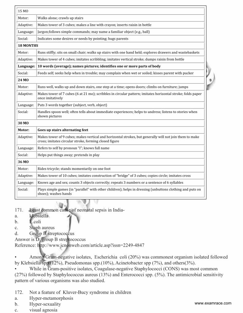
15 MO
Motor: Walks alone; crawls up stairs
Adaptive: Makes tower of 3 cubes; makes a line with crayon; inserts raisin in bottle
Language: Jargon;follows simple commands; may name a familiar object (e.g., ball)
Social: Indicates some desires or needs by pointing; hugs parents
18 MONTHS
Motor: Runs stiffly; sits on small chair; walks up stairs with one hand held; explores drawers and wastebaskets
Adaptive: Makes tower of 4 cubes; imitates scribbling; imitates vertical stroke; dumps raisin from bottle
Language: 10 words (average); names pictures; identifies one or more parts of body
Social: Feeds self; seeks help when in trouble; may complain when wet or soiled; kisses parent with pucker
24 MO
Motor: Runs well, walks up and down stairs, one step at a time; opens doors; climbs on furniture; jumps
Adaptive: Makes tower of 7 cubes (6 at 21 mo); scribbles in circular pattern; imitates horizontal stroke; folds paper once imitatively
Language: Puts 3 words together (subject, verb, object)
Social: Handles spoon well; often tells about immediate experiences; helps to undress; listens to stories when shown pictures
30 MO
Motor: Goes up stairs alternating feet
Adaptive: Makes tower of 9 cubes; makes vertical and horizontal strokes, but generally will not join them to make cross; imitates circular stroke, forming closed figure
Language: Refers to self by pronoun “I”; knows full name
Social: Helps put things away; pretends in play
36 MO
Motor: Rides tricycle; stands momentarily on one foot
Adaptive: Makes tower of 10 cubes; imitates construction of “bridge” of 3 cubes; copies circle; imitates cross
Language: Knows age and sex; counts 3 objects correctly; repeats 3 numbers or a sentence of 6 syllables
Social: Plays simple games (in “parallel” with other children); helps in dressing (unbuttons clothing and puts on shoes); washes hands
171. Least common cause of neonatal sepsis in India- a. klebsiellab. E.colic. Staph aureusd. Group B streptococcusAnswer is D: group B streptococcusReference: http://www.jcnonweb.com/article.asp?issn=2249-4847
• Among Gram-negative isolates, Escherichia coli (20%) was commonest organism isolated followed by Klebsiella spp.(12%), Pseudomonas spp.(10%), Acinetobacter spp (7%), and others(3%). • While in Gram-positive isolates, Coagulase-negative Staphylococci (CONS) was most common (27%) followed by Staphylococcus aureus (13%) and Enterococci spp. (5%). The antimicrobial sensitivity pattern of various organisms was also studied.
172. Not a feature of Kluver-Bucy syndrome in childrena. Hyper-metamorphosisb. Hyper-sexualityc. visual agnosia
www.examrace.com
d. SeizuresAnswer is C: Visual AgnosiaReference: API
Kluver bucy syndrome indicate the involvement of the medial temporal lobe regions and bilateral lesions of Ammon’s horn as a sine qua non for the production of the syndrome. Involvement of temporal lobes by hydrocephalus in tubercular meningitis and the inflammatory reaction of the brain to the antigen liberated by the degenerating cysticercal cyst may probably explain the clinical features observed in our patients with tuberculous meningitis and NCC. The patient with KBS in association with NCC probably suggests that in certain clinical settings this syndrome can be reversible.
Features of KBS 1. Self stimulation of genitals and hyper-orality) are common to children with severe learning difficul-ties arising from a variety of causes. 2. Temporal lobe epilepsy and improvement with temporal lobectomy. As many as 71% of the patients with temporal lobe epilepsy exhibit altered sexual behavior associated with ictal events. Inter-ictal hypo-sexuality occurs in 80% of the patients; 20% exhibit a variety of paraphilias. Perhaps episodes of seizures reflect transient bilateral dysfunction of the temporal lobe. Such temporary functional bilateral lobectomy could cause the same syndrome as an anatomical lobectomy and would be similar to the pathophysiological mechanism, which may account for our patient’s behavior. 3. Hypermetamorphosis is exaggerated response to all stimuli present in the visual field.
Carbamazepine and leuprolides have been found to decrease the sexual behavioral abnormality in some indi-viduals with KBS. Other medications such as haloperidol and anti-cholinergics may also be useful in treat-ing behavioral abnormalities associated with KBS
173. Child with croup, well hydrated, feeding well, consolable. T/t is a) Dexamethasoneb) Racemic epinephrinec) Antibioticsd) Nasal washing for influenza and RSVAns : A: DexamethasoneReference: chapter 382.1 nelson textbook• The effectiveness of oral corticosteroids in viral croup is well established. Corticosteroids decrease the edema in the laryngeal mucosa through their anti-inflammatory action. Oral steroids are beneficial, even in mild croup, as measured by reduced hospitalization, shorter duration of hospitalization, and reduced need for subsequent interventions such as epinephrine administration. Most studies that demonstrated the efficacy of oral dexamethasone used a single dose of 0.6 mg/kg; a dose as low as 0.15 mg/kg may be just as effec-tive. Intramuscular dexamethasone and nebulized budesonide have an equivalent clinical effect; oral dos-ing of dexamethasone is as effective as intramuscular administration. The only adverse effect described in the treatment of croup with corticosteroids was the development of Candida albicans laryngotracheitis in a patient who received dexamethasone, 1 mg/kg/24 hr, for 8 days. Corticosteroids should not be administered to children with varicella or tuberculosis (unless the patient is receiving appropriate antituberculosis therapy) because they worsen the clinical course.• Nebulized racemic epinephrine is an accepted treatment for moderate or severe croup. The mecha-nism of action is believed to be constriction of the precapillary arterioles through the β-adrenergic receptors, causing fluid resorption from the interstitial space and a decrease in the laryngeal mucosal edema. Tradition-ally, racemic epinephrine, a 1 : 1 mixture of the d- and l-isomers of epinephrine, has been administered. A dose of 0.25–0.75 mL of 2.25% racemic epinephrine in 3 mL of normal saline can be used as often as every 20 min. Racemic epinephrine was initially chosen over the more active and more readily available l-epi-nephrine to minimize anticipated cardiovascular side effects such as tachycardia and hypertension. There is
www.examrace.com
evidence that l-epinephrine (5 mL of 1 : 1,000 solution) is equally effective as racemic epinephrine and does not carry the risk of additional adverse effects. This information is both practical and important, because racemic epinephrine is not available outside the United States.• The indications for the administration of nebulized epinephrine include moderate to severe stridor at rest, the possible need for intubation, respiratory distress, and hypoxia. The duration of activity of racemic epinephrine is <2 hr. Therefore, observation is mandated. The symptoms of croup may reappear, but racemic epinephrine does not cause rebound worsening of the obstruction. Patients can be safely discharged home af-ter a 2–3 hr period of observation provided they have no stridor at rest; have normal air entry, normal color, and normal level of consciousness; and have received steroids (see later). Nebulized epinephrine should still be used cautiously in patients with tachycardia, heart conditions such as tetralogy of Fallot, or ventricular outlet obstruction because of possible side effects.• Antibiotics are not indicated in croup. A helium-oxygen mixture (Heliox) may be effective in chil-dren with severe croup who may need intubation. Children with croup should be hospitalized for any of the following: progressive stridor, severe stridor at rest, respiratory distress, hypoxia, cyanosis, depressed mental status, poor oral intake, or the need for reliable observation.
174. In a child with rickets with deformity, when should a decision to undertake corrective surgery be undertaken?a. When vitamin D levels return to normalb. when growth plate healing is seen radiographicallyc. when bone specific alkaline phosphatase is normald. when serum calcium is normal.
Answer: C: when bone specific alkaline phosphatise is normalReference: Nelson chapter 48 http://emedicine.medscape.com/article/985510• In nutritional rickets, the phosphorus level rises in 96 hours and radiographic healing is visible in 6-7 days. Neither happens with Familial Hypophosphatemic Rickets. If severe deformities have occurred, ortho-pedic correction may be required after healing.• Also lets see the status of serum chemistry values in rickets
Serum calcium This level is often within the reference range at the time of diagnosis, as a conse-quence of increased parathyroid hormone secretion. Hence it cannot be used for monitoring response
Serum phosphate The phosphorus level is invariably low for age, unless recent partial treatment or recent exposure to sunlight has occurred. It rises in 96 hours after treatment but may not rise in resistant rickets.
Serum vitamin D3 Although Calcidiol (25-hydroxy vitamin D) is low and parathyroid hormone is elevated, determining Calcidiol and parathyroid hormone levels is typically not necessary in order to establish a diagnosis. Also remember that values would be normal in resistant rickets and normalcy of values hence would not be indicative of response to treatment.
S.A.P Alkaline Phosphatase levels are uniformly elevated and indicate metabolic bone disease. Values returning to baseline indicate healing process.
• Treatment for rickets may be administered gradually over several months or in a single-day dose of 15,000 mcg (600,000 U) of vitamin D. If the gradual method is chosen, 125-250 mcg (5000-10,000 U) is given daily for 2-3 months until healing is well established and the alkaline phosphatase concentration is approaching the reference range. Because this method requires daily treatment, success depends on compli-ance. • If the vitamin D dose is administered in a single day, it is usually divided into 4 or 6 oral doses. An intramuscular injection is also available. Vitamin D (cholecalciferol) is well stored in the body and is gradu-ally released over many weeks. Because both calcitriol and calcidiol have short half-lives, these agents are unsuitable for treatment and they bypass the natural physiologic controls of vitamin D synthesis
www.examrace.com
• NELSON STATES in chapter 48- Most children have an excellent response to treatment, with radio-logic healing occurring within a few months. Laboratory test results should also normalize rapidly. Many of the bone malformations improve dramatically, but children with severe disease may have permanent defor-mities. Short stature does not resolve in some children. Rarely, patients may benefit from orthopedic inter-vention for leg deformities, although this is generally not done until the metabolic bone disease has healed, there is clear evidence that the deformity will not self-resolve, and the deformity is causing functional prob-lems.ANAESTHESIA175. Correct about hypothermia in anesthesia?a. Seen in all patientsb. Corrected by warm salinec. Maximum loss due to conductiond. Can not be corrected by “bair hugger”Ans . b) corrected by warm salineRef : Lee’s synopsis of anesthesia, 12th ed. , pg. 291-93; frca.co.uk/documents/117/perioperative hypothermia,prevention& managementExplanation : A.High risk patients include:• Children and elderly• Pre-operative temperature <36 C• Combined general and regional anaesthesia• Major or intermediate surgery• Prolonged surgery• Patients at risk of cardiovascular complications• ASA 3-5 patients
b. Use warmed irrigation fluids. Connect a blood and fluid warmer if large amounts of fluid and blood prod-uct use are anticipated.
c. Mechanism of heat lossRadiation: contributes to most of heat loss - approximately 40% and is proportional to the environment/core temperature difference (to the power of four).• Convection up to 30% and is due to loss of heat to air immediately surrounding the body. It is proportional to the velocity of the air.• Conduction: up to 5% and is due to heating surfaces in contact with the body such as theatre table or cold fluids.• Evaporation contributes to 8-15% and occurs from cleaning fluids, skin, respiratory, bowel and wound surfaces.• Respiratory 8-10% enhanced by cooling effect of cold anaesthetic gases.
d.Active warming using forced air warmers such as the “Bair hugger”, that are devices that blow hot air into a blanket on top of the patient.
176. A patient was posted for laparoscopic surgery, and received iv antibiotic following which the patient become pulseless and unresponsive. Which of the following is the next best step for the patient?a. Call ambulanceb. Check for breathingc. Give 2 breathsd. Cardiac compressionsAns. d) Cardiac compressionsRef. 2010 AHA Guidelines for CPRExp: The correct sequence of resuscitation is C→A→B. The compressions should be given at rate of 100/min & breaths at 8-10/min., if the patient is already intubated www.examrace.com
177. I/V induction agent contraindicated in epilepsy- a. Propofolb. Thipentonec. Ketamine d. Midazolam Ans.A) propofol, C)KetamineRef. Hand book of anesthesia & co-existing Disease, Stoelting&dierdorf, 2nd ed. ,pg. 213; Short Textbook of Anesthesia, Ajay Yadav, 2nd ed., pg. 135
Explanation : Both drugs are contraindicated in an epileptic patient, I personally feel that if one option needs to be chosen, it should be ketamine; other drugs that are contraindicated are Enflurane, Atracurium, Metho-hexitone
178. Lithium potentiates non depolarising muscle relaxants .How many days prior to surgery, lithium should be stopped- a. 1 day b. 2 day c. 3 day d. 4 dayAns. b) 2 dayRef. Lee’s synopsis of anesthesia, 12 ed. , pg. 10Explanation : Lithium should be stopped 2 days before major surgery as it potentiates the non depolarizing group of ms.Relaxants
179. Vasopressor of choice used for t/t of hypotension during anesthesia in aortic stenosis- a. Dopamine b. Dobutamine c. Ephedrine d. Phenylephrine
Ans. D) PhenylephrineRef. Anesthesia & Analgesia Journal,2011, Vol.X,NumberX,Review Article, The Clinical Implications of Isolated Alpha1Adrenergic Stimulation,pg 3 Explanation :Phenylephrine is commonly recommended as the mostappropriate first-line treatment for hypotension in thepatient with aortic stenosis. The rationale is 3-fold: (a) if left ventricular (LV) afterload is relatively fixed by the stenotic valve, increasing peripheral vascular resistance will have less of an effect on myocardial work than it would on an “unloaded” left ventricle; (b) increases in diastolic blood pressure will presumably in-crease CPP, and thus myocardial DO2and (c) reflexive bradycardia reduces myocardial consumption of oxygen (VO2).
180. Suxamethonium is used as an intravenous clear aqueous solution. what is its shelf life- a. 30 min b. 1 yrc. 2 yrd. 3 yr
Ans. B) 1 yrRef. mhra.gov.uk/home/groups/l-unit1/documents/websiteresources/con2032933.pdf; Pg 6Explanation : Finished product stability studies have been conducted in accordance with current guidelines. Based on the results, a shelf-life of 18 months has been set, with the storage conditions “Do not freeze”, “Keep container in the outer carton” and “Store in refrigerator (2-8°C)” www.examrace.com
181. A 57 yr old male presenting with intracranial surgery .Inhalational agent of choice- a. Sevoflurane b. Isoflurane c. Desflurane d. Halothane
Ans. b) IsofluraneRef. Short Text book of Anesthesia, Ajay Yadav, 2nded. ,pg 62Explanation: Rise in ICP is minimum with isoflurane among inhalational agents, making it an agent of choice in neurosurgery
182. All of the following statements are correct about the treatment of prolonged suxamethoniumapnoea due to plasma cholinesterase deficiency (after a single dose of suxamethonium) excepta. Reversal with incremental doses of neostigmine b. Continue anaesthesia and mechanical ventilation till recovery c. Transfusion of fresh frozen plasma d. PlasmapheresisAns.A) Reversal with incremental doses of neostigmineRef : Short text book of anesthesia, Ajay yadav, 2nd Ed. , Pg 87-88Explanation : Neostigmine trial can worsen the block, it can be given only if the cause of prolonged block-ade is Phase II block (i.e, excessive dose of succinylcholine), still sufficient evidence is lacking 183. True about Celiac block?a. Block at L3 levelb. Done usually b/l using alchol ,phenolc. Most common side effect is diarrhea and hypotensiond. Done for lower abdominal malignanciesAnswer: (c) Most common side effect is diarrhea and hypotensionRef: Lee’s Synopsis of Anaesthesia, 12 th Ed, Pg : 657-660Explanation: common indication: CA PANCREAS supplies Upper GIT lower esophagus,liver pancreas etc The injection consists of a local anesthetic. On occa-sion, epinephrine, clonidine or a steroid medication may be added to prolong the effects of the celiac plexus block.
ndicationsIndicated to control pain of the epigastric viscera, especially due to primary or metastatic upper abdominal cancers.The most frequent pathology associated with use of this block is pancreatic cancer and associated metastasis.Not frequently used for chronic pancreatitis or to provide anesthesia for intraabdominal surgery.
Drugs utilizedFor a sensory block:0.25% Bupivacaine with or without 1:200,000 epi.For a neurolytic block: 50% - 100% Alcohol diluted with sterile water or local anesthetic. Total volume to be no more than 15-20 ml for each injection. Overfilling the space may cause the alcohol to leak and spread posteriorly, resulting in alcohol neuritis.
The celiac plexus is situated retroperitoneally in the upper abdomen.It is at the level of the T12 and L1 ver-tebrae, anterior to the crura of the diaphragm.It encases the anterolateral surface of the abdominal aorta and the celiac and superior mesenteric arteries.It continues inferiorly as the superior mesenteric plexus and then as the inferior mesenteric plexus.The vena cava lies anteriorly on the right, the aorta anteriorly on the left.IndicationsIndicated to control pain of the epigastric viscera, especially due to primary or metastatic upper abdominal
www.examrace.com
cancers.The most frequent pathology associated with use of this block is pancreatic cancer and associated metastasis.Not frequently used for chronic pancreatitis or to provide anesthesia for intraabdominal surgery.
Drugs utilizedFor a sensory block:0.25% Bupivacaine with or without 1:200,000 epi.For a neurolytic block:50% - 100% Alcohol diluted with sterile water or local anesthetic. Total volume to be no more than 15-20 ml for each injection.Overfilling the space may cause the alcohol to leak and spread posteriorly, resulting in alcohol neuritiRADIOLOGY 184. T.E. E is superior to Transthoracic therapy because:a. Better evaluates Left ventricular thrombusb. Left atrial appendage thrombusc. Thrombus in left ventricled. Ventricular structureAnswer: (b) thrombus in atrial appendage.Reference: (the practice of clinical echocardiography Elsevier)Explanation:TTE--u look at the heart from the front of the chest and actually the transducer must pass bones,muscles,lungs so any of these structures can affect the quallity of the imageTEE--u look at the heart from the esophagus which is just in the back of the left atrium so u dont have all those structures in between and plus u see the posterior of the heart much clear compaired to TTE(atrium) It depends on the situation. Atriums and mitral and tricuspide valves are better seen by the TEE. To detect a thrombus in the left ventricle, both will do. For IE, the sensitivity is around 95% for TEE and 65% for TTE.
185. Which stage of neuro-cysticercosis in CT is not associated with peri-lesionaledema?a. nodular calcified stage b. Vesicular stage c. Colloidalversicular stage d. Granular modular stage Answer: (b) Vesicular stage Reference: .( Neurocysticercosis: Radiologic-Pathologic Correlation)Explanation: Active parenchymal NCC is the most common form of disease. The viable cyst appears as a thin-walled fluid-filled cyst with a mural nodule (live scolex); the cyst causes no inflammatory reaction or edema, and it does not enhance (see the image below). Symptomatic infection develops when the cysticer-cus loses the ability to control the host’s inflammatory and immune responses186. Tc 99 pertechnate scan produces hot spots in salivary glands. The most probable diagnosis is :a. Adenolymphomab. Warthintumorc. Adenoid cystic tumord. pleomorphicAnswer: warthin or adenolymphoma Reference: (Textbook of radiology david Sutton, oral medicine ch09)Explanation:99mTcO4 - (pertechnetate ion) is distributed like Cl- and is concentrated in the salivary glands. Therefore sodium pertechnetate (Na99mTcO4) is used to ; (a) scan the salivary glands, especially the parotids; and (b) assess salivary gland function. Salivary tumours: All salivary tumours produce a ‘cold’ spot on 99mTc (technetium) scan except adenolymphoma (Warthin’s tumour), which is a form of monomorphic adenoma. The latter takes up more isotope than the rest of the gland to give a ‘hot’ spot on scan, so that a definite pre-operative diagnosis can be made without biopsy.
187. not a radio feature of acute pyeloneph-a. perinephric edema on ct
www.examrace.com
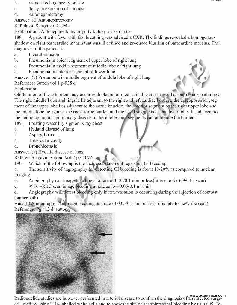
b. reduced echogenecity on usgc. delay in excretion of contrastd. AutonephrectomyAnswer: (d) Autonephrectomy Ref: david Sutton vol 2 p944Explanation : Autonephrectomy or putty kidney is seen in tb.188. A patient with fever with fast breathing was advised a CXR. The findings revealed a homogenous shadow on right paracardiac margin that was ill defined and produced blurring of paracardiac margins. The diagnosis of the patient is a. Pleural effusionb. Pneumonia in apical segment of upper lobe of right lungc. Pneumonia in middle segment of middle lobe of right lungd. Pneumonia in anterior segment of lower lobeAnswer: (c) Pneumonia in middle segment of middle lobe of right lungReference: Sutton vol 1 p-935 d. Explanation Obliteration of these borders may occur with pleural or mediastinal lesions as well as pulmonary pathology. The right middle l obe and lingula lie adjacent to the right and left cardiac borders, the apicoposterior ,seg-ment of the upper lobe lies adjacent to the aortic knuckle, the anterior segment of the right upper lobe and the middle lobe lie against the right aortic border, and the basal segments of the lower lobes lie adjacent to the hemidiaphragms. pulmonary disease in these lobes and segments can obliterate the borders 189. Froating water lily sign on X ray chesta. Hydatid disease of lungb. Aspergillosisc. Tubercular cavity d. BronchiectasisAnswer: (a) Hydatid disease of lungReference: (david Sutton Vol-2 pg-1072)190. Which of the following is the incoreect statement regarding GI bleedinga. The sensitivity of angiography for detecting GI bleeding is about 10-20% as compared to nuclear imagingb. Angiography can image bleeding at a rate of 0.05/0.1 min or less( it is rate for tc99 rbc scan)c. 99To –RBC scan image bleeding at rate as low 0.05-0.1 ml/mind. Angiography will detect bleeding only if extravasation is occurring during the injection of contrast (sumer seth)Ans: (b) Angiography can image bleeding at a rate of 0.05/0.1 min or less( it is rate for tc99 rbc scan)Reference: Pg 412 d. sutton
Radionuclide studies are however performed in arterial disease to confirm the diagnosis of an infected surgi-cal graft by using “I In-labelled white cells and to show the site of gastrointestinal bleeding by using 99”Tc-
www.examrace.com
labelled colloid or red cells (Fig. above ).
DERMATOLOGY191. A 7 year old boy with boggy swelling of the scalp with multiple discharge sinuses with cervical lymphadenopathy with easily pluckable hair. What would be done for diagnosis?a. KOH mountb. Pus culturec. Biopsyd. patch testAnswer (A) KOH mountReference: Roxburgh 17 e p. 41Exp: Diagnosis is tenia capitis and KOH mount is done for diagnosis.192. A child has pin head silvery papules on hands, forearms and skin of penis. The diagnosis is a. Molluscum contagiosumb. Lichen planusc. Lichen nitidusd. ScabiesAnswer (c) Lichen nitidus Reference: Roxburgh 17 e p. 274-278Exp:Lichen nitidus most commonly presents as an incidental finding on physical examination or after the patient notices an insidious onset of the lesions.1 It is characteristically asymptomatic.2 Physical examination re-veals pinpoint- to pinhead-sized skin-colored papules that may be scaly or have a central depression.3 They usually are found on the forearms, trunk, and the glans and shaft of the penis. Lichen nitidus can be discrete or generalized. In the discrete form, papules typically do not co-alesce; however, they may form or group at sites of trauma or skin pressure (the isomorphic or “Koebner” phenomenon).2 In the generalized (confluent) form, papules coalesce into red-yellow to brown plaques, especially in joint flexures, wrist and forearm ventral surfaces, or inframammary areas, making the clinical diagnosis more challenging.3 Biopsy may be helpful because lichen nitidus has a characteristic histologic appearance. The etiology of lichen nitidus is unknown, and no laboratory abnormalities or associations with systemic disease have been established. Because lichen nitidus is rare, definitive establishment of the epide-miology is difficult. One study2 of 43 cases demonstrated a male-to-female ratio of almost 4:1, although the generalized (confluent) form may be more common in women.3 No racial predisposition or known genetic inheritance pattern has been noted.1,2 Lichen nitidus primarily affects children and young adults, with a median age of seven years in males and 13 years in females.193. A 30 yr old man is having multiple ulcers in mouth with epididymitis with complains of blurring of vision. The probable diagnosis isa. Behcet syndromeb. Ritter syndromec. Xerodermad. Oculo cutaneous pemphigusAnswer (A) Behcet syndromeReference: Roxburgh 17 e p. 179-180Explanation: Behcet’s syndrome The manifestations of the syndrome can be broadly divided into the following:Mucocutaneous: (i) oral aphthous ulceration, present in 98% of patients with similar genital (scrotal/labial) ulceration in 80%; and (ii) skin lesions (acneiform folliculitis, erythema nodosum and occasionally ulcer-ation) and pathergy reaction [8].Arthritis/arthralgia: in up to 50% of patients, usually non-destructive.Ophthalmic involvement: panuveitis and retinal vasculitis in 50% of patients.Systemic vasculitis: thrombophlebitis, arteritis and aneurysm formation.
www.examrace.com

Additionally, gastrointestinal ulceration, epididymitis, central nervous system lesions and pulmonary lesions may all occur on the basis of the underlying vasculitis
194. Multiple vesicular painful lesions on dermatome T3 are duea. Herpes simplexb. Herpes zosterc. Scabiesd. Dermatitis herpitiformisAnswer (B) Herpes zosterReference: Neena khanna 3/e p 72Herpes zosteri. vesicles are usually unilateral and limited to dermatomes (M.C. Thoracic)ii. pain in course of nerve precedes vesicles.iii. Caused by varicella zosteriv. One attack gives life long immunity
www.examrace.com