Embed Size (px)
Citation preview

East African Scholars Journal of Agriculture and Life Sciences Abbreviated Key Title: East African Scholars J Agri Life Sci ISSN 2617-4472 (Print) | ISSN 2617-7277 (Online)
Published By East African Scholars Publisher, Kenya
Volume-3 | Issue-3 | Mar-2020 | DOI:10.36349/EASJALS.2020.v03i03.002
*Corresponding Author: Komal Javed 76
Research Article
Agrobacterium Mediated Delivery of Multiplex CRISPR/Cas9 System
in Cotton for Cotton leaf curl virus Disease Resistance
Komal Javed 1M.Phil Student, Department of Biochemistry, University of Agriculture Faisalabad Pakistan
Article History
Received: 04.02.2020
Accepted: 09.03.2020
Published: 25.03.2020
Journal homepage:
http://www.easpublisher.com/easjals/
Quick Response Code
Abstract: In Pakistan, cotton crop contributes 23% to the GDP and its massive export provides 60% of the total profit in trading business. Unfortunately, the Gemini viruses destroy the cotton crop at
an alarming rate. Now, the CLCuVs causes the Cotton Leaf Curl Virus Disease in cotton crop and
reduce its productivity. This viral attack on cotton plant resulted 5 billion US dollars loss to the
Pakistan. Well, in that regard, some conventional methodologies were used like the plant breeding
and specific RNA editing. At the same time, biotechnology introduced some very attractive
techniques which carry the massive potential for the eradication of this disease. These techniques include the ZFNs, TALENs and CRISPR/Cas9. In my research, CRISPR methodology was adopted
because of its marvelous efficiency of site specific mutagenesis. The conserved locations of the rep
gene of the different CLCuVs were identified as the target sites. These specific sites provided the information for the establishment of the 3 gRNAs. The single expression vector pHSE-401 having
the multiple guided RNAs and 1Cas9 were cloned. The specific cotton variety coker-312 was used.
The transformation of the vector was performed by hypocotyls excision of the cotton plant and delivery infection was done by agrobacterium EHA-105 Strain. Then, 1000 infectious hypocotyls
were shifted on the MSB having respective antibiotics and then, on the regeneration media which
converted the hypocotyls edges into the callus. Only two transgenic calli were obtained, the percentage of transgenic calli was 0.02% which were screened by PCR and run on gel. The bands of
200 bp confirmed the presence of the CRISPR/Cas9 construct in cotton calli.
Keywords: CRISPR/Cas9, Multiplex vector, 3gRNA and Cas9, Agrobacterium, Transformation,
Callus. Copyright © 2020 The Author(s): This is an open-access article distributed under the terms of the Creative Commons Attribution 4.0 International
License (CC BY-NC 4.0) which permits unrestricted use, distribution, and reproduction in any medium for non-commercial use provided the original
author and source are credited.
1. INTRODUCTIONThe economy of Pakistan majorly relays on the sector
of agriculture. Its percentage in GDP is 19.82% but the
participation of the agriculture sector dropped from
50% in 1950 to the about 21% in 2014. The money
which is earned by the agriculture products, labor force
of forestry, farming of the livestock and the crops
transformation feed the 64% of the village population of
Pakistan. According to statistics, 66% of Pakistan
people have employment in agriculture sector. In the
decade of the 20th century, the growth rate of the
agriculture products was 2.5% annually. In the
agriculture package of 1997, the yield of the crop was
developed up to 5.9%. To increase the productivity of
the agriculture, the annual production was increased
from 0.2% in the 2010 and the 2.9% rise happened in
2015 (Anonymous, 2014-2015).
Without any doubt, the progress of the country
depends on the development of the sector of the
agriculture. The rural population of the Pakistan uses
the cotton crop as an earning source. Approximately, in
80 countries, the cotton crop provides the labor
opportunity to the workers. Total 90% of the financial
aid is provided by the upland cotton. 25 million metric
tons of fiber was produced over the 34.4 million
hectares land (Anonymous, 2008).
In the 1996, United States produced the
genetically modified cotton which had the Bt gene.
After introducing the Bt gene in the cotton plant, just
25% of the pesticides were utilized (Pannetier et al.,
1997). A pivotal role of the genetic engineering has
been observed in the development of the insect
resistance and herbicide resistance characteristics in the
cotton plants. Begomoviruses introduced severe types
of diseases which damage the economy of Pakistan.
Begomoviruses complex with the alpha and beta
satellites molecules cause the CLCuD in cotton crop
and reduced its productivity. CLCuD is a major threat
for cotton crop (Sattar et al., 2013).
Different types of cotton leaf curl viruses are as
under:
Cotton leaf curl Alabad virus (CLCuAIV)
Cotton leaf curl Kokhran virus (CLCuKOV)
Cotton leaf curl Bangalore virus (CLCuBAV)
Cotton leaf curl Multan virus (CLCuMUV)
Cotton leaf curl Rajasthan virus (CLCuRAV)

Komal Javed; East African Scholars J Agri Life Sci; Vol-3, Iss- 3 (Mar, 2020): 76-90
© East African Scholars Publisher, Kenya 77
Thickening of the vein, reduction downward or upward
leaf area, puckering and decline in the growth rate of
the cotton are the symptoms of the CLCuD (Sattar et
al., 2013).
2. RESEARCH OBJECTIVE Induction of the infection of Agrobacterium in
hypocotyls of cotton crop by transformation
Development of the transgenic callus of cotton
plant
PCR screening of the callus to identify the
multiplex CRISPR/Cas9 construct
3. MATERIALS AND METHODS The present research work was conducted in
Cotton Biotechnology Laboratory (CBL), Center for
Advanced Studies in Agriculture and Food Security
(CAS-AFS), using the standard protocols of the
laboratory.
3.1 Synthesis of Primers for gRNA
According to the sequence of the gRNA,
primers were designed by CHOPCHOP primer
designing tool. Primers had the restriction sites and
produced the only sticky ends. The Oligos annealing
was done through PCR, oligos were annealed by
fluctuating the temperature of the system. After every
minute, the temperature was decreased from 95°C to the
4°C by decreasing 10 degree.
3.2 Confirmation through digestion
The pHSE-401 plasmid was restricted with the
restriction enzymes (XbaI, EcoRI) and then, run on the
agarose gel. Restriction digestion has the purpose to
obtain two fragments. These were almost 11738bp and
4916bp. The sizes of these specific fragments
confirmed the plasmid of pHSE-401 (Green and
Sambrook et al., 2012).
3.3 Reagents and materials
Restriction enzymes (XbaI and EcoRI).
Plasmid (pHSE_401).
10x Buffer.
Water (d3H2O).
3.4 Protocol
All the following reagents were added in an
Eppendorf tube with such concentration.
Restriction enzymes = 1µl
Plasmid (pHSE- 401) DNA = 2µl
10x buffer = 1µl
D3H2O = 6µl
Total = 10µl
3.5 Running of Agarose Gel (Green and Sambrook,
2012)
Agarose
6x sample loading buffer
1x TAE buffer
1kb DNA ladder
Ethidium Bromide
3.6 Preparation of 50x TAE Buffer
Glacial Acetic acid = 57.1ml
Tris–base = 242grms
0.5 M EDTA = 100ml
3.7. 1x TAE Buffer preparation
For preparing the 100ml volume, 98ml of d2H2O and
2ml of 50x TAE buffer were used.
3.7.1 Preparation of agarose gel
In a clean 250ml flask, 100ml of 1x TAE was added
and 1grm of agarose gel powder was included to make
1% of gel (W/V).
The gel solution was kept in microwave until the
solution became transparent.
The solution was kept outside the oven to about
60°C.
Now, 3µl (EtBr) Ethidium Bromide was added into
the gel.
In a casting tray, the hot liquid was poured and the
comb was kept by inserting the sides into the
notches.
After the polymerization of the gel, the comb was
taken out from the gel to expose the wells for the
sample.
3.7.2 Preparation of the loading buffer
Glycerol in water 30% (v/v)
Bromophenol Blue 0.25% (w/v)
Xylene Cyanol 0.25% (w/v)
3.8 Agarose Gel Loading
In every 20µl reaction mixture, 4µl of 6x loading
buffer was added.
On gel, samples were loaded and then, order was
recorded.
3.9 Gel Running
In a chamber, the gel was kept.
Electric supply was provided to the chamber of
electrophoresis.
Voltage was set at 80V.
The samples migrated from the negative electrode to
the positive electrode.
Electricity source was removed.
The lid of the tank was taken away.
As EtBr was added, then blue strain showed the
movement of DNA directly.
In gel documentation machine, gel was placed (Bio-
Rad) and samples were compared with the ladder
DNA.
3.10 Miniprep technique for Plasmid Isolation
3.10.1 Reagents and Materials.
Komal Javed; East African Scholars J Agri Life Sci; Vol-3, Iss- 3 (Mar, 2020): 76-90
© East African Scholars Publisher, Kenya 78
Antibiotics (Kanamycin, Tetracycline, Rifampicin) for
the isolation of plasmid.
Colonies of E.coli
LB media (500 ml)
Ethanol (70%, 100%)
D3H2O , containing 20µl (m) RNA ase
Alkaline solution I
Alkaline solution II
Alkaline solution III
3.11 Synthesis of L.B Media
Yeast extract =5grms
NaCl =10grms
Tryptone =5grms
3.12 Preparation of Re-suspension solution
Tris-Chloride (pH 8.0) =25mM
Glucose =50mM
EDTA (pH8.0) =10mM
After synthesis, solution I was autoclaved and stored at
4°C (Green and Sambrook, 2012).
Synthesis of alkaline lysis solution II (Lysis solution)
(Green and Sambrook, 2013)
SDS =1% (w/v)
NaOH =0.2N
Synthesis of alkaline lysis solution III (Green
and Sambrook, 2012)
Glacial acetic 11.5mL
Water 28.5mL
Potassium acetate 5M (60mL)
3.13 Cells preparation (Green and Sambrook, 2012)
One colony of the mutated bacteria is inoculated in
10ml LB media having Kanamycin, Rifampicin and
Tetracycline culture was kept at 37°C with fast
shaking.
The mutated bacteria in LB media was taken 1.5ml
only and poured in Eppendorf tubes. Then, it was
spun for 30 seconds at 15000rpm at the 4°C
temperature.
.The supernatant was discarded and the remained
part was utilized for the next stage.
By dynamic vortexing, the deposited bacterial cells
were kept again in 100µl of ice-cold alkaline lysis
solution I.
In every solution, 200µl of freshly synthesized
alkaline lysis solution II was appended. Tubes were
covered and all the contents were mixed by rapidly
inverting the tubes approximately five times.
Then, used 150µl of ice col alkaline lysis solution
III and it was intensified and inverted for many
times.
At 4°C, the bacterial solution was spun at the high
speed for 5 minutes.
The supernatant was transferred to a new tube,
In supernatant, 100% ethanol was added kept at -
20°C for half an hour.
The essence was spun at the 4oc for two minutes
only in a new tube aqueous layer was transferred for
next step, pellet was proceeded.
In pellet, 70% ethanol was added and closed tube
was upended for multiple times.
The deionized distilled water having RNA are was
used for dissolving the DNA and at 20°C, it was
stored.
3.14 Cotton transformation and regeneration of
plant
Cotton (Coker-312) seeds were sown in peat moss
and covered with aluminum foil for 2 to 3 days.
On daily bases, watered the plants.
Hypocotyls of cotton were appeared after 6 days.
The hypocotyls sterilization.
3.15 Process of sterilization
Commercial 12% bleach is available, 1% or 1.5% is
used mostly for cotton.
8.33ml of 12% bleach and prepare volume up to
10ml.
5ul of tween 20 is used.
The sterilization solution is added into the cotton’s
hypocotyls.
In shaker, shaking should be done for 15 minutes.
For five times, washing with D2H2O water.
Drying of hypocotyls with the filter paper.
3.16 Agrobacterium culture Preparation
For the purpose of transformation.
The Agrobacterium tumefaciens strain EHA-105
was used. This strain had pHSE -401 plasmid
transformed.
The growth of the vector occurred in LB media at
28°C in the shaking incubator.
Centrifugation of the culture was done at 4°C at
15000rpm.
Pellet was re-suspended in the MGL for the
infection of the explants.
The hypocotyls were sliced with sharp blade and
then, they were inoculated with the agrobacterium
suspension for approximately 30 minutes.
Sterile filler paper were used and then, dried them
carefully.
What man filter paper were used for the transfer of
the dry hypocotyls segments.
Shift the hypocotyls on the plates which carry the
co-cultivation media.
For induction of the callus, after two or three days,
shifted them to selection media.
The callus developed on the edge of the hypocotyls
of cotton.
3.17 Preparation of LB Media
5g/L extract of Yeast
10g/L of NaCl
5g/L of Tryptone

Komal Javed; East African Scholars J Agri Life Sci; Vol-3, Iss- 3 (Mar, 2020): 76-90
© East African Scholars Publisher, Kenya 79
3.18 MGL Preparation
5g/L Tryptone.
0.1g/L of MgSO4
5g/L of NaCl
1g/L of Glycine
0.25g/L of KH2 PO4
pH 5.8
3.19 MSB Media Preparation
The Murashige and Skoog Basal medium w/
Vitamins
0.1 mg/l Kinetin
3% (w/v) Glucose
20mg/ L Acetosyringone
1g/ L MgCl2
0.1mg/ L 2.4-D
0.25% (w/v) Phytagel pH 5.8
3.20 Selection on Callus Induction media (CIM)
0.1 mg/L 2, 4-D
3% (w/v) Glucose
0.1 mg/L Kinetin
40mg/L Cefotaxime
50 mg/L Kanamycin
25% (w/v) Phytagel, pH 5.8
3.21 Preparation of chemically competent cells of E.
coli
3.21.1 Chemicals, Reagents and Apparatus
Calcium Chloride
Potassium Acetate
Manganese Chloride
Glycerol
Rubidium Chloride/ Potassium Hydroxide
MOPS/ PIPES
3.22 250ml TFBI
10mM CaCl2
30mM Potassium Acetate
100mM Rubidium Chloride
50mM Manganese Chloride
15% of Glycerol
Maintain pH to 5.8 by adding the 1M
acetic acid
Filter sterilize through 0.2mm filter paper
3.22.1 Protocol (Green and Sambrook, 2012)
From the LB plate a single colony of E.coli strain was
selected and picked and inoculated 2.5ml of LB
medium shaking was done at 220rpm at the 37oC in the
incubator shaker overnight.
The culture of agrobacterium was utilized to
inoculate 250ml of the LB medium.
Then, cells were grown in a 1 liter flask for three
hours until the 0D600 increases up to 0.4 – 0.6.
Through centrifugation, the cells were collected at
5000rpm at 4°C for 5 minute.
The supernatant was discarded.
In 0.4 volume of ice-cold TFBI, cells were gently
re-suspended.
On ice surface, the re-suspended cells were
incubated for 5 minutes.
Now, again supernatant was discarded.
In 0.4 volume of ice-cold TFB2, the cells were
gently re-suspended.
For 30 minutes, the cells were incubated on ice and
then, aliquoted into the pre-chilled tubes.
Rapidly, freeze the tubes by dipping into the liquid
Nitrogen and then, store at -80°C.
Protocol for the preparation of the E.coli calcium
chloride competent cells.
In 50ml falcon tube, inoculate a single colony into
the 5ml LB media.
At 37°C, grow O/N.
In the next morning, utilize the 1ml to inoculate
100ml of LB in the 250ml bottle.
For 4-6 hours, shakes at 37°C.
For 10 minutes, keep the cells on ice (keep cold
form now on).
Now centrifuge the cells and collect the cells in the
big centrifuge for just three minutes at 6000 rpm.
Discard the supernatant and then, gently re-suspend
on 10ml cold 0.1m Calcium Chloride. (Cells are
very delicate and sensitive to the mechanical
disruption, so handle them nicely).
For 20 minutes, incubate the cells on ice.
Centrifuge the cells
Discard the supernatant and carefully re-suspend
5ml cold 0.1 M Calcium Chloride / 15% Glycerol
Now, dispense in micro tubes (300 µl/ tube)
Freeze at the -80°C
3.23 E.coli Transformation
Keep lu of plasmid DNA in a micro tube.
Carefully, add the 100µl of the competent cells.
DNA control tube having cells but no DNA is
incubated for 30 minutes in ice.
At 42°C, heat shock it for two minutes.
Keep them back on ice.
In tubes, now add 900µl of LB media.
Incubate for 30 minutes at 37°C.
In LB Amp or LB carb plates, plate 100-1000µl of
the cells.
In a blood plate, plate 100µl of the NO DNA control
(For checking the quality of cells). Now, grow the
O/N, the rest is saved in the cold room or even
freeze with 15% of Glycerol. This is the case when
you do not get the colonies.
If, there is a need of multiple colonies or the ligation
is not quite efficient, then centrifuge the cells for
one minute at 8000rpm, discard 900µl supernatant,
100µl left is re-suspend and whole lot is plated.
3.24 Preparation of the agrobacterium competent
cells
Take the 8ml overnight culture of the
Agrobacterium and grow it in LB media having
5ug/ml Tetracycline +25 ug/ml Kanamycin +50
ug/m Rifampicin.

Komal Javed; East African Scholars J Agri Life Sci; Vol-3, Iss- 3 (Mar, 2020): 76-90
© East African Scholars Publisher, Kenya 80
The LB must has the 5g/L NaCl at all the
stages.
Do not add antibiotics in 192ml LB media,
inoculate 8ml overnight culture.
At28°C, shake it until an OD reaches up to 0.5.
At 4°C, centrifuge at the 4000rpm for fifteen
minutes.
Now use 10mm tris solution for the re-
suspension of the pellet.
At 4°C, centrifuge for 15 minutes at 4000rpm.
In 20ml chilled LB, re-suspend the pellets and
store it in liquid nitrogen. These cells get freeze
at -80°C, they can stay competent for
approximately for 6 months.
3.25 Agrobacterium transformation
Take out the agrobacterium competent cells
from the -80°C freeze and now leave them on
ice for 20-30 minutes.
Stored agar plates are removed from the 4oC
freezer and let them warm up to the temperature
of the room and incubated these plates at 37°C
in an incubator.
In a micro centrifuge or falcon tube, mix 1-5 µl
of DNA into the 20-50µl of the agrobacterium
competent cells, carefully mix by using the
slight flicking of the fingers at the bottom of the
tube for few times.
3.26 Protocol of freezing The competent cell /DNA is incubated on ice
for 20-30 minutes.
Water bath having the temperature of 42°C is
used because heat shock the every
transformation tube by putting the bottom ½ to
2 /3 of the tube for 30-60 seconds (for most of
the time 45 seconds are considered as an ideal
period.
Leave the tubes again on ice for two minutes.
Add 250-1000 µl of LB media into the bacteria
and for 45 minutes, grow them in shaking
incubator at the 37°C.
The agar plate must contain the appropriate
selection antibiotics.
So plate few or all of the transformation onto a
10cm LB agar plate.
At 37°C, incubate the plates overnight.
3.27 Protocol for plasmid purification (Green and
Sambrook, 2012)
Take the 3ml liquid LB medium having respective
antibiotic then, culture the single colony of E.coli
and grow overnight at the 37°C temperature.
The culture of E.coli was centrifuged in 1.5ml
(micro) centrifuge tubes for 30 seconds at the
13500rpm.
In 250µl of the re-suspension solution, re-suspend
the pelleted cells now transfer the suspension of the
cells to a micro centrifuge tube. Through vortexing
or pipetting up and down, the bacteria must be re-
suspended thoroughly until where is no any clump
remains.
The 250µl of the Lysis solution is added and mix it
properly by investing the tube.
Invent the tube 4-6 times, so the solution becomes
first viscous and them little clear.
The 350µl neutralization solution is added and then
rapidly mix. Invert the tube 4-6 times properly and
thoroughly.
For minutes, centrifuge the debris if pellet cell and
DNA of chromosomes.
The supernatant is transferred to the, gene JET spin
column through decanting or by the pipetting.
Try to prevent the transfer of the white precipitate
For one minute, centrifuge remove the flow-through
and put the column again into the same collection
tube.
The gene JET spin column is washed by the
addition of the 500µl of the wash solution I and
then, centrifuge for approximately 30 to 60 seconds
and then remove the flow through.
The 500µl of wash solution is added to the gene Jet
spin column the dilution is done by adding the
ethanol before the use. Centrifuge for 30 to 60
seconds and remove the flow-through. Put the
column again into the collection tube.
The 500µl of the wash solution is used for the
washing process.
Remove the flow, through and then centrifuge for
one more extra minute to remove the residues.
Wash the solution, this step is mandatory for the
removal of the residual ethanol in preps of plasmid.
Take a fresh 1.5ml micro centrifuge tube and shift
the Gene JET spin column into this tube. In the
center of the Gene JET spin column, Elution Buffer
is added to elute the plasmid DNA. Just try to avoid
the touching of the pipette tip with the membrane.
For 2 minutes incubate at the room temperature and
for 2 minutes centrifuge then
Through the column in trash bin and store this
purified plasmid having DNA at 20°C.
3.28 Agarose Gel Preparation Method
1g of agarose is weighted.
In a flask, add this agarose powder into the
100mml of the 1*TAE buffer.
Heat it in oven for 3 minutes and wait until it
dissolved completely.
For cooling the gel, leave it for few minutes.
Et Br is added with respect to the final volume
of the gel which is 2µl.
Now put the comb into the tray of gel.
The gel is poured into the already settled comb
tray.
Solidification is done by leaving it for some
time.
3.29 1X TAE Buffer
Tris Base is 4.84g.
Glacial Acetic Acid is 1.14ml.

Komal Javed; East African Scholars J Agri Life Sci; Vol-3, Iss- 3 (Mar, 2020): 76-90
© East African Scholars Publisher, Kenya 81
2ml of 0.5 M EDTA (pH=8).
With water, bring the volume up to one Liter.
Samples loading and running of the Gel.
Buffer is added for preparing the sample.
In electrophoresis unit, keep the agarose get
tray.
Gently add the TAE Buffer into the
electrophoretic unit just to cover the gel
carefully remove the combs from the agarose
gel.
Loading the ladder into the first well of the gel.
The, remembers the number of the well and
load your sample. Now, set the voltage
between the 80-150V and then run the gel.
After 30 minutes power off the unit.
Now, remove the gel gently and observed it
under the system of gel documentation.
PCR confirmation
Reagents
Green Master Mix 6µl
Reverse primer 1µl
Forward primer 1µl
Water 4µl
Template 1µl
Primers
Forward primer GACAAGAAGTACTCGATCGGCCTC
Reverse Primer AATGGCCCCTGAACTTAATCATG
PCR profile
Steps Temp. Time No. of cycles
Initial Denaturation 940C 5 min
35 cycles
Denaturation 940C 30 sec
Annealing 540C 45 sec
Extension 720C 1 min
Final Extension 720C 5 min
Protocol
13µl reaction mixture was added in the PCR tubes.
After added mixture, PCR tubes were incubated in
thermos-cycler.
PCR product was visualized on 1% agarose gel
under gel documentation system.
1kb ladder was used to estimate the product size.
4. RESULTS 4.1Map of cloning vector
In this research, multiplex vector pHSE-401
was used. It is the first vector which is brilliant in
multiplexing. The map of the pHSE-401 cloning vector
was obtained through Addgene website.
Figure a: The map of the cloning vector pHSE-401.

Komal Javed; East African Scholars J Agri Life Sci; Vol-3, Iss- 3 (Mar, 2020): 76-90
© East African Scholars Publisher, Kenya 82
4.2 Map of the pHSE-401 expression vector
The map of the vector was taken from the
Vector Builder. There are the 3gRNAs, 3gRNAs
scaffold and 1Cas9 in this pHSE-401 vector. The vector
has complete 16935 bp, U6-26p and U6-26t are also the
parts of the expression vector. Cas9 which consists of
5645 bp. This vector was transformed into the
agrobacterium, kanamycin, rifampicin and tetracycline
are the specific antibiotics for this vector.
Figure b: Map of pHSE-401 plant expression vector having 3gRNAs and Cas9.
4.3. Transformed E. coli cells and their colonies Preparation
The transformation of plasmid in the Top-10 strain E. coli was done by the heat shock method by using the
standard protocol of the laboratory. Multiple colonies of the E. coli were obtained over single plate.
Figure c: Transformation of the vector pHSE-401 in E.coli.
4.4. Formation of the colonies of the transformed
Agrobacterium cells
The Agrobacterium cells of EHA-105 strain
were transformed with the vector pHSE-401 by the
freeze thaw method. The whole procedure was
performed on ice and then, kept in liquid nitrogen
container. Agrobacterium cells were stored in the
glycerol stock and they were spread on the agar plate
for making the colonies of the transformed
Agrobacterium.

Komal Javed; East African Scholars J Agri Life Sci; Vol-3, Iss- 3 (Mar, 2020): 76-90
© East African Scholars Publisher, Kenya 83
Figure d: Colonies of transformed pHSE-401 into Agrobacterium.
4.5. Plasmid Confirmation
The expression vector Phse-401, containing
plant codon optimized Cas9 was obtained by the
VectorBuilder. The pHSE-401 plasmid was restricted
with the restriction enzymes XbaI and EcoRI and then,
run on the 1% of agarose gel. By the restriction
digestion process, two fragments were obtained. These
were almost 11738bp and 4916bp. The sizes of these
specific fragments confirmed the plasmid of
pHSE_401.
Figure e: pHSE-401 plasmid confirmation. (M) 1kb DNA ladder. (1) Undigested fragment of pHSE-401 plasmid. (2) Digested
fragments of pHSE-401 plasmid.
4.6. Preparation of the nursery of cotton plants in
laboratory The linted cotton seeds were taken and the lint
was removed with the commercial sulphuric acid due to
which lint was completely removed. This step was
performed in the fume hood because of the commercial
sulphuric acid. Now, the de-linted cotton seeds were
sterilized by using the ethanol and HgCl2. The ethanol
sterilized the cotton seeds, then these seeds were sown
in the peat moss for three days and covered with
aluminum foil and the covered pots were uncovered
after three days. So, the immature plant came out which
became mature after 4 days, these plants are the less
mature in age.
4916 bp
500 bp
1000 bp
2000 bp
3000 bp
11738 bp
1 2 M 16654 bp

Komal Javed; East African Scholars J Agri Life Sci; Vol-3, Iss- 3 (Mar, 2020): 76-90
© East African Scholars Publisher, Kenya 84
Figure f: Nursery development of Coker-312.
4.6.1. Hypocotyls sterilization and excision
All the cotton plants were taken out from the
peat moss after seven days and their leaves and roots
were separated from hypocotyls and then, hypocotyls
were shifted into the falcon having sterilization
solution. And after that shake it gently and then,
removed the sterilization solution from the petri plate.
Now, 1000 hypocotyls were cut into the smaller
segments. The hypocotyls were cut into the small pieces
with the sharp sterile blade. After cutting into short
segments, they were placed on filter paper. Again dry
the smaller fragments of cotton hypocotyls with the
autoclaved filter paper.
Figure g: Excision and drying of hypocotyls.
4.6.2. MGL addition
Agrobacterium primary and secondary culture
was prepared and then, secondary culture was
centrifuged at 8000rpm for 05 minutes. The secondary
culture was taken, then the pellet formation occurred
and supernatant was discarded and the MGL solution
was added in the pellet and then, pellet was dissolved.
For this step, ice was used and intense shaking was
done in this step for the complete pellet dissolving in
the MGL solution. This dissolved solution was added
into the cotton hypocotyls and waited for 20 minutes
and then, the solution was removed.
Figure h: Poured MGL solution having dissolved pellet of agrobacterium.
4.6.3 Shifting of hypocotyls on MSB media
All the hypocotyls were transferred on the
MSB media which carried the hormones and kanamycin
selection antibiotic and the plates were covered and
kept in dark for 2 days for giving suitable environment
for agrobacterium infection development in hypocotyls.
The purpose of the shifting on MSB is the initiation of
the growth in the hypocotyls edges. Darkness is the
major requirement for this step. After 48 hours,
hypocotyls are shifted to the next media.

Komal Javed; East African Scholars J Agri Life Sci; Vol-3, Iss- 3 (Mar, 2020): 76-90
© East African Scholars Publisher, Kenya 85
Figure i: Shifting of hypocotyls on MSB media.
4.6.4. Transferring of hypocotyls on CIM media All the hypocotyls were taken out from MSB
media after the 48 hours and then, they were shifted on
CIM media. CIM media had the respective antibiotics,
glucose, glycine and necessary nutrients which
stimulated the growth of the hypocotyls because callus
started to appear at the edges of segments. At the edges,
after 4 weeks, 2 calli appeared and again hypocotyls
were shifted to new CIM plates for further growth.
Figure j: Hypocotyls shifting on Callus induction media.
4.6.5. Formation of callus After the four weeks, callus started to appear at
the edges of the hypocotyls of the cotton crop. The
undifferentiated cells started to appear at the sides of
the 2 hypocotyls portions. After every four weeks
period, new plate of freshly prepared CIM media was
used for the proper and efficient growth of the calli. The
efficiency of result was 0.02%. Only two transgenic
calli were obtained.
Figure k. Callus appeared on excised edges of hypocotyls.
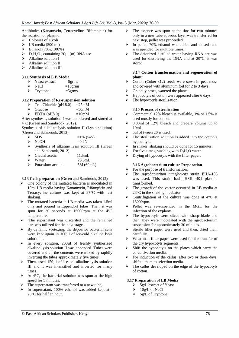
4.6.6. PCR based screening of callus
DNA was extracted from 2 transformed callus
grown on selection media. For this method, Mini prep
kit was used and DNA was isolated. Then, PCR
technique was used by following the standard protocol.
For that purpose, primers were designed for gRNA
through CHOPCHOP bio-informatics tool and fragment
was amplified through this process. After PCR, product
was run on 1% agarose gel for getting the band for the
identification and confirmation of the gRNA. So, the
band of 200 base pairs confirmed the presence of the
gRNA in the callus of cotton. This cotton callus was
transgenic callus with transformed vector.
Komal Javed; East African Scholars J Agri Life Sci; Vol-3, Iss- 3 (Mar, 2020): 76-90
© East African Scholars Publisher, Kenya 86
M 3 2 1
200bp
Figure l: Confirmation of gRNA in plants (M) 1kb DNA ladder and bright bands of 1 show the amplified fragment of gRNA.
DISCUSSION
The economy of Pakistan majorly depends
upon the cotton products like lint and cotton seeds. Its
products play role in the 10% GDP and 55% in the
export business. Nowadays, synthetic substance and
fertilizers are used. The crop cultivation land is
increased which increased the production of cotton. The
cotton crop has become susceptible to pests, insects and
viruses. Cotton leaf curl virus disease is a dangerous
threat for the cotton production because leaves get curly
and inverted. There is only DNA A component because
the genome of these viruses is monopartite which is
equal to the genome of the Begomoviruses of family
Geminiviridae. The complex of these viruses cause the
80% reduction in the cotton production last year.
Another option to conventional GMO (transgenic) crop
and plant rearing strategies to enhance trim plant is
focused on genome designing or genome altering
(Khaoula et al., 2003).
Through site particular nucleases, the directed
transgene reconciliation technique fulfilled in a
persuasive and unequivocal way. With the help of these
built nucleases we develop perfectly DSBs (2 fold
stranded breaks) to focus DNA and absolutely provoke
the DNA repair component (Luisa et al., 2007). Among
these Nucleases, there is Zinc finger nuclease (ZFNs)
has a built exhibit of zinc fingers which then, converge
in to the synergist region of Fok nuclease, acts about as
a dimers to acquaint DSBs (Fenola et al., 2002). The
second nuclease is TALEN (Transcription activator like
effector nucleases consists of DNA restricting area of a
TALE joined with non-particular FoK nuclease, form a
grouping specific nuclease that was utilized to develop
a DSBs. From these two units of grouping specifically,
TAL Effector Nuclease attach to the objective DNA and
then, engineer a spacer locale of 2-9 bp to permit the
dimerization of endonuclease areas and develop DSBs
(Jieliang et al., 2003). A new technique of present days
has come as a game changer for this reason. This is
Cluster regularly interspaced short palindromic repeats
(CRISPR) related to Cas endonuclease. This magic has
quite recently been uncovered. A little planner which is
the guided RNA accomplish the endonuclease Cas9
towards the target DNA and chop the particular
grouping vital to the guide RNA (Haft et al., 2018).
For Pakistan economy, from the last 30 years,
CLCuD has become a critical threat. The rep gene of
these viruses is targeted, Multiple techniques like ZFNs,
TALENs and CRISPR/Cas9 have been utilized to
inhibit this disease by targeting the specific site which
is mandatory for the process of- rolling circle
replication mechanism in the Begomoviruses in the field
of genome engineering , for triggering the genome of
CABs, CRISPR/Cas9 system has become a gene
changer CRISPR/Cas9 system is taken as a most
advanced methodology because it has the ability to
trigger the different genes simultaneously. There are the
multiple gRNAs and Cas9 in single vector (Ayan et al.,
2019).
In our research, firstly, the designed vector
map was taken from VectorBuilder. The vector pHSE-
401 is the first vector which is brilliant for multiplexing,
it has 3 gRNA and 1 Cas9. All the 3 RNAs perform the
role as a guide and help the Cas9 towards its target area,
ultimately for the development of the cuts at the
respective sites. The sequence of the vector pHSE-401
was confirmed through the restriction digestion method.
For this purpose, two restriction enzymes were used.
They cut the vector DNA into the two fragments of
specific sizes which confirmed the identity of the vector
when we run it on the gel. NCBI was used to find the
sequence of genome of CLCuK. The vector DNA was
isolated through mini prep kit and its concentration was
checked by running it on the gel. Gel Documentation
image was captured, the clear bands came which
showed the good concentration of the plasmid DNA and
after this, in Agrobacterium vector was transformed
(Naeem et al., 2018).
Coker 312 cotton variety was used for
transformation because Coker 312 has good
regeneration capacity. We have established a rapid and
technically efficient transformation system. Well, there

Komal Javed; East African Scholars J Agri Life Sci; Vol-3, Iss- 3 (Mar, 2020): 76-90
© East African Scholars Publisher, Kenya 87
are several methodologies are available but they are
quite complex and time consuming. The agrobacterium
strain EHA105 was selected because it gave good
results in previous cotton studies. For that purpose,
cotton seeds were grown in peat moss because studies
showed that it has more water retention capacity. After
3 days, immature plants started to appear. When these
plants became mature on the 7th
day, then they were
used for transformation, we used to keep them in dark
instead of light because previous studies show that
green hypocotyls give excellent and rapid callus
forming result.
The cultures of Agrobacterium were prepared,
firstly, primary culture was developed and then,
secondary culture was formed with respective
antibiotics kanamycin, rifampicin and tetracycline,
OD600 was 0.8-1.0. The agrobacterium culture was
centrifuged at 8000rpm. On the day of transformation,
15 hypocotyls were cut with sharp blade and then, used
for the purpose of the transformation. The hypocotyls
were co-cultivated with the Agrobacterium pellet
dissolved in the MGL solution. Then, 1000 hypocotyls
were shifted to MSB media, for this, hypocotyls were
kept for two days in dark with fully covered aluminum
foil. Then, CIM media was prepared, hypocotyls were
transferred from the MSB to the CIM media which has
glycine, glucose and agar etc. It gave stable
environment for agrobacterium infection. We added
Plant Tissue Culture and carbenicillin, it gave
contamination free media to the hypocotyls and after
the process of the shifting, 2calli appeared. There was
the 0.02% of calli. After this, PCR Screening was the
major step. For PCR Screening, for primer designing,
CHOPCHOP bioinformatics tool was used. Two
primers were designed, one of them is forward and the
other is reverse, these primers were designed for the
confirmation of the gRNA. The callus DNA was
extracted through mini prep kit. The step of DNA
isolation was performed for getting the pure DNA.
Now, the PCR protocol was used for PCR Screening,
there were 35 cycles run. Then, PCR product was
obtained and run on the gel against 1kb ladder, two
bright bands of 200 bp were observed and they
confirmed the presence of the gRNA. So, it means that
CRISPR/Cas9 construct has successfully transformed
into the cotton crop. Now, this crop has ability to tackle
the CLCuVs by chopping the genome of the virus with
Cas9 (Sattar et al., 2010).
This study is quite different from other studies
because it has the involvement of the multiplex
CRISPR vector with 3 gRNAs and 1 Cas9. We have
developed an efficient and quite competent derived
transformation methodology which has enough
potential to protect the cotton crop from CLCuD. The
advanced techniques of the CRISPR/Cas9 works in a
marvelous way. It targets the replication encoding genes
of virus by introducing the cleavage that was the major
reason to perform the cotton transformation for
developing the resistance against the cotton leaf curl
virus disease in cotton plant. Therefore, the CLCuD
cascade can be targeted through this techniques by
aligning approaches for the induction of resistance
along with CRISPR/Cas9 tool. Our method can be used
for the resistance development against crop damaging
viruses (Zaidi et al., 2018).
3. CONCLUSION Cotton crop provides us food, feed and
economical support. In 2006, approximately 34.3
million hectares (Mha) cultivation land was yielding
the almost 25 million metric tons (MMT) of fiber. For
20 million farmers among 80 countries of are
producing cotton as a cash crop (De Onis et al., 1993).
Cotton lint is quit valuable, therefore China (26.40%)
then, India (20.5%). After that United States of
America (13.9%) and Pakistan (8.5%). Different other
countries like Brazil (6.3%), Uzbekstan (4%).
Australia (3.8%) and Turkey (3.3%). These countries
are considered as the major producing bodies of the
cotton lint. Cotton crop is attacked by many pests,
insects, bacteria, viruses and fungi. So, these
pathogens are the big threats for the cotton crop. In
different stages of plant life like in shoot development,
feed and food and flowering are also affected through
the antibiotics stresses, drought, cold, high temperature
and salinity. For the economy of the country, CLCuD
is the big danger for the transmission of the virus to the
cotton plant, whitefly acts as a vector. The viral
genome is targeted at the specific genes included in
replication or the life cycles Begomoviruses. For this
purpose, multiplex editing with CRISPR/Cas9 system
is surely preferred. In this study, pHSE_401 vector
was used which has the 3 gRNA and 1 Cas9. This
construct was transferred into the agrobacterium and
cultures having respective antibiotics were developed.
Then, cotton hypocotyls fragments were prepared and
interacted with the agrobacterium culture. Then, MSB
and CIM media were used. Callus appeared and PCR
was done which showed that the CRISPR/Cas9 –
3gRNAs complex was completely transformed into the
cotton plants through Agrobacterium, the plants having
the CRISPR/Cas9 construct did not show the
symptoms of the cotton leaf curl virus disease. The
purpose of the study was to inhibit the replication of
the cotton leaf curl virus by targeting the viral genome.
REFRENCES 1. Ali, M. (1997). Breeding of cotton varieties for
resistance to cotton leaf curl virus. Pak. J.
Phytopathol, 9(1), 1-7.
2. Ali, Z., Abulfaraj, A., Idris, A., Ali, S., Tashkandi,
M., & Mahfouz, M. M. (2015). CRISPR/Cas9-
mediated viral interference in plants. Genome
biology, 16(1), 238.
3. Ali, Z., Ali, S., Tashkandi, M., Zaidi, S. S., &
Mahfouz, M. M. (2016). CRISPR/Cas9-mediated

Komal Javed; East African Scholars J Agri Life Sci; Vol-3, Iss- 3 (Mar, 2020): 76-90
© East African Scholars Publisher, Kenya 88
immunity to geminiviruses: differential
interference and evasion. Sci Rep 6: 26912.
4. Anonymous. (2008). FAO statistics.
http://www.fao.org.accessed 7 Jan 2008.
5. Anonymous. (2014-15). Economic Survey of
Pakistan. 2014-15. 2:25.
6. Baltes, N. J., Hummel, A. W., Konecna, E., Cegan,
R., Bruns, A. N., Bisaro, D. M., & Voytas, D. F.
(2015). Conferring resistance to geminiviruses with
the CRISPR–Cas prokaryotic immune
system. Nature Plants, 1(10), 1-4.
7. Baltes, N. J., J. Gill-Humanes, T., Cermak, P. A.
Atkins &Voytas, D.F. (2014). NA replicons for
plant genome engineering. Plant Cell. 26: 151-163
8. Barrangou, R. & van der Oost, J. (2013). CRISPR-
Cas systems: RNA-Mediated Adaptive Immunity
in Bacteria and Archaea. 82, 237-66.
9. Barrangou, R., Fremaux, C., Deveau, H., Richards,
M., Boyaval, P., Moineau, S., ... & Horvath, P.
(2007). CRISPR provides acquired resistance
against viruses in prokaryotes. Science, 315(5819),
1709-1712.
10. Beerli, R. R., Segal, D. J., Dreier, B., & Barbas, C.
F. (1998). Toward controlling gene expression at
will: specific regulation of the erbB-2/HER-2
promoter by using polydactyl zinc finger proteins
constructed from modular building
blocks. Proceedings of the National Academy of
Sciences, 95(25), 14628-14633.
11. Bernstein, E., Caudy, A. A., Hammond, S. M., &
Hannon, G. J. (2001). Role for a bidentate
ribonuclease in the initiation step of RNA
interference. Nature, 409(6818), 363-366.
12. Bibikova, M., Golic, M., Golic, K. G., & Carroll,
D. (2002). Targeted chromosomal cleavage and
mutagenesis in Drosophila using zinc-finger
nucleases. Genetics, 161(3), 1169-1175.
13. Bolotin, A., Quinquis, B., Sorokin, A., & Ehrlich,
S. D. (2005). Clustered regularly interspaced short
palindrome repeats (CRISPRs) have spacers of
extrachromosomal origin. Microbiology, 151(8),
2551-2561.
14. Böttcher, B., Unseld, S., Ceulemans, H., Russell,
R. B., & Jeske, H. (2004). Geminate structures of
African cassava mosaic virus. Journal of
virology, 78(13), 6758-6765.
15. Briddon, R. W., & Markham, P. G. (2001). Cotton
leaf curl virus disease. Virus research, 71(1-2),
151-159.
16. Briddon, R. W., Bull, S. E., Amin, I., Mansoor, S.,
Bedford, I. D., Rishi, N., ... & Markham, P. G.
(2004). Diversity of DNA 1: a satellite-like
molecule associated with monopartite
begomovirus–DNA β complexes. Virology, 324(2),
462-474.
17. Briddon, R. W., Mansoor, S., Bedford, I. D.,
Pinner, M. S., & Markham, P. G. (1999). Clones of
cotton leaf curl geminivirus induce symptoms
atypical of cotton leaf curl disease. Virus
Genes, 20(1), 19-26.
18. Briddon, R. W., Mansoor, S., Bedford, I. D.,
Pinner, M. S., Saunders, K., Stanley, J., ... &
Markham, P. G. (2001). Identification of DNA
components required for induction of cotton leaf
curl disease. Virology, 285(2), 234-243.
19. Carroll, D. (2011). Genome engineering with zinc-
finger nucleases. Genetics. 188: 773-782.
20. Ceasar, S. A., Rajan, V., Prykhozhij, S. V.,
Berman, J. N., & Ignacimuthu, S. (2016). Insert,
remove or replace: a highly advanced genome
editing system using CRISPR/Cas9. Biochimica et
Biophysica Acta (BBA)-Molecular Cell
Research, 1863(9), 2333-2344.
21. Čermák, T., Baltes, N. J., Čegan, R., Zhang, Y., &
Voytas, D. F. (2015). High-frequency, precise
modification of the tomato genome. Genome
biology, 16(1), 232-246.
22. Chandrasekaran, J., Brumin, M., Wolf, D.,
Leibman, D., Klap, C., Pearlsman, M., ... & Gal‐On, A. (2016). Development of broad virus
resistance in non‐transgenic cucumber using
CRISPR/Cas9 technology. Molecular plant
pathology, 17(7), 1140-1153.
23. Cheng, X., Li, F., Cai, J., Chen, W., Zhao, N., Sun,
Y., ... & Wu, X. (2015). Artificial TALE as a
convenient protein platform for engineering broad-
spectrum resistance to
begomoviruses. Viruses, 7(8), 4772-4782.
24. Dalmay, T., Hamilton, A., Rudd, S., Angell, S., &
Baulcombe, D. C. (2000). An RNA-dependent
RNA polymerase gene in Arabidopsis is required
for posttranscriptional gene silencing mediated by a
transgene but not by a virus. Cell, 101(5), 543-553.
25. Damalas, C. A. (2009). Understanding benefits and
risks of pesticide use. Res. Essays. 4(10), 945-949.
26. Endo, M., Mikami, M., & Toki, S. (2016). Bi-
allelic gene targeting in rice. Plant Physiol, 170:
666-677.
27. Fauser F., Schiml, S., & Puchta. H. (2014). Both
CRISPR/Cas-bascd nucleases and nickases can be
used efficiently for genome engineering in
Arabidopsis thaliana. Plant J. 79: 348-359.
28. Feng, Z., Mao, Y., Xu, N., Zhang, B., Wei, P.,
Yang, D. L., ... & Zeng, L. (2014). Multigeneration
analysis reveals the inheritance, specificity, and
patterns of CRISPR/Cas-induced gene
modifications in Arabidopsis. Proceedings of the
National Academy of Sciences, 111(12), 4632-
4637.
29. Finnegan, E. J., Margis, R., & Waterhouse, P. M.
(2003). Posttranscriptional gene silencing is not
compromised in the Arabidopsis CARPEL
FACTORY (DICER-LIKE1) mutant, a homolog of
Dicer-1 from Drosophila. Current Biology, 13(3),
236-240.
30. Fonfara, I., Richter, H., Bratovič, M., Le Rhun, A.,
& Charpentier, E. (2016). The CRISPR-associated
DNA-cleaving enzyme Cpf1 also processes
precursor CRISPR RNA. Nature, 532(7600), 517-
521.

Komal Javed; East African Scholars J Agri Life Sci; Vol-3, Iss- 3 (Mar, 2020): 76-90
© East African Scholars Publisher, Kenya 89
31. Gamborg, O. L., R. A. Miller and K. Ojima. 1968.
Nutrient requirements of suspension culture of
soybean roots cells. Exp. Cell. Res. 50: 151-158.
32. Gao, J., G. Wang, S. Ma, X. Xie , X. Zhang, Y.
Wu, P. Zhao and Q. Xia . 2015. CRISPR/Cas9-
inediated targeted mutagenesis in Nicotiana
tabacum. Plant Mol. Biol, 87(1-2): 99-1 10.
33. Gao, J., G. Wang, S. Ma, X. Xie, X. Wu, X. Zhang,
Y. Wu, P. Zhao and Q. Xia. 2015. CRISPR/Cas9-
mediated targeted mutagenesis in Nicotiana
tabacum. Plant Mol Biol, 87: 99-1 10.
34. Garneau, J. E., M. E. Dupuis, M. Villion, D. A.
Romero, R. Barrangou, P. Boyaval, C.
Fremaux, P. Horvath, A. H. Magada'n n and S.
Moineau. 2010. The CPJSPR/Cas bacterial immune
system cleaves bacteriophage and plasmid DNA.
Nature. 468: 67-71.
35. Hull, R., S. N. covey and A. J. Maule.1987.
Structure and replication of caulimovirus genomes.
J. cell sci. suppl, 7: 213-229.
36. Hussain, T. and M. Ali. 1975. A review of cotton
diseases of Pakistan. Pak. Cottons. 19: 71-86.
37. Ilyina T. V. and E. V. Koonin. 1992. Conserved
sequence motifs in the initiator proteins for rolling
circle DNA replication encoded by diverse
replicons from eubacteria, eukaryotes and archae
bacteria. Nucleic Acids Res. 20: 3279-3285.
38. Iqbal, Z., M. N. Sattar and M. Shafiq. 2016.
CRISPR/Cas9: A Tool to Circumscribe Cotton
Leaf Curl Disease. Front. Plant Sci. 7: 475.
39. Ishino, Y., H. Shinagawa, K. Makino, M.
Amemura and A. Nakata. 1987. Nucleotide
sequence of iap gene, responsible for alkaline
phosphatase isozyme conversion in Escherichia
coli, and identification of the gene product, J. Gen.
Vir. 76: 2915-2922.
40. Jin, S. X., X. L. Zhang, S. G. Liang, Y. C. Nic, X.
P. Guo and C. Huang. 2005. Factors affecting
transformation efficiency of embryogenic callus of
upland cotton (Gossypium hirsutum L.) with
Agrobacterium tumefaciens. Plant Cell Tiss org.
81: 229-237.
41. Jin, S. X., X. L. Zhang, Y. C. Nie, X. P. Guo, S. G.
Liang and H. G. Zhu. 2006b. Identification of a
novel elite genotype for in vitro culture and genetic
transformation of cotton. Biology Plant. 50: 519-
524.
42. Jinek, M., K. Chylinski, l. Fonfara, M. Hauer, J. A.
Doudna and E. Charpentier. 2012. A programmable
dual-RNA-guided DNA endonuclease in adaptive
bacterial immunity. Science. 337: 816-821.
43. Joung J. K. and J. D. Sander. 2013. TALENs: a
widely applicable technology for targeted genome
editing. Nat Rev Mol Cell Biol. 14: 49-55.
44. Kanchiswamy, C. N. 2016. DNA-free genome
editing methods for targeted crop improvement.
Plant Cell Rep. .35: 1469-1474.
45. Karginov, F. V. and G. J. Hannon. 2010. The
CRISPR system: small RNA-guided defence in
bacteria and archaea. Mol. Cell. 37 (l): 7-19.
46. Kheyr-Pour, A. M. Bendahmane, V. Matzeit, G. P.
Accotto, S. crespi, and B. Gronenborn. 1991.
Tomato yellow leaf curl virus from Sardinia is a
whitefly-transmitted monopartite geminivirus.
Nucleic Acids Res. 19: 6763-6769.
47. Koonin E. V. and T. V. llyina. 1992. Geminivirus
replication proteins are related to the Prokaryotic
plasmid rolling circle DNA replication initiator
proteins. J. Gen. Virol. 73: 2763-2766.
48. Kumar, A., J. Kumar and J. Khan. 2010. Sequence
characterization of cotton leaf curl virus from
Rajasthan: phylogenetic relationship with other
members of geminiviruses and detection of
recombination. Virus Gen. 40: 282-289.
49. Lai, H., J. He, M. Engle, M.S. Diamond and Q.
Chen. 2012. Robust production of virus-like
particles and monoclonal antibodies with
geminiviral replicon vectors in lettuce. Plant
Biotechnol J. 10: 95-104.
50. Laufs J., S. Schumacher, N. Geisler, l. Jupin and B.
Gronenborn. 1995. Identification of the nicking
tyrosine of geminivirus Rep protein. FEBS Lett.
377: 258-262.
51. Li, Z. S., Z. B. Liu, A. Q. Xing, B. P. Moon, J. P.
koellhoffer, L. X. Huang, R. T. ward. E. Clifton. S.
C. Falco and A. M. Ciqan.2015. Cas9-guide RNA
directed genome editing in soybean. Plant Physiol.
169: 960-70.
52. I,ico, C., Q. Chen and L. Santi. 2008. Viral vectors
for production of recombinant proteins in plants. J.
Cell. Physiol. 216: 366-377.
53. Makarova, K. S., D. H. llaft, R. Barrangou, S. J.
Brouns , E. Charpentier , P. Horvath , S. Moineau ,
F. J. Mojica , Y. l. Wolf , A. F. Yakunin , J. van der
Oost and E. V. Koonin . 201 1. Evolution and
clessification of the CRISPR-Cas systems. Nat.
Rev. Microbiol. 9(6):467-477
54. Makarova, K. S., N. V. Grishin, S. A. Shabalina, Y.
l. Wolf and E. V. Koonin. 2006. A putative RNA-
interference-based immune system in prokaryotes:
computational analysis of the predicted enzymatic
machinery, functional analogies with eukaryoti
RNAi, and hypothetical mechanisms of action. Biol
Direct. l: 7.
55. Makarova, K. S., Y. I. Wolf', O. S. Alkhnbashi, F,
Costa, S. A. Shah, S. J. Saunders, R. Barrangou S.
J. Brouns, E. Charpentier, D. H. Haft, P. Horvath,
S. Moineau, F. J. Mojica, R.M. Terns, M. P. Terns,
M. F. White, A.F. Yakunin, R. A. Garrett, J.
Vander Oost, R. Backofen and E. V. Koonin.2015.
An updated evolutionary classification of CRISPR-
Cas systems. Nat. Rev. Microbiol.13 (11): 722-736.
56. Mali, P., L. Yang, K. M. Esvelt, J. Aach, M. Guell,
J. E. DiCarlo, J. E. Norville and G. M. Church.
2013a. RNA-guided human genome engineering
via Cas9. Science. 339: 823-826.
57. Mansoor, S. R. W. Briddon, Y. Zafar and J.
Stanelye. 2003. Geminivirus disease complexes: an
emerging threat. Trends Plant Sci, 8: 128-1 34.

Komal Javed; East African Scholars J Agri Life Sci; Vol-3, Iss- 3 (Mar, 2020): 76-90
© East African Scholars Publisher, Kenya 90
58. Mao, Y. Y., Zhang, Z. Feng, P. Wei, H. Zhang, J.
R. Botella and J-K. Zhu. 2015. Development of
germ-line-specific CRISPR-Cas9 systems to
improve the production of heritable gene
modifications in Arabidopsis. Plant Biotechnol J.
14:519-532.
59. Mao, Y., Z. Zhang, Z. Feng, P. Wei, H. Zhang, J.
R. Botella and J. K. Zhu. 2015. Development of
germ-line-specificCRlSPR-Cas9 systems to
improve the production of heritable gene
modifications in Arabidopsis. Plant Biotechnol. J,
14(2): 519-532.
60. Marraffini, L. A. and E. J. Sontheimer. 2008.
CRISP R interference limits horizontal gene
transfer in staphylococci by targeting DNA.
Science. 322: 1843-1845.
61. Marraffini, L, A. and E. J. Sontheimer. 2010.
CRISPR interference: RNA-directed adaptive
immunity in bacteria and archaea. Nat. Rev. Genet.
I l (3): 181-190.
62. Mercer, A. C., T. Gaj, R. P. Fuller and C. F. 3rd.
Barbas. 2012. Chimeric TALE recombinases with
programmable DNA sequence specificity. Nucleic
Acids Res. 40: 11163-11172.
63. Nakayama, T., M. B. Fish, M. Fisher, J. Oomen-
Hajagos, G. H. Thomsen and R. M. Grainger .
2013. Simple and efficient CRISPR/Cas9-mediated
targeted mutagenesis in Xenopus tropicalis.
Genesis. 51(12): 835-843.
64. Navot, N., E. Pichersky, M. Zeidan, D. Zamir and
H. Czosnek. 1991. Tomato yellow leaf curl virus: a
whitefly-transmitted geminivirus with a single
genomic component. Virology. 185: 151-161.
65. Nekrasov, V., B. Staskawicz, D. Weigel, J. D. G.
Jones and S. Kamoun. 2013. Targeted mutagenesis
in the model plant Nicoatina benthamiana using
Cas9 RNA-guided endonuclease. Nat Biotechnol.
31: 691-693.
66. pannetier, C., M. Giband, P. Couzi, V. L. Tan, M.
Mazier, J. Tourneur and B. Hau. 1997. Introduction
of new traits into cotton through genetic
engineering: insect resistance as example.
Euphytica. 96: 163-166.
67. Piatek, A. and M. M. Mahfouz. 2016. Targeted
genome regulation via synthetic programmable
transcriptional regulators. Crit.Rev.Biotechnol.
37(4): 429-440.
68. Schiml, S., F. Fauser and H. Puchta. 2014. The
CRISPR/Cas system can be used as nuclease for in
planta gene targeting and as paired nickases for
directed mutagenesis in Arabidopsis resulting in
heritable progeny. Plant J. 80: 1139-50.
69. schmid-Burgk, J. L., T. Schmidt, V. Kaiser, K.
Honing and V. 2013. A ligation independent
cloning technique for high-throughput assembly of
transcription activator- like effector genes. Nat.
Biotechnol. 31: 76-81.
70. settlage, S. B., R. G. See and L. Hanley-Bowdoin.
2005. Geminivirus C3 protein: replication
enhancement and protein interactions. J. Virol. 79:
9885-9895.


![Generation of Targeted Knockout Mutants in Arabidopsis ... · Keywords: CRISPR/Cas9, Genome editing, Arabidopsis thaliana, Plants, Knockout [Background] The CRISPR/Cas9 system (Cas9)](https://img.pdfslide.us/doc/110x75/5fcbdfb69ddbe939ee10f004/generation-of-targeted-knockout-mutants-in-arabidopsis-keywords-crisprcas9.jpg)