Embed Size (px)
Citation preview

Aging Reduces Total Neuron Number in the DorsalComponent of the Rodent Prefrontal Cortex
Alexis M. Stranahan, Nicole T. Jiam, Amy M. Spiegel, and Michela Gallagher*
Department of Psychological and Brain Sciences, Johns Hopkins University, Baltimore, Maryland 21218
ABSTRACTFor many years, aging was thought to be accompanied
by significant decreases in total neuron number across
multiple brain regions. However, this view was revised
with the advent of modern quantification methods, and
it is now widely accepted that the hippocampus and
many regions of the cortex show substantially pre-
served numbers of neurons during normal aging. None-
theless, age-related changes in neuron number do
occur in focal regions of the primate prefrontal cortex
(PFC), but the question of whether age-related neuron
loss is an exclusive characteristic of the PFC in prima-
tes remains relatively unexplored. To investigate the
loss of neurons with normal aging in rodents, we used
unbiased stereological methods to quantify the number
of principal neurons and interneurons in the PFC of young
and aged rats. We observed a significant age-related
decline in the number of principal neurons in the dorsal
PFC. The number of interneurons positively stained with
antibodies to glutamic acid decarboxylase 67 was also
reduced in the dorsal PFC of aged rats. These observa-
tions indicate that the dorsal PFC is susceptible to neuron
loss with aging in rodent brain and suggest some com-
mon basis for vulnerability in cortical circuits across spe-
cies. J. Comp. Neurol. 520:1318–1326, 2012.
VC 2011 Wiley Periodicals, Inc.
INDEXING TERMS: cingulate cortex; stereology; GAD67; interneuron
Age-related alterations in prefrontal cortical structure
have been reported in humans, in terms of both regional
volume (Tisserand et al., 2002) and neuronal morphology
(de Brabander et al., 1998). Similar changes, including
synaptic atrophy, reductions in dendritic spine density,
and loss of neurons have been detected in non-human
primates (Peters et al., 1998; Dumitriu et al., 2010; Smith
et al., 2004). Neuronal loss occurs focally in area 8a, a
component of the dorsolateral prefrontal cortex (PFC)
with particular relevance for attention and working mem-
ory (Levy and Goldman-Rakic, 1999). The selective vul-
nerability of area 8a is supported by the maintenance of
total neuron number across other components of the dor-
solateral prefrontal cortex (dPFC), such as area 46 (Smith
et al., 2004). However, the question of whether neuronal
loss also occurs in the PFC of aging rodents has received
comparatively less attention.
Although clear species differences exist in PFC anat-
omy in primates and rodents (Uylings et al., 2003), pre-
frontal regions in the rat can be subdivided into a dorsal
and ventral component based on cytoarchitectural and
hodological criteria. The dorsal component, comprised of
cingulate areas 1 and 2, is differentiated in a laminar pat-
tern, whereas the ventral regions, which include the infra-
limbic and prelimbic areas, exhibit less clear lamination
(Uylings et al., 2003). The connectivity also differs
between the dPFC and the ventral prefrontal cortex
(vPFC) in a manner consistent with primate connectional
anatomy (Fuster, 2008). The dPFC receives and recipro-
cates projections from motor, somatosensory, visual, and
retrosplenial cortices, whereas the vPFC is distinguish-
able by relatively greater connectivity with the hippocam-
pus and associated cortical structures (Uylings et al.,
2003). In this regard, the rat dPFC and vPFC share many
characteristics with the primate, and may exhibit similar
features of structural change with aging.
Here we evaluated age-related alterations in prefrontal
cortical neuron number, by using a well-characterized rat
model of cognitive decline (Gallagher et al., 1993). We
observed that aging was associated with loss of neurons
in the dPFC, with maintenance of neuron number in the
Grant sponsor: National Institutes of Health National Research ServiceAward; Grant number: F32AG03481801; Grant sponsors: Ford Foundation/National Research Council (to A.M.S.); Provost’s Undergraduate ResearchAward, Johns Hopkins University (to N.T.J.); program project grant, JohnsHopkins University; Grant number: P01AG009973-18 (to M.G).
*CORRESPONDENCE TO: Michela Gallagher, Ph. D., Department ofPsychological and Brain Sciences, Johns Hopkins University, 3400 N.Charles St., Baltimore, MD 21218. E-mail: [email protected]
VC 2011 Wiley Periodicals, Inc.
Received June 21, 2011; Revised August 9, 2011; Accepted October 4,2011
DOI 10.1002/cne.22790
Published online 20 October 2011 in Wiley Online Library(wileyonlinelibrary.com)
1318 The Journal of Comparative Neurology | Research in Systems Neuroscience 520:1318–1326 (2012)
RESEARCH ARTICLE
vPFC. The number of cells stained with antibodies against
glutamic acid decarboxylase 67 (GAD67) was also
reduced in aged rats. The changes in prefrontal cortical
neuron number were not correlated with behavioral
impairment in the assessment of hippocampus-depend-
ent spatial memory, consistent with the functional disso-
ciation between hippocampal-mediated and prefrontally
mediated memory systems reported in other studies
(Barense et al., 2002).
MATERIALS AND METHODS
Animals and tissue preparationAll animal procedures followed NIH guidelines and were
approved by the Animal Care and Use Committee of Johns
Hopkins University. Male Long-Evans rats (Charles River
Laboratories, Wilmington, MA) were 6 (adult) or 24 (aged)
months of age at the time of the studies. Aged rats were
obtained at 8–9 months old and were housed in a Johns
Hopkins University vivarium until they were 24 months old.
For euthanasia, rats were anesthetized with isoflurane and
perfused transcardially with sterile saline, followed by 4%
paraformaldehyde in phosphate buffer (pH 7.4). After 24
hours of postfixation at 4�C, brains were moved into 10%
glycerol in phosphate buffer for 24 hours at 4�C, followed
by an additional 24 hours in 20% glycerol at 4�C. Brains
were then sectioned on the coronal plane in a 1:10 series
at 50 lm thickness by using a freezing microtome. Sec-
tions were stored in cryoprotectant at�80�C.
Behavioral categorizationThe rats used in this study were behaviorally catego-
rized by using the water maze task, as described (Strana-
han et al., 2011). Behavioral testing took place during the
light phase, with training over 8 days in sessions of three
trials per day. At the start of each trial, rats were placed
in the water at the perimeter of the pool, with starting
locations varied across trials. Each trial lasted for 90 sec-
onds or until the rat successfully located the platform,
with a 60-second intertrial interval. Every sixth trial was a
probe trial to assess the rat’s spatial bias during its
search. Rats were permitted to escape on probe trials
when a retracted platform was made available after 30
seconds for completion of those trials. An index score,
derived from the proximity of the rat to the escape plat-
form location during the 30-second free swim on probe
trials, was used to characterize performance of the rats in
the maze for the purpose of neurobiological analyses.
This index is the sum of the weighted proximity scores
measured during the probe trials, with lower scores
reflecting better spatial memory as indicated by shorter
average distances from the platform location (Gallagher
et al., 1993).
Histology and immunohistochemistryFor cresyl violet staining, a 1:5 series of sections was
mounted onto coated slides, dried, and stained in 2.5%
cresyl violet acetate (Sigma, St. Louis, MO). Stained
slides were then dehydrated through increasing concen-
trations of ethanol, cleared with Citrisolv, and cover-
slipped under Permount. For immunohistochemistry, we
used a mouse monoclonal antibody against GAD67
(MAB5406, clone 1G10.2; lot #LV1721349; Millipore, Bed-
ford, MA). This antibody was raised against a recombinant
fusion protein containing the N-terminal regions (amino
acids 4–101) of human GAD67. The N-terminal region is
not shared by GAD65, and the antibody showed no cross-
reactivity with the 65-kDa isoform of GAD in rat brain
lysates (Millipore datasheet). The staining showed only the
expected pattern of cytoplasmic labeling in neurons.
Expected labeling pattern was based on comparison with
immunolabeling in areas with a high density of c-aminobu-tyric acid (GABA)ergic neurons, such as the basal ganglia
(Gonzales et al., 1991). Western blot analysis of rat hippo-
campal protein extracts revealed a single band at 67 kDa.
Sections were washed in 0.1 M Tris-buffered saline (TBS;
pH 7.6) to remove cryoprotectant, and endogenous peroxi-
dases were quenched in 0.3% H202 in TBS. After additional
TBS washes, sections were blocked in 5% normal horse se-
rum in TBS with 0.3% Tween-20. After blocking, sections
were incubated with primary antibody at 1:1,000 dilution in
TBS containing 0.15% Tween-20 and 3% normal horse se-
rum for 2 days at 4�C with shaking. Following primary anti-
body incubation, sections were washed in TBS, and reacted
with biotinylated secondary antibody goat anti-mouse IgG
(Vector, Burlingame, CA) diluted 1:500 in TBS with 0.15%
Tween-20 and 5% normal horse serum for 1 hour. After
additional washes in TBS, the bound secondary antibody
was detected with avidin-biotin complex (ABC Elite; Vector)
for 1 hour. Sections were then washed in TBS, and the avi-
din-biotin complex was visualized with nickel-enhanced dia-
minobenzadine (Vector). After another set of TBS rinses, tis-
sue sections were mounted onto coated slides and dried.
To detect regional boundaries, GAD67-stained sections
were counterstained with 1% Neutral Red. After Neutral
Red staining, sections were dehydrated, cleared, and cover-
slipped as described above for cresyl violet staining.
Unbiased stereologyThe rostrocaudal extent of the prefrontal cortex was
defined according to cytoarchitectural criteria (Van Eden
and Uylings, 1985), with reference to Paxinos and Watson
(1996). Cingulate areas 1 and 2 (Cg1 and Cg2) were taken
to represent the dPFC, and the infralimbic and prelimbic
areas were grouped to represent the vPFC (Fig. 1). The bor-
der between the prelimbic and Cg1 areas is marked by a
widening of layer V and sparser distribution of layer III cells
Aging reduces prefrontal cortical neuron number
The Journal of Comparative Neurology | Research in Systems Neuroscience 1319

in the prelimbic area, compared with the Cg1. The posterior
border of the Cg1 and Cg2 is defined by an increase in the
density of cells in layer II, and by the presence of a clearly
distinguishable layer III. The mediolateral border of the Cg1
and the secondary motor cortex (M2, also known as the
medial precentral area; Van Eden and Uylings, 1985) is
identifiable by a transition between the orderly laminar hem
separating layers I and II in the Cg1, and the broader, less
regular appearance of layer II in the M2 (Fig. 1). All cytoarch-
itectural boundaries were assessed at 10� magnification
before being outlined at 5� magnification with the aid of
StereoInvestigator software (MBF Bioscience, Williston, VT).
Unbiased stereological analysis of total neuron num-
bers and GAD67-positive neuron numbers were carried
out with the optical fractionator technique by using Ster-
eoInvestigator software. Stereological sampling parame-
ters are given in Table 1. At each systematically selected
site, cells were visualized by using a 100� oil immersion
objective. Principal neurons were readily identifiable on
cresyl violet-stained sections based on their pyramidal
morphology with the apex pointed toward the pial sur-
face, and a clearly defined nucleolus. Although soma size
was not assessed systematically in this analysis, pyrami-
dal cell diameters typically ranged between 10 and
20 lm, allowing them to be clearly distinguished from the
smaller, darker glial cells, Section thickness was recorded
at each site, and the average section thickness per ani-
mal was computed for stereological estimate calcula-
tions. StereoInvestigator software was used to calculate
the total neuron number, the numbers of counted neu-
rons, and the corresponding coefficient of error. For
GAD67, we also measured antibody penetration by re-
cording the average z-depth of labeled cells. Volume esti-
mates for the dPFC and vPFC were computed via the Cav-
alieri method. Counting criteria for GAD67 were modified
because the staining obscured the nucleus. Instead of
using the top of the nucleus as the unique point for
marker placement, the most superficial point at which
the cell soma was visibly filled with the diaminobenzadine
reaction product was identified and marked.
Figure 1. Anatomical characteristics of the rodent prefrontal cortex. A: Sagittal view of the rat forebrain showing the rostrocaudal extent
of the dorsal (cingulate areas 1 and 2 [Cg1, Cg2]) and ventral (prelimbic and infralimbic areas [PreL, IL]) prefrontal cortex. B: Coronal
schematic showing the position of the dorsal and ventral prefrontal regions, as well as secondary motor cortex (M2), which borders the
dorsal prefrontal cortex (dPFC) mediolaterally. C: The mediolateral border between the dPFC and M2 is clearly distinguishable based on
the clear laminar demarcation between layer I and II in dPFC relative to M2. D: Cytoarchitectural characteristics of the ventral prefrontal
cortex (vPFC), the dPFC, and the M2 region. Scale bar ¼ 1.0 mm in A; 250 lm in C,D.
Stranahan et al.
1320 The Journal of Comparative Neurology |Research in Systems Neuroscience
Photomicrographs were acquired by using StereoInves-
tigator, stored as .tiff files, and adjusted for brightness
and contrast by using Adobe Photoshop (Adobe Systems,
San Jose, CA).
Statistical analysisTotal neuron numbers and GAD67-positive cell num-
bers were compared across young (n ¼ 8) and aged (n ¼16) rats by using bidirectional Student’s t-tests in Graph-
pad Prism version 5.0 (La Jolla, CA). To evaluate whether
water maze performance was associated with prefrontal
cortical neuron number, aged rats were classified as
aged-unimpaired (relative to young; Gallagher et al.,
1993), or aged-impaired (n ¼ 8 aged-unimpaired, n ¼ 8
aged-impaired). The resulting three groups were analyzed
by using one-way ANOVA with Tukey’s post hoc test. For
all statistical analyses, significance was set at P < 0.05.
RESULTS
Aging reduces total neuron number in thedorsal prefrontal cortex
Aged rats had fewer neurons in the dorsal component of
the prefrontal cortex (Figs. 2A, 3A,B; t23 ¼ 3.78, P ¼ 0.001;
total neuron numbers, mean 6 SEM, young ¼ 694,624 6
23,714, aged ¼ 505,3036 34,887). Neuronal loss was not
accompanied by global atrophy, as the volume of the dPFC
was similar across young and aged rats (Fig. 2B; t23 ¼ 0.39,
P ¼ 0.70; regional volume in mm3, mean 6 SEM, young ¼2.416 0.11, aged¼ 2.346 0.11). Section thickness, num-
ber of sampling sites, and corresponding coefficients of
error derived from this analysis are shown in Table 1.
The Cg1 and Cg2 (also known as dorsal and ventral cin-
gulate cortices) were grouped together to represent the
dPFC. This parcellation was determined on the basis of
previously published work demonstrating connectional
similarities between the two cingulate areas (Vertes
et al., 2006). However, we also quantified total neuron
number in the Cg1 and Cg2 separately. This analysis
revealed that both the Cg1 and Cg2 exhibit neuron loss
with aging (for Cg1, total neuron numbers, mean 6 SEM,
young ¼ 388,212 6 16,709, aged ¼ 286,959 6 19,916,
t23 ¼ 3.43, P ¼ 0.002; for Cg2, total neuron numbers,
mean 6 SEM, young ¼ 306,411 6 11,301, aged ¼218,3436 16,529, t23 ¼ 3.71, P¼ 0.001). Because simi-
lar trends were observed for the Cg1 and Cg2, these two
regions were grouped together in subsequent analyses.
There was no change in total neuron number in the ven-
tral prefrontal cortex (Figs. 2A, 3C,D; t17 ¼ 0.36, P ¼ 0.72;
total neuron numbers, mean 6 SEM, young ¼ 286,614 6
12,740, aged¼ 291,8266 7,772). Likewise, there was no
change in the volume of the ventral prefrontal cortex (Fig.
2B; t17¼ 0.17, P¼ 0.86; regional volume in mm3, mean6
SEM, young¼ 1.786 0.12, aged¼ 1.756 0.07). Section
thickness, number of sampling sites, and coefficients of
error in this analysis are shown in Table 1.
Aging reduces the number of GAD67-positive cells in the dorsal prefrontal cortex
In order to evaluate whether specific neuronal popula-
tions might be more susceptible to aging, we analyzed
the number of cells that expressed GAD67. Again, aging
reduced GAD67-positive cell number in the dorsal pre-
frontal cortex (Figs. 2C, 4A,B; t21 ¼ 4.86, P ¼ 0.001; total
GAD67-positive cell numbers, mean 6 SEM, young ¼174,115 6 12,543, aged ¼ 103,813 6 8,201). Section
thickness, number of sampling sites, and corresponding
coefficients of error are shown in Table 1. The relation-
ship between principal neurons and GAD67-positive cell
number in the dPFC was therefore unchanged (Fig. 2D;
t21 ¼ 1.08, P ¼ 0.29; GAD67-positive cells as a percent
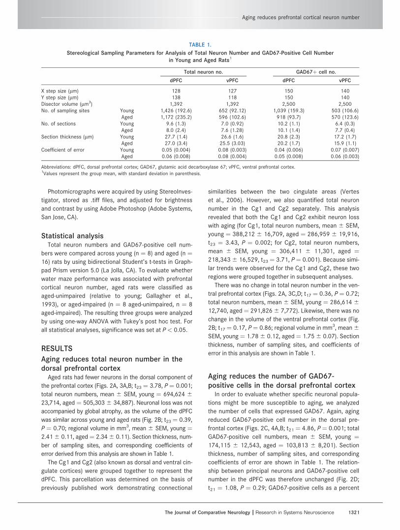
TABLE 1.
Stereological Sampling Parameters for Analysis of Total Neuron Number and GAD67-Positive Cell Number
in Young and Aged Rats1
Total neuron no. GAD67þ cell no.
dPFC vPFC dPFC vPFC
X step size (lm) 128 127 150 140Y step size (lm) 138 118 150 140Disector volume (lm3) 1,392 1,392 2,500 2,500No. of sampling sites Young 1,426 (192.6) 652 (92.12) 1,039 (159.3) 503 (106.6)
Aged 1,172 (235.2) 596 (102.6) 918 (93.7) 570 (123.6)No. of sections Young 9.6 (1.3) 7.0 (0.92) 10.2 (1.1) 6.4 (0.3)
Aged 8.0 (2.4) 7.6 (1.28) 10.1 (1.4) 7.7 (0.4)Section thickness (lm) Young 27.7 (1.4) 26.6 (1.6) 20.8 (2.3) 17.2 (1.7)
Aged 27.0 (3.4) 25.5 (3.03) 20.2 (1.7) 15.9 (1.1)Coefficient of error Young 0.05 (0.004) 0.08 (0.003) 0.04 (0.006) 0.07 (0.007)
Aged 0.06 (0.008) 0.08 (0.004) 0.05 (0.008) 0.06 (0.003)
Abbreviations: dPFC, dorsal prefrontal cortex; GAD67, glutamic acid decarboxylase 67; vPFC, ventral prefrontal cortex.1Values represent the group mean, with standard deviation in parenthesis.
Aging reduces prefrontal cortical neuron number
The Journal of Comparative Neurology | Research in Systems Neuroscience 1321

of principal neuron number, mean 6 SEM, young ¼ 25.4
6 1.72, aged ¼ 21.66 2.41).
There was no change in GAD67-positive cell number in
the ventral prefrontal cortex (Figs. 2C, 4C,D; t21 ¼ 0.46,
P ¼ 0.63; total GAD67-positive cell numbers, mean 6
SEM, young ¼ 34,923 6 5,847, aged ¼ 32,202 6
3,064). Section thickness, number of sampling sites, and
corresponding coefficients of error are shown in Table 1.
The percent of GAD67-positive cells relative to total neu-
ron number was likewise unchanged (Fig. 2D; t16 ¼ 1.47,
P ¼ 0.16; GAD67-positive cells as a percent of principal
neuron number, mean 6 SEM, young ¼ 13.81 6 2.35,
aged ¼ 10.25 6 1.24). Antibody penetration, defined as
the average z-depth of the counted cells, was similar
across young and aged rats (data not shown).
Reductions in neuron number occurindependently of spatial memory impairment
Rats were behaviorally categorized based on their perform-
ance in the hippocampus-dependent version of the water
maze, as described (Stranahan et al., 2011). Aged rats that
performed within the range of young, as measured by using
an index score derived from proximity to the goal platform
location during interpolated probe trials, were classified as
aged-unimpaired (AU), whereas rats that performed outside
of the range of young were designated aged-impaired (AI).
Neuronal loss in the dorsal prefrontal cortex occurred
in both AU and AI rats (Fig. 5A; F2,22 ¼ 8.84, P ¼ 0.005;
total neuron numbers, mean6 SEM, young ¼ 694,6246
23,714, AU ¼ 502,556 6 56,310, AI ¼ 508,050 6
45,201). Neuron number was maintained in the ventral
prefrontal cortex of aged rats, irrespective of spatial
memory performance (F2,16 ¼ 0.66, P ¼ 0. 53; total neu-
ron numbers, mean 6 SEM, young ¼ 286,614 6 12,740,
AU ¼ 280,914 6 9,104, AI ¼ 300,919 6 11,394). The
number of GAD67-positive cells was reduced with aging,
and this reduction was not associated with spatial mem-
ory performance (F2,22 ¼ 12.87, P ¼ 0.003; total GAD67-
positive cell numbers, mean6 SEM, young ¼ 174,115 6
12,543, AU ¼ 114,9646 14,757, AI¼ 94,0566 7,728).
To gain greater insight into mechanisms that might be
associated with spatial memory performance in aged
rats, we analyzed the percent of GAD67-positive neurons
relative to total neuron number. Age-related cognitive
Figure 2. Aging reduced total neuron number and the number of GAD67-positive cells in the dorsal component of the rat prefrontal cor-
tex. A: Unbiased stereological estimates of total neuron number reveal that aging is associated with neuronal loss in the dorsal prefrontal
cortex (dPFC). There was no effect of aging on neuron number in the ventral prefrontal cortex (vPFC). B: Changes in total neuron number
are not accompanied by gross regional atrophy, as indicated by volume estimates by using the method of Cavalieri. C: The number of
GAD67-positive cells is reduced with aging in the dPFC. There was no change in the number of GAD67-positive cells in the vPFC. D:
Because total neuron number is also reduced in the dorsal prefrontal cortex with aging, the ratio of GAD67-immunoreactive cells to princi-
pal neurons (expressed here as percent GAD67-positive cells relative to total pyramidal cell number on adjacent series of sections) is
unchanged. *, Significant difference at P < 0.05 following Student’s t-test.
Stranahan et al.
1322 The Journal of Comparative Neurology |Research in Systems Neuroscience

impairment was not associated with any difference in this
relationship (F2,22 ¼ 1.53, P ¼ 0.24; ratio of total neurons
to GAD67-positive cell number, mean 6 SEM, young ¼25.46 1.73, AU ¼ 24.66 4.87; AI¼ 19.06 1.34).
DISCUSSION
In the current report, we observed neuronal loss in the
dorsal component of the rat PFC. This loss occurred
among principal neurons and GAD67-immunoreactive
cells, such that a stable ratio of principal neurons to puta-
tive interneurons was maintained. Changes were confined
to the dPFC, as no alterations in total neuron number or
GAD67-positive cell number were observed in the vPFC,
and age-related neuronal loss was not associated with
spatial memory performance. We used behavioral charac-
terization in a hippocampal-dependent task because indi-
vidual differences in this model were previously shown to
correlate with age-related reductions in both hippocam-
pal and prefrontal cortical glucocorticoid receptor
Figure 3. Histological evidence for neuronal loss in the dorsal component of the prefrontal cortex in aged rats. A,B: Principal neuron num-
ber is greater in the dorsal prefrontal cortex of young rats (A), relative to aged rats (B). C,D: The number of principal neurons is unchanged
in the ventral prefrontal cortex of young (C) and aged (D) rats. Insets depict higher magnification images of Cresyl violet-stained cells in
the dorsal (A,B) and ventral (C,D) prefrontal cortex. Scale bar ¼ 100 lm in A (applies to A,B) and C (applies to C,D). Inset scale bar ¼ 20
lm in A (applies to A,B) and C (applies to C,D).
Aging reduces prefrontal cortical neuron number
The Journal of Comparative Neurology | Research in Systems Neuroscience 1323
expression (Bizon et al., 2001). We believe that when these
data are taken together with the current observation that
both aged-impaired and aged-unimpaired rats exhibit
decreased neuron number in the dorsal prefrontal region, to
the extent that such neuron loss is detrimental in this be-
havioral model, some compensatory mechanisms might be
recruited to maintain the performance of unimpaired rats.
In addition to interactions of the PFC with the medial
temporal lobe, the PFC participates in the default mode
network, defined as a set of anatomically connected cort-
ical regions that exhibited correlated activation patterns
at rest. Recent reports have demonstrated the existence
of a default mode network in humans (Honey et al.,
2009), non-human primates (Vincent et al., 2007), and
rodents (Liang et al., 2011). Correlations between levels
of activation across the default mode network and cogni-
tive impairment with aging have also been demonstrated
(Sperling et al., 2010), opening the possibility that neuron
loss in one component of this system could disrupt the
coordination of activity and information processing, with
consequences for cognitive functions not limited to hip-
pocampal-dependent memory.
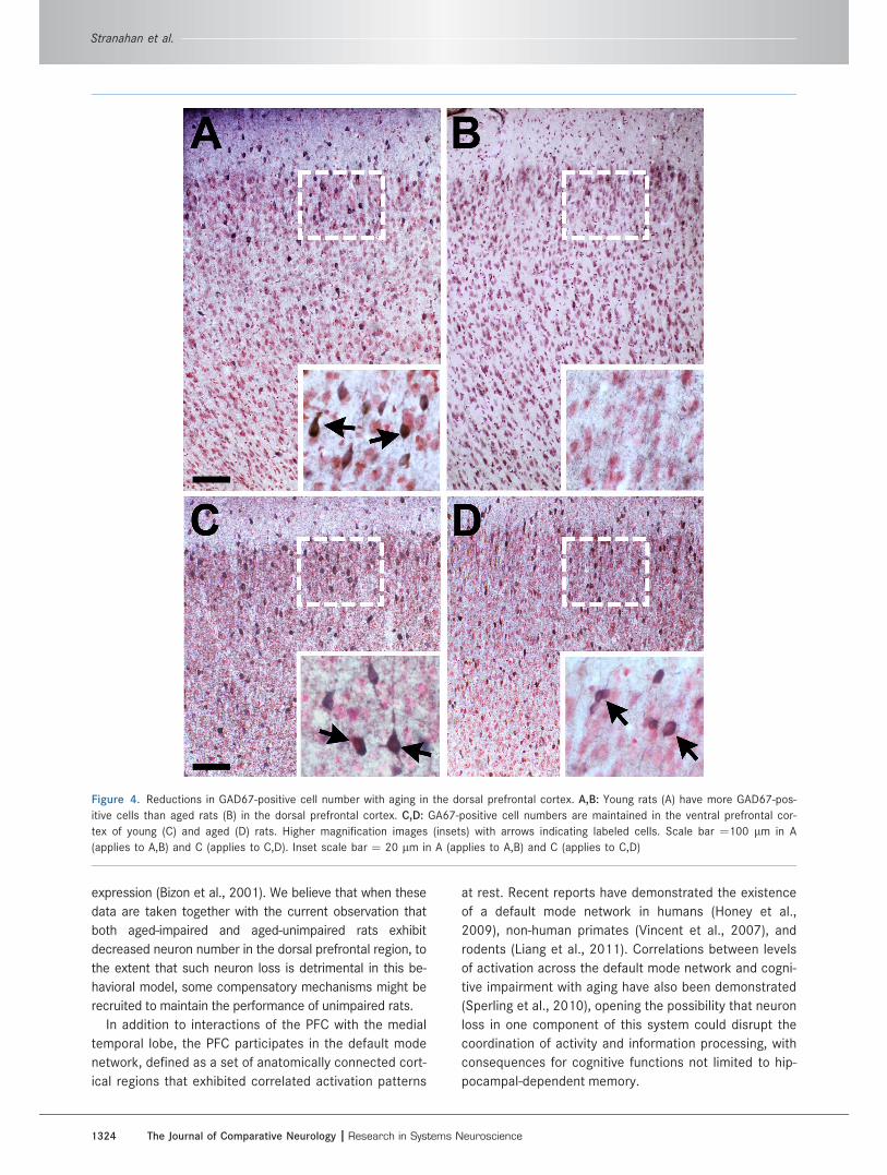
Figure 4. Reductions in GAD67-positive cell number with aging in the dorsal prefrontal cortex. A,B: Young rats (A) have more GAD67-pos-
itive cells than aged rats (B) in the dorsal prefrontal cortex. C,D: GA67-positive cell numbers are maintained in the ventral prefrontal cor-
tex of young (C) and aged (D) rats. Higher magnification images (insets) with arrows indicating labeled cells. Scale bar ¼100 lm in A
(applies to A,B) and C (applies to C,D). Inset scale bar ¼ 20 lm in A (applies to A,B) and C (applies to C,D)
Stranahan et al.
1324 The Journal of Comparative Neurology |Research in Systems Neuroscience
Independent effects of aging across neurobehavioral
systems have been reported and could be relevant to the
current findings. Aged rats perform poorly during atten-
tional set-shifting (Barense et al., 2002) in a task that has
been demonstrated to depend on the integrity of the pre-
frontal cortex (Birrell and Brown, 2000). It is noteworthy
that, in aged rats, individual differences in spatial memory
performance are uncorrelated with set-shifting ability,
suggesting that prefrontal cortical and medial temporal
memory systems exhibit some dissociable effects of
aging (Barense et al., 2002). The extent to which age-
related reductions in prefrontal cortical neuron number
and behavioral deficits in attentional set-shifting may be
attributable to changes in activation of the default mode
network remain to be determined.
Moreover, the question of whether neuron loss
observed in the current study contributes to age-related
deficits in attentional set-shifting has not yet been
addressed. The behavioral significance of neuron loss
detected in the cortex would be important to determine
because cognitive deficits in hippocampal-dependent
tasks are observed in the absence of altered neuron num-
ber in the medial temporal lobe but are correlated with
functional alterations in largely intact circuits (Rapp and
Gallagher, 1996; for review, see Wilson et al. 2006).
Our observation of neuronal loss in the dorsal region
and preservation of neuron number in the ventral region
differs from a previous report (Yates et al., 2008). Yates
and colleagues (2008) reported an age-dependent reduc-
tion in the vPFC, but not the dPFC. Although the regional
boundaries identified in the current study were reliably
detected on the same sections by two different observ-
ers, the possibility that this disparity arises from differen-
ces in regional parcellation across the two studies cannot
be ruled out. However, genetic divergence between the
Long-Evans hooded rats used by Yates et al. (2008),
derived from an in-house breeding colony, and the Long-
Evans rats purchased from Charles River Laboratories
for use in the current study is another possible reason
for differences in the results. Because genetic differen-
ces in the effect of ventral prefrontal lesions on behavior
have been demonstrated (Chang and Maren, 2010), it is
possible that genetic drift could account for distinct tra-
jectories of neuronal loss in subregions of the prefrontal
cortex.
With respect to the current findings, the stereological
data and prefrontal regional volumes derived from young
rats in our study fall closely within the range of esti-
mates from previous studies using unbiased stereologi-
cal methods in frozen tissue (Cerqueira et al., 2005;
Yates et al., 2008). Because using frozen sections could
affect counts through the z-axis, the current observa-
tions might underestimate absolute neuron numbers.
However, to our knowledge, no comparison values from
tissue prepared by using Vibratome sectioning exists for
these cortical regions, so it is difficult to assess what
effect sectioning on a freezing microtome might have
had on total neuron numbers. In that context, the
numerical trends observed in the current study may also
under-represent the absolute number of prefrontal
GAD67-positive interneurons, and numbers of cells
expressing GAD65 were not assessed.
Disinhibition of the PFC leads to deficits in cognitive
flexibility (Gruber et al., 2010), in a manner reminiscent of
the impaired reversal learning reported in a subset of
aged rats (Schoenbaum et al., 2006). We quantified the
number of GAD67-positive cells and expressed the ratio
of GAD67-positive cells to principal neurons to estimate
whether this population might be specifically vulnerable
to aging in the prefrontal cortex. Because both the num-
ber of principal neurons and the number of GAD67-
positive cells was reduced, the numerical relationship
between excitatory and GAD67-expressing inhibitory neu-
ron number was maintained. This suggests that neuronal
loss in the aging dPFC is not limited to a specific neuronal
phenotype. Perhaps most importantly, a focal age-related
decline in prefrontal cortical neuron number appears to
be conserved across rodents in the current study and in
primate species (Smith et al., 2004), suggesting that a
common basis for such vulnerability in cortical circuits
may exist in the mammalian brain.
ACKNOWLEDGMENTS
M.G. is the founder of AgeneBio Incorporated, a bio-
technology company that is dedicated to commercializing
therapies to treat cognitive impairment in aging, and she
has a financial interest in the company.
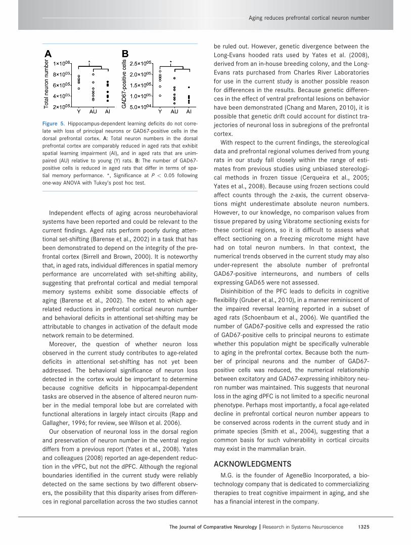
Figure 5. Hippocampus-dependent learning deficits do not corre-
late with loss of principal neurons or GAD67-positive cells in the
dorsal prefrontal cortex. A: Total neuron numbers in the dorsal
prefrontal cortex are comparably reduced in aged rats that exhibit
spatial learning impairment (AI), and in aged rats that are unim-
paired (AU) relative to young (Y) rats. B: The number of GAD67-
positive cells is reduced in aged rats that differ in terms of spa-
tial memory performance. *, Significance at P < 0.05 following
one-way ANOVA with Tukey’s post hoc test.
Aging reduces prefrontal cortical neuron number
The Journal of Comparative Neurology | Research in Systems Neuroscience 1325
LITERATURE CITEDBarense MD, Fox MT, Baxter MG. 2002. Aged rats are impaired
on an attentional set-shifting task sensitive to medial frontalcortex damage in young rats. Learn Mem 9:191–201.
Birrell JM, Brown VJ. 2000. Medial frontal cortex mediatesperceptual attentional set shifting in the rat. J Neurosci20:4320–4324.
Bizon JL, Helm KA, Han JS, Chun HJ, Pucilowska J, Lund PK,Gallagher M. 2001. Hypothalamic-pituitary-adrenal axisfunction and corticosterone receptor expression in behav-iorally categorized young and aged Long-Evans rats. Eur JNeurosci 14:1739–1751.
Cerqueira JJ, Pego JM, Taipa R, Bessa JM, Almeida OF, SousaN. 2005. Morphological correlates of corticosteroid-induced changes in prefrontal cortex-dependent behaviors.J Neurosci 25:7792–7800.
Chang CH, Maren S. 2010. Strain difference in the effect ofinfralimbic cortex lesions on fear extinction in rats. BehavNeurosci 124:391–397.
de Brabander JM, Kramers RJ, Uylings HB. 1998. Layer-specificdendritic regression of pyramidal cells with ageing in thehuman prefrontal cortex. Eur J Neurosci 10:1261–1269.
Dumitriu D, Hao J, Hara Y, Kaufmann J, Janssen WG, Lou W,Rapp PR, Morrison JH. 2010. Selective changes in thinspine density and morphology in monkey prefrontal cortexcorrelate with aging-related cognitive impairment. J Neuro-sci 30:7507–7515.
Fuster J. 2008. The prefrontal cortex (Fourth ed.). AcademicPress.
Gallagher M, Burwell R, Burchinal M. 1993. Severity of spatiallearning impairment in aging: development of a learningindex for performance in the Morris water maze. BehavNeurosci 107:618–626.
Gonzales C, Kaufman DL, Tobin AJ, Chesselet MF. 1991. Dis-tribution of glutamic acid decarboxylase (Mr 67,000) in thebasal ganglia of the rat: an immunohistochemical studywith a selective cDNA-generated polyclonal antibody. JNeurocytol 20:953–961.
Gruber AJ, Calhoon GG, Shusterman I, Schoenbaum G, RoeschMR, O’Donnell P. 2010. More is less: a disinhibited pre-frontal cortex impairs cognitive flexibility. J Neurosci 30:17102–17110.
Honey CJ, Sporns O, Cammoun L, Gigandet X, Thiran JP, MeuliR, Hagmann P. 2009. Predicting human resting-state func-tional connectivity from structural connectivity. Proc NatlAcad Sci U S A 106:2035–2040.
Levy R, Goldman-Rakic PS. 1999. Association of storage andprocessing functions in the dorsolateral prefrontal cortexof the nonhuman primate. J Neurosci 19:5149–5158.
Liang Z, King J, Zhang N. 2011. Uncovering intrinsic connec-tional architecture of functional networks in awake ratbrain. J Neurosci 31:3776–3783.
Paxinos, G, Watson C. 1998. The Rat Brain Stereotaxic Coor-dinates. Academic Press.
Peters A, Sethares C, Moss MB. 1998. The effects of agingon layer 1 in area 46 of prefrontal cortex in the rhesusmonkey. Cereb Cortex 8:671–684.
Rapp PR, Gallagher M. 1996. Preserved neuron number in thehippocampus of aged rats with spatial learning deficits.Proc Natl Acad Sci U S A 93:9926–9930.
Rapp PR, Deroche PS, Mao Y, Burwell RD. 2002. Neuron num-ber in the parahippocampal region is preserved in agedrats with spatial learning deficits. Cereb Cortex 12:1171–1179.
Schoenbaum G, Setlow B, Saddoris MP, Gallagher M. 2006.Encoding changes in orbitofrontal cortex in reversal-impaired aged rats. J Neurophysiol 95:1509–1517.
Smith DE, Rapp PR, McKay HM, Roberts JA, Tuszynski MH.2004. Memory impairment in aged primates is associatedwith focal death of cortical neurons and atrophy of sub-cortical neurons. J Neurosci 24:4373–4381.
Sperling RA, Dickerson BC, Pihlajamaki M, Vannini P, LaVio-lette PS, Vitolo OV, Hedden T, Becker JA, Rentz DM, Sel-koe DJ, Johnson KA. 2010. Functional alterations inmemory networks in early Alzheimer’s disease. NeuromolMed 12:27–43.
Stranahan AM, Haberman RP, Gallagher M. 2011. Cognitivedecline is associated with reduced reelin expression inthe entorhinal cortex of aged rats. Cereb Cortex 21:392–400.
Tisserand DJ, Pruessner JC, Sanz Arigita EJ, van Boxtel MP,Evans AC, Jolles J, Uylings HB. 2002. Regional frontal corti-cal volumes decrease differentially in aging: an MRI studyto compare volumetric approaches and voxel-based mor-phometry. Neuroimage 17:657–669.
Uylings HB, Groenewegen HJ, Kolb B. 2003. Do rats have aprefrontal cortex? Behav Brain Res 146:3–17.
Van Eden CG, Uylings HB. 1985. Cytoarchitectonic develop-ment of the prefrontal cortex in the rat. J Comp Neurol241:253–267.
Vertes RP, Hoover WB, Do Valle AC, Sherman A, Rodriguez JJ.2006. Efferent projections of reuniens and rhomboid nucleiof the thalamus in the rat. J Comp Neurol 499:768–796.
Vincent JL, Patel GH, Fox MD, Snyder AZ, Baker JT, Van EssenDC, Zempel JM, Snyder LH, Corbetta M, Raichle ME. 2007.Intrinsic functional architecture in the anaesthetized mon-key brain. Nature 447:83–86.
Wilson IA, Gallagher M, Eichenbaum H, 2006. Tanila H. Neuro-cognitive aging: prior memories hinder new hippocampalencoding. Trends Neurosci 29:662–670.
Yates MA, Markham JA, Anderson SE, Morris JR, Juraska JM.2008. Regional variability in age-related loss of neuronsfrom the primary visual cortex and medial prefrontal cortexof male and female rats. Brain Res 1218:1–12.
Stranahan et al.
1326 The Journal of Comparative Neurology |Research in Systems Neuroscience