Embed Size (px)
DESCRIPTION
Agent-based modelling of epithelial cells. An example of rule formulation and extension Dr Dawn Walker, University of Sheffield, UK. What determines cell behaviour?. Environmental factors Extracellular matrix Calcium concentration Growth medium. Other cells Intercellular bonds - PowerPoint PPT Presentation
Citation preview
Agent-based modelling of epithelial cells
An example of rule formulation and extension
Dr Dawn Walker, University of Sheffield, UK
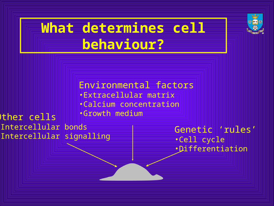
What determines cell behaviour?
Other cells•Intercellular bonds•Intercellular signalling
Environmental factors•Extracellular matrix•Calcium concentration•Growth medium
Genetic ‘rules’•Cell cycle•Differentiation
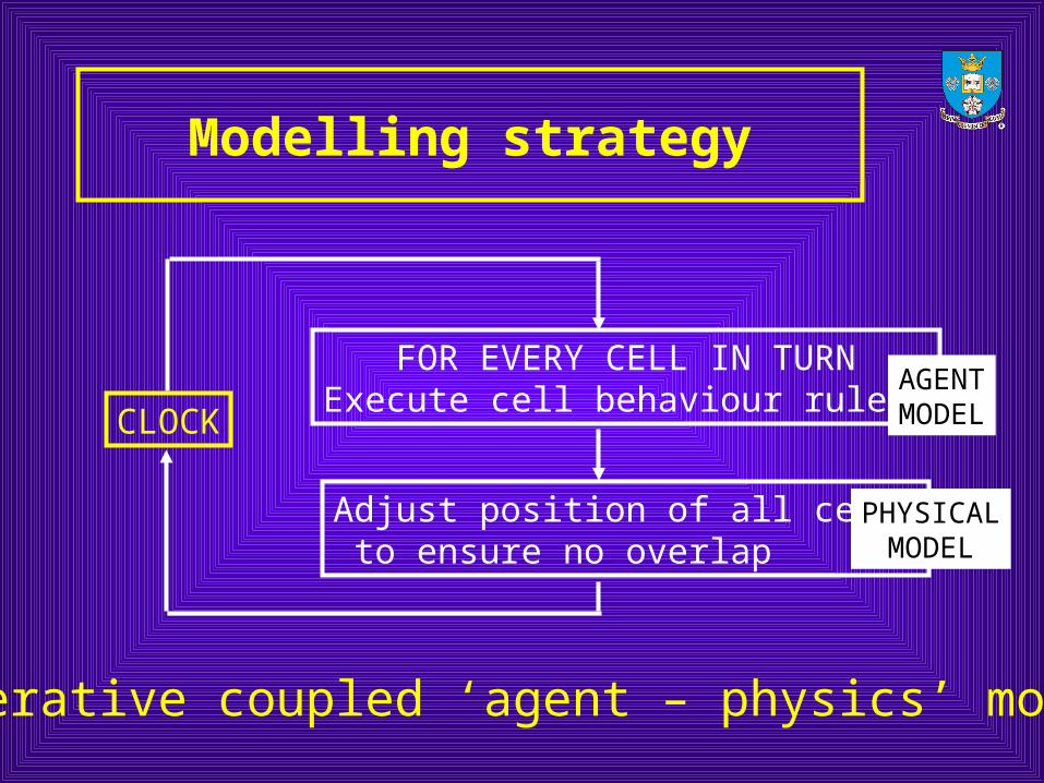
Modelling strategy
CLOCK
FOR EVERY CELL IN TURNExecute cell behaviour rules
Adjust position of all cells to ensure no overlap
AGENTMODEL
PHYSICALMODEL
Iterative coupled ‘agent – physics’ model
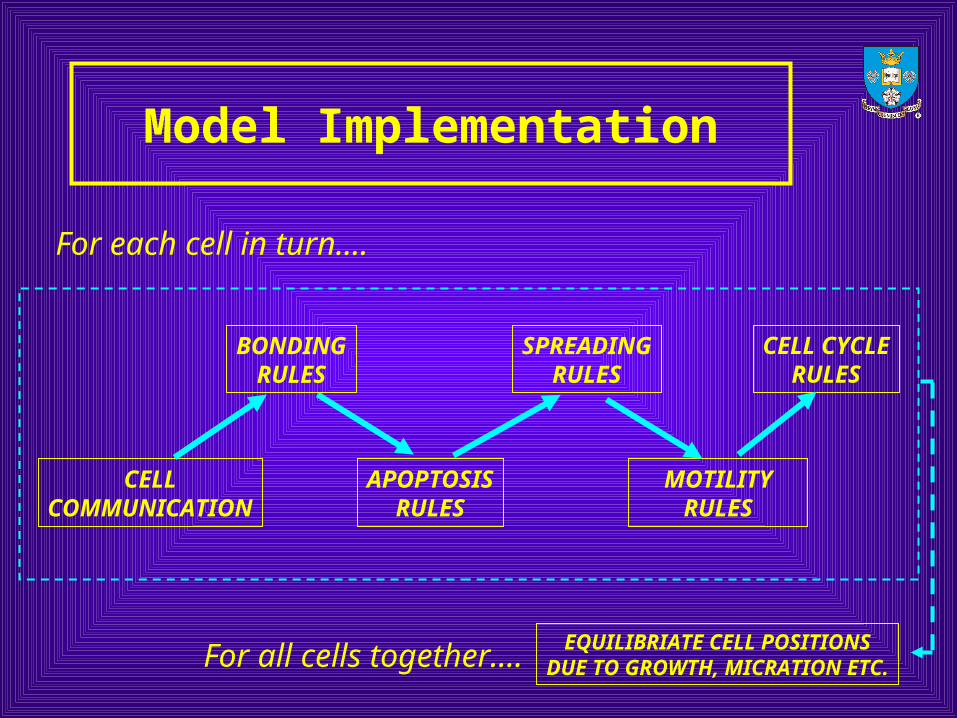
Model Implementation
CELLCOMMUNICATION
APOPTOSISRULES
MOTILITYRULES
BONDINGRULES
SPREADINGRULES
CELL CYCLERULES
For each cell in turn….
For all cells together…. EQUILIBRIATE CELL POSITIONSDUE TO GROWTH, MICRATION ETC.
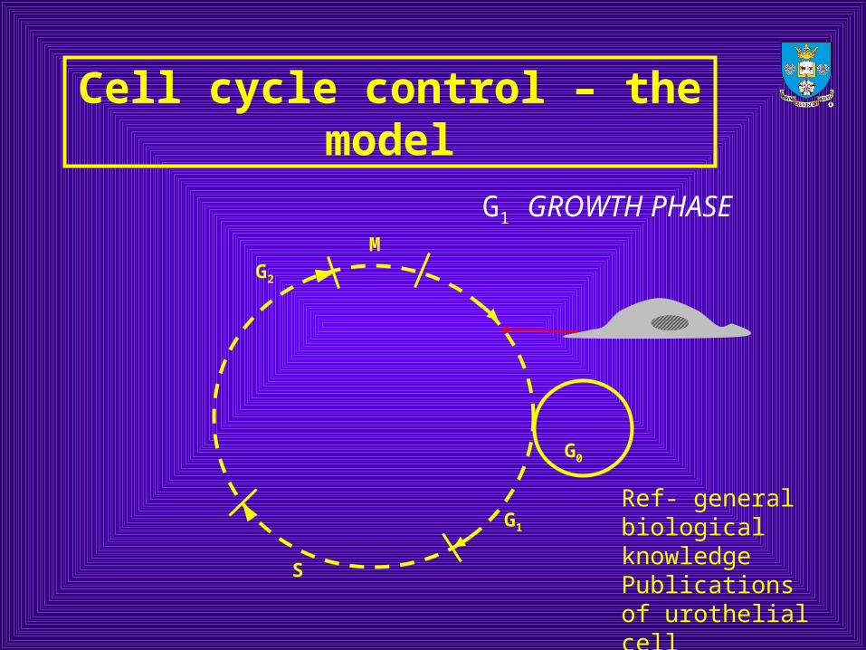
Cell cycle control – the model
M
G2
S
G1
G0
G1 GROWTH PHASE
Ref- general biological knowledgePublications of urothelial cell proliferation time
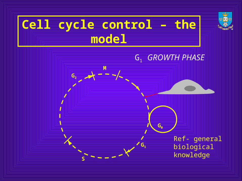
Cell cycle control – the model
M
G2
S
G1
G0
G1 GROWTH PHASE
Ref- general biological knowledge
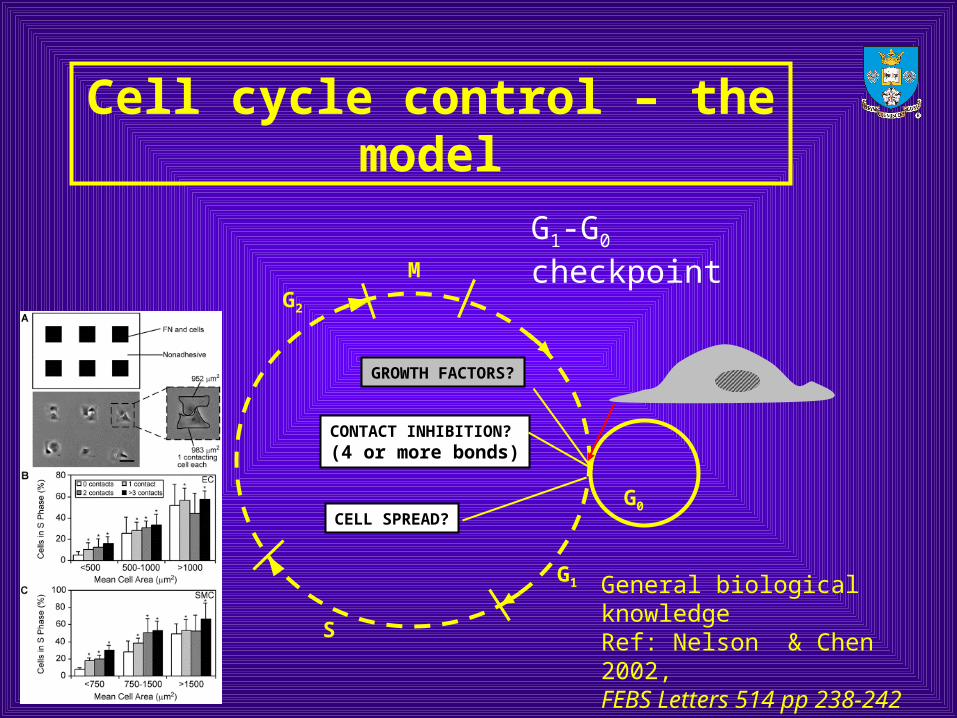
Cell cycle control – the model
M
G2
S
G1
G0
CONTACT INHIBITION?(4 or more bonds)
CELL SPREAD?
G1-G0 checkpoint
GROWTH FACTORS?
General biological knowledgeRef: Nelson & Chen 2002,FEBS Letters 514 pp 238-242
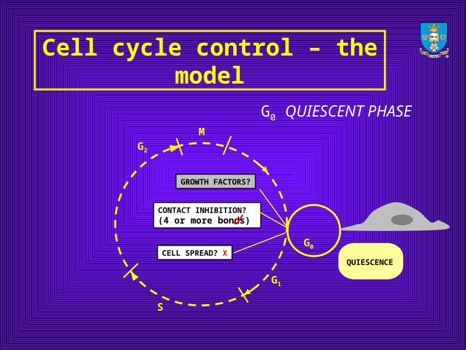
Cell cycle control – the model
M
G2
S
G1
G0
CONTACT INHIBITION?(4 or more bonds)
CELL SPREAD? XQUIESCENCE
G0 QUIESCENT PHASE
GROWTH FACTORS?
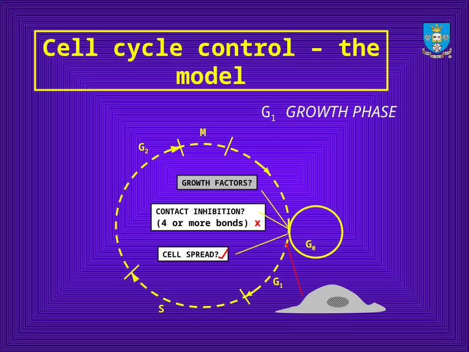
Cell cycle control – the model
M
G2
S
G1
G0
CONTACT INHIBITION?
(4 or more bonds) x
CELL SPREAD?
G1 GROWTH PHASE
GROWTH FACTORS?
Cell cycle control – the model
M
G2
S
G1
G0
S PHASE – (CHROMOSOMEREPLICATION)
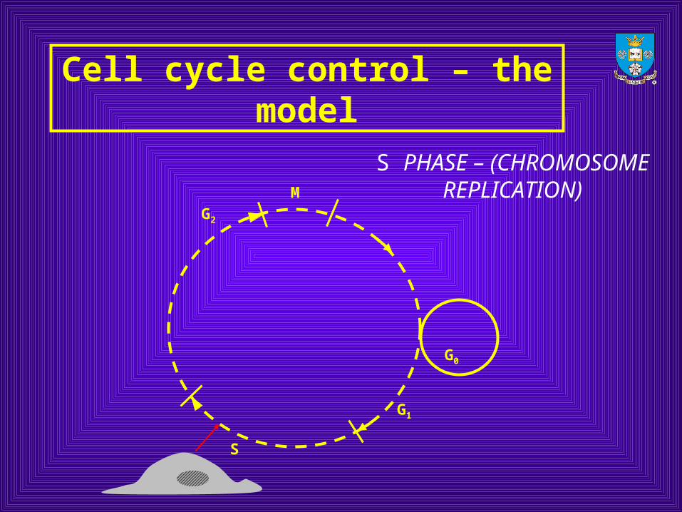
Cell cycle control – the model
M
G2
S
G1
G0
G2 PHASE – (HOUSEKEEPING)
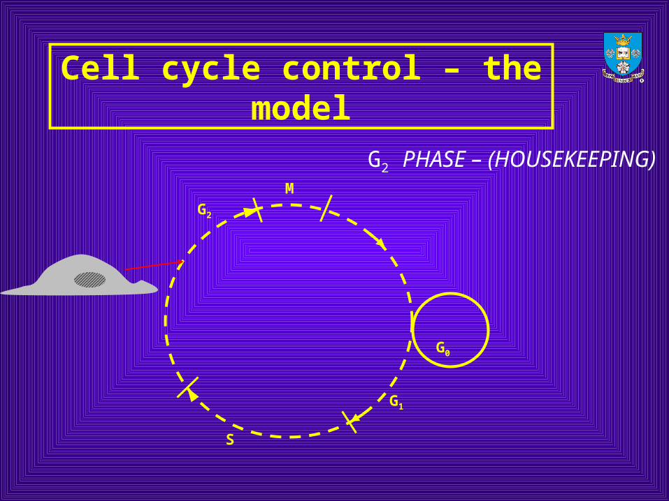
Cell cycle control – the model
M
G2
S
G1
G0
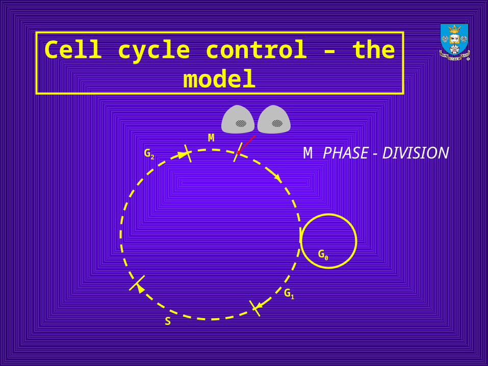
M PHASE - DIVISION
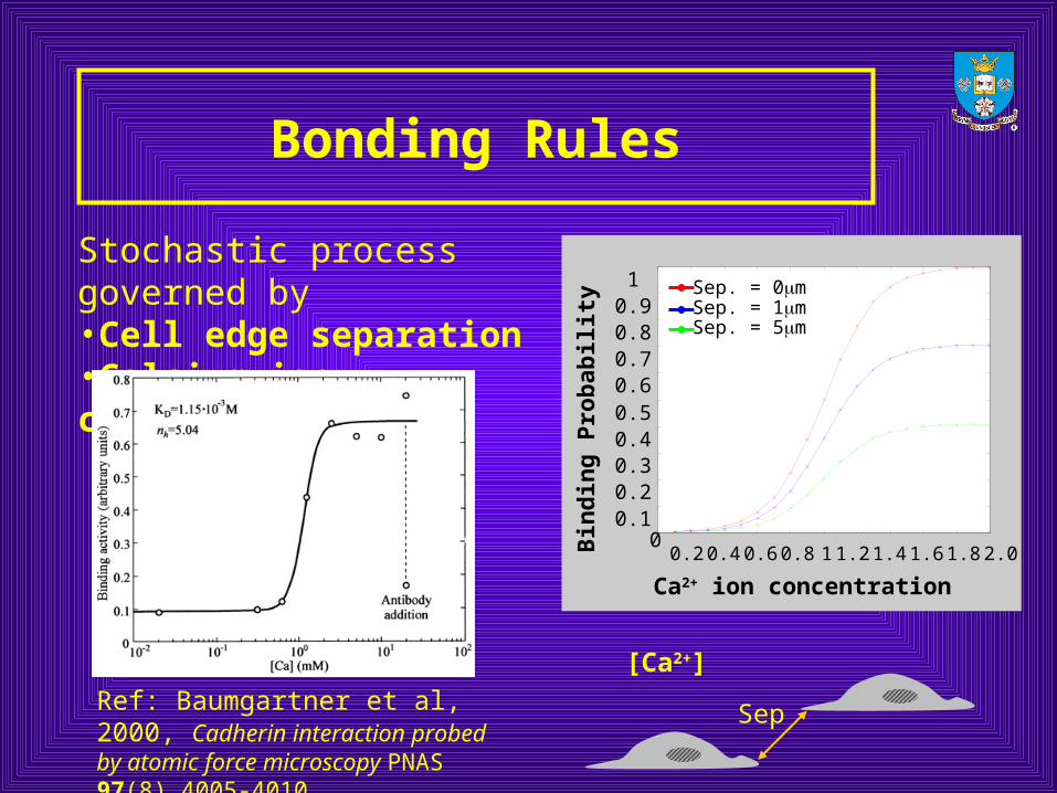
Bonding Rules
Stochastic process governed by•Cell edge separation•Calcium ion concentration
0.10.20.30.40.50.60.70.80.91
0.2 0.4 0.6 0.8 1 1.2 1.4 1.6 1.8 2.00
Bin
din
g P
rob
abili
tyCa2+ ion concentration
Sep. = 0mSep. = 1mSep. = 5m
Sep
[Ca2+]
Ref: Baumgartner et al, 2000, Cadherin interaction probed by atomic force microscopy PNAS 97(8) 4005-4010.
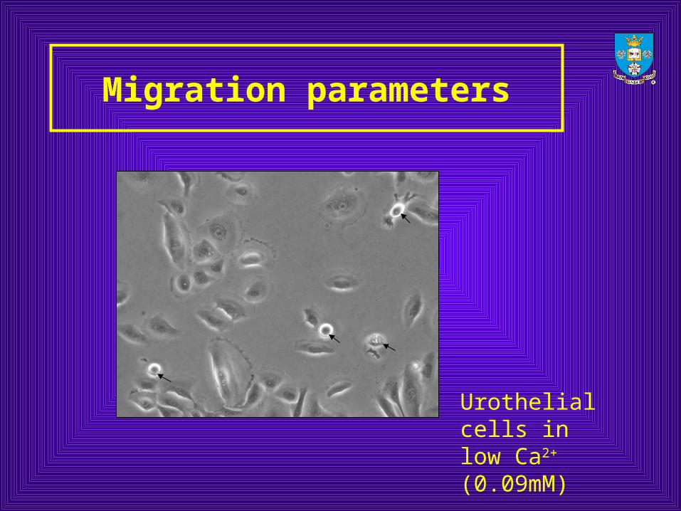
Migration parameters
Urothelial cells in low Ca2+ (0.09mM)
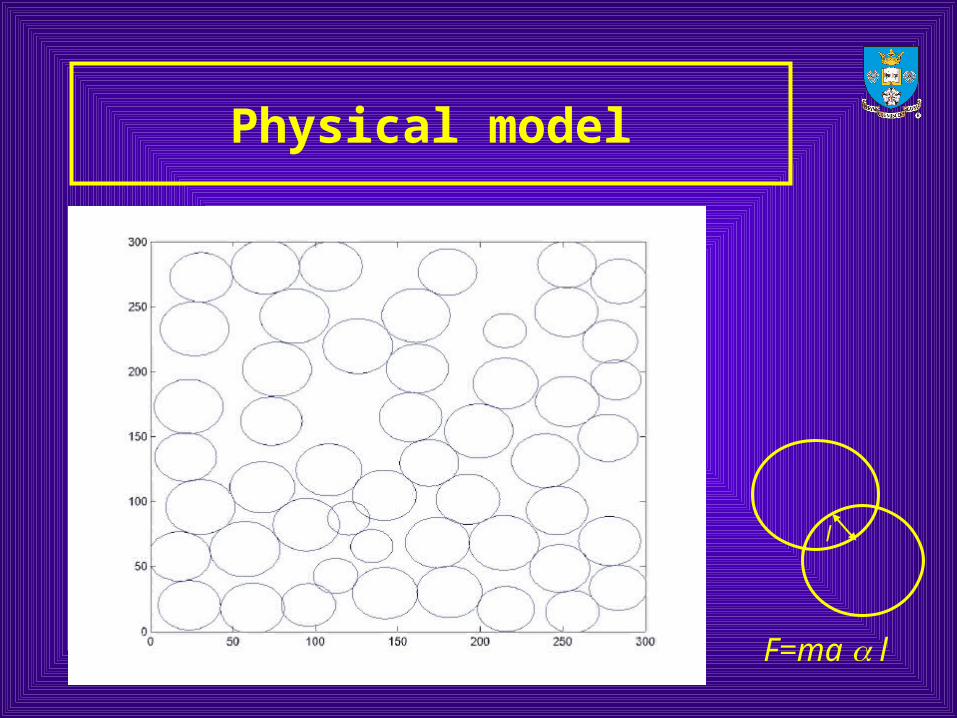
Physical model
l
F=ma l
Ca2+ dependent behaviour - In Vitro vs. In Virtuo
• Intercellular bonds require the presence of Ca2+ ions
• In Ca2+ conc.> 1mM many bonds are formed
• Cells with several intercellular bonds become contact-inhibited (stop cycling)
• WHAT IS THE EFFECT OF Ca2+ ON GROWTH AND PROLIFERATION?
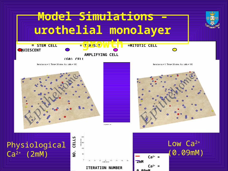
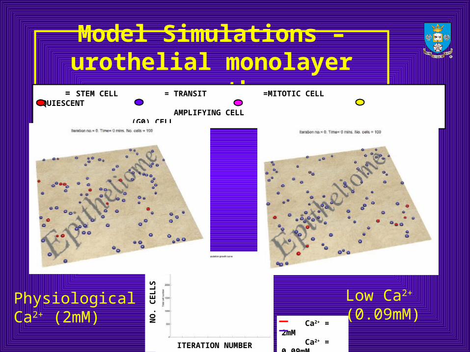
= STEM CELL = TRANSIT =MITOTIC CELL =QUIESCENT AMPLIFYING CELL (G0) CELL
Model Simulations – urothelial monolayer growth
Physiological Ca2+ (2mM)
Low Ca2+ (0.09mM)
ITERATION NUMBER
NO
. CE
LL
S
Ca2+ = 2mM Ca2+ = 0.09mM
Model Simulations – urothelial monolayer growth
Physiological Ca2+ (2mM)
Low Ca2+ (0.09mM)
ITERATION NUMBER
NO
. CE
LL
S
Ca2+ = 2mM Ca2+ = 0.09mM
= STEM CELL = TRANSIT =MITOTIC CELL =QUIESCENT AMPLIFYING CELL (G0) CELL

In virtuo wound healing (urothelium)
Physiological Ca2+ (2mM) Low Ca2+ (0.09mM)
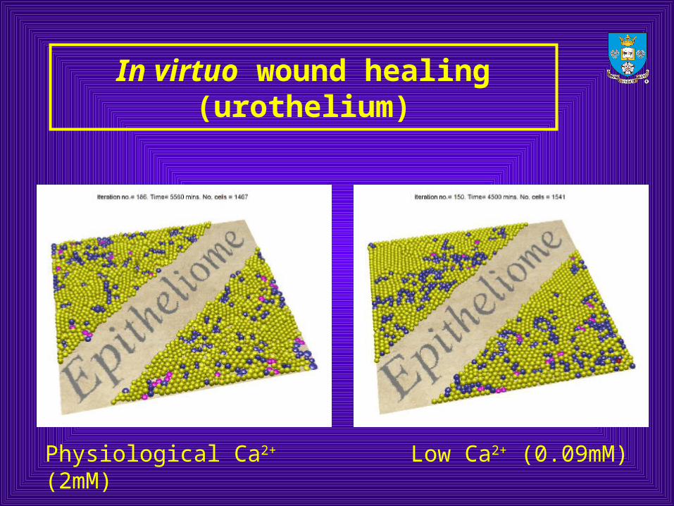
In virtuo wound healing (urothelium)
Physiological Ca2+ (2mM) Low Ca2+ (0.09mM)
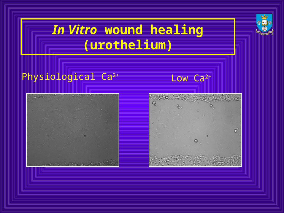
In Vitro wound healing (urothelium)
Low Ca2+Physiological Ca2+
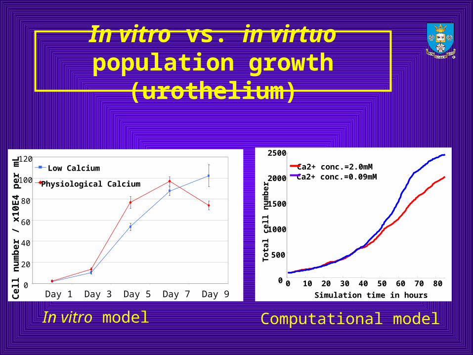
In vitro vs. in virtuo population growth (urothelium)
In vitro model Computational model
0
500
1000
1500
2000
2500
0 10 20 30 40 50 60 70 80
Simulation time in hours
To
tal
ce
ll n
um
be
r
Ca2+ conc.=2.0mMCa2+ conc.=0.09mM
0
20
40
60
80
100
120
Day 1 Day 3 Day 5 Day 7 Day 9
Cel
l n
um
ber
/ x
10E
4 p
er m
L Low Calcium
Physiological Calcium
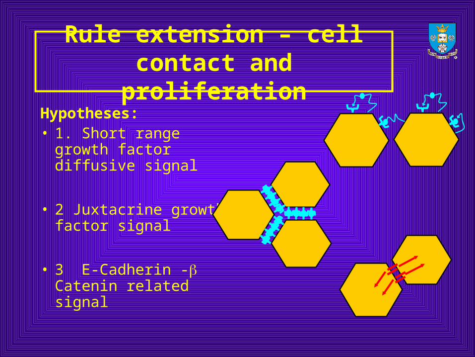
Rule extension – cell contact and proliferation
Hypotheses:• 1. Short range growth
factor diffusive signal
• 2 Juxtacrine growth factor signal
• 3 E-Cadherin - Catenin related signal
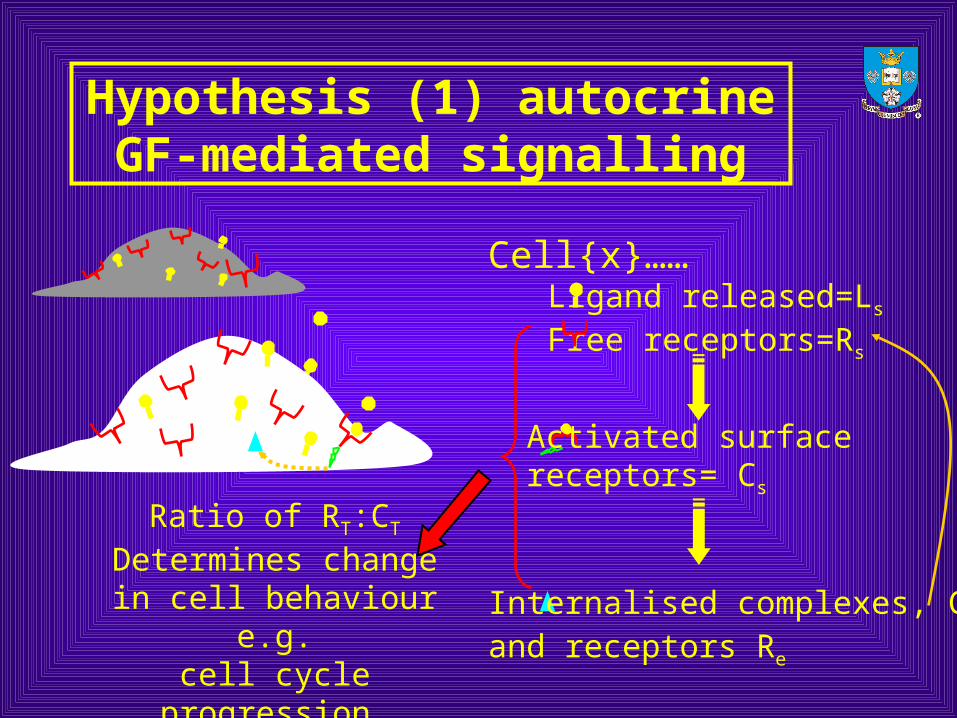
Hypothesis (1) autocrine GF-mediated signalling
Cell{x}……Ligand released=Ls
Free receptors=Rs
Ratio of RT:CT
Determines change in cell behaviour e.g.
cell cycle progression, migration
Internalised complexes, Ce
and receptors Re
Activated surface receptors= Cs
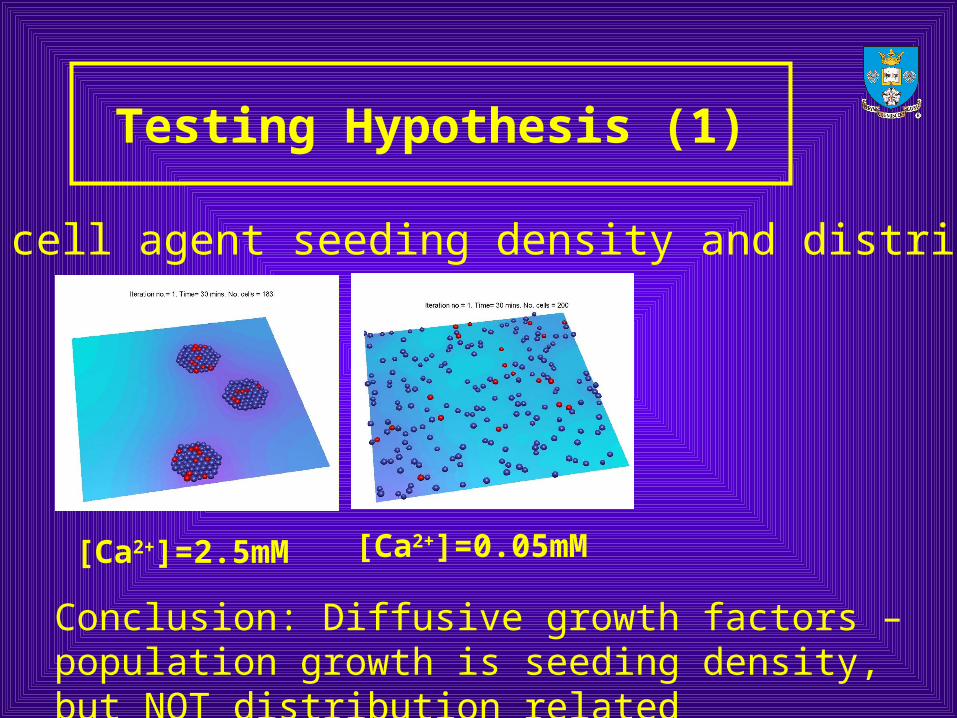
Testing Hypothesis (1)
[Ca2+]=0.05mM[Ca2+]=2.5mM
Initial cell agent seeding density and distribution
Conclusion: Diffusive growth factors – population growth is seeding density, but NOT distribution related
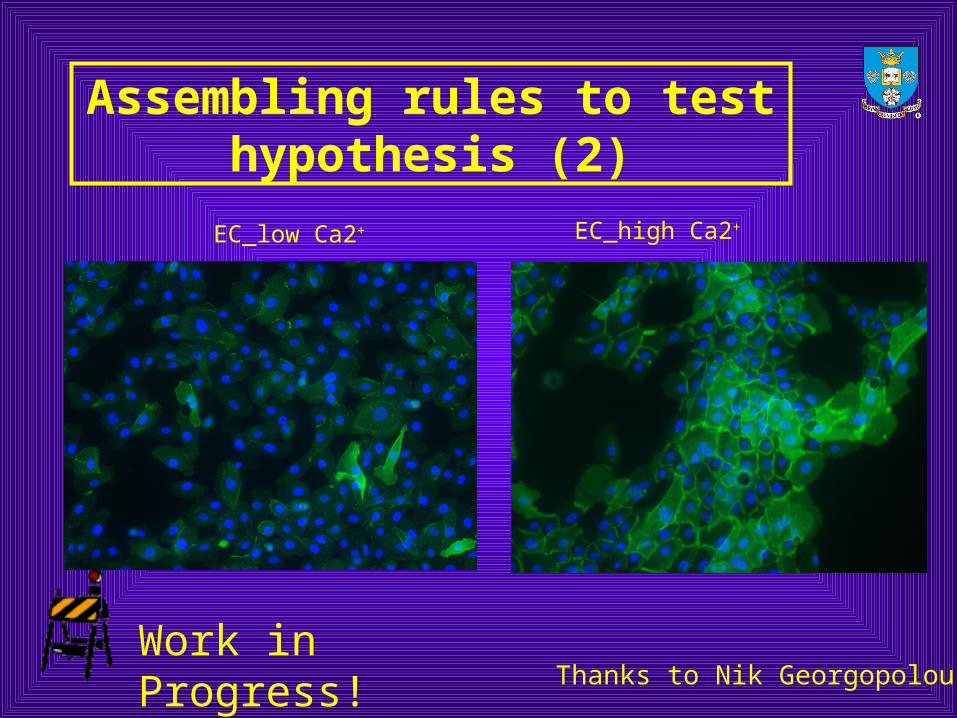
Assembling rules to test hypothesis (2)
EC_high Ca2+EC_low Ca2+
Work in Progress!Thanks to Nik Georgopolous
Summary
• Initial rule formulation can be based on simplifications and abstractions of known biological behaviour
• Iterative comparison with experimental data can improve the accuracy of the model and direct experimental investigation
• The rule set can be extended to model additional aspects of cell behaviour (e.g. differentiation, stratification)
• Rules can be replaced by more complex models (e.g. inter- and intra- cellular signalling)

Thank you for listening
Any Questions?