Embed Size (px)
DESCRIPTION
Age-Related macular degeneration & retinitis pigmentosa Ayesha S abdullah. 17.01.2014. Learning outcomes. By the end of this lecture the students would be able to Describe the epidemiology of ARMD - PowerPoint PPT Presentation
Citation preview

AGE-RELATED MACULAR DEGENERATION
& RETINITIS PIGMENTOSA
AYESHA S ABDULLAH
17.01.2014

Learning outcomes
By the end of this lecture the students would be able to
1. Describe the epidemiology of ARMD2. Correlated the clinical presentation of age-
related macular degeneration (ARMD) with the underlying pathophysiology
3. Outline the principles of treatment of ARMD4. Describe the epidemiology and clinical
presentation of retinitis pigmentosa

Case
A 72 year old man presented to the OPD with the complaints of difficulty in recognizing faces and distortion of vision for the last one year.
When he reads the words seem distorted and wavy with the left eye.
He is a known hypertensive and smoker for the last 40 years. His elder brother has similar problem.

Physical examination
VA: 6/12 OD 6/24 OS Anterior segment: bilateral early
cataract Pupils : normal Posterior segment: fundus photograph Defect on Amsler Grid


How is ARMD caused?
What is the function of RPE? Absorb light, transport oxygen & nutrients to the
outer retina Transport cellular wastes from the outer retina to
choroid In ARMD RPE dysfunction leads to accumulation
of the waste products under the retina at the interface between the Bruch’s membrane and RPE in the form of yellowish deposits
They can be seen on ophthalmoscopy as “Drusen”


Aaetiology
Oxidative damage
Microvascular disease
Genetic predisposition
Known risk factors


Choroid
RPEBruch’s


Choroid
Bruch’sRPE

Choroid
Bruch’sRPE


It is a slow process RPE continues to slow down transport of nutrients and
wastes Overlying photoreceptors and RPE get atrophic It may continue as a slowly progressive “dry” form of the
disease In some it assumes a more aggressive “wet form” New vessels grow from the choroid underneath the retina
forming a neovascular membrane. It leaks and can bleed Resulting in severe visual damage The macula may undergo fibrosis and scarring- “diskiform
scar”


EpidemiologyWhat is the magnitude of ARMD?
Leading cause of blindness in developed countries over the age of 50 years
Whites are twice at risk as compared to blacks

Who is at risk?
Age Family history Smoking doubles the risk Hypertension Cardiovascualr disease Hyperlipidemias Variants of complement factor H Excessive exposure to sunlight

Can it be prevented?
No Risk modification

How can it be treated Risk modification Monitoring – Amsler Grid Diet . Age-Related Eye Disease Study
(AREDS), showed that those who are at high risk for developing advanced age-related macular degeneration, may be helped by taking a specific combination of antioxidants and zinc.
Vitamin C, Vitamin E, Vitamin A as beta-carotene*, Zinc , Copper in a specific dose
*with caution in smokers, increases the risk of lung cancer

Specific treatment
Anti-VEGF intravitreal injections Photodynamic therapy Thermal Laser treatment Surgical excision of the membrane

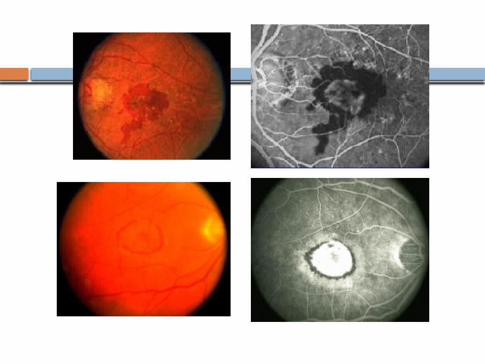
Rehab
Low vision aids

Electronic devices


Directions for using the Amsler grid
1. If you wear reading glasses, put them on for this test.
2. Hold this book at a comfortable reading distance.
3. Cover one of your eyes.
4. Look at the grid. Keep your eye focused on the white dot at the center of the grid throughout the test.
5. Without moving your eye from the center dot, notice the lines that make up the grid. All of the lines should be straight and all of the squares should look the same.
6. There shouldn’t be any blank, dark, or distorted areas on the grid.
7. Call your eye doctor right away if you notice anything unusual or abnormal in your vision.
8. Use the same procedure to test your other eye.

Retinitis pigmentosa
A degenerative retinal disease Varying pattern of inheritance
Autosomal dominant. 20% of cases (AD) Autosomal recessive (AR) X- linked. Rare with worst prognosis (XL) Isolated cases
Photoreceptor dysfunction primarily affecting rods Systemic associations and deafness 1:5000 prevalence Can have a typical or atypical clinical presentation

How can it be diagnosed?
Bilateral involvement Night blindness Loss of peripheral vision Signs
Arteriolar narrowing Bone specule pigmentary changes Waxy pale disc
ERG Perimetry


How progressive is the visual loss
Starts in childhood Progresses very slowly Rarely the person is blind by the age of 30
year ( X-linked) Rarely proceeds to complete blindness AD-best prognosis with retention of
central vision beyond the 6th decade XL has the worst prognosis with severe
visual loss by the 4th decade

Ocular associations
Cataract Glaucoma Myopia Keratoconus Macular oedema Vitreous changes Optic disc drusen Coats-like disease with exudative RD

Is it treatable?
No The associated ocular diseases like,
cataract, macular oedema , myopia and glaucoma can be treated
The person would be needing low vision aids when the visual field loss becomes more.
Needs to be differentiated from certain drug induced retinopathies ( chloroquine & thioridazine), infections (syphilis) and severe posterior uveitis