Embed Size (px)
Citation preview
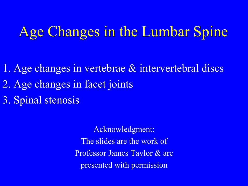
Age Changes in the Lumbar Spine
1. Age changes in vertebrae & intervertebral discs
2. Age changes in facet joints
3. Spinal stenosis
Acknowledgment:
The slides are the work of
Professor James Taylor & are
presented with permission
Age changes in lumbar vertebrae:
Osteoporosis
• Lumbar vertebrae are shorter & wider with more
concave endplates in old age.
• Selective loss of transverse trabeculae from the
cancellous bone within vertebral bodies ->
• Loss of the stiffness provided by transverse ties ->
collapse of the vertical load bearing beams ->
• Vertebral end plates collapse -> increased concavity
• Loss of stature with ageing
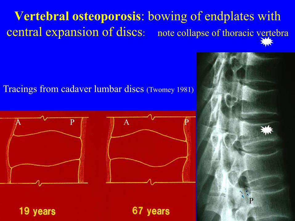
Vertebral osteoporosis: bowing of endplates with
central expansion of discs: note collapse of thoracic vertebra
Tracings from cadaver lumbar discs (Twomey 1981)
A P A P
A P
AGEING IN LUMBAR DISCS
MORPHOLOGY AND CHEMISTRY
• Increased convexity at vertebral end plates
• Loss of proteoglycans (PG) & water
• Disc bulging at posterior margin
• Disc fissuring: circumferential + radial
• Loss of disc height in 33% of L4-5 & L5-S1
Lumbar disc degeneration (LDD)
• Bulge or Prolapse: usually asymptomatic
• Herniation (less common): may result from
trauma and produce nerve root irritation
(chemical) or nerve root compression (stenosis)
• Internal disruption: acute fissuring of the
innervated outer 1/3rd of the annulus is the
most common cause of discogenic pain (Crock
Spine 11:650, 1986)
Lumbar Disc Degeneration (LDD)
& Symptoms
• Accelerated by lifetime overloading, in
heavy manual work or some elite sports
• LDD shows significant genetic predisposition
• Discography in a fissured disc may provoke a
patient’s pain
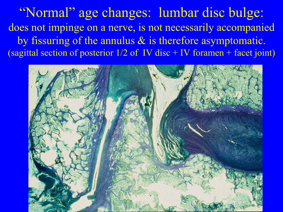
“Normal” age changes: lumbar disc bulge: does not impinge on a nerve, is not necessarily accompanied
by fissuring of the annulus & is therefore asymptomatic. (sagittal section of posterior 1/2 of IV disc + IV foramen + facet joint)
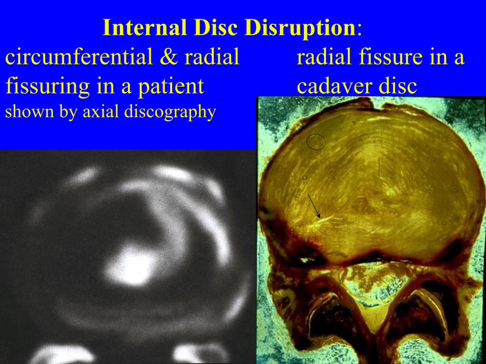
Internal Disc Disruption:
circumferential & radial radial fissure in a
fissuring in a patient cadaver disc shown by axial discography
AGEING IN LUMBAR DISCS
FUNCTIONAL CHANGES
• Increased disc stiffness
• Decreased ranges of movement
• Increased creep – progressive deformation (strain) of a structure under
prolonged loading (stress)
• Increased hysteresis – the recovery from distortion: the lag of recovery from
deformation (strain) after the load (stress) has been removed
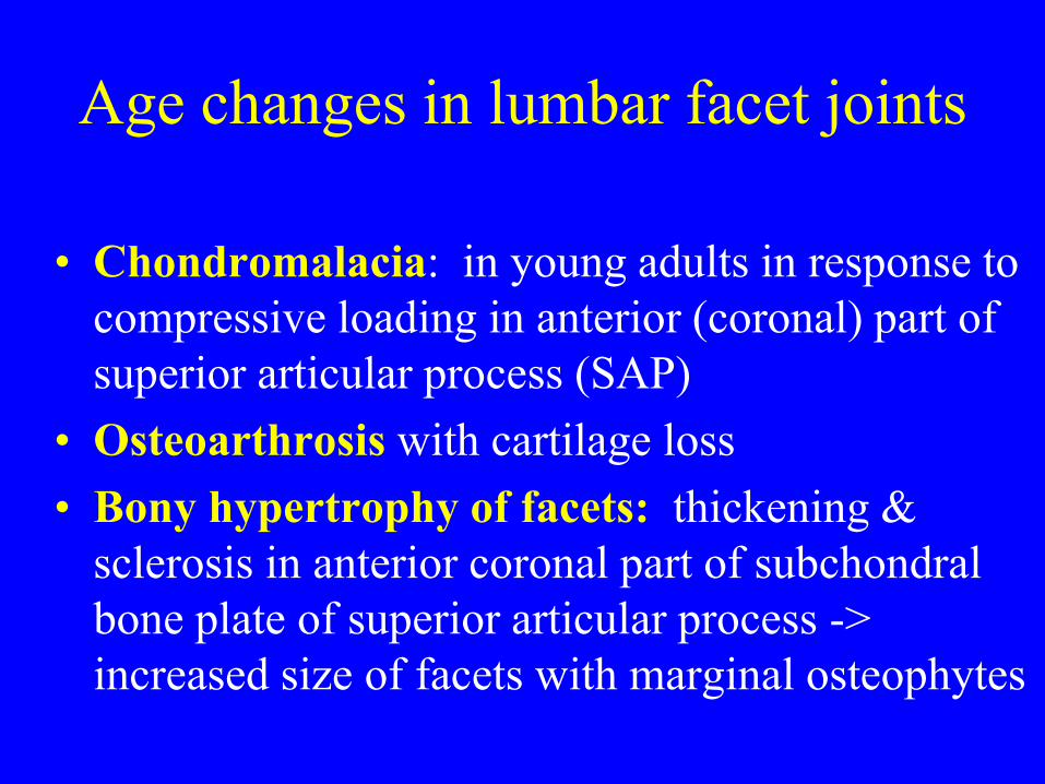
Age changes in lumbar facet joints
• Chondromalacia: in young adults in response to
compressive loading in anterior (coronal) part of
superior articular process (SAP)
• Osteoarthrosis with cartilage loss
• Bony hypertrophy of facets: thickening &
sclerosis in anterior coronal part of subchondral
bone plate of superior articular process ->
increased size of facets with marginal osteophytes
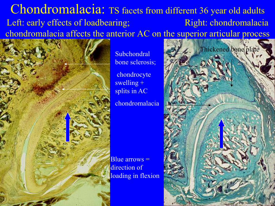
Chondromalacia: TS facets from different 36 year old adults Left: early effects of loadbearing; Right: chondromalacia
chondromalacia affects the anterior AC on the superior articular process
Subchondral
bone sclerosis;
chondrocyte
swelling +
splits in AC
chondromalacia
Thickened bone plate
Blue arrows =
direction of
loading in flexion
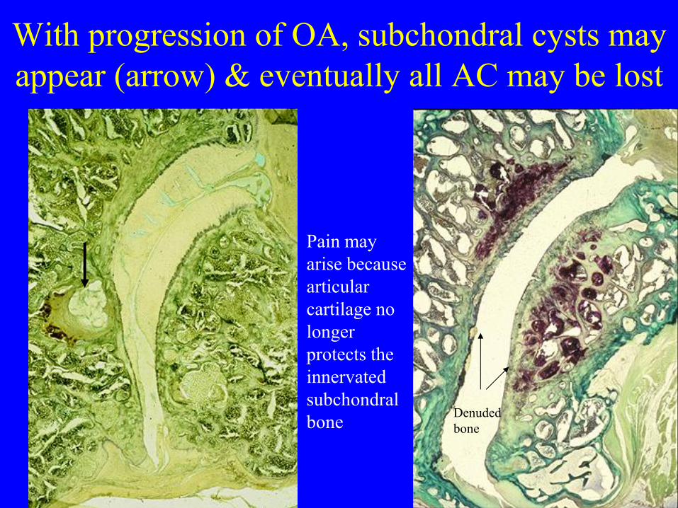
With progression of OA, subchondral cysts may
appear (arrow) & eventually all AC may be lost
Denuded
bone
Pain may
arise because
articular
cartilage no
longer
protects the
innervated
subchondral
bone
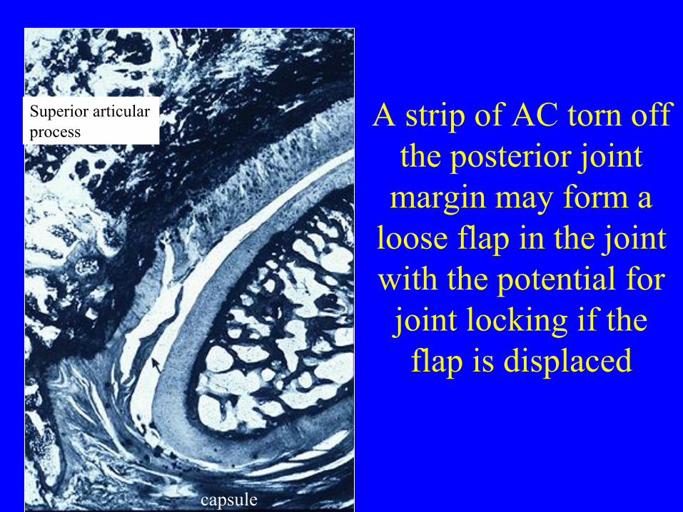
A strip of AC torn off
the posterior joint
margin may form a
loose flap in the joint
with the potential for
joint locking if the
flap is displaced
capsule
Superior articular
process
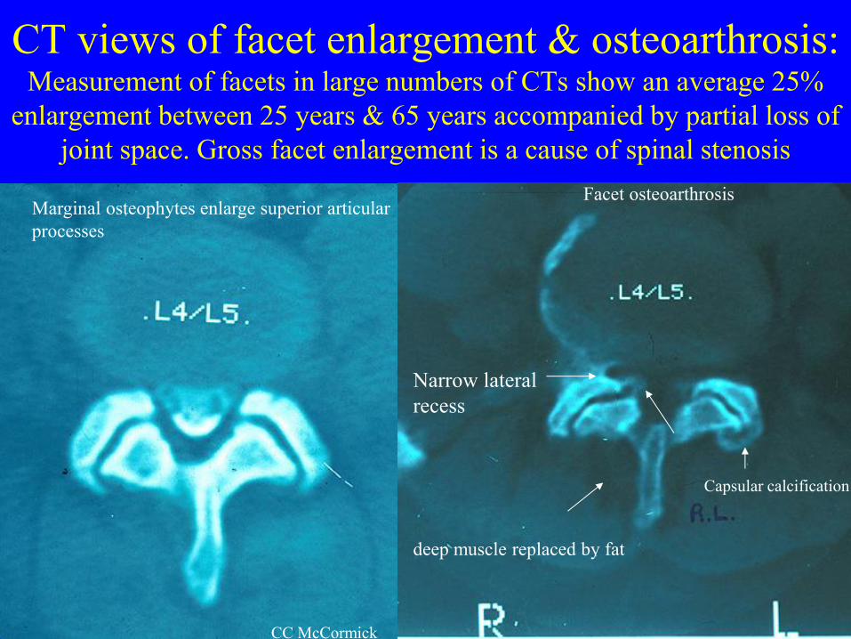
CT views of facet enlargement & osteoarthrosis: Measurement of facets in large numbers of CTs show an average 25%
enlargement between 25 years & 65 years accompanied by partial loss of
joint space. Gross facet enlargement is a cause of spinal stenosis
Capsular calcification
deep muscle replaced by fat
Narrow lateral
recess
Marginal osteophytes enlarge superior articular
processes
Facet osteoarthrosis
CC McCormick
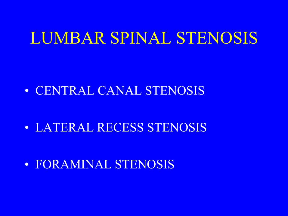
LUMBAR SPINAL STENOSIS
• CENTRAL CANAL STENOSIS
• LATERAL RECESS STENOSIS
• FORAMINAL STENOSIS
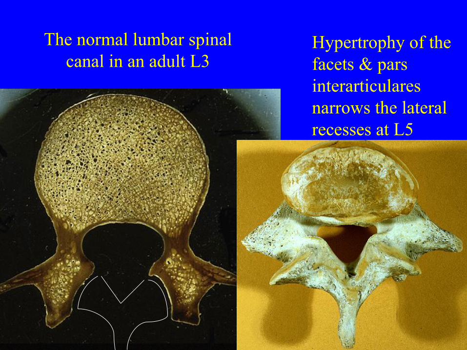
The normal lumbar spinal
canal in an adult L3
Hypertrophy of the
facets & pars
interarticulares
narrows the lateral
recesses at L5
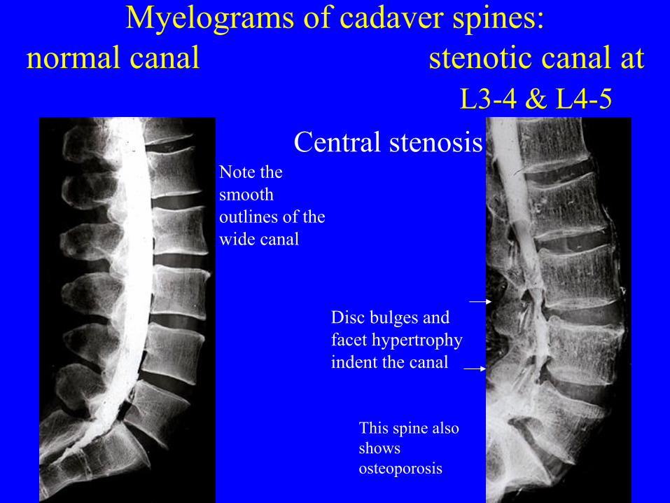
Myelograms of cadaver spines:
normal canal stenotic canal at
L3-4 & L4-5
Disc bulges and
facet hypertrophy
indent the canal
Note the
smooth
outlines of the
wide canal
This spine also
shows
osteoporosis
Central stenosis
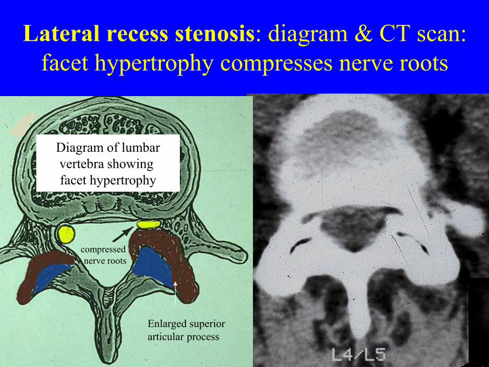
Lateral recess stenosis: diagram & CT scan:
facet hypertrophy compresses nerve roots
Enlarged superior
articular process
compressed
- nerve roots
Diagram of lumbar
vertebra showing
facet hypertrophy
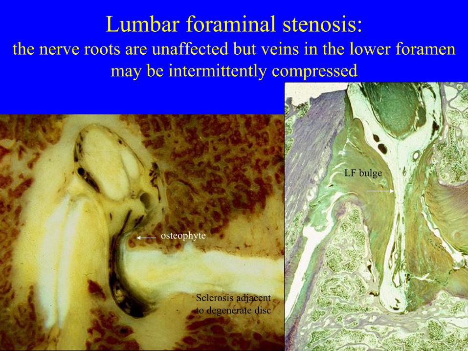
Lumbar foraminal stenosis: the nerve roots are unaffected but veins in the lower foramen
may be intermittently compressed
Sclerosis adjacent
to degenerate disc
osteophyte
LF bulge
Central stenosis may cause multilevel
radicular signs & symptoms;
Lateral recess stenosis usually affects a
specific nerve root with single segment
sensory ± motor effects
Foraminal stenosis is less likely to impinge
on a nerve due to the high position of the
nerve in the foramen but it may affect veins
Clinical Note on Ageing
The importance of movement in maintaining good muscle
tone & good posture
• Reduced bone density & reduced ranges of movement
with ageing are not inevitable
• Exercise can strengthen bone & reduce the likelihood of
osteoporosis
• Exercise with reduced lordosis may benefit patients
with spinal stenosis
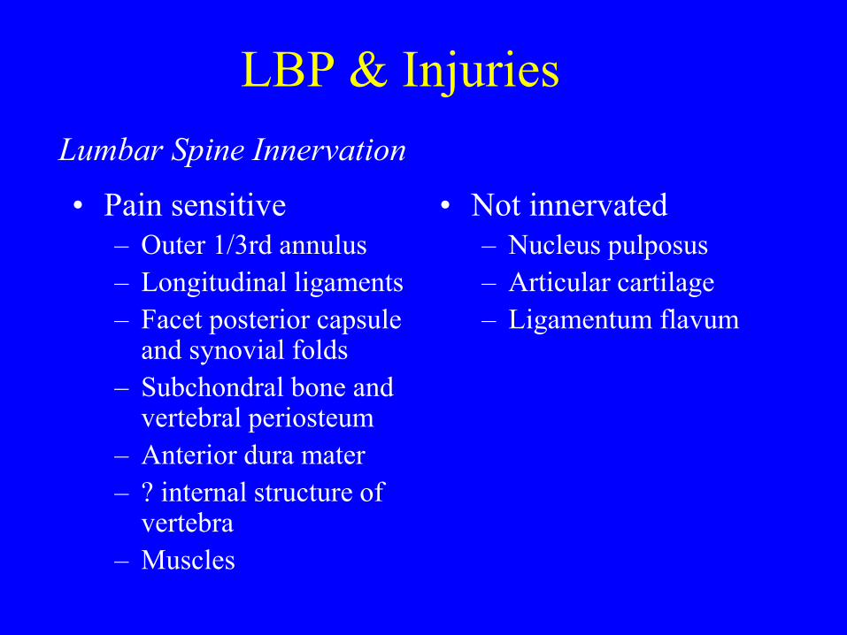
LBP & Injuries
• Pain sensitive
– Outer 1/3rd annulus
– Longitudinal ligaments
– Facet posterior capsule and synovial folds
– Subchondral bone and vertebral periosteum
– Anterior dura mater
– ? internal structure of vertebra
– Muscles
• Not innervated
– Nucleus pulposus
– Articular cartilage
– Ligamentum flavum
Lumbar Spine Innervation
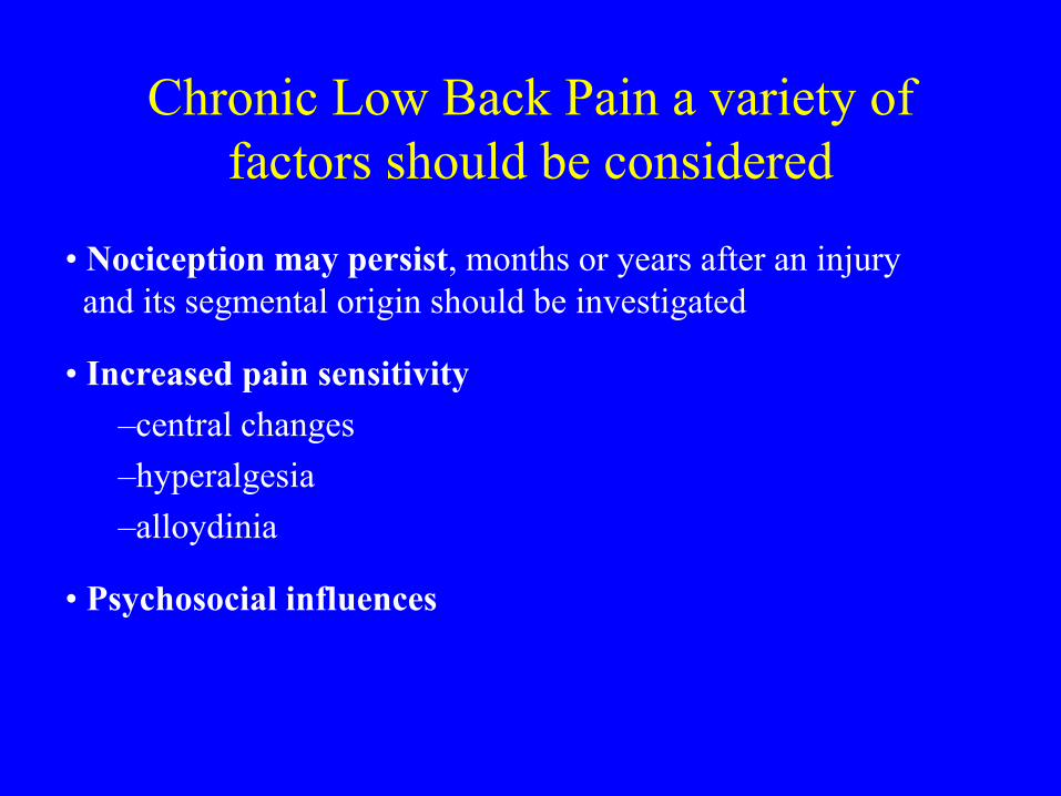
Chronic Low Back Pain a variety of
factors should be considered
• Nociception may persist, months or years after an injury
and its segmental origin should be investigated
• Increased pain sensitivity
–central changes
–hyperalgesia
–alloydinia
• Psychosocial influences
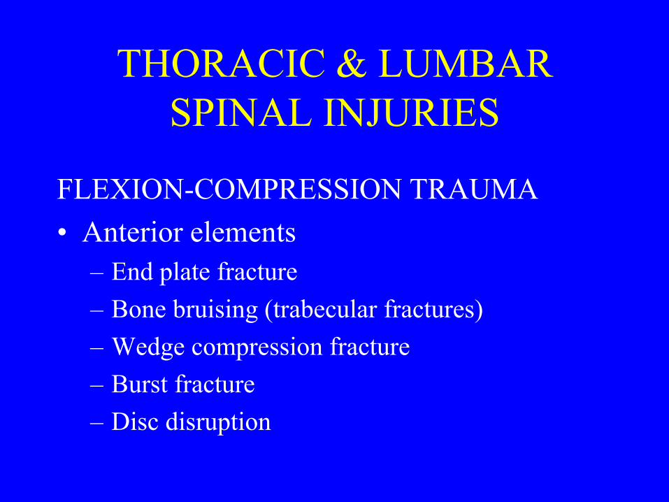
THORACIC & LUMBAR
SPINAL INJURIES
FLEXION-COMPRESSION TRAUMA
• Anterior elements
– End plate fracture
– Bone bruising (trabecular fractures)
– Wedge compression fracture
– Burst fracture
– Disc disruption
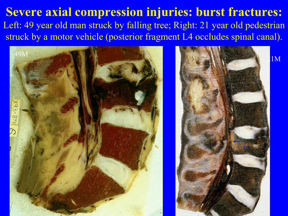
Severe axial compression injuries: burst fractures: Left: 49 year old man struck by falling tree; Right: 21 year old pedestrian
struck by a motor vehicle (posterior fragment L4 occludes spinal canal).
49M 21M
L4
LUMBAR DISC INJURIES
• The Disc is vulnerable to ROTATIONAL STRAIN
IN FLEXION as the facets offer less protection:
• Circumferential or radial tear of posterior annulus
is more common than disc herniation
• In axial compression or flexion compression injury a
disc injury may be associated with an end plate # or
vertebral wedge #. The fracture is visible on x-ray
but the disc injury may not be visible. This may
cause accelerated disc degeneration.
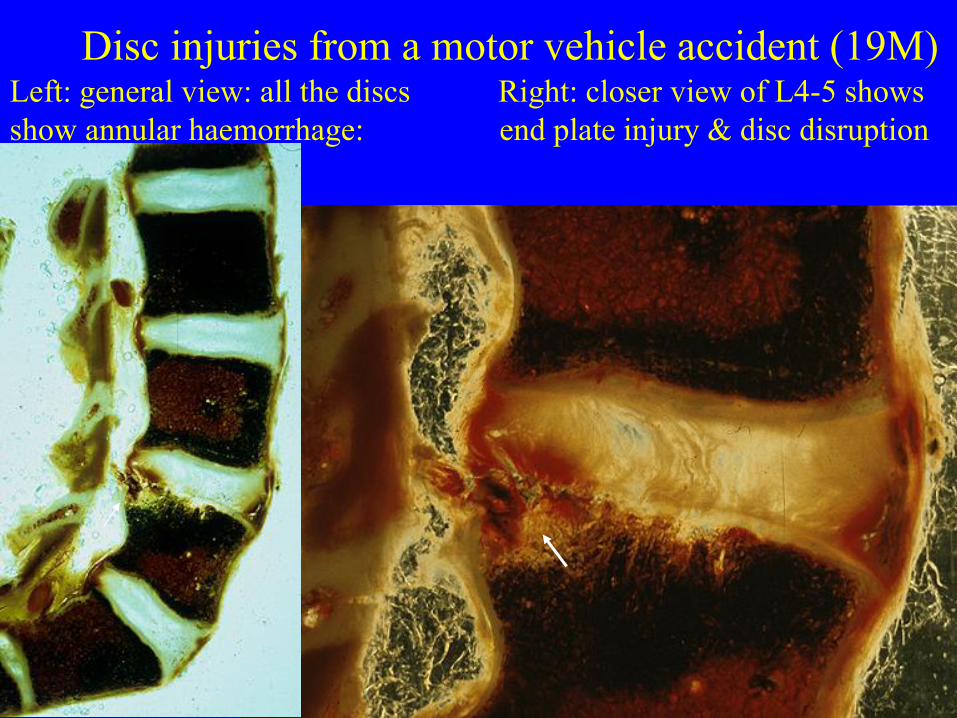
Disc injuries from a motor vehicle accident (19M) Left: general view: all the discs Right: closer view of L4-5 shows
show annular haemorrhage: end plate injury & disc disruption
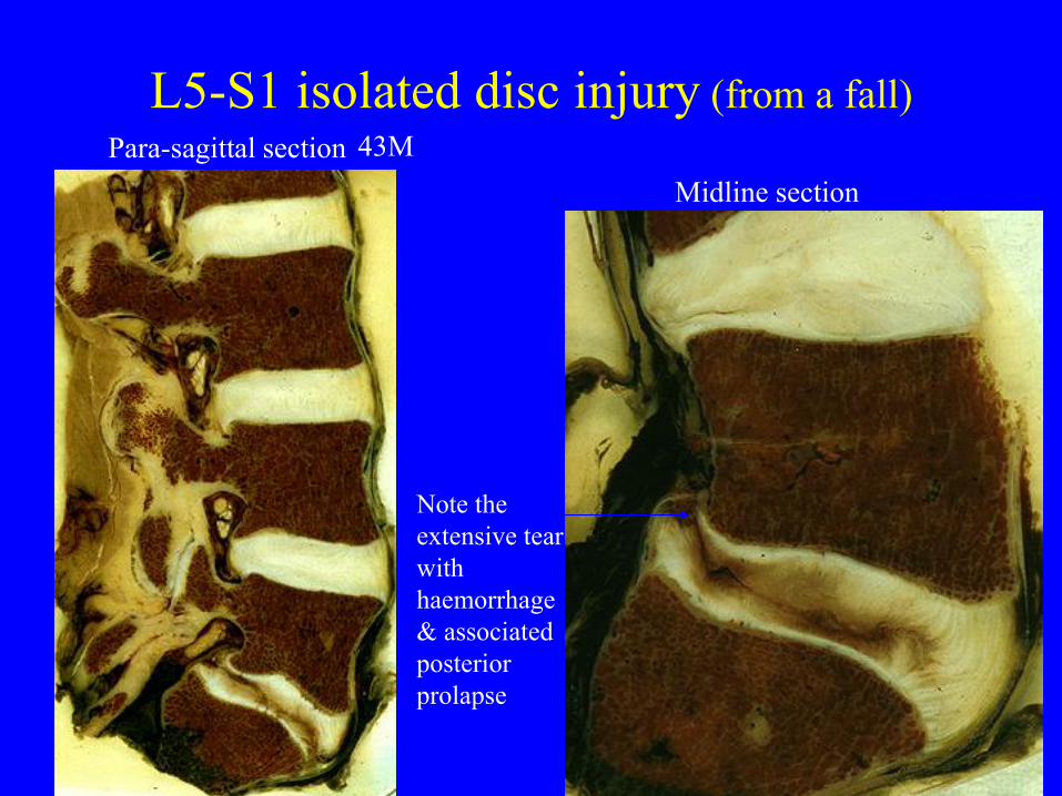
L5-S1 isolated disc injury (from a fall)
43M
Note the
extensive tear
with
haemorrhage
& associated
posterior
prolapse
Para-sagittal section
Midline section
LUMBAR FACET JOINT INJURIES
• Soft tissue injury with haemarthrosis
• Articular surface micro-fracture + haemarthrosis
• Central vertical fracture to Superior Articular Process (SAP)
• Mamillary process fracture
These injuries were not visible on
post-mortem x-rays
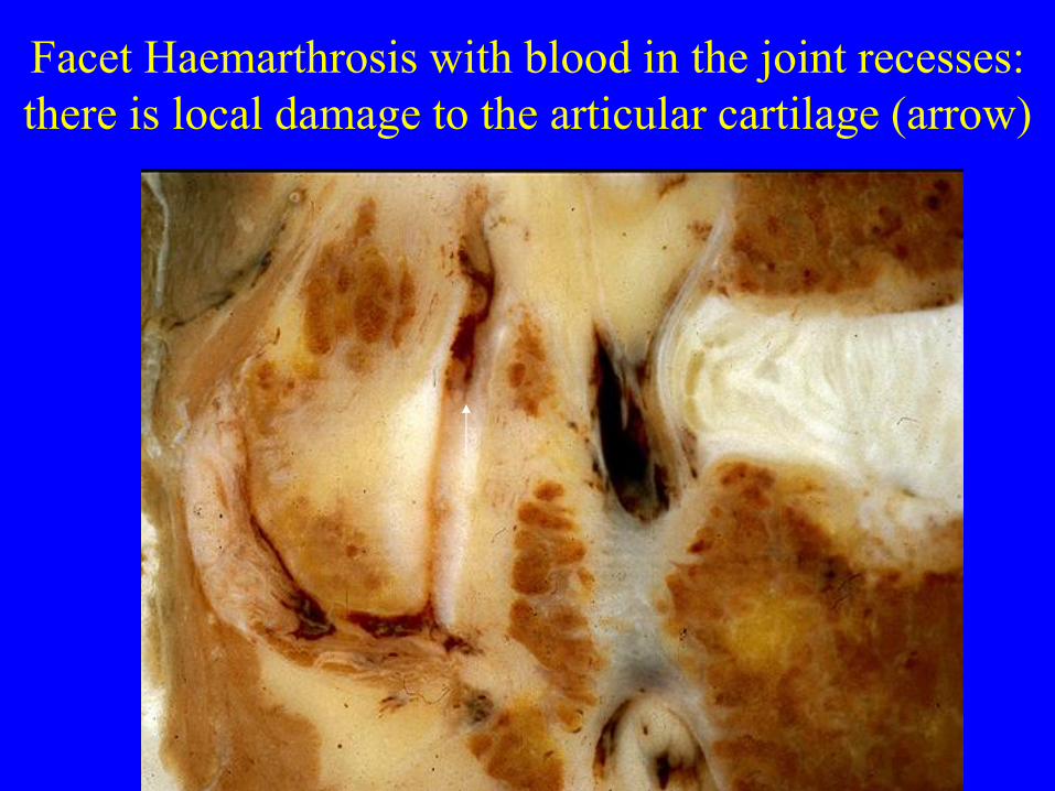
Facet Haemarthrosis with blood in the joint recesses:
there is local damage to the articular cartilage (arrow)
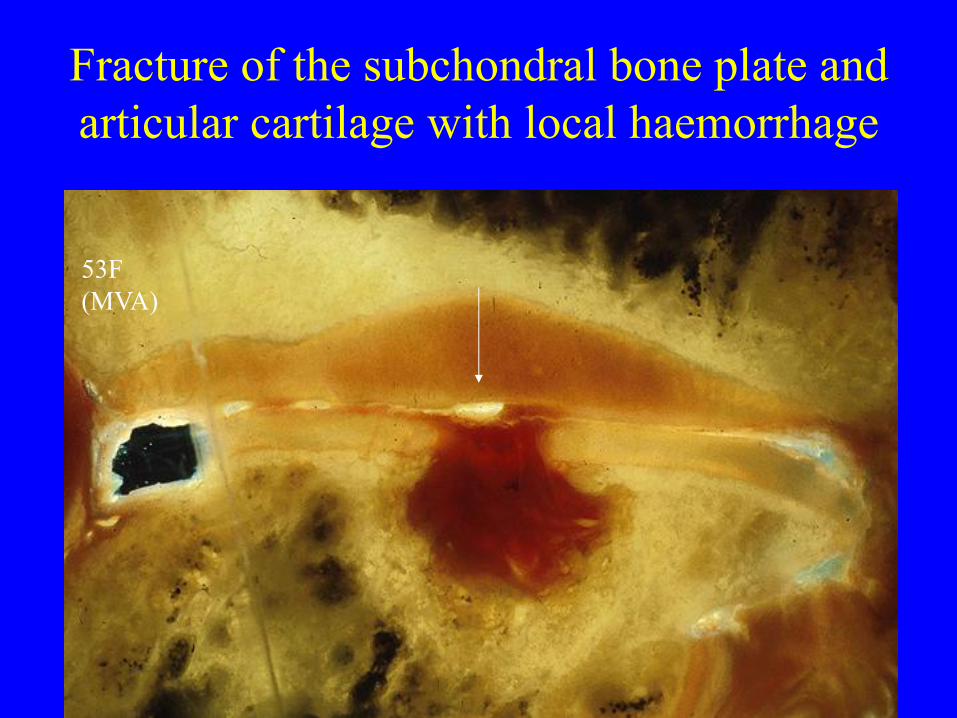
Fracture of the subchondral bone plate and
articular cartilage with local haemorrhage
53F
(MVA)
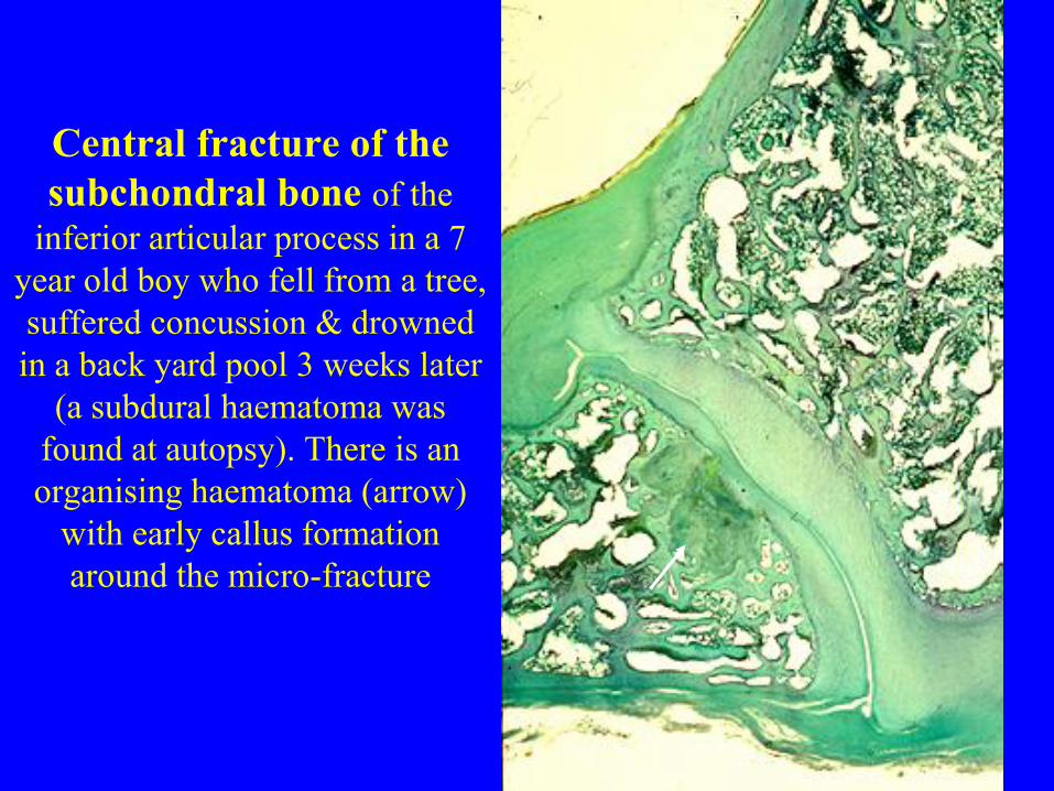
Central fracture of the
subchondral bone of the
inferior articular process in a 7
year old boy who fell from a tree,
suffered concussion & drowned
in a back yard pool 3 weeks later
(a subdural haematoma was
found at autopsy). There is an
organising haematoma (arrow)
with early callus formation
around the micro-fracture
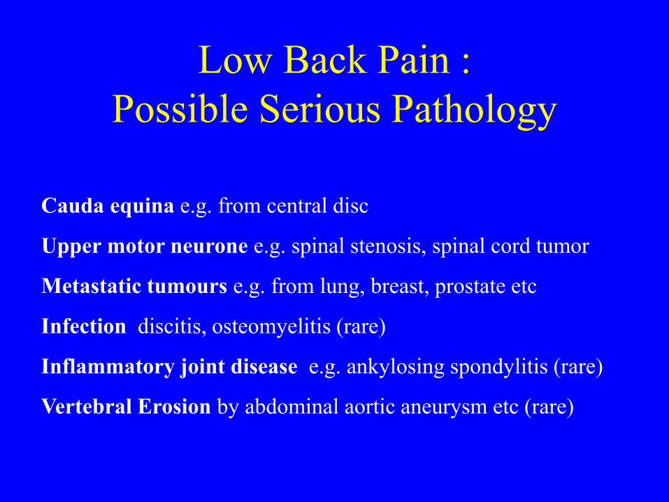
Low Back Pain :
Possible Serious Pathology
Cauda equina e.g. from central disc
Upper motor neurone e.g. spinal stenosis, spinal cord tumor
Metastatic tumours e.g. from lung, breast, prostate etc
Infection discitis, osteomyelitis (rare)
Inflammatory joint disease e.g. ankylosing spondylitis (rare)
Vertebral Erosion by abdominal aortic aneurysm etc (rare)
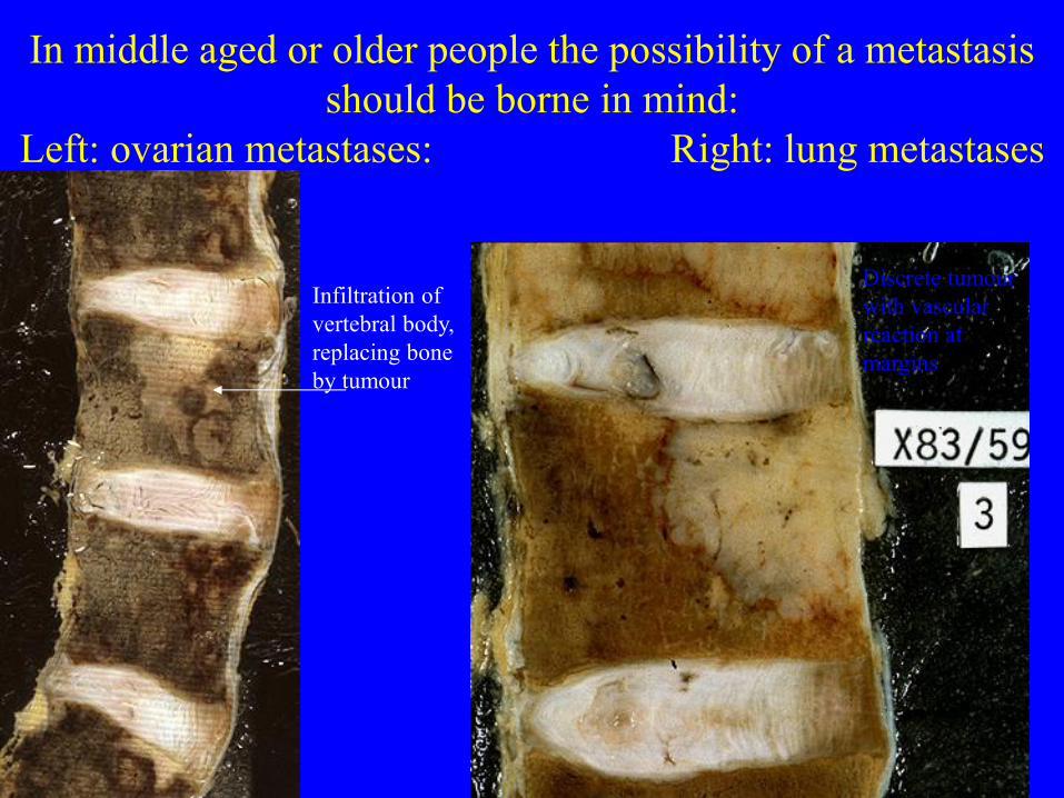
In middle aged or older people the possibility of a metastasis
should be borne in mind:
Left: ovarian metastases: Right: lung metastases
Discrete tumour
with vascular
reaction at
margins
Infiltration of
vertebral body,
replacing bone
by tumour