Embed Size (px)
Citation preview

JARO 03: 174–184 (2001)DOI: 10.1007/s101620020035
Age- and Size-Related Changes in the Inner Ear and HearingAbility of the Adult Zebrafish (Danio rerio)
DENNIS M. HIGGS,1 MARCY J. SOUZA,1,2 HEATHER R. WILKINS,1,3 JOELLE C. PRESSON,1 ANDARTHUR N. POPPER1
1Department of Biology, University of Maryland, College Park, MD 20742, USA2College of Veterinary Medicine, North Carolina State University, Raleigh, NC 27695, USA3Department of Cell and Molecular Physiology, University of North Carolina, Chapel Hill, NC 27599, USA
Received: 13 July 2001; Accepted: 29 August 2001; Online publication: 12 November 2001
ABSTRACT INTRODUCTION
Fishes, unlike most other vertebrate groups, continueNew hair cells are produced in the ears of fishes forto add sensory hair cells to their ears for much of theirmonths or years after hatching (Corwin 1981, 1983;lives. However, it is not clear whether the addition everPopper and Hoxter 1984; Lombarte and Popperstops or how the addition of sensory cells impacts1994). However, it is still not known whether prolifera-hearing ability. In this article, we tested both questionstion continues for the duration of a fish’s life. Signifi-using the zebrafish, Danio rerio. Our results not onlycant postembryonic auditory hair cell addition inhave important implications for understanding thefishes is clearly different from findings in birds andconsequences of adding sensory receptors, but thesemammals. Birds and mammals do not add hair cellsresults for normal zebrafish also serve as valuable base-to the auditory end organ after final maturation underline information for future studies of select mutationsnormal conditions (Ruben 1967; Corwin and Cotan-on the ear and hearing of this species. Our resultsche 1988; Ryals and Rubel 1988) although birds doshow that hair cell production continues in uncrowdedadd hair cells throughout life to vestibular organszebrafish up to 10 months of age (about one-third of(Jørgensen and Mathiesen 1988; Roberson et al. 1992)a normal life span), but despite this addition there isand to auditory organs after damage (Cotanche 1987;no change in hearing sensitivity or bandwidth. There-Cruz et al. 1987; Corwin and Cotanche 1988; Ryalsfore, hearing is not related to the number of sensoryand Rubel 1988).cells in the ear in juvenile and adult animals. We also
Related to the postembryonic addition of sensoryshow that despite no net addition of hair cells afterhair cells is the question of whether these changesabout 10 months, hair cells are still being produced,affect hearing abilities. Hearing has been shown tobut at a lower rate, presumably to replace cells that areimprove in some vertebrates with age (e.g., frogs: Boat-dying. Moreover, crowding of zebrafish has a markedright–Horowitz and Megela Simmons 1995; geckos:impact on the growth of the fish and on the additionWerner et al. 1998; birds: Golubeva and Tikhonovof sensory cells to the ear. We also demonstrate that1985; mammals: Rubsamen 1992; McFadden et al.fish size, not age, is a better indicator of developmental1996; Reimer 1996; Hill et al. 1998), at least untilstate of zebrafish.maturation of the auditory region of the ear is com-Keywords: auditory hair cells, ABR, zebrafish, cell death,plete. There are few studies, however, relating hearingneurogenesis, developmentand growth in fish. The addition of sensory hair cellsto an auditory end organ in elasmobranchs is corre-lated with an increase in sensitivity of the nerve branchinnervating that end organ (Corwin 1983). Other stud-
Correspondence to: Dennis M. Higgs, Ph.D. • Department of Biologyies examining auditory abilities in fish either found a• University of Maryland • College Park, MD 20742. Telephone:
(301) 405-6903; fax: (301) 314-9358; email: [email protected] change in hearing ability with growth (Kenyon 1996;
174

HIGGS ET AL.: Hearing Ability of Adult Zebrafish 175
Iwashita et al. 1999; Wysocki and Ladich 2001) or Fish were prepared for microscopic analysis byfound no change between different sizes of fish (Pop- euthanizing with MS-222 (an anesthetic for cold-per 1971). However, only the elasmobranch study blooded vertebrates). The total length (tip of snout(Corwin 1983) directly correlated the responses of the to tip of tail) was determined for each animal used inear with changes in the number of sensory hair cells. this study. The jaw and tissue covering the otic capsules
Zebrafish have become a particularly important were then removed and the remaining tissue, con-model for vertebrate genetic studies, and there is an taining the ear, was placed in 2.0% paraformaldehydeincreasing number of studies on the genetics and and used as described below.development of the ear in this species (Granato et al. Phalloidin and hair cell counts. The number of saccular1996; Riley and Grunwald 1996; Whitfield et al. 1996; hair cells was determined for uncrowded zebrafishRiley and Moorman 2000). In spite of this importance, aged 3, 6, 10, 15, and 18 months and for crowdedthere have been few studies on the zebrafish auditory zebrafish aged 10 and 27 months. The saccule issystem (Waterman and Bell 1984; Platt 1993; Haddon thought to be the primary auditory endorgan in thisand Lewis 1996) and no studies examining zebrafish group (reviewed in Platt and Popper 1981; Fay 1988b),hearing ability. The purpose of the current study was so only saccular hair cells were considered here. Atto examine developmental changes in the number of least three fish were used for each age group, with upsensory hair cells in the saccule of zebrafish (Danio to six for the youngest animals where more specimensrerio) aged three months to over two years, represent- were available. Fish were sacrificed and the tissue fixeding animals from sexual immaturity to adulthood. We in 2% paraformaldehyde for 30 minutes. The sacculesalso examined the hearing ability of zebrafish to inves- were then removed from the heads, rinsed in phos-tigate possible relationships between hair cell number phate buffer, and placed in Oregon Green Phalloidinand hearing ability. In addition, we wanted to test the (Molecular Probes, Inc., Eugene, OR) for 30 minutes.hypothesis that hair cell addition slows or stops when Phalloidin selectively binds to the actin in hair celloverall growth of the fish stops. This hypothesis is par- bundles, making them easy to identify. The tissue wasticularly important if zebrafish become models for rinsed in phosphate buffer, mounted using Prolonggenetic effects on adult or aging ears since mainte- Antifade (Molecular Probes, Inc.), and examinednance of fish under conditions where growth is artifi- using a Biorad MC 1024 confocal microscope. Lowcially slowed (e.g., crowded conditions) could have power images (40X objective) of the saccular epithe-profound effects on interpretation of results. lium were taken and then manually merged to get a
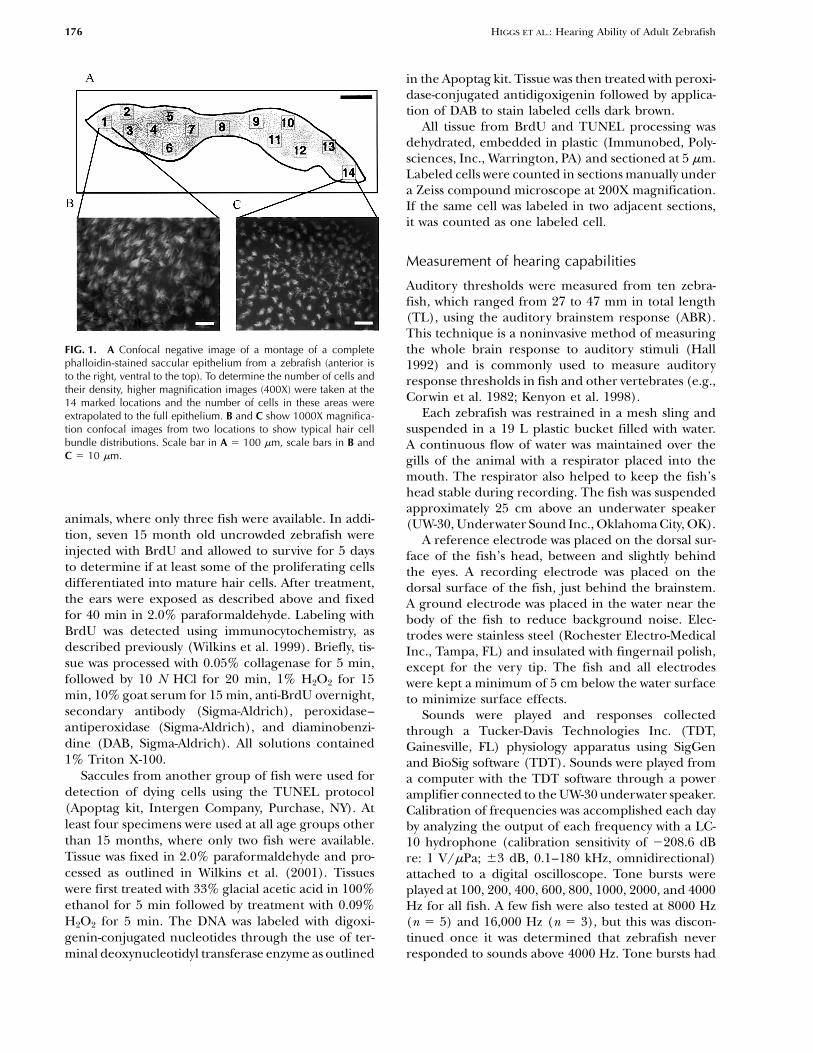
view of the total epithelium (Fig. 1). The total area ofthe epithelium was determined using a computerizedMATERIALS AND METHODSdigitizer and Arc View planimetric software.
Since there are several thousand hair cells on eachInner ear structure saccular epithelium, it was necessary to estimate total
numbers of cells rather than attempt to count eachZebrafish between the ages of three weeks and sixcell. This method, while giving only an estimate of hairmonths were obtained from Eckwell Water Life (Gib-cell number, is reasonable to use when working withinsonton, FL). The animals were hatched and raised,a single species where density of hair cells is similaruntil the time of shipping, in large uncrowded pondsbetween specimens (Popper and Hoxter 1984) andwhere the growth rate was unrestricted. Once in thedoes not change as the fish grow (see Results). Hair celllaboratory, the fish were divided into uncrowdedcounts were obtained from 14 preselected locations on(fewer than 10 fish per 37.85 L tank) and crowdedthe magnified montage of the epithelium (Fig. 1).(more than 30 fish per 37.85 L tank) groups and keptEach counting area was a 2 mm square box on thein these groups until used in experiments. As fish weremagnified montage representing approximately 3–5%used in experiments they were replaced so that theof the total saccular area, depending on the size ofdensity of the fish in the tank remained unchanged.the saccule; the total hair cell count was extrapolatedWhile it is possible that fish in the uncrowded tanksby a simple ratio. The number of cells was counted bybecame more crowded with growth, we do not feeltwo different investigators to ensure accuracy andthis was a significant concern in the current studynever differed by more than 3% between investigators.because of the small size of the fish relative to the tankValues reported are the averages of the two counters.in which they were housed (Innes 1956). Fish were
BrdU and TUNEL labeling. Saccules were dissectedfed daily and the tanks maintained at 23�C. The fishfrom zebrafish that had received one injection of BrdUwere kept on a 12:12 light:dark cycle and monitored(1 mg/10 g body weight; Sigma-Aldrich, St Louis, MO)daily. Animal use was under the supervision of the3 hours prior to sacrifice. At least six specimens wereUniversity of Maryland Institutional Animal Care and
Use Committee. used for each age group other than 27 month old
176 HIGGS ET AL.: Hearing Ability of Adult Zebrafish
in the Apoptag kit. Tissue was then treated with peroxi-dase-conjugated antidigoxigenin followed by applica-tion of DAB to stain labeled cells dark brown.
All tissue from BrdU and TUNEL processing wasdehydrated, embedded in plastic (Immunobed, Poly-sciences, Inc., Warrington, PA) and sectioned at 5 �m.Labeled cells were counted in sections manually undera Zeiss compound microscope at 200X magnification.If the same cell was labeled in two adjacent sections,it was counted as one labeled cell.
Measurement of hearing capabilities
Auditory thresholds were measured from ten zebra-fish, which ranged from 27 to 47 mm in total length(TL), using the auditory brainstem response (ABR).This technique is a noninvasive method of measuring
FIG. 1. A Confocal negative image of a montage of a complete the whole brain response to auditory stimuli (Hallphalloidin-stained saccular epithelium from a zebrafish (anterior is 1992) and is commonly used to measure auditoryto the right, ventral to the top). To determine the number of cells and response thresholds in fish and other vertebrates (e.g.,their density, higher magnification images (400X) were taken at the
Corwin et al. 1982; Kenyon et al. 1998).14 marked locations and the number of cells in these areas wereEach zebrafish was restrained in a mesh sling andextrapolated to the full epithelium. B and C show 1000X magnifica-
tion confocal images from two locations to show typical hair cell suspended in a 19 L plastic bucket filled with water.bundle distributions. Scale bar in A � 100 �m, scale bars in B and A continuous flow of water was maintained over theC � 10 �m. gills of the animal with a respirator placed into the
mouth. The respirator also helped to keep the fish’shead stable during recording. The fish was suspendedapproximately 25 cm above an underwater speaker
animals, where only three fish were available. In addi- (UW-30, Underwater Sound Inc., Oklahoma City, OK).tion, seven 15 month old uncrowded zebrafish were A reference electrode was placed on the dorsal sur-injected with BrdU and allowed to survive for 5 days face of the fish’s head, between and slightly behindto determine if at least some of the proliferating cells the eyes. A recording electrode was placed on thedifferentiated into mature hair cells. After treatment, dorsal surface of the fish, just behind the brainstem.the ears were exposed as described above and fixed A ground electrode was placed in the water near thefor 40 min in 2.0% paraformaldehyde. Labeling with body of the fish to reduce background noise. Elec-BrdU was detected using immunocytochemistry, as trodes were stainless steel (Rochester Electro-Medicaldescribed previously (Wilkins et al. 1999). Briefly, tis- Inc., Tampa, FL) and insulated with fingernail polish,sue was processed with 0.05% collagenase for 5 min, except for the very tip. The fish and all electrodesfollowed by 10 N HCl for 20 min, 1% H2O2 for 15 were kept a minimum of 5 cm below the water surfacemin, 10% goat serum for 15 min, anti-BrdU overnight, to minimize surface effects.secondary antibody (Sigma-Aldrich), peroxidase– Sounds were played and responses collectedantiperoxidase (Sigma-Aldrich), and diaminobenzi- through a Tucker-Davis Technologies Inc. (TDT,dine (DAB, Sigma-Aldrich). All solutions contained Gainesville, FL) physiology apparatus using SigGen1% Triton X-100. and BioSig software (TDT). Sounds were played from
Saccules from another group of fish were used for a computer with the TDT software through a powerdetection of dying cells using the TUNEL protocol amplifier connected to the UW-30 underwater speaker.(Apoptag kit, Intergen Company, Purchase, NY). At Calibration of frequencies was accomplished each dayleast four specimens were used at all age groups other by analyzing the output of each frequency with a LC-than 15 months, where only two fish were available. 10 hydrophone (calibration sensitivity of �208.6 dBTissue was fixed in 2.0% paraformaldehyde and pro- re: 1 V/�Pa; �3 dB, 0.1–180 kHz, omnidirectional)cessed as outlined in Wilkins et al. (2001). Tissues attached to a digital oscilloscope. Tone bursts werewere first treated with 33% glacial acetic acid in 100% played at 100, 200, 400, 600, 800, 1000, 2000, and 4000ethanol for 5 min followed by treatment with 0.09% Hz for all fish. A few fish were also tested at 8000 HzH2O2 for 5 min. The DNA was labeled with digoxi- (n � 5) and 16,000 Hz (n � 3), but this was discon-genin-conjugated nucleotides through the use of ter- tinued once it was determined that zebrafish never
responded to sounds above 4000 Hz. Tone bursts hadminal deoxynucleotidyl transferase enzyme as outlined

HIGGS ET AL.: Hearing Ability of Adult Zebrafish 177
a 2 ms rise and fall time, were 10 ms in duration, andwere gated through a Hanning window—conditionssimilar to those used in other ABR studies (e.g., McFad-den et al. 1996; Yan et al. 2000; Mann et al. 2001).While there were somewhat broad sidebands in thesignals below 800 Hz, the level of the second harmonicwas at least 15 dbV lower than the fundamental outputfrequency, even at the lowest frequencies used.
Responses to each tone burst at each sound pressurelevel were collected by the BioSig software package,with 400 responses averaged for each presentation.Sound intensity levels at each frequency wereincreased in 5 dB steps until a stereotypical ABR spikewas achieved. Responses were judged by eye, and detec-tion was defined as being able to see a spike abovebackground in the stereotypical ABR responses (Fig.2). This qualitative assessment is commonly used inABR studies (Hall 1992; Kenyon et al. 1998), andresults obtained in this manner do not differ fromresults using more quantitative methods (Mann et al.2001). We have also found that this method gives con-sistent results between investigators in our laboratory.For presentation, response averages were high-pass fil-tered at 30 Hz and low-pass filtered at 3000 Hz toremove high frequency noise. Threshold was definedas the lowest sound level giving a defined response.Since results of physiological hearing tests can varybetween laboratories (Popper et al. 1973; Fay 1988a),we needed to be able to have some basis for comparingour results with thresholds determined for otherfishes. As there is a large body of data on hearingsensitivity in goldfish (Carassius auratus), we deter-mined thresholds for this species (n � 3, TL approxi-mately 70 mm) using the identical procedures usedfor zebrafish as a way to compare data for zebrafishwith other species in the literature.
As a final control to assure that the responses wereactually from the auditory system, we made recordingsfrom both whole dead zebrafish and isolated trunkmusculature of dead fish. In no case did theserecordings give responses that in any way resembledthe responses from the brain of living fish (Fig. 2C).
Statistical analyses
All statistical analyses were done as analyses of variance(ANOVA) with the Tukey post hoc test as followup whensignificant differences were found (Zar 1984), except
�
FIG. 2. Auditory brainstem response traces (after low-pass filteringat 3000 Hz) to an (A) 100 Hz tone burst and an (B) 800 Hz toneburst in an adult zebrafish. Auditory threshold was judged as thelowest intensity showing a defined response (140 dB at 100 Hz and125 dB at 800 Hz for this individual). Dead fish controls never gavea response similar to an ABR (C ). All intensity values are dB re 1 �Pa.

178 HIGGS ET AL.: Hearing Ability of Adult Zebrafish
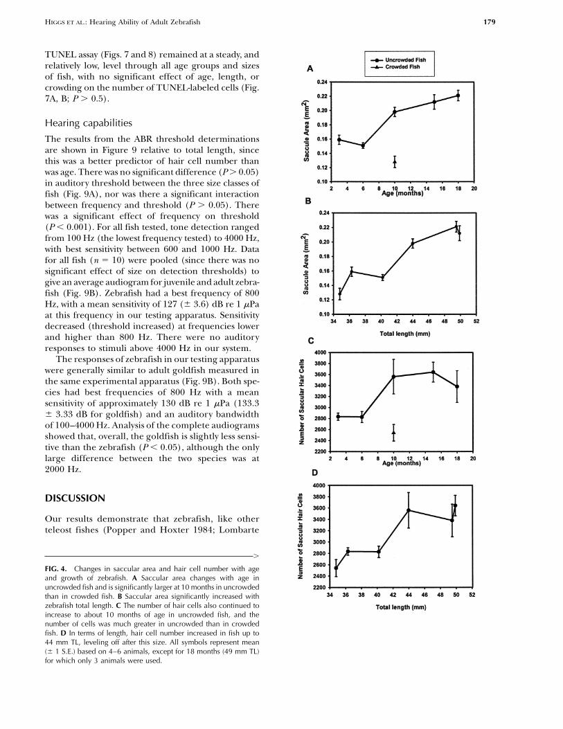
parameters relative to fish total length. Whenexpressed as a function of fish length, with crowdedand uncrowded animals considered together, therewas a significant increase in saccular area with growthof the fish (P � 0.001), with fish smaller than 42 mmhaving significantly smaller saccules than those largerthan 42 mm (P � 0.01, Fig. 4B).
In uncrowded fish, there was a significant increasein the number of hair cells with age (P � 0.02), withnumbers changing little after 10 months of age (Fig.4C). There were significantly fewer (P � 0.05) hair
FIG. 3. Mean (� 1 S.E.) fish growth for crowded and uncrowded cells in 10 month old crowded fish (2541 � 75) thanzebrafish. Between 11 and 17 animals were used for each mean,in the same-age fish raised in uncrowded conditionsexcept for 27 months for which only 6 animals were available.(3560 � 175, Fig. 4C). When crowded and uncrowdedanimals were considered together and hair cell num-ber was expressed as a function of fish total length,
for comparisons of crowded versus uncrowded ani- there was a significant increase (P � 0.01) in hair cellmals, which were compared with a Student’s t-test. For number with total length in zebrafish. Animals smallercomparisons of auditory threshold, animals were put than 42 mm tended to have fewer hair cells than thoseinto one of three size classes: 25–34 mm (n � 4), larger than 42 mm, although there were few significant35–44 mm (n � 4), and 45–50 mm (n � 2). For differences between individual size classes (Fig. 4D).auditory thresholds, a 2-factor ANOVA was performed Confocal images suggested that there were densitywith frequency and size class as the independent vari- differences in hair cells along the length of the saccularables. Means are given with plus or minus one standard epithelium. These differences were compared by plot-error when provided. For the morphological parame- ting the number of cells in each of the 14 regionsters, all ANOVAs were done as single factor. For all examined (Fig. 1). Figure 5 shows that the density oftests, � � 0.05 was used as the significance level. cells was considerably greater at the caudal end of the
epithelium than at the rostral end for all of the sizegroups examined. Crowded and uncrowded fish had
RESULTS the same density distribution within the counted areas,and the relative densities at different regions did notchange as the fish grew (Fig. 5).Growth
Fish kept in the uncrowded tanks showed significant Cell division and deathincreases in size up to 15 months of age (P � 0.01)but no significant change in size between 15 and 18 Cells labeled with BrdU were found in the saccular
epithelia of all fish examined (Fig. 6A, B), and whatmonths (P � 0.05; Fig. 3). Fish in crowded conditionsshowed a significant increase in length between 10 appeared to be labeled mature hair cells were found in
the animals allowed to survive for 5 days after injectionand 27 months of age (P � 0.001), but they neverreached the length of 10 month old uncrowded fish (Fig. 6C). In the animals with 3 h postinjection expo-
sure, there was a significant effect of age on the num-(Fig. 3). What is most interesting, however, is that atthe one time period for which statistical comparisons ber of BrdU-labeled cells in uncrowded animals (P �
0.01) but no effect of age between the two groupsare possible for uncrowded and crowded fishes—10months of age, the crowded fish averaged 35 (�0.11) of crowded animals (P � 0.05, Fig. 7A). There were
significantly more (P � 0.01) BrdU-labeled cells in themm in total length, while uncrowded fish averaged44 (�0.36) mm. This difference was significant in a 10 month old uncrowded (9.8 � 1.5) than in the 10
month old crowded (3.5 � 0.8) animals. When theStudent’s t-test (P � 0.001).The total saccular area of uncrowded fish increased number of BrdU-labeled cells was expressed as a func-
tion of fish total length regardless of age or growingup to 10 months of age (Fig. 4A). Although there wassome continued growth after 10 months, changes in conditions (Fig. 7B), there was a significant increase
in the number of labeled cells with fish length (P �area after 10 months were not significant (P � 0.05).The saccular area of 10 month crowded fish was signifi- 0.03). Animals smaller than 42 mm had fewer labeled
cells than those larger than 42 mm, although therecantly smaller (0.13 � 0.004 mm2) than that ofuncrowded animals (0.20 � 0.014 mm2) (P � 0.001). were few significant differences between individual
size classes.Due to the variability in length at a given age (Fig. 3and see Discussion), we also examined morphological The number of dying cells detected using the
HIGGS ET AL.: Hearing Ability of Adult Zebrafish 179
TUNEL assay (Figs. 7 and 8) remained at a steady, andrelatively low, level through all age groups and sizesof fish, with no significant effect of age, length, orcrowding on the number of TUNEL-labeled cells (Fig.7A, B; P � 0.5).
Hearing capabilities
The results from the ABR threshold determinationsare shown in Figure 9 relative to total length, sincethis was a better predictor of hair cell number thanwas age. There was no significant difference (P � 0.05)in auditory threshold between the three size classes offish (Fig. 9A), nor was there a significant interactionbetween frequency and threshold (P � 0.05). Therewas a significant effect of frequency on threshold(P � 0.001). For all fish tested, tone detection rangedfrom 100 Hz (the lowest frequency tested) to 4000 Hz,with best sensitivity between 600 and 1000 Hz. Datafor all fish (n � 10) were pooled (since there was nosignificant effect of size on detection thresholds) togive an average audiogram for juvenile and adult zebra-fish (Fig. 9B). Zebrafish had a best frequency of 800Hz, with a mean sensitivity of 127 (� 3.6) dB re 1 �Paat this frequency in our testing apparatus. Sensitivitydecreased (threshold increased) at frequencies lowerand higher than 800 Hz. There were no auditoryresponses to stimuli above 4000 Hz in our system.
The responses of zebrafish in our testing apparatuswere generally similar to adult goldfish measured inthe same experimental apparatus (Fig. 9B). Both spe-cies had best frequencies of 800 Hz with a meansensitivity of approximately 130 dB re 1 �Pa (133.3� 3.33 dB for goldfish) and an auditory bandwidthof 100–4000 Hz. Analysis of the complete audiogramsshowed that, overall, the goldfish is slightly less sensi-tive than the zebrafish (P � 0.05), although the onlylarge difference between the two species was at2000 Hz.
DISCUSSION
Our results demonstrate that zebrafish, like otherteleost fishes (Popper and Hoxter 1984; Lombarte
�
FIG. 4. Changes in saccular area and hair cell number with ageand growth of zebrafish. A Saccular area changes with age inuncrowded fish and is significantly larger at 10 months in uncrowdedthan in crowded fish. B Saccular area significantly increased withzebrafish total length. C The number of hair cells also continued toincrease to about 10 months of age in uncrowded fish, and thenumber of cells was much greater in uncrowded than in crowdedfish. D In terms of length, hair cell number increased in fish up to44 mm TL, leveling off after this size. All symbols represent mean(� 1 S.E.) based on 4–6 animals, except for 18 months (49 mm TL)for which only 3 animals were used.
180 HIGGS ET AL.: Hearing Ability of Adult Zebrafish
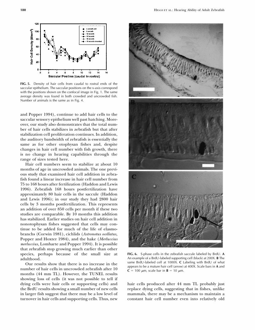
FIG. 5. Density of hair cells from caudal to rostral ends of thesaccular epithelium. The saccular positions on the x-axis correspondwith the positions shown on the confocal image in Fig. 1. The sameaverage density was found in both crowded and uncrowded fish.Number of animals is the same as in Fig. 4.
and Popper 1994), continue to add hair cells to thesaccular sensory epithelium well past hatching. More-over, our study also demonstrates that the total num-ber of hair cells stabilizes in zebrafish but that afterstabilization cell proliferation continues. In addition,the auditory bandwidth of zebrafish is essentially thesame as for other otophysan fishes and, despitechanges in hair cell number with fish growth, thereis no change in hearing capabilities through therange of sizes tested here.
Hair cell numbers seem to stabilize at about 10months of age in uncrowded animals. The one previ-ous study that examined hair cell addition in zebra-fish found a linear increase in hair cell number from75 to 168 hours after fertilization (Haddon and Lewis1996). Zebrafish 168 hours postfertilization haveapproximately 80 hair cells in the saccule (Haddonand Lewis 1996); in our study they had 2800 haircells by 3 months postfertilization. This representsan addition of over 850 cells per month if these twostudies are comparable. By 10 months this additionhas stabilized. Earlier studies on hair cell addition innonotophysan fishes suggested that cells may con-tinue to be added for much of the life of elasmo-branchs (Corwin 1981), cichlids (Astronotus ocellatus,Popper and Hoxter 1984), and the hake (Merlucciusmerluccius, Lombarte and Popper 1994). It is possiblethat zebrafish stop growing much earlier than other
FIG. 6. S-phase cells in the zebrafish saccule labeled by BrdU. Aspecies, perhaps because of the small size atAn example of a BrdU-labeled supporting cell (black) at 200X. B Theadulthood.same BrdU-labeled cell at 1000X. C Labeling with BrdU of whatOur results show that there is no increase in theappears to be a mature hair cell (arrow) at 400X. Scale bars in A and
number of hair cells in uncrowded zebrafish after 10 C � 100 �m, scale bar in B � 10 �m.months (44 mm TL). However, the TUNEL resultsshowing loss of cells (it was not possible to tell ifdying cells were hair cells or supporting cells) and hair cells produced after 44 mm TL probably just
replace dying cells, suggesting that in fishes, unlikethe BrdU results showing a small number of new cellsin larger fish suggest that there may be a low level of mammals, there may be a mechanism to maintain a
constant hair cell number even into relatively oldturnover in hair cells and supporting cells. Thus, new

HIGGS ET AL.: Hearing Ability of Adult Zebrafish 181
FIG. 8. Using the TUNEL procedure, we find that there is a popula-tion of cells in the saccular epithelium undergoing apoptosis, asshown at arrow. Preliminary evidence suggests that apoptosis occursin saccules from zebrafish aged 3, 6, 10, 15, 18, and 21 months.Scale bar � 10 �m.
conditions under which fish are kept. While we cannotstipulate that fish in our uncrowded groups grew asmuch as would fish in the wild, it is clear that the fishin our uncrowded conditions grew far faster and farlarger than fish under our crowded conditions. Theimplications of this finding are significant for all futurestudies of growth and aging in zebrafish, and possiblyeven for early developmental studies. Clearly, factorssuch as fish size, rate of cell proliferation, and otherchanges will be affected by the extent of crowding ofthe fish being studied. It should also be noted that thenumber of fish in our crowded tanks is very closeto the level recommended for general zebrafish care(Westerfield 1995).
Our results also point out the need for a bettermetric than age for studies of fish development. OurFIG. 7. Changes in BrdU- and TUNEL-labeled cells in the zebrafish
saccular epithelium. A The number of BrdU-labeled cells increased two groups (crowded vs. uncrowded) of 10 month oldup to 10 months of age and then stabilized in uncrowded animals fish had significantly different numbers of hair cellswhile there was no significant change in BrdU label in crowded and significantly different numbers of proliferatinganimals. B When crowded and uncrowded animals were considered
cells. Thus, saying a fish is 10 months old tells littletogether, the number of BrdU-labeled cells increased with length,about the developmental state of the animal, as a 10with the largest increase occurring after 42 mm TL. The number of
TUNEL-labeled cells stayed constant and small throughout ontogeny month old fish could represent a wide range of devel-(A and B). All symbols represent mean (� 1 S.E.). opmental stages and have vastly different sensory mor-
phology depending on rearing conditions. Totallength is a much better indicator of developmental
age. The nonsignificant increase in saccular area after progress than age in posthatching fish (Fuiman et al.44 mm may be due to increases in supporting cell 1998), and the use of age makes comparison betweennumbers, since hair cell numbers have stabilized species especially problematic. As shown by our datawhile cell proliferation is continuing, or to increases on the effects of crowding and by previous studiesin size of supporting or hair cells. (Zweifel and Lasker 1976; Chambers and Leggett 1987;
Vollestad 1992), it would be helpful if researchers notuse age as a metric in studies of posthatching fishImplications for crowding on zebrafish growthdevelopment but instead use length or, better, theontogenetic index of Fuiman et al. (1998), whichOur results also demonstrate that the size of zebrafish,expresses larval development as a percentage of thethe number of sensory hair cells, and the rate of cell
proliferation in the ear are dramatically affected by larval period elapsed.

182 HIGGS ET AL.: Hearing Ability of Adult Zebrafish
study and yet no discernible effect on auditory thresh-old. There are several possible explanations for thechanges encountered in previous studies and the lackof change in our animals. First, and most obviously,there are differences in the species used. Second, thetechniques used for the elasmobranch and those usedin the current study were very different. Corwin (1983)looked at the specific output of the ear using com-pound action potentials measured in the eighth cra-nial nerve, while we looked at the evoked responsefrom the brainstem. Evoked brainstem responses pre-sumably integrate information from the whole ear,processing in the brainstem, and, possibly, the outputof both ears to the brain and influences from higherbrain centers on the brainstem. Third, the fish usedin other studies may have been younger than thoseused here.
The significant question to ask concerns the lackof correlation between hair cell increase in our fishesand change in hearing capabilities. One explanationis that while there may potentially be a greaterresponse in the eighth nerve with number of hair cellsin the ear (as per the findings in the ray), the resultof higher level processing and combination of inputsfrom various receptors may eliminate the differences.Indeed, one model of fish hearing predicts that theremust be an increase in hair cell number in order tomaintain a stable hearing threshold as fish grow andthe distance between the ear and peripheral structures(e.g., the swim bladder) increases (Popper et al. 1988;Rogers et al. 1988; Fineran and Hastings 2000). Analternative though less compelling hypothesis is that
FIG. 9. Audiograms based on ABR for (A) fish of 25–34 mm (n �the increase in hair cells impacts other aspects of hear-4), 35–44 mm (n � 4), and 45–50 mm (n � 2) and (B) a compositeing, such as discrimination capabilities, and this wouldaudiogram from juvenile and adult zebrafish based on pooling of the
individual audiograms shown in A (n � 10). Zebrafish were slightly not have shown up in our audiometric studies.more sensitive (lower thresholds) than goldfish (B) examined using The zebrafish audiogram does not show any appre-the identical methods, including electrode placement. All symbols ciable differences from that for goldfish in our testingrepresent mean (� 1 S.E.).
apparatus. This was not surprising since both speciesare in the same taxonomic family (Cyprinidae) andhave similar inner ear anatomy and ultrastructure(Platt 1977, 1993). Most importantly, the zebrafish,Hearing capabilitieslike other otophysan fishes, have a series of bones,the Weberian ossicles, which connect the pressure-The current study is the first that provides data on
zebrafish hearing abilities. There were no differences detecting swim bladder to the inner ear. This specialconnection is known to enhance hearing in otophysanin auditory threshold, bandwidth, or best frequency
for zebrafish over the size range examined in this study fishes as compared with other species (Fay 1988b; Pop-per and Fay 1999), so it is not surprising that zebrafish(34–50 mm TL), despite differences in hair cell num-
ber and saccular area. That there were no changes in have good hearing.There are differences in auditory sensitivity, but notauditory sensitivity with changes in fish size is interest-
ing. Rays (Raja clavata) show an increase in physiologi- bandwidth, of goldfish in our apparatus as comparedwith previous reports. Kenyon et al. (1998), also usingcal sensitivity of the auditory nerve that is concomitant
with an increase in hair cell number (Corwin 1983), ABR, found a similarly shaped audiogram as ours forgoldfish, with response bandwidth from 100 to 5000and other species show increased auditory sensitivity
with age in very young fish (Kenyon 1996; Wysocki Hz and a best frequency of 1000 Hz, but thresholdswere 20–60 dB lower in their system. This is probablyand Ladich 2001). There was a significant increase in
hair cell number in zebrafish examined in the current due to differences in testing apparatus. We feel that

HIGGS ET AL.: Hearing Ability of Adult Zebrafish 183
J, Fay RR, Popper AN, Tavolga WN, (eds) Sensory Biology ofcomparisons of auditory sensitivity are difficultAquatic Animals. Springer-Verlag New York, 1988b, p. 712–731.between laboratories, whether with ABR or with other
FINERAN JJ, HASTINGS MC. A mathematical analysis of the peripheralmeasures. Fay (1988a) found a 50 dB difference in auditory system mechanics in the goldfish (Carassius auratus). J.sensitivity and Popper et al. (1973) found up to 30 dB Acoust. Soc. Am. 108:1308–1321, 2000, DOI: 10.1121/1.1286099.
FUIMAN LA, POLING KR, HIGGS DM. Quantifying developmentaldifference in thresholds between experimenters withprogress for comparative studies of larval fishes. Copeia 1998:602–goldfish. In our apparatus we have found sensitivity611, 1998.differences up to 40 dB in tests on the American shad
Golubeva TB, Tikhonov AV (1985) The voice and hearing of birds(Alosa sapidissima) simply by varying the depth of elec- in ontogeny. In: Acta XVIII International Ornithology Congresstrode placement (unpublished data). Thus, measures Nauka, Moscow, pp. 259–274.
GRANATO M, VAN EEDEN FJM, SCHACH U, TROWE T, BRAND M, FURU-of auditory sensitivity are useful only on a comparativeTANI–SEKEI M, HAFFTER P, HAMMERSCHMIDT M, HEISENBERG C-basis in the same experimental conditions. What is ofP, JIANG Y-J, KANE DA, KELSH RN, MULLINS MC, ODENTHAL J,interest between investigators is the bandwidth, theNUSSLEIN–VOLHARD C. Genes controlling and mediating locomo-
shape of the audiogram, and the frequency of best tion behavior of the zebrafish embryo and larva. Developmentsensitivity. In these three respects there is good agree- 123:399–413, 1996.
HADDON C, LEWIS J. Early ear development in the embryo of thement between our goldfish data and previous investiga-zebrafish, Danio rerio. J. Comp. Neurol. 365:113–128, 1996, DOI:tions (Popper 1971; Fay 1988a; Kenyon et al. 1998),10.1002/(SICI)1096-9861(19960129)365:1�113::AID-validating our results for zebrafish.CNE9�3.3.CO;2-A.
HALL JW III. Handbook of auditory evoked responses. Allyn & BaconBoston, MA, 1992.
HILL KG, CONE–WESSON B, LIU GB. Development of auditory func-ACKNOWLEDGMENTStion in the tammar wallaby Macropus eugenii. Hear. Res. 117:97–106, 1998, DOI: 10.1016/S0378-5955(97)00211-6.The work reported here was supported by grant AG015681
INNES WT. Exotic aquarium fishes. Innes Publishing Philadelphia,from the National Institute on Aging of the NIH (ANP) and PA, 1956.grant DC04502-01 from the National Institute on Deafness IWASHITA A, SAKAMOTO M, KOJIMA T, WATANABE Y, SOEDA H. Growthand other Communicative Disorders of the NIH (DMH). effects on the auditory threshold of red sea bream. Nippon SuisanAdditional support was provided for DMH and HRW by Gakkaishi 65:833–838, 1999.training grant DC-00046 from the National Institute on Deaf- JØRGENSEN JM, MATHIESEN C. The avian inner ear: continuous pro-
duction of hair cells in vestibular sensory organs but not in theness and Other Communicative Disorders of the NIH. Weauditory papilla. Naturwissenschaften 75:319–320, 1988.want to thank Tim Maugel for help with microscopy and
KENYON TN. Ontogenetic changes in the auditory sensitivity of dam-two anonymous reviewers for comments on the manuscript.selfishes (pomacentridae). J. Comp. Physiol. A 179:553–561, 1996.
KENYON TN, LADICH F, YAN HY. A comparative study of hearingability in fishes: the auditory brainstem response approach.
REFERENCES J. Comp. Physiol. A 182:307–318, 1998, DOI: 10.1007/s003590050181.
LOMBARTE A, POPPER AN. Quantitative analyses of postembryonicBOATRIGHT–HOROWITZ SS, MEGELA SIMMONS A. Postmetamorphichair cell addition in the otolithic end organs of the inner ear ofchanges in auditory sensitivity of the bullfrog midbrain. J. Comp.the European hake, Merluccius merluccius (Gadiformes, Teleostei).Physiol. A 177:577–590, 1995.J. Comp. Neurol. 345:419–428, 1994.CHAMBERS RC, LEGGETT WC. Size and age at metamorphosis in
MANN DA, HIGGS DM, TAVOLGA WN, SOUZA MJ, POPPER AN. Ultra-marine fishes: an analysis of laboratory-reared winter floundersound detection by clupeiform fish. J. Acoust. Soc. Am. 109:3048–(Pseudopleuronectes americanus) with a review of variation in other3054, 2001, DOI: 10.1121/1.1368406.species. Can. J. Fish. Aquat. Sci. 44:1936–1947, 1987.
MCFADDEN SL, WALSH EJ, MCGEE J. Onset and development ofCORWIN JT. Postembryonic production and aging in inner ear hairauditory brainstem response in the Mongolian gerbil (Merionescells in sharks. J. Comp. Neurol. 201:541–553, 1981.unguiculatus). Hear. Res. 100:68–79, 1996, DOI: 10.1016/0378-CORWIN JT. Postembryonic growth of the macula neglecta auditory5955(96)00108-6.detector in the ray, Raja clavata: Continual increases in hair cell
PLATT C. Hair cell distribution and orientation in goldfish otolithnumber, neural convergence, and physiological sensitivity. J.organs. J. Comp. Neurol. 172:283–297, 1977.Comp. Neurol. 217:345–356, 1983.
PLATT C. Zebrafish inner ear sensory surfaces are similar to thoseCORWIN JT, COTANCHE DA. Regeneration of sensory hair cells afterin goldfish. Hear. Res. 65:133–140, 1993.acoustic trauma. Science 240:1772–1774, 1988.
PLATT C, POPPER AN. Fine structure and function of the ear. In:CORWIN JT, BULLOCK TH, SCHWEITZER J. The auditory brain stemTavolga WN, Popper AN, Fay RR, (eds) Hearing and Sound Com-response in five vertebrate classes. Electroencephalogr. Clin. Neu-munication in Fishes. Springer-Verlag New York, 1981, p. 4–38.rophysiol. 54:629–641, 1982.
POPPER AN. The effects of size on auditory capacities of the goldfish.COTANCHE DA. Regeneration of hair cell stereociliary bundles inJ. Aud. Res. XI:239–247, 1971.the chick cochlea following severe acoustic trauma. Hear. Res.
POPPER AN, HOXTER B. Growth of a fish ear: 1. Quantitative analysis30:181–195, 1987.of sensory hair cell and ganglion cell proliferation. Hear. Res.CRUZ RM, LAMBERT PR, RUBEL EW. Light microscopic evidence of15:133–142, 1984.hair cell regeneration after gentamicin toxicity in chick cochlea.
POPPER AN, FAY RR. The auditory periphery in fishes. In: Fay RR,Arch. Otolaryngol. Head Neck Surg. 130:1058–1062, 1987.Popper AN, (eds) Comparative Hearing: Fish and Amphibians.FAY RR. Hearing in Vertebrates: a Psychophysics Databook. Hill-FaySpringer-Verlag New York, 1999, p. 43–100.Winetka, IL, 1998a.
FAY RR. Peripheral adaptations for spatial hearing in fish. In: Atema POPPER AN, CHAN ATH, CLARKE NL. An evaluation of methods for

184 HIGGS ET AL.: Hearing Ability of Adult Zebrafish
behavioral investigations of teleost audition. Behav. Res. Meth. WATERMAN RE, BELL DH. Epithelial fusion during early semicircularInstr. 5:470–472, 1973. canal formation in the embryonic zebrafish, Brachydanio rerio.
POPPER AN, ROGERS PH, SAIDEL WM, COX M. The role of the fish Anat. Rec. 210:101–114, 1984.ear in sound processing. In: Atema J, Fay RR, Popper AN, Tavolga WERNER YL, MONTGOMERY LG, SAFFORD SD, IGIC P, SAUNDERS JC.WN, (eds) Sensory Biology of Aquatic Animals. Springer-Verlag How body size affects middle-ear structure and function and audi-New York, 1988, p. 687–710. tory sensitivity in gekkonoid lizards. J. Exp. Biol. 201:487–502,
REIMER K. Ontogeny of hearing in the marsupial, Monodelphis domes- 1998.tica, as revealed by brainstem auditory evoked potentials. Hear. WESTERFIELD M. The Zebrafish Book, 3rd ed. University of OregonRes. 92:143–150, 1996, DOI: 10.1016/0378-5955(95)00213-8. Eugene, OR, 1995.
RILEY BB, GRUNWALD DJ. A mutation in zebrafish affecting a local- WHITFIELD TT, GRANATO M, VAN EEDEN JM, SCHACH U, BRAND M,ized cellular function required for normal ear development. Dev. FURUTANI–SEIKI M, HAFFTER P, HAMMERSCHMIDT M, HAISENBERGBiol. 179:427–435, 1996, DOI: 10.1006/dbio.1996.0272. C-P, JIANG Y-J, KANE DA, KELSH RN, MULLINS MC, ODENTHAL J,
RILEY BB, MOORMAN SJ. Development of utricular otoliths, but not NUSSLEIN–VOLHARD C. Mutations affecting development of thesaccular otoliths, is necessary for vestibular function and survival zebrafish inner ear and lateral line. Development 123:241–254,in zebrafish. J. Neurobiol. 43:329–337, 2000, DOI: 10.1002/1097- 1996.4695(20000615)43:4�329::AID-NEU2�3.0.CO;2-H. WILKINS HR, PRESSON JC, POPPER AN. Proliferation of vertebrate
ROBERSON DF, WEISLEDER P, BOHRER PS, RUBEL EW. Ongoing pro-inner ear supporting cells. J. Neurobiol. 39:527–535, 1999, DOI:
duction of sensory cells in the vestibular epithelium of the chick.10.1002/(SICI)1097-4695(19990615)39:4�527::AID-
Hear. Res. 57:166–174, 1992.NEU6�3.3.CO;2-B.ROGERS PH, POPPER AN, COX M, SAIDEL WM. Processing of acoustic
WILKINS HR, PRESSON JC, POPPER AN, RYALS BM, DOOLING RJ. Hairsignals in the auditory system of bony fish. J. Acoust. Soc. Am.cell death in a hearing-deficient canary. J. Assoc. Res. Otolaryngol.83:338–349, 1988.2:79–86, 2001.RUBEN RJ. Development of the inner ear of the mouse: A radioauto-
WYSOCKI LE, LADICH F. The ontogenetic development of auditorygraphic study of terminal mitoses. Acta Otolaryngol. Suppl. 220:1–sensitivity, vocalization and acoustic communication in the laby-44, 1967.rinth fish Trichopsis vittata. J. Comp. Physiol. A 187:177–187, 2001.RUBSAMEN R. Postnatal development of central auditory frequency
YAN HY, FINE ML, HORN NS, COLON WE. Variability in the role of themaps. J. Comp. Physiol. A 170:129–143, 1992.gasbladder in fish audition. J. Comp. Physiol. A 186:435–445, 2000.RYALS BM, RUBEL EW. Hair cell regeneration after acoustic trauma
ZAR JH. Biostatistical Analysis, 2nd ed. Prentice-Hall Englewoodin adult Coturnix quail. Science 240:1772–1774, 1988.Cliffs, NJ, 1984.VOLLESTAD LA. Geographic variation in age and length at metamor-
ZWEIFEL JR, LASKER R. Prehatch and posthatch growth of fishes—aphosis of maturing European eel: environmental effects and phe-notypic plasticity. J. Anim. Ecol. 61:41–48, 1992. general model. Fish. Bull. US 74:609–621, 1976.