Embed Size (px)
Citation preview
AGA Technical Review on Treatment of PatientsWith Dysphagia Caused by Benign Disordersof the Distal Esophagus
This literature review and the recommendations therein were prepared for the American Gastroenterological Association ClinicalPractice and Practice Economics Committee. The paper was approved by the committee on September 27, 1998.
Dysphagia is the perception that there is an impedi-ment to the normal passage of swallowed material.
Odynophagia is the sensation of pain on swallowing.Dysphagia can be caused by a number of disorders,benign and malignant, that involve either the oropharynxor the esophagus. The purpose of this report is to developa rational approach to the treatment of adult patients whohave dysphagia caused by benign disorders of the distalesophagus. The report provides a critical review ofpertinent literature on which to base this approach.Patient care strategies that emerge from the review aresummarized in the accompanying American Gastroenter-ological Association (AGA) Medical Position Statement.For this report, the distal esophagus is defined, somewhatarbitrarily, as the segment of esophagus that extends fromthe level of the aortic arch to the gastric cardia. Themuscularis propria in this esophageal segment is com-posed predominantly of smooth muscle.1 Thus the distalesophagus is susceptible to three general categories ofdisease processes that can cause dysphagia (Table 1): (1)mucosal (intrinsic) diseases that narrow the lumen of theesophagus through inflammation, fibrosis, or neoplasia;(2) mediastinal (extrinsic) diseases that encase and ob-struct the esophagus by direct invasion or through lymphnode enlargement; and (3) diseases affecting the esopha-geal smooth muscle and its innervation that disruptperistalsis, interfere with lower esophageal sphincterrelaxation, or both. This review considers clinical reportson these disorders that have been published in peer-reviewed journals since 1966. The reports were identifiedprimarily by a MEDLINE search using the followingMeSH terms: deglutition disorders, esophageal dysphagia,esophageal stenosis, esophageal motility disorders, and esopha-geal achalasia. Clinical studies published only in abstractform are not included. However, even the peer-reviewedliterature on the treatment of patients with dysphagiadue to benign esophageal disorders consists predomi-nantly of retrospective, uncontrolled studies of small,heterogeneous patient populations who were followed uponly briefly. The conclusions that can be drawn fromthese reports often are limited, and the serious deficien-
cies in study design and execution often preclude mean-ingful meta-analyses. This report highlights the strengthsand weaknesses of the most relevant studies.
Diagnosis
History
It has been estimated that the cause of dysphagiacan be determined with an accuracy of approximately80% on the basis of a careful history alone.2 Some keyelements of the history for patients with dysphagia arehighlighted below.
Is the dysphagia for solid foods, liquids, or both?Mucosal and mediastinal diseases that involve the distalesophagus cause dysphagia by narrowing the esophageallumen. Such narrowings usually pose little barrier to thepassage of liquids, and consequently these diseases charac-teristically cause dysphagia only for solid foods.3 Incontrast, diseases that disrupt peristalsis by affecting thesmooth muscle and its innervation may cause dysphagiafor both solids and liquids. In achalasia, persistentcontraction of the lower esophageal sphincter (LES)causes complete mechanical obstruction of the esophagusthat persists until either the sphincter relaxes or thehydrostatic pressure of the retained material exceeds thepressure generated by the sphincter muscle. Even in theabsence of peristalsis, gravity often can empty theesophagus of liquid material effectively if the LES isrelaxed. Therefore, patients who have disordered peristal-sis and profoundly hypotensive LESs often experience nodysphagia or dysphagia only for solid foods.4,5
Where does the patient perceive that ingestedmaterial sticks? Patients with esophageal obstructionoften perceive that swallowed material sticks at a pointthat is either above or at the level of the lesion causing theobstruction.6–8 In a recent radiographic study of 12patients with lower esophageal mucosal rings that im-peded the passage of a marshmallow bolus, the perceivedlevel of the obstruction was localized to the neck in 7, tothe sternal angle in 2, to the midchest in 2, and to thelower chest in 1.7 It is uncommon for patients to perceive
GASTROENTEROLOGY 1999;117:233–254
that swallowed material sticks at a level substantiallybelow that of the obstructing lesion. In a recent endo-scopic study of 139 patients with dysphagia caused byesophageal strictures, the patients’ perception of the levelof obstruction agreed with the endoscopists’ localizationof the stricture with an accuracy of 64 cm in 74% ofcases.8 Fifteen percent of patients with strictures of thedistal esophagus localized obstruction to the proximalesophagus, whereas only 5% of patients with proximalesophageal strictures perceived obstruction in the distalesophagus. Thus, the perception that a swallowed bolussticks above the suprasternal notch is of little value forlocalization of the obstruction because this sensationcould be caused by a lesion located anywhere from thepharynx to the most distal esophagus. However, if thepatient localizes the obstruction to a point below thesuprasternal notch, the chances are excellent that thedysphagia is caused by a disorder that involves the distalesophagus.
Are there symptoms of oropharyngeal dysfunc-tion? Dysphagia caused by oropharyngeal dysfunction isthe subject of another AGA Technical Review9 and is notdiscussed in detail in this report. Oropharyngeal dyspha-gia often results from diseases that affect the striatedmuscles of the oropharynx or their innervation (e.g.,muscular dystrophies, cerebrovascular accidents). Pa-tients with these neuromuscular diseases may experiencedifficulty in initiating swallowing, and swallowing maybe accompanied by nasopharyngeal regurgitation, pulmo-nary aspiration, and a sensation that residual materialremains in the pharynx. If any of these symptoms areprominent, evaluation for oropharyngeal dysfunctionmay precede tests for esophageal disorders.
Is the dysphagia intermittent or progressive?Patients who have lower esophageal mucosal (Schatzki)rings typically complain of dysphagia that is intermittentand nonprogressive. These patients characteristically ex-
perience discrete, short-lived episodes of dysphagia forsolid foods, often during a meal in a restaurant (hence theterm ‘‘steak-house syndrome’’) or at a social function.Episodes may be separated by weeks, months, or years,and the patient typically experiences no swallowingdifficulty between discrete episodes. In contrast, esopha-geal strictures usually cause dysphagia that is progressivein frequency and severity. With benign strictures, theprogression is typically slow and insidious (over a periodof months to years), and weight loss is minimal. Malig-nant esophageal strictures usually cause dysphagia thatprogresses rapidly (over a period of weeks to months), andweight loss may be profound.
Is there a history of chronic heartburn? Heart-burn is the cardinal symptom of gastroesophageal refluxdisease (GERD), and a history of chronic heartburnsupports the possibility that dysphagia may be caused bya peptic esophageal stricture. However, the history ofpyrosis should be interpreted with caution because thesensation of burning, substernal chest discomfort is notspecific for GERD. For example, patients with achalasiafrequently complain of a heartburn sensation that may becaused by abnormal motor activity as well as by esopha-geal acid exposure.10 Conversely, many patients whodevelop peptic strictures as a result of GERD have noantecedent history of heartburn. In one study of 154patients with benign (mostly peptic) esophageal stric-tures, for example, only 75% of the patients related ahistory of chronic heartburn.11 Finally, approximatelytwo thirds of patients with dysphagia caused by adenocar-cinoma in Barrett’s esophagus have a history of long-standing heartburn.12 Although the history of heartburnprovides useful clinical information, conclusions regard-ing the etiology of dysphagia in an individual patientshould not be based primarily on the presence or absenceof heartburn.
Has the patient taken medications likely tocause pill esophagitis? A number of medications takenin pill form are potentially caustic to the esophagus andcan cause deep ulceration with stricture formation if theyhave prolonged contact with the esophageal mucosa.Although the list of medications that can cause pillesophagitis is long, most cases reported in the UnitedStates have been caused by antibiotics (e.g., doxycycline),potassium chloride preparations, nonsteroidal anti-inflammatory drugs (NSAIDs), or quinidine.13 Recently,a number of cases of pill esophagitis have been attributedto alendronate sodium, an agent used in the treatment ofosteoporosis in postmenopausal women.14
Is there a history of collagen-vascular disease?Collagen-vascular diseases such as scleroderma, rheuma-
Table 1. Diseases of the Distal Esophagus That CauseDysphagia
Mucosal diseasesGERD (peptic stricture)Esophageal ringsEsophageal tumorsCaustic injury (e.g., lye ingestion, pill esophagitis, sclerotherapy)Radiation injuryInfectious esophagitis
Mediastinal diseasesTumors (e.g., lung cancer, lymphoma)Infections (e.g., tuberculosis, histoplasmosis)
Diseases affecting smooth muscle and its innervationAchalasiaSclerodermaOther motility disorders
234 AMERICAN GASTROENTEROLOGICAL ASSOCIATION GASTROENTEROLOGY Vol. 117, No. 1
toid arthritis, and systemic lupus erythematosus canaffect the distal esophagus and cause disordered motil-ity.15 Esophageal dysmotility in these disorders is often,but not invariably, associated with Raynaud’s phenom-enon.15,16 In scleroderma and related collagen-vasculardisorders, fibrosis and vascular obliteration in gut smoothmuscle cause poor esophageal contractility and weaknessof the LES that predisposes to severe GERD.17 Inaddition, patients with collagen-vascular disease often aretreated with medications such as NSAIDs that can causepill esophagitis. Consequently, dysphagia associated withcollagen-vascular disease may be the result of disorderedesophageal motility, severe GERD, pill esophagitis, orsome combination of these abnormalities. Although itseems logical to assume that pill esophagitis should occurmore frequently in patients with esophageal motordisorders, there are few published data to support thisnotion.
Is the patient immunosuppressed? Infectiousesophagitis occurs frequently in patients whose immunesystem has been compromised severely by infection withthe human immunodeficiency virus, by advanced malig-nancy, or by organ transplantation with the administra-tion of potent immunosuppressive drugs. It has been esti-mated that 30%–40% of patients who have the acquiredimmunodeficiency syndrome develop symptoms of esopha-geal disease.18 Most esophageal infections are caused byone or a combination of only three organisms: candida,cytomegalovirus, and herpes simplex virus.19 Odynopha-gia is usually the predominant symptom for patients withinfectious esophagitis, but most patients experiencedysphagia as well.19 Esophageal stricturing is an uncom-mon late complication of infectious esophagitis.20
Physical Examination
Physical examination is important for assessmentof the patient’s nutritional status and ability to toleratethe invasive procedures that may be necessary to managethe esophageal disorder. However, the physical examina-tion infrequently provides specific clues to the cause ofdysphagia. For patients with dysphagia caused by collagen-vascular diseases, physical examination may show charac-teristic features such as joint abnormalities, calcinosis,telangiectasias, sclerodactyly, and rashes. A palpable leftsupraclavicular (Virchow’s) lymph node suggests dyspha-gia caused by a malignancy within the abdomen (e.g.,adenocarcinoma of the esophagogastric junction). Diffusedental erosions may be a sign of GERD.21 Finally, thephysical examination may show evidence of neuromuscu-lar disorders that can interfere with swallowing such asParkinson’s disease, although most neuromuscular dis-
eases that cause dysphagia do so by involving the striatedmuscle of the oropharynx and proximal esophagus (notthe smooth muscle of the distal esophagus).
Barium Swallow
For decades, physicians have debated whetherbarium swallows should be performed early in theevaluation of esophageal dysphagia, or whether it is morecost-effective to bypass the radiographic examinationentirely and proceed directly to endoscopic evaluation.Proponents of the latter approach argue that endoscopy isalmost always required in the evaluation of esophagealdysphagia for both diagnostic and therapeutic purposesand that barium swallows usually do not provide enoughadditional information to justify the expense, inconve-nience, and potential risk from radiation exposure. Thosewho advocate the former approach contend that a welldone barium swallow provides valuable anatomic informa-tion about the esophagus that may help to direct therapyand prevent procedural complications. In the absence ofstudies validating the cost-efficacy of either approach,this debate will continue.
Despite the lack of data on cost-efficacy, a number ofobservations suggest that the practice of early radio-graphic evaluation for patients with esophageal dyspha-gia is useful. Barium contrast examination appears to bemore sensitive than endoscopy for the detection of subtlenarrowings of the esophagus such as those caused by ringsand by peptic strictures that are .10 mm in diam-eter.22–24 In one study of 60 patients with lower esopha-geal rings, for example, barium swallows showed therings in 95% of cases, whereas rings were demonstratedby endoscopic examination in only 58% of the patients.22
However, this study is more than 12 years old, and fewdata have been published that confirm these observations.Having the patient perform a Valsalva maneuver orswallow a solid bolus such as a marshmallow may increasethe sensitivity of the radiographic evaluation for detec-tion of structural and functional lesions of the esopha-gus.25,26 Furthermore, fluoroscopic examination can iden-tify abnormalities in esophageal motility.27–29 When thepatient swallows barium while in the supine or rightoblique position, the fluoroscopist can assess the efficacyof esophageal peristalsis. Barium swallow may be espe-cially helpful in suggesting the diagnoses of achalasia anddiffuse esophageal spasm, conditions that may be difficultto identify endoscopically in early cases. In one study thatassessed the accuracy of esophageal radiography in pa-tients with manometrically verified esophageal motilitydisorders, for example, barium swallows identified achala-sia in 18 of 19 cases (95%) and diffuse esophageal spasm
July 1999 AMERICAN GASTROENTEROLOGICAL ASSOCIATION 235
in 5 of 7 cases (71%).28 Early radiographic demonstrationof achalasia may prevent the situation in which endo-scopic examination is performed initially for diagnosticpurposes and then repeated later for therapy because theendoscopist either did not recognize the disorder or wasnot prepared to perform a pneumatic dilation or botuli-num toxin injection on the initial evaluation. Bariumswallow can identify lesions that may pose potentialhazards or create confusion for the endoscopist such aslarge Zenker’s or epiphrenic diverticula or large para-esophageal hernias. For patients with esophageal stric-tures, barium esophagram can provide information on thelength and tightness of the lesion that may be helpful inchoosing the type of dilator to be used for treatment andin deciding whether dilation should be done withfluoroscopic guidance (see below). Finally, initial bariumswallow provides an objective baseline record of theesophagus that can be useful in assessing the response totherapy or progression of disease. Despite all theseproposed advantages of early radiographic evaluation,however, no study yet has verified the contention thatbarium swallow performed before endoscopy decreasescomplications or improves outcome.
Endoscopy
Unless contraindicated by serious comorbidity,endoscopic evaluation is recommended for most patientswith dysphagia of esophageal origin to establish orconfirm a diagnosis, to seek evidence of esophagitis, toexclude malignancy, and when appropriate to implementtherapy. Unlike the radiologist, the endoscopist canobtain biopsy and brush cytology specimens of esopha-geal lesions that may establish a diagnosis of neoplasms orspecific infections. Endoscopy is more sensitive thanradiology for identification of subtle mucosal lesions ofthe esophagus (e.g., mild esophagitis caused by gastro-esophageal reflux or infection).30 The precise sensitivityof endoscopy for identification of mucosal lesions is notentirely clear, however, because endoscopy often has beenused as the gold standard for establishment of thepresence of mucosal disease. Therefore, if a subtle mucosallesion is missed by endoscopic examination, it wouldprobably be missed or dismissed as a spurious finding onan alternative diagnostic study such as barium swallow.The acceptance of endoscopy as a gold standard test formucosal disease also results in bias in evaluation of thesensitivity of other diagnostic modalities. For example,reported estimates on the sensitivity of barium swallowfor identification of moderate esophagitis range between79% and 93%.30 Because endoscopy was used as the goldstandard for mucosal disease in these studies, the sensitiv-
ity of radiology could not possibly exceed that ofendoscopy.
Esophageal Manometry
The clinical use of esophageal manometry is thesubject of a recent AGA technical review, and the readeris referred to that report for details about the procedure.31
Esophageal manometry is the gold standard test foresophageal motility disorders. Esophageal manometryhas been shown to be especially useful for establishmentof diagnoses of achalasia and diffuse esophageal spasm andfor detection of esophageal motor abnormalities associ-ated with collagen-vascular diseases.31
For patients with dysphagia of esophageal origin,history and results of barium swallow or endoscopy can beused to decide whether esophageal manometry is neces-sary. Esophageal motility study usually is not needed atall for patients with mechanical causes of dysphagia suchas strictures or rings. These patients can be treated withesophageal dilation and antireflux therapy if necessary,and manometry can be considered for those whosedysphagia persists despite adequate treatment of themechanical and inflammatory lesions. For patients thoughtto have dysphagia caused by motility disorders other thanachalasia, it usually is not critical that manometryprecede endoscopy because there are no specific endo-scopic therapies for these disorders. Therefore, thesepatients generally will not require second endoscopysolely for therapeutic purposes, as might occur if thediagnosis of achalasia were not established before theendoscopic evaluation. Endoscopy is performed in pa-tients with motility disorders to assess the degree ofesophagitis and to seek mechanical lesions (e.g., esopha-geal rings) that might be contributing to the dysphagia.Any mechanical lesions noted at endoscopy can be treatedduring or immediately after the procedure, irrespective ofthe precise nature of the underlying motility problem.
For patients thought to have dysphagia as a result ofmotility abnormalities associated with collagen-vasculardiseases, manometry need not be performed routinely ifdysphagia disappears with treatment of any associatedreflux esophagitis and esophageal stenoses. For patientswhose dysphagia persists despite such treatment, manom-etry can establish the nature of the motility problem.31
However, it is not clear that the information provided byesophageal manometry justifies the expense and inconve-nience of the procedure, even in this setting. There are nospecific treatments for motility disorders other thanachalasia and its variants, so esophageal manometry oftendoes not alter patient treatment. One might argue thatthe results of the motility study can be used to directtherapy with prokinetic agents. According to this argu-
236 AMERICAN GASTROENTEROLOGICAL ASSOCIATION GASTROENTEROLOGY Vol. 117, No. 1
ment, prokinetic agents that augment smooth musclecontraction would be used only for patients with disor-ders characterized by weak esophageal motility such asscleroderma and would be avoided for patients withspastic motility disorders such as diffuse esophagealspasm. However, the esophageal effects of the fewavailable prokinetic agents often are only marginal forpatients with weak esophageal motility, and the effectsare not specific for any individual disorder.32 Although itseems logical to assume that prokinetic agents wouldhave detrimental effects for patients with spastic motilitydisorders, few published data support this notion. Manypatients with dysphagia caused by esophageal motilitydisorders are treated empirically with prokinetic agentswithout the benefit of a motility study, and it is not clearthat documentation of the disorder by manometricexamination has a substantial influence on patient man-agement or outcome. One older study of 363 patientsreferred for esophageal manometry concluded that theprocedure changed the course of treatment in only 4% ofcases.33 One recent report provides a more optimisticassessment of the procedure, reporting that esophagealmanometric examination resulted in a change in patienttreatment in 49% of 268 patients referred to a motilitylaboratory.34 However, this report provides no details onprecisely how the results of manometry changed treat-ment. Furthermore, the investigators considered mano-metric confirmation of certain clinical diagnoses to be achange in treatment. Thus it is not clear that earlyesophageal manometry for patients with conditions otherthan achalasia is preferable to a course of empiric therapywith prokinetic agents.
As discussed above, an esophageal motility studyideally should precede endoscopy for patients thought tohave achalasia. Ordinarily, the motility catheter is passedblindly through the nose or mouth into the stomach. Inpatients with achalasia whose esophagus is dilated andtortuous, however, it may not be possible to advance themotility catheter blindly into the stomach because theflexible catheter curls in the capacious esophagus ratherthan passing through the hypertensive sphincter. In thissituation, the information on LES function necessary toestablish a manometric diagnosis of achalasia is notavailable. Few published data guide clinicians on how todeal with this problem. The options are (1) to pass themotility catheter using fluoroscopic guidance; (2) to passthe motility catheter over a guidewire placed endoscopi-cally in the stomach; (3) to drag the motility catheter intothe stomach using endoscopic techniques; and (4) todispense with the motility study altogether. The firstthree options for catheter passage can be cumbersome anduncomfortable, and the success rate is not well docu-
mented. Furthermore, endoscopic methods for catheterplacement often require the administration of sedativesthat can influence esophageal motility. When the clini-cian is unable to position the motility catheter success-fully in a patient with suspected achalasia, it seemsreasonable to review the case critically before usinginvasive techniques for catheter passage. If the diagnosisof achalasia appears clear-cut based on typical clinical andradiographic features, the motility study usually can bedeferred and endoscopy can be performed for diagnosticand therapeutic purposes. For such patients, the diagnos-tic problem usually involves distinction among primary(idiopathic) achalasia, secondary achalasia (e.g., caused bymalignancy), and hypocontraction disorders (e.g., sclero-derma) with peptic stricture formation. Manometrycannot reliably distinguish primary and secondary achala-sia, and endoscopy usually can differentiate betweenachalasia (in which the esophageal narrowing caused bythe sphincter muscle can be traversed easily with gentlepressure on the endoscope) and peptic stricture (in whichthe fibrotic, and typically inflamed, stricture posessubstantial resistance to passage of the endoscope). Inva-sive techniques for positioning of the manometry cathetercan be considered for patients who have clinical orradiographic features that are suggestive but not entirelytypical of achalasia.
Videofluoroscopy
Videofluoroscopy, in which a motion recording ismade of swallows involving barium suspensions andbarium-coated materials, is an excellent technique forassessment of oropharyngeal function. However, thistechnique is of little value for evaluation of esophagealdisorders.35
Esophageal Transit Scintigraphy
In esophageal transit scintigraphy, patients swal-low a radiolabeled liquid (e.g., water mixed with techne-tium-99m sulfur colloid), and radioactivity within theesophagus is measured over time using a gamma cam-era.36 Patients with esophageal motility disorders typi-cally have delayed disappearance of the radiolabel fromthe esophagus. The test provides a quantitative estimateon the efficacy of emptying in different regions of theesophagus and is well tolerated by patients because itrequires no intubation. Radionuclide transit studies areless sensitive and specific than manometry for establish-ment of the diagnosis of specific esophageal motilitydisorders, but scintigraphy provides information on bolustransit through the esophagus that can complementmanometric data.29,36,37 Esophageal transit scintigraphypresently is used primarily for research, and routine
July 1999 AMERICAN GASTROENTEROLOGICAL ASSOCIATION 237
application of this test is not yet recommended forclinical purposes.
Treatment of Specific Disorders
Benign Esophageal Strictures
In most cases, benign strictures of the esophagusare presumed to develop as sequelae of deep esophagealulcerations that stimulate fibrous tissue production andcollagen deposition.38 However, the precise mechanismsthat lead to stricture formation are not known, and it isconceivable that some strictures develop as a result ofchronic esophageal inflammation even without mucosalulceration. It has been estimated that 60%–70% of allbenign esophageal strictures in the United States arepeptic in origin, i.e., a consequence of reflux esophagi-tis.39 Stricture formation also may complicate the esopha-geal ulceration that can occur with caustic ingestion, pillesophagitis, and infectious esophagitis. It is not clear thatmucosal ulceration is a requisite factor in the develop-ment of radiation strictures of the esophagus. Mostreported series on the treatment of benign esophagealstrictures are composed predominantly or exclusively ofpatients with peptic strictures, and few reports havefocused specifically on the treatment of fibrotic stricturesthat are not peptic in origin.40 Consequently, publishedguidelines on the management of benign esophagealstrictures are based primarily on the results of studies ofpatients with peptic lesions.
In the era before proton pump inhibitors, pepticstrictures were widely regarded as fixed, fibrotic lesionsthat would respond only to a mechanical therapy aimed atstretching or tearing the fibrous tissue. Antireflux therapywas used to control the symptoms of esophagitis and toprevent progression of the stricture, but there was littleexpectation that elimination of reflux esophagitis wouldwiden the established stenosis or improve dysphagia.Clinical trials comparing treatment with H2-receptorantagonists with placebo for patients with peptic esopha-geal stenoses supported this notion of stricture immuta-bility.41–43 These studies showed a significant decrease inreflux esophagitis in the patients treated with H2-blockers but no reduction in the need for stricturedilation. Although a small surgical series published in1975 had shown a significant improvement in dysphagiaand a substantial increase in stricture diameter forpatients treated with antireflux surgery alone (withoutstricture dilation), this report received relatively littleattention from gastroenterologists.44 Recent studies ofpatients with peptic strictures have shown that chronic,aggressive acid suppression therapy with proton pumpinhibitors both improves dysphagia and decreases the
need for subsequent esophageal dilations.45–47 For ex-ample, in one study of 366 patients with peptic esopha-geal strictures who were randomly assigned to receivemedical therapy with either omeprazole (20 mg daily) orranitidine (150 mg twice daily) for 1 year after baselinestricture dilation, repeat dilation was required in only30% of patients in the omeprazole group compared with46% in the ranitidine group (P , 0.01).47 In a studycorrelating esophageal stricture diameter (measured radio-graphically), grade of esophagitis (determined endoscopi-cally), and severity of dysphagia (estimated using anumerical scoring system), Dakkak et al.48 found thatstricture diameter alone could explain only 30% of thevariation in dysphagia score, whereas the combination ofstricture diameter and severity of esophagitis couldaccount for 66% of that variation. The authors concludedthat the degree of esophagitis is as important as stricturediameter in causing dysphagia. In summary, these studiesshow a reversible component of reflux esophagitis thatcontributes to dysphagia in some patients with pepticesophageal strictures.45–48 This esophagitis can be con-trolled with either proton pump inhibitors or antirefluxsurgery. For patients who remain symptomatic despitetreatment with proton pump inhibitors or fundoplica-tion, 24-hour esophageal pH monitoring can be used todocument the adequacy of therapy in controlling acidreflux.
In addition to aggressive antireflux therapy, patientswith benign esophageal strictures usually are treated (atleast initially) with dilation. Esophageal dilation has beenpracticed since the 16th century, when physicians usedtapered wax wands to dislodge material stuck in theesophagus.49 Indeed, the word bougie is derived fromBoujiyah, the name of an Algerian city that was the centerof the medieval wax candle trade. Today, three majortypes of esophageal dilating devices are used com-monly50: (1) mercury-filled bougies that are passedblindly through the mouth (e.g., tapered-tipped Maloneydilators, blunt-tipped Hurst dilators); (2) polyvinyl bou-gies that can be passed over a fine guidewire that ispositioned within the stricture using either fluoroscopicor endoscopic guidance (e.g., Savary dilators); and (3)balloon dilators that are passed either over a guidewire orthrough the endoscope (so called ‘‘through-the-scope,’’ orTTS, balloons). The first two types of dilators are pushedthrough the stenotic segment and thus deliver axiallydirected shearing forces as well as radially directeddilating forces to the stricture. In contrast, balloondilators deliver only radially directed dilating forces. Intheory, therefore, balloon dilators should stretch thestricture uniformly while eliminating complications asso-ciated with the application of shearing stresses.51 Despite
238 AMERICAN GASTROENTEROLOGICAL ASSOCIATION GASTROENTEROLOGY Vol. 117, No. 1
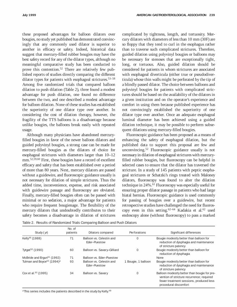
these proposed advantages for balloon dilators overbougies, no study yet published has demonstrated convinc-ingly that any commonly used dilator is superior toanother in efficacy or safety. Indeed, historical datasuggest that mercury-filled rubber bougies may have thebest safety record for any of the dilator types, although nomeaningful comparative study has been conducted toprove this contention.52 There are relatively few pub-lished reports of studies directly comparing the differentdilator types for patients with esophageal strictures.53–58
Among five randomized trials that compared balloondilation to push dilation (Table 2), three found a modestadvantage for push dilation, one found no differencebetween the two, and one described a modest advantagefor balloon dilation. None of these studies has establishedthe superiority of one dilator type over another. Inconsidering the cost of dilation therapy, however, thefragility of the TTS balloons is a disadvantage becauseunlike bougies, the balloons break easily with repeatedusage.
Although many physicians have abandoned mercury-filled bougies in favor of the newer balloon dilators andguided polyvinyl bougies, a strong case can be made formercury-filled bougies as the dilators of choice foresophageal strictures with diameters larger than 10–12mm.52,59,60 First, these bougies have a record of excellentefficacy and safety that has been established over a periodof more than 80 years. Next, mercury dilators are passedwithout a guidewire, and fluoroscopic guidance usually isnot necessary for dilation of simple strictures. Thus theadded time, inconvenience, expense, and risk associatedwith guidewire passage and fluoroscopy are obviated.Finally, mercury-filled bougies often can be passed withminimal or no sedation, a major advantage for patientswho require frequent bougienage. The flexibility of themercury dilators that undoubtedly contributes to theirsafety becomes a disadvantage in dilation of strictures
complicated by tightness, length, and tortuosity. Mer-cury dilators with diameters of less than 10 mm (30F) areso floppy that they tend to curl in the esophagus ratherthan to traverse such complicated strictures. Therefore,guided dilation using polyvinyl bougies or balloons maybe necessary for stenoses that are exceptionally tight,long, or tortuous. Also, guided dilation should beconsidered for patients in whom strictures are associatedwith esophageal diverticula (either true or pseudodiver-ticula) whose thin walls might be perforated by the tip ofa blindly passed dilator. The choice between balloons andpolyvinyl bougies for patients with complicated stric-tures should be based on the availability of the dilators ina given institution and on the operator’s experience andcomfort in using them because published experience hasnot convincingly established the superiority of onedilator type over another. Once an adequate esophagealluminal diameter has been achieved using a guideddilation technique, it may be possible to perform subse-quent dilations using mercury-filled bougies.
Fluoroscopic guidance has been proposed as a means ofenhancing the safety of esophageal dilation, but thepublished data to support this proposal are few andunconvincing.52 Fluoroscopic guidance usually is notnecessary in dilation of esophageal strictures with mercury-filled rubber bougies, but fluoroscopy can be helpful inselected cases to ensure that the dilator has traversed thestricture. In a study of 145 patients with peptic esopha-geal strictures or Schatzki’s rings treated with Maloneydilators, fluoroscopy was found to alter the dilationtechnique in 24%.61 Fluoroscopy was especially useful forensuring proper dilator passage in patients who had largehiatal hernias. Fluoroscopic guidance is used commonlyfor passing of bougies over a guidewire, but recentretrospective studies have challenged the need for fluoros-copy even in this setting.62–64 Kadakia et al.62 usedendoscopy alone (without fluoroscopy) to pass a marked
Table 2. Results of Randomized Trials Comparing Balloon and Push Dilators
Study ( yr)No. of
patients Dilators compared Perforations Significant differences
Kelly49 (1988) 71 Balloon vs. Celestin andEder–Puestow
0 Bougie modestly better than balloon forreduction of dysphagia and maintenanceof stricture patency
Tytgat50 (1990) 60 Balloon vs. Savary–Gilliard 0 Bougie modestly better than balloon forreduction of dysphagia
McBride and Ergun51 (1992) 71 Balloon vs. Eder–Puestow NoneTulman and Boyce52 (1994)a 93 Balloon vs. Celestin and
Eder–Puestow1 Bougie, 1 balloon Bougie modestly better than balloon for
reduction of dysphagia and maintenanceof stricture patency
Cox et al.53 (1995) 34 Balloon vs. Savary 0 Balloon modestly better than bougie for pre-vention of stricture recurrence; requiredfewer treatment sessions, produced lessprocedural discomfort
aThis series includes the patients described in the study by Kelly.49
July 1999 AMERICAN GASTROENTEROLOGICAL ASSOCIATION 239
guidewire through esophageal strictures in 138 patientsand successfully performed bougienage over the guidewirewithout fluoroscopic guidance and without complica-tions in all of these cases. However, it is not clear whetherthe relatively simple strictures in these patients couldhave been dilated successfully even with no guidewireusing only mercury-filled rubber bougies. Kozarek etal.64 found that fluoroscopic guidance was necessary inonly 8% of more than 300 patients who had esophagealstrictures dilated successfully with polyvinyl bougies.Thus it appears that fluoroscopically guided dilationshould be necessary only in the minority of patients whohave complicated esophageal strictures or large hiatalhernias.
Esophageal dilators are sized using the French gaugesystem in which the dilator’s diameter can be estimatedusing the following formula: Dilator Diameter (in mm)5 French Gauge Size 4 3. For example, a 30F dilator hasa diameter of 10 mm. There is no clear consensus on theoptimal size to which a peptic stricture should be dilated.For patients with Schatzki rings, dysphagia is the rulewhen the ring diameter is less than 13 mm (39F) anduncommon when the ring diameter exceeds 20 mm(60F).65 In a number of published series of patients withpredominantly peptic esophageal strictures, progressivedilation to a gauge size between 40 and 60F resulted ingood relief of dysphagia in approximately 85% of caseswith a very low rate of complications.11,66–70 In theory,successful dilation might decrease the mechanical barrierto gastroesophageal reflux imposed by the stricture andthereby result in an increase in heartburn and regurgita-tion. One investigation on this issue did not find asignificant overall postdilatation increase in acid refluxby protracted esophageal pH monitoring in a group of 10patients with peptic strictures, although certain individu-als in that group experienced a marked exacerbation ofacid reflux after the dilating procedure.71 With theavailability of highly effective antisecretory medicationssuch as proton pump inhibitors, concerns about exacerba-tion of reflux disease should not be a major factor inlimiting the extent of esophageal dilation. Nevertheless,it seems preferable to individualize dilation therapyrather than to aim for the same arbitrary dilator size forall patients. Although no study on benign strictures hasdocumented clearly that the risk of esophageal perfora-tion increases with dilator sizes up to 60F, it seems logicalto assume that fibrotic strictures have a critical sizebeyond which they cannot stretch without rupturing.Furthermore, the extent of initial stricture dilation doesnot seem to influence either stricture recurrence or therequirement for subsequent dilation39; therefore, there islittle support for the concept that strictures should be
dilated aggressively to prevent recurrence. For a patientwho has experienced complete relief of dysphagia withdilation to 48F, there is little to be gained by dilation tolarger diameters. Most patients experience good relief ofdysphagia when dilated to a bougie size between 40 and54F. The extent of dilation in an individual patientshould be based on the symptomatic response to therapyand on difficulties encountered during the dilationprocedure.
When dilating a stricture with bougies, the initialchoice of dilator size is based on an estimate of stricturediameter provided by a barium swallow or endoscopicexamination. A more physiological approach to estima-tion of stricture diameter involves having the patientswallow barium spheres of known diameter,53 but thistechnique is seldom used in clinical practice and has notbeen shown to improve the results of dilation. It isgenerally recommended that the first bougie passed havea diameter approximately equal to that estimated for thestricture.60 The ‘‘rule of threes’’ is a clinical maxim thatholds that no more than three bougies of progressivelyincreasing size should be passed at any one dilationsession to minimize the risks of esophageal perforationand hemorrhage.52 Mercury-filled rubber bougies (forwhich the rule of threes originally was formulated)increase in size by 2F units, so the diameter of the thirdbougie is only 1.3 mm larger than the first (e.g., dilationof a stricture using 36, 38, and 40F bougies increases thestricture diameter 12–13.3 mm). Although the rule ofthrees seems reasonable as a clinical guideline, no studiesverify that adherence to the rule improves dilationefficacy or safety. Furthermore, with balloon dilation,strictures are routinely dilated in one session to adiameter far greater than that which could be achievedwith the passage of three bougies. Balloons are designedto burst if a certain pressure is exceeded during dilation,but it is not clear that the burst pressure is less than thatrequired to rupture the diseased esophagus. One reportdescribed no perforations among 35 patients with a meanstricture diameter of 7.6 mm who underwent dilation inone session using a 60F balloon.53 This represents a meanincrease in stricture diameter of 12.4 mm. To achieve thisdegree of dilation with mercury dilators of progressivelyincreasing diameter would require the passage of at least18 bougies. No study has systematically evaluated thesafety of passing so many bougies in one sitting. If oneelects to dilate a stricture with mercury-filled bougiesrather than balloons, it seems a reasonable concession tothe unvalidated rule of threes to pass bougies of progres-sively increasing diameter until resistance is first encoun-tered, and to pass no more than two bougies after that inthe same session. This may not be a reasonable approach
240 AMERICAN GASTROENTEROLOGICAL ASSOCIATION GASTROENTEROLOGY Vol. 117, No. 1
when using polyvinyl dilators passed over a guidewire,because these dilators may not provide the operator witha meaningful tactile impression of stricture resistance.50
With polyvinyl dilators, the resistance to passage per-ceived by the operator may be more a function of frictionproduced by the guidewire than of resistance provided bythe esophageal stenosis. Nevertheless, esophageal dilationusing polyvinyl dilators has an excellent safety record,and it is not clear that adherence to the rule of threesenhances safety. In one recent study, benign esophagealstrictures in more than 400 patients were dilated usingeither a single, large polyvinyl dilator ($45F) or multiplepolyvinyl dilators so that the stricture diameter increasedby .2 mm in one session.64 Despite this flagrantviolation of the rule of threes, only one perforation wasobserved in 662 dilation sessions. If one elects to dilate astricture using balloon dilators, it seems prudent to limitthe initial dilation to no more than 45F. Although apreviously mentioned report found no complications in asmall series of patients dilated initially with 60F bal-loons,53 it seems preferable to be more conservative untilresults of future studies on the safety and efficacy ofballoon dilation are available.
The major complications of esophageal dilation areperforation and bleeding. These two complications ap-pear to occur with approximately equal frequency, al-though there is substantial variation among the reportedseries. For example, the 1974 American Society forGastrointestinal Endoscopy survey found rates of perfora-tion and bleeding of 0.1% and 0.3%, respectively, among13,139 esophageal dilations performed with mercury-filled rubber bougies.72 In contrast, Patterson et al.11
observed 5 perforations and only 1 hemorrhage duringesophageal dilation in 154 patients, most of whom hadmultiple sessions of bougienage performed over a periodof up to 87 months. In another American Society forGastrointestinal Endoscopy survey conducted in 1984,10 hemorrhages and 2 perforations were reported among456 patients treated with balloon dilation of esophagealstrictures for an overall complication rate of 2.5%.73 It isdifficult to provide precise estimates on the rate ofcomplications for esophageal dilation because of inconsis-tencies in the available studies. The patient populationsin most reported series were heterogeneous, composed ofpatients with strictures of varying complexity caused by anumber of different disease processes. It is likely that thecomplication rate is highest for dilations performed forstrictures that are exceptionally tight, long, or tortuous,but most reports do not supply specific informationregarding these stricture variables. If guided dilationtechniques are used preferentially for patients withcomplex strictures (those most likely to be associated
with procedural complications), then guided dilationmay spuriously appear to be more dangerous than blindbougienage. Many reports do not specify precisely thecriteria used for the choice of dilation technique, fewstudies are randomized, and it is difficult to perform ablinded trial of dilation therapy. With one or more ofthese deficiencies present in virtually every reportedstudy on esophageal dilation, it is difficult to perform ameaningful meta-analysis. Based on a review of theflawed studies available, it appears that serious complica-tions can be expected in approximately 0.5% of alldilation procedures. With such a low rate of complica-tions, a meaningful comparative study seeking to demon-strate a significant safety benefit of one procedure overanother will require large numbers of patients, numbersfar greater than those included in any comparative studyreported to date (Table 2).
Bacteremia complicates esophageal dilation more oftenthan any other procedure performed by gastroenterolo-gists.51 A number of reports suggest that bacteremiaaccompanies esophageal dilation in 20%–45% ofcases.74–76 Despite the high frequency of bacteremia,clinically recognizable infectious complications of esopha-geal dilation such as endocarditis and brain abscesses haverarely been reported.77–80,81 Although antibiotic prophy-laxis for esophageal dilation generally is recommendedroutinely only for patients at high risk for endocarditisaccording to the American Heart Association’s guide-lines,82,83 some authorities recently have suggested thatsuch prophylaxis should be given routinely even topatients with ‘‘intermediate risk’’ lesions such as mitralvalve prolapse with insufficiency.75
After initial dilation, stricture recurrence is a frequentphenomenon. Before proton pump inhibitors becameavailable, a number of investigations suggested that onlyapproximately 40% of patients would experience pro-tracted relief of dysphagia after a single dilation session,and approximately 60% would require multiple dila-tions.11,69,84,85 With proton pump inhibitor therapy, asfew as 30% of patients may require repeat dilationswithin 1 year.47 For patients with peptic strictures,factors associated with the need for multiple dilationsinclude a history of weight loss and absence of heartburnat the time of initial dilation.86 Neither the severity of theinitial stenosis nor the type and size of dilator usedappears to a have major influence on the likelihood ofstricture recurrence.11,69,86 Therefore, for individual pa-tients there is no reliable method to predict the need forrepeated dilation. Patients require close follow-up afterinitial dilation, and the procedure should be repeated ifdysphagia returns.
The role of surgical treatment for benign esophageal
July 1999 AMERICAN GASTROENTEROLOGICAL ASSOCIATION 241
strictures remains disputed. There are two major ap-proaches to the surgical treatment of esophageal stric-tures: (1) antireflux surgery with intraoperative stricturedilation for patients with peptic strictures caused byGERD (for patients whose peptic strictures are associatedwith substantial esophageal shortening, a lengtheningprocedure such as a Collis gastroplasty may be necessaryfor successful fundoplication), and (2) esophageal recon-struction for patients with strictures of any etiology thatare not responsive to dilation. Esophageal reconstructionmay rarely involve a procedure that widens the stricturewithout a resection (e.g., Thal fundic patch) or, morecommonly, the stenotic esophagus may be resected andreconstructed either by use of a gastric pull-up procedureor by interposition of a loop of bowel (colon or jejunum)between the remaining esophagus and the stomach. Forpatients with peptic strictures treated by antirefluxsurgery and intraoperative dilation, the success rate forrelief of dysphagia is similar to that reported for nonsurgi-cal dilation therapy.87–92 The major advantage of thisapproach is that successful antireflux surgery obviateslifelong medical therapy with its attendant expense andinconvenience. However, there is no clear benefit forsurgical treatment of esophageal strictures over medicaltherapy for relieving dysphagia per se, and there is a smalloperative mortality rate (usually ,1%) associated withfundoplication. The requirement for repeated dilationafter antireflux surgery for strictures ranges between 1%and 31% and appears to be somewhat smaller than thatdescribed for medical treatment of esophageal steno-ses.87–92 One might argue that the small risk of operativemortality might be offset by the reduced need forrepeated esophageal dilation with its attendant mortalityrate. However, there has been no meaningful comparativestudy of medical and surgical treatments for pepticesophageal strictures, so it is not clear that surgery istruly superior to medical therapy in prevention ofstricture recurrence. Physicians should be especiallycautious in recommending antireflux surgery for patientswith peptic esophageal strictures caused by esophagealmotility disorders such as scleroderma. The combinationof abnormal esophageal motor function and mechanicalobstruction imposed by fundoplication can result insevere postoperative dysphagia. However, scleroderma isnot an absolute contraindication to antireflux surgery,and some reports have described excellent outcomes forfundoplication in small series of selected patients withpeptic esophagitis and strictures caused by scleroderma.93
Finally, in rare cases, intractable esophageal strictures willrequire surgical resection and reconstruction. Operativemorbidity and mortality are substantially higher for
esophageal resection and reconstruction procedures thanfor antireflux surgery.
There are several reports on the use of endoscopicsteroid injection for the treatment of patients withrefractory esophageal strictures.94–96 All of these studiesinvolve small numbers of patients with benign stricturesof diverse etiology that had not responded to at least oneattempt at esophageal dilation. Some patients appeared torespond dramatically to steroid injection by exhibitingsubstantial improvement in dysphagia and decreases intheir requirement for repeated dilations. These smallstudies were not randomized or controlled, and theconclusions that can be drawn regarding the efficacy ofsteroid injection are very limited. Also, the mechanismsby which steroid injection might improve esophagealstenoses are not clear. Nevertheless, steroid injectionappears to be a relatively safe procedure, and a trial of thisunproved therapy seems reasonable for those rare patientswith benign esophageal strictures who derive no orshort-lived relief of dysphagia despite repeated attemptsat stricture dilation and aggressive control of refluxesophagitis. Finally, the technique of self-bougienage canbe taught to patients who require very frequent esopha-geal dilation despite intensive medical therapy andsteroid injection and for whom surgery is either contrain-dicated or unacceptable. The very limited published dataavailable on self-bougienage suggest that the techniquecan be both safe and effective.97
Lower Esophageal (Schatzki) Rings
In 1953, two independent groups of investigatorspublished descriptions of patients who had dysphagiaassociated with ringlike constrictions of the distal esopha-gus.98,99 The investigators had differing opinions aboutthe nature of these lower esophageal rings. Ingelfingerand Kramer98 proposed that the ringlike narrowings werecaused by contraction of an overactive band of esophagealmuscle, whereas Schatzki and Gary99 believed that esopha-geal rings were fixed structures that were not contractile.Although it is now clear that lower esophageal rings arequite common, controversy persists regarding the precisenature and pathogenesis of these structures.100 Based onan extensive review of the literature and on an autopsystudy of the distal esophagus, Goyal et al.101,102 con-cluded that some of the controversy had arisen becauseseveral different disorders had been included under therubric ‘‘lower esophageal ring’’ (e.g., muscular rings,mucosal rings, and ringlike peptic strictures). Muscularrings (also called A rings) are caused by a thickened bandof esophageal muscle fibers. The location of these ringscorresponds with an annular thickening in the muscularispropria of the distal esophagus that anatomists have
242 AMERICAN GASTROENTEROLOGICAL ASSOCIATION GASTROENTEROLOGY Vol. 117, No. 1
called the inferior esophageal sphincter (a structure thatshould not be confused with the LES described function-ally by physiologists).101 Muscular rings are locatedapproximately 2 cm above the esophagogastric junctionand rarely cause dysphagia. Most of the discussion thatfollows pertains to lower esophageal mucosal rings (alsocalled Schatzki rings or B rings), which are by far themost common type of lower esophageal ring found inpatients with dysphagia.101
The lower esophageal mucosal ring is a thin, dia-phragm-like, circumferential fold of mucosa that pro-trudes into the lumen of the distal esophagus, therebyposing a physical barrier to the passage of solid material.Mucosal rings usually are located at the squamocolumnarjunction with squamous epithelium lining the uppersurface and columnar epithelium lining the lower aspectof the ring. Fibrous tissue often can be found in thelamina propria. Some authorities have challenged thecontention that mucosal rings occur primarily at thesquamocolumnar junction, but the contrary evidencepresented is unconvincing.103 Schatzki rings are bestappreciated on barium swallow, where they are usually, ifnot invariably, associated with hiatal hernias. Withcareful radiological techniques aimed at distending thedistal esophagus, a lower esophageal ring can be found inapproximately 15% of all patients who have bariumswallows.104 However, few of these rings cause dysphagia.
The pathogenesis of the lower esophageal mucosal ringis not clear, but a number of hypotheses have beenproposed.101,105 One hypothesis holds that the ring ismerely a pleat of redundant mucosa that forms when theesophagus shortens either transiently (during contractionof the longitudinal muscle) or permanently (from un-known cause). Another hypothesis suggests that rings arecongenital in origin. Few data either strongly support orclearly refute these two hypotheses. A third hypothesissuggests that lower esophageal mucosal rings are thinpeptic strictures that develop as a consequence of GERD.If this hypothesis is correct, treatment of patients withrings might be directed at controlling reflux esophagitis.However, data on the association of GERD and Schatzkirings are inconclusive and contradictory. Goyal et al.102
found no evidence of reflux esophagitis associated withany of 9 esophageal mucosal rings identified at postmor-tem examination. Jamieson et al.105 found that GERDsymptoms and abnormal acid reflux (identified by esopha-geal pH monitoring) were less frequent among 32patients with Schatzki rings than among 32 controlpatients who had hiatal hernias without rings. In con-trast, Marshall et al.106 found evidence of GERD (abnor-mal acid reflux by 24-hour pH monitoring or endoscopicsigns of reflux esophagitis) in 13 of 20 patients with
symptomatic Schatzki rings. Also supporting a potentialrole for GERD in the pathogenesis of lower esophagealmucosal rings are the observations that demonstrablerings are virtually always associated with hiatal herniasand that serial radiographic examinations in some pa-tients with rings have shown progression in the esopha-geal stenoses over time to the point that they resembledpeptic strictures more than rings.107 Finally, a role for pillesophagitis has been suggested in the pathogenesis ofSchatzki rings. In one study, 62% of patients with ringswho had no signs or symptoms of GERD had a history ofingestion of medications known to cause pill esophagi-tis.105
The hypotheses on the pathogenesis of lower esopha-geal mucosal rings need not be mutually exclusive. Forexample, a congenital ring might be narrowed further byscarring from reflux or pill esophagitis. It is also conceiv-able that subtle rings do not cause symptoms unless thereis a supervening disorder that further interferes withesophageal clearance such as esophagitis or dysmotility.This could explain an apparent association between ringsand GERD, even if GERD plays no role in the pathogen-esis of the rings per se.
Treatment of patients with dysphagia caused by loweresophageal mucosal rings begins with reassurance thatthe condition is benign and with advice that food bechewed slowly and carefully.100 However, there are nodata to show that this advice is beneficial, and dilationtherapy is recommended for most patients. Traditionally,initial dilation therapy for Schatzki rings involves thepassage of a single large bougie or balloon (45–60F)aimed at fracturing (rather than merely stretching) themucosal fold.108 This approach differs from that discussedabove for peptic strictures, which are treated by gradualstretching for fear of rupturing the fibrotic esophaguswith a single, abrupt dilation. The safety of abruptdilation for esophageal rings has been well established,and most reported series limited to patients with Schatzkirings describe no complications of the procedure. How-ever, the contention that abrupt dilation is more effectivethan gradual dilation for relief of dysphagia has not beenverified. Furthermore, the notion that ring fracture byabrupt dilation should result in a low rate of recurrentdysphagia has not been substantiated by publishedexperience. Indeed, a number of reports suggest thatrecurrence is the rule rather than the exception afterdilation of symptomatic Schatzki rings.105,109,110 Forexample, one recent report describes 33 patients withsymptomatic Schatzki rings who were treated by abruptdilation with the passage of a single, large bougie(46–58F) and were followed up for a mean duration of 2years.109 Initial results were excellent, with all patients
July 1999 AMERICAN GASTROENTEROLOGICAL ASSOCIATION 243
reporting complete relief of dysphagia at a 4-weekfollow-up examination. However, actuarial life-table analy-sis showed that only 68% would remain free of dysphagiaafter 1 year, whereas only 11% of patients were estimatedto be symptom-free by year 5. In another study of 61patients with symptomatic Schatzki rings who werefollowed up for a mean duration of 75 months, 63%developed recurrent dysphagia after initially successfuldilation.110
No clinical features have been identified that areconsistently useful for predicting which patients willneed repeated dilations for Schatzki rings. The initialdiameter of the ring has not been found to correlatesignificantly with recurrence.109,110 Some investigatorshave suggested that recurrence is more likely in patientswho have GERD associated with their Schatzki rings,105
whereas others have found no correlation between thepresence of GERD and the need for repeated ringdilations.109 For patients who have both Schatzki ringsand GERD, antireflux therapy aimed at eliminating thesigns and symptoms of reflux esophagitis clearly isappropriate despite the lack of proof that such treatmentreduces the frequency of recurrent dysphagia. All patientswith rings who are treated with dilation should beadvised that recurrence is likely and that dilation mayneed to be repeated if dysphagia returns.
A number of different treatments have been used forpatients with ‘‘defiant’’ rings that do not respond toabrupt dilation using standard balloons and bougies, orthat recur quickly after initial relief of dysphagia. Thereare anecdotal reports on the successful use of pneumaticdilation with large balloon dilators such as those usuallyreserved for the treatment of achalasia.111 Some investiga-tors have performed endoscopic electrosurgical incision ofthe rings with good results.112,113 Others have usedsurgery either to rupture or excise the rings, and to repairthe associated hiatal hernias.114,115 However, one report ofsuch surgical therapy describes the recurrence of symp-toms in 14 of 36 patients (39%).114 A concomitantmotility disorder could explain the frequent recurrence ofsymptoms in some patients with defiant Schatzki rings,but few studies have systematically searched for esopha-geal dysmotility in these patients. The possibility of amotility disorder should be explored with a manometricexamination before one of the potentially hazardoustherapies mentioned above is used for patients withdefiant lower esophageal mucosal rings.
Physicians occasionally encounter patients who haveintermittent dysphagia for solid foods suggestive of aSchatzki ring but have no demonstrable abnormality onbarium swallow or endoscopic examination of the esopha-gus. Few reports have addressed the treatment of such
patients. One study explored the role of empiric esopha-geal dilation in patients who had ‘‘esophageal dysphagia’’and a normal esophagoscopy, barium swallow, or both.116
Among 20 such patients who had dysphagia for solidfoods only, empiric bougienage to 54F resulted inimmediate and complete resolution of dysphagia in 19cases (95%). During a median follow-up period of 20months, furthermore, 13 of those 19 patients experiencedno recurrence of dysphagia. In contrast, complete resolu-tion of dysphagia was seen after empiric dilation in only 2of 17 patients (12%) who had dysphagia for both solidsand liquids. It seems likely that empiric dilation waseffective in the former patients because they had subtlerings, webs, or strictures that were missed by thediagnostic studies, whereas the latter patients probablyhad motility disorders. Although the design of this studywas far from ideal, the report suggests that a trial ofempiric bougienage is reasonable for patients who com-plain of dysphagia for solid food and who have normalfindings on endoscopic examinations.
Achalasia
Primary achalasia is an esophageal disease ofunknown cause in which there is degeneration of neuronsin the wall of the esophagus.117,118 This degenerativeprocess preferentially involves the nitric oxide–producinginhibitory neurons that effect the relaxation of esophagealsmooth muscle.119,120 In some patients, degenerativechanges are found in brainstem ganglion cells in thedorsal motor nucleus of the vagus, and Wallerian degen-eration has been observed in vagal fibers that supply theesophagus.121 However, the disordered motility thatcharacterizes achalasia appears to result primarily fromthe degeneration of inhibitory neurons within the esopha-gus itself. The smooth muscle of the LES is tonicallycontracted at rest and relaxes when intramural neuronsrelease their inhibitory neurotransmitters. Loss of inhibi-tory innervation in the LES causes basal sphincterpressures to increase and renders the sphincter muscleincapable of normal relaxation. Unlike the LES, thesmooth muscle of the esophageal body does not exhibitresting tone; therefore, the loss of inhibitory neurons haslittle effect on resting pressure in the body of theesophagus. However, inhibitory influences are necessaryfor normal peristalsis, and the loss of inhibitory neuronsresults in aperistalsis.
Although the etiology of primary achalasia is notknown, certain recognized diseases can cause esophagealmotor abnormalities similar or identical to those ofprimary achalasia. This condition is called secondaryachalasia or pseudoachalasia. In Chagas’ disease, seen inCentral and South America, for example, esophageal
244 AMERICAN GASTROENTEROLOGICAL ASSOCIATION GASTROENTEROLOGY Vol. 117, No. 1
infection with the protozoan parasite Trypanosoma cruzican result in a loss of intramural ganglion cells that causesaperistalsis and incomplete LES relaxation.122 Malignan-cies rarely can cause pseudoachalasia either through directinvasion of esophageal neural plexuses (e.g., adenocarci-noma of the esophagogastric junction) or through releaseof uncharacterized humoral factors that disrupt esopha-geal function as part of a paraneoplastic syndrome.123 It isimportant to exclude the diagnosis of pseudoachalasiacaused by malignancy before invasive therapies such aspneumatic dilation or surgical myotomy (see below) areimplemented. It seems likely that these procedures wouldbe especially hazardous for patients with infiltratingneoplasms of the distal esophagus, although few pub-lished data are available to support this contention. Inmost cases, a careful history and endoscopic examinationare sufficient to exclude the diagnosis of pseudoachalasia.However, if the clinical history is strongly suggestive ofachalasia caused by malignancy (e.g., onset in old age,rapid progression of symptoms, profound weight loss),additional tests such as computed tomography or endo-sonography might be necessary to exclude an infiltratingneoplasm.
Presently, no therapy can reverse or even halt thedegeneration of enteric neurons that occurs in primaryachalasia. Therefore, the treatment of this disorder isfunctional, aimed at decreasing resting pressure in theLES (by pharmacological or mechanical means) to thepoint that the sphincter no longer poses a substantialbarrier to the passage of ingested material. This therapeu-tic attack on the LES does not reliably restore function inthe body of the esophagus, although the return ofperistaltic activity has been observed in some patientsafter the administration of various therapies designedsolely to decrease LES pressure.124–126 Nitrates andcalcium channel blockers have been shown to relax thesmooth muscle of the LES both in normal individuals andin patients with achalasia, and these agents have beenused to treat the disorder with variable success.127–135
Sublingual isosorbide dinitrate causes a substantial de-crease in LES pressure within minutes, and the effectoften lasts for more than 1 hour.127 Limited studiessuggest that more than 75% of patients with achalasiaexperience substantial improvement in dysphagia whenthey take isosorbide in a dose of 5–10 mg sublingually 10minutes before meals.127,128 One comparative trial in 15patients with achalasia found that nitrates were superiorto nifedipine both for decreasing LES pressure (64%decrease from baseline for nitrates vs. 47% decrease fornifedipine) and for relieving dysphagia (swallowing im-proved in 87% of patients on nitrates vs. 53% of patientson nifedipine).128 However, nitrate therapy often must be
discontinued because many patients experience intoler-able side effects (predominantly headache) and becausesome patients become refractory to the nitrates after aninitial good response.127,128 Like nitrates, calcium chan-nel blockers administered sublingually also result insubstantial decreases in LES pressure for more than 1hour.135 However, the results of studies on the effects ofcalcium channel blockers on the symptoms of achalasiaare contradictory; some investigators report good,133,135
marginal,130 and no134 relief of dysphagia in small groupsof patients treated with these agents. Although verapamiland diltiazem have been used, most published experiencehas involved nifedipine given in a dose of 10–20 mgsublingually 30 minutes before meals. Adequate plasmalevels have been observed when calcium channel blockersare administered orally (rather than sublingually) topatients with achalasia,134 but the absorption of orallyadministered drugs may be erratic for patients withadvanced disease in whom the pills can linger for hours inthe flaccid esophagus. In summary, pharmacotherapy forachalasia is inconvenient, often ineffective, and frequentlyassociated with side effects and tachyphylaxis. It appearsthat pharmacotherapy is best reserved for patients whoare unwilling or unable to tolerate the more effectiveinvasive forms of therapy discussed below.
At one time, authorities recommended esophagealdilation using large, mercury-filled rubber bougies (50–60F) as the initial therapy for achalasia.136 As techniquesfor forceful dilation of the LES in achalasia becamepopular, bougienage was dismissed as a procedure that, atbest, provided only transient and incomplete relief ofdysphagia.137 In 1982, Mandelstam et al.136 reported thatbougienage to 58F resulted in relief of dysphagia formonths to years in 4 of their 5 patients with achalasia andcalled for a reappraisal of the role of bougienage in thetreatment of this disorder. More recently, McJunkin etal.138 reviewed their experience with achalasia and re-ported that bougienage was successful when used asinitial therapy in 10 of 20 patients. Despite these reports,most modern authors continue to dismiss bougienage as aprocedure that provides only temporary and incompleterelief for patients with achalasia. Consequently, fewmodern gastroenterologists have any experience withbougienage in the treatment of achalasia. In the absenceof well-designed, prospective studies, it is difficult todraw firm conclusions regarding the efficacy of bougie-nage for achalasia. Presently, it appears that bougienage,like pharmacotherapy, is best reserved as an alternativetreatment for patients who are unwilling or unable totolerate the more invasive forms of therapy discussedbelow.
For decades, pneumatic dilation of the LES using large
July 1999 AMERICAN GASTROENTEROLOGICAL ASSOCIATION 245
balloons has been a popular form of treatment forachalasia.139–155 This therapy was designed to weaken theLES by tearing its muscle fibers, although experimentalevidence that the procedure indeed works by inducingmuscular tears is scant.148 Many different balloon dilatorshave been used over the years for treatment of achalasia(e.g., Mosher bag, Sippy dilators, Brown–McHardy dila-tor, Rider–Moeller dilator), but most are no longer beingmanufactured. Presently, the two most popular pneu-matic dilators in the United States are the Rigiflex dilator(similar in design to the Gruntzig angioplasty catheter),which is passed over a guidewire and positioned fluoro-scopically,150 and the Witzel dilator, which is mounted onan endoscope and inflated under direct vision.144
There is no clear consensus on the optimal method forperforming pneumatic dilation, and reported protocolshave varied widely with regard to the types of medica-tions used for sedation (which theoretically could affectthe outcome if the sedatives cause LES relaxation, andwhich have ranged from no sedatives whatsoever togeneral anesthesia), the types of dilators used (e.g.,Mosher, Sippy, Brown–McHardy, Rider–Moeller, Rigi-flex, Witzel), the maximum diameter of the balloon(reported range, 2.4–5.0 cm), the pressure to which theballoon is inflated (reported range 105 to .1000 mmHg), the rate of balloon inflation (rapid vs. gradual), theduration of balloon inflation (reported range, severalseconds to more than 5 minutes), and the number ofballoon inflations per dilating session (reported range,1–5).155 Most investigators have used a single pneumaticdilator at each treatment session, with the need forsubsequent dilations determined empirically by thesymptomatic response to the initial dilation.154 Othershave used the method of progressive pneumatic dilationadvocated by Vantrappen and Janssens148 in which aseries of balloons of progressively increasing diameter areinflated until there is manometric and radiographicevidence of adequate LES disruption. With so manypossible permutations and combinations of the variouscomponents of pneumatic dilation, and with so fewreports of prospective studies, it is difficult to perform ameaningful meta-analysis on the outcome of the proce-dure.
Despite the many variations in technique describedabove, most studies (primarily retrospective reviews of aninstitution’s experience) describe good to excellent short-term results in 60%–85% of patients with achalasiatreated with a single session of pneumatic dilation. Theduration of follow-up in most of these retrospectivereports is relatively brief; consequently, few data areavailable on the long-term outcome of pneumatic dila-tion. One large retrospective study found that only 65%
of 313 patients with achalasia treated with one or morepneumatic dilations still reported good to excellentresults after a median follow-up of 11 years.140 In one ofvery few prospective studies on patients with achalasiatreated initially with pneumatic dilation, 28 of 54subjects (52%) were found to require repeat dilationsduring a median follow-up of 4.1 years.156 The estimatedprobability of remaining in remission after a singledilation was 59% at 1 year and 26% at 5 years. In aretrospective investigation of 123 patients treated ini-tially with pneumatic dilation, Parkman et al.157 foundthat 42% required further treatment during a meanfollow-up of 4.7 years. These investigators also noted thatrepeat pneumatic dilations became progressively lesseffective than the initial procedure. For example, 58% ofpatients who had a second pneumatic dilation requiredfurther treatment, whereas 73% required additionaltherapy after a third dilation. In a recent, more optimisticreport of a retrospective study, Katz et al.158 found thatpneumatic dilation was successful in relieving the symp-toms of achalasia in 85% of 72 patients who werefollowed up for a mean of 6.5 years, and only 4 patientsrequired more than one dilation procedure. Overall, thesestudies suggest that approximately 50% of patients withachalasia treated initially with a single pneumatic dila-tion will require further therapy within 5 years and thatsubsequent pneumatic dilations are progressively lesslikely to result in a sustained remission. Some authorshave recommended that other forms of therapy beconsidered after two or three unsuccessful pneumaticdilations.117
A number of studies have sought to identify clinicaland technical factors that might help clinicians predictresponses to pneumatic dilation.156–161 Young age consis-tently has been shown to be associated with a poorresponse.156–159 In one prospective study, for example, the2-year sustained remission rate for patients under age 40was only 29%, compared with 67% for those age 40 yearsand older.156 Perhaps young muscle is less susceptible todamage by forceful dilation than old muscle. Among thetechnical factors, LES pressure after dilation appears to bethe best predictor of outcome. In the aforementionedprospective study, the 2-year remission rate was 100% forpatients whose postdilation LES pressure was ,10 mmHg, 71% for those whose postdilation LES pressure was10–20 mm Hg, and 23% for those with postdilation LESpressures of $20 mm Hg.156 The size of the dilatingballoon also appears to influence the outcome (i.e., thelong-term remission rate may be higher when largerdiameter balloons are used), although few studies havespecifically explored this issue.151,154,156 Factors that donot appear to have a substantial influence on the response
246 AMERICAN GASTROENTEROLOGICAL ASSOCIATION GASTROENTEROLOGY Vol. 117, No. 1
to pneumatic dilation include gender, duration of esopha-geal symptoms before treatment, diameter of the esopha-gus, pretreatment LES pressure, duration of ballooninflation,161 number of balloon inflations per dilatingsession, maximum inflation pressure, and chest painpresent during the procedure. Finally, although manyclinicians have been taught to look for blood on thedilating balloon as proof that the procedure has beenconsummated, this finding has not been found to be auseful predictor of outcome.157,160
Despite the wide variations in equipment and tech-niques used for pneumatic dilation, reported complica-tion rates are remarkably similar. Esophageal perforationis the most common serious complication of the proce-dure, and most large series describe rates of perforation inthe range of 2%–6%.117,155 Mortality from pneumaticdilation is rare and has been estimated at approximately0.2%.117 The new Rigiflex dilators do not appear to haveany safety advantage over the older balloons, and someinvestigators even suggest that perforation rates arehigher with the newer instruments.162 There are very fewreports of studies that have prospectively compared thedifferent dilator types for safety and efficacy, and thosethat have been published are primarily of historicalinterest because some of the dilators compared are nolonger available.163 Achalasia is an uncommon disease,and the perforation rate for pneumatic dilation is rela-tively low. Consequently, it is difficult to conduct ameaningful study on factors that might predispose toesophageal perforation. A number of potential predispos-ing factors have been suggested, including malnutrition,weight loss, low LES pressure, high-amplitude contrac-tions in the distal esophagus, previous pneumatic dila-tions, administration of anesthesia, large balloon size, andhigh inflation pressures.164–166 However, none of thesefactors has been shown consistently to influence the safetyof pneumatic dilation. In addition, the presence of a largeepiphrenic diverticulum has been regarded as a contrain-dication to pneumatic dilation because of anecdotalreports of perforations in this setting.147 However, therisk of perforation imposed by these diverticula has notbeen well established. Although it once was standardclinical practice to hospitalize patients for pneumaticdilation, it appears that performing the procedure in anoutpatient setting for otherwise healthy individuals doesnot substantially increase the risk of complications.167,168
A number of authorities recommend that esophagraphybe performed shortly after pneumatic dilation (usually assoon as the sedation has worn off) to seek evidence ofperforation.117 Although water-soluble contrast (e.g.,Gastrografin) is commonly recommended for this study,such hypertonic agents can cause a chemical pneumonitis
if they are aspirated into the lungs. Consequently, theseagents should not be used in sedated patients or inneurologically impaired individuals who are at risk foraspiration. Indeed, it may be preferable to use dilutebarium rather than water-soluble contrast agents rou-tinely for these postdilation esophagrams, but this conten-tion has not been validated by formal investigation.Finally, it has been estimated that approximately 2% ofpatients treated with pneumatic dilation will developreflux esophagitis because of the resulting LES hypoten-sion.117 Indeed, aggressive pneumatic dilation essentiallychanges the manometric features of achalasia to those ofscleroderma, a disorder often associated with severeGERD. One recent study describes abnormal acid refluxby 24-hour esophageal pH monitoring in 6 of 17 patientswho had pneumatic dilation.169 With the availability ofhighly effective antireflux therapies such as proton pumpinhibitors, concerns about the induction of GERD bypneumatic dilation need not be overriding.
Surgical myotomy, in which the surgeon weakens theLES by cutting its muscle fibers, traditionally has beenviewed as the primary alternative to pneumatic dilationfor achalasia. Cardiomyotomy was first performed in1913 by Ernst Heller, and modern surgeons now usemodifications of Heller’s original procedure.117 The stan-dard ‘‘open’’ myotomy can be performed using either anabdominal or, more commonly, a thoracic approach.170–
184 Recently, a number of centers have reported theirpreliminary results with minimally invasive surgery forachalasia in which laparoscopic or thoracoscopic tech-niques are used to perform the myotomy.185–190 Otherimportant differences in technique among centers includevariations in the length and depth of the myotomyincision and whether or not the myotomy is combinedwith an antireflux procedure. Surgical myotomy resultsin good to excellent relief of symptoms in 70%–90% ofpatients, with few serious complications and a mortalityrate similar to that reported for pneumatic dilation(approximately 0.3%).117 Reflux esophagitis (which maybe complicated by esophageal ulceration, stricture, andBarrett’s esophagus) has been found to develop in approxi-mately 11% of patients treated by surgical myotomy,117
and surgeons continue to debate the need for the additionof an antireflux procedure. The addition of a fundoplica-tion appears to reduce the rate of symptomatic, postmy-otomy GERD to approximately 4% but may increase thefrequency of postoperative dysphagia by imposing a valvebetween the aperistaltic esophagus and the stomach.191 Inthe absence of well-designed prospective studies, thedebate over the need for fundoplication in this settingremains unresolved. However, the recent availability ofhighly effective medical antireflux therapies such as
July 1999 AMERICAN GASTROENTEROLOGICAL ASSOCIATION 247
proton pump inhibitors should simplify the managementof postoperative GERD considerably and perhaps obviateroutine fundoplication.
Late recurrence of dysphagia after surgical myotomy orpneumatic dilation may be caused by a return of tone inthe damaged LES muscle, by GERD with peptic strictureformation, or very rarely by squamous cell carcinoma ofthe esophagus that develops with increased frequency inpatients with achalasia.192 Endoscopic examination shouldreadily differentiate these disorders. Late recurrence ofdysphagia appears to be less common after surgicalmyotomy than after pneumatic dilation, but postopera-tive recurrence of dysphagia is not unusual after surgicaltreatment. For example, Jara et al.176 describe a sustainedremission rate of 81% after 10 years for patients treatedwith esophagomyotomy. Ellis et al. performed a Kaplan–Meier analysis on the results of esophagomyotomy in 81patients and estimated sustained remission rates of 86%at 15 years and 67% at 20 years.193 Malthaner et al.194
also found a deterioration of surgical results over timewith good to excellent results reported by 21 of 22patients (95%) at 1 year, 17 of 22 (77%) at 5 years, 15 of22 (68%) at 10 years, 11 of 16 (69%) at 15 years, and 6 of9 (67%) at 20 years or more.
Relatively few studies have directly compared theresults of myotomy and pneumatic dilation. In oneretrospective study, Okike et al.177 reported good toexcellent relief of dysphagia in 85% of 468 patientstreated with myotomy compared with only 65% of 431patients who underwent pneumatic dilation for achalasia.In another retrospective review, Anselmino et al.195
concluded that surgery was safer and more effective thanpneumatic dilation, although the outcome of pneumaticdilation in this study was far worse than that reported bymost investigators (i.e., 15% rate of esophageal rupture,39% rate of dysphagia relief after a mean follow-up of 55months). In contrast, Abid et al.196 reviewed theirexperience with pneumatic dilation (36 patients) andsurgical myotomy (9 patients) in patients followed up fora mean interval of approximately 2 years and concludedthat the procedures are equally effective (success rate forboth procedures, approximately 90%) if both are per-formed by skilled operators. In one of the two prospec-tive, randomized trials of myotomy and pneumaticdilation reported to date, Csendes et al.197 describeexcellent results after a median follow-up of approxi-mately 5 years in 40 of 42 (95%) patients in the surgicalgroup, compared with 24 of 37 (65%) patients whounderwent pneumatic dilation. This study has beencriticized for using a pneumatic dilation protocol thatmay have been suboptimal (i.e., use of atropine aspremedication, low balloon inflation pressures, short
duration of inflation),198 but as noted above, thesetechnical factors have not been shown to affect theoutcome of pneumatic dilation. A recent, smaller random-ized trial of balloon dilation and myotomy found the twoprocedures equally effective in relieving dysphagia for upto 3 years.199 Despite the limitations of the availablestudies, it appears that surgery generally is superior topneumatic dilation for both short-term and long-termrelief of dysphagia. Furthermore, the rate of seriouscomplications such as esophageal perforation appears tobe less for surgically treated patients, and the mortalityrates for the two procedures are approximately equal. Themajor disadvantages for surgery are the high initial cost,protracted recovery period, and frequent development ofGERD postoperatively. A recent cost analysis comparingopen surgery and pneumatic dilation concluded thatinitial pneumatic dilation (with surgery reserved fordilation failures) was the most cost-effective approach.157
However, this analysis did not consider myotomy per-formed by minimally invasive techniques. The short-term relief of dysphagia by minimally invasive surgeryappears to be at least as good as that described for theopen procedure, but long-term results are not yet avail-able because the procedures are so new. Prospective,comparative studies are sorely needed in this area. Iflaparoscopic or thoracoscopic techniques enable surgeonsto perform myotomy at reduced cost and with shorterrecovery periods, if the operation is shown to be durable,and if the incidence of severe GERD is acceptably low,then myotomy by minimally invasive techniques maywell be more cost-effective than pneumatic dilation.Pending the results of long-term studies on minimallyinvasive surgery, however, the decision between pneu-matic dilation and myotomy as initial therapy forachalasia should be based on a consideration of thepatient’s preferences and on the availability of personnelexperienced in the two techniques.
Recently, an interesting new approach to the treatmentof achalasia has been introduced by Pasricha et al.200–203
These investigators noted that botulinum toxin, a potentinhibitor of the release of acetylcholine from nerveendings, has been used successfully for decades to treatcertain spastic disorders of skeletal muscle such asblepharospasm. They hypothesized that the local, peren-doscopic injection of botulinum toxin into the LES ofpatients with achalasia would poison the excitatory(acetylcholine-releasing) neurons that contribute to LESsmooth muscle tone, thereby effecting a therapeuticdecrease in LES pressure. The investigators tested theirhypothesis in an animal model and verified that localbotulinum toxin injection caused a significant decrease inLES pressure in piglets.200 Later, a pilot study in 10
248 AMERICAN GASTROENTEROLOGICAL ASSOCIATION GASTROENTEROLOGY Vol. 117, No. 1
patients with achalasia suggested a beneficial therapeuticeffect for botulinum toxin injection of the LES,201 aneffect that was confirmed in a double-blind, placebo-controlled trial in 21 patients.202 Most recently, thisgroup has described the results of botulinum toxininjection in 31 patients with achalasia.203 Twenty-eightof the 31 patients (90%) showed immediate symptomaticimprovement, but this effect was short-lived in manycases. Despite repeated injections of the toxin whensymptoms returned, only 20 of the 31 patients (65%)remained in remission 6 months after the initial treat-ment. In these 20 responders, botulinum toxin injectioncaused resting LES pressure to decrease by 45% comparedwith baseline values. During a median follow-up of 2.4years, however, 19 of the 20 responders experienced arelapse of symptoms that required further therapy. Among15 of these patients who were treated with anotherbotulinum toxin injection, only 9 had sustained remis-sions. Kaplan–Meier analysis suggested that only 68% ofpatients in remission at 6 months would remain inremission at the end of 1 year. The median duration ofremission after initial treatment was 16 months, and thelongest remission observed was 28 months. Other groupshave confirmed the short-term efficacy of botulinumtoxin injection.204,205 Pasricha et al.200–203 note that likepneumatic dilation, botulinum toxin injection was moreeffective in older patients (82% response rate for patientsage .50 years vs. 43% in younger patients). They alsonote that the toxin was 100% effective for patients withvigorous achalasia (defined by the presence of pressurewaves in the esophageal body with amplitudes .40 mmHg), whereas only 52% of patients with classic achalasiashowed a clinical response at 6 months. However, anothergroup of investigators did not find a difference inresponse to botulinum toxin injection between patientswith vigorous and classic achalasia.204 Botulinum toxininjection appears to be remarkably safe. Approximately25% of patients experience transient, mild chest painimmediately after the procedure, and fewer than 5% ofpatients develop symptomatic GERD. The most seriouscomplication reported to date is a case report of a patientwho developed severe, ulcerative esophagitis (probablycaused by acid reflux) after toxin injection.206 Thispatient also was found to have adhesions and periesopha-geal inflammation when he subsequently underwentsurgical treatment for achalasia. These studies suggestthat botulinum toxin injection of the LES is a remarkablysafe procedure that can induce a clinical remission for atleast 6 months in approximately two thirds of patientswith achalasia. However, most patients need repeatedinjections to maintain the remission, and only approxi-mately two thirds of those patients in remission at 6
months will remain in remission at 1 year despiterepeated injections. Vaezi et al.207 recently reported theresults of a prospective study in which 42 patients withachalasia were randomly assigned to receive either botuli-num toxin injection or pneumatic dilation. At 12months, only 32% of patients treated with botulinuminjection were in symptomatic remission, compared with70% of patients in the pneumatic dilation group.
Castell and Katzka208 point out that botulinum toxintherapy is expensive (approximately $300 just for thebotulinum toxin used at each treatment session), thefailure rate is substantial, patients frequently need re-peated injections, and the long-term efficacy of theprocedure is unknown. These concerns, combined withthe very poor response rate found in patients under theage of 50, suggest that botulinum toxin injection maynot be the ideal choice of therapy for young patients withachalasia. Further studies are needed before the procedurecan be recommended for clinical application outside ofresearch protocols. Presently, botulinum toxin injectioncan be considered for the treatment of patients who haveserious comorbidity and for whom pneumatic dilation orsurgical myotomy poses inordinate risks.
Other experimental therapies have been proposed forthe treatment of achalasia, including endoscopic my-otomy of the LES using a needle-knife209 and perendo-scopic injection of ethanolamine into the LES.210 Al-though preliminary reports are interesting, far moreinvestigation is needed before these potentially hazardoustreatments can be recommended for clinical use. In veryrare patients in whom all reasonable therapies have failed,esophageal resection and reconstruction may be consid-ered as a last resort.191 Finally, a feeding gastrostomy canbe considered for patients in whom all reasonable primarytherapies for achalasia have failed or for very elderly andinfirm patients for whom other therapies may be hazard-ous. Few published data are available on the use ofgastrostomy for patients with achalasia, but many neuro-logically intact patients undoubtedly will find thistherapy unacceptable.
STUART JON SPECHLER, M.D.Dallas Department of Veterans Affairs Medical Center
and University of Texas Southwestern Medical CenterDallas, Texas
References1. Meyer GW, Austin RM, Brady CE III, Castell DO. Muscle anatomy
of the human esophagus. J Clin Gastroenterol 1986;8:131–134.
2. Castell DO, Donner MW. Evaluation of dysphagia: a carefulhistory is crucial. Dysphagia 1987;2:65–71.
July 1999 AMERICAN GASTROENTEROLOGICAL ASSOCIATION 249
3. Castell DO, Knuff TE, Brown FC, Gerhardt DC, Burns TW, GaskinsRD. Dysphagia. Gastroenterology 1979;76:1015–1024.
4. Poirier TJ, Rankin GB. Gastrointestinal manifestations of progres-sive systemic scleroderma based on a review of 364 cases. AmJ Gastroenterol 1972;58:30–44.
5. Lock G, Holstege A, Lang B, Scholmerich J. Gastrointestinalmanifestations of progressive systemic sclerosis. Am J Gastroen-terol 1997;92:763–771.
6. Edwards DAW. History and symptoms of esophageal disease. In:Vantrappen G, Hellemans J, eds. Diseases of the esophagus.Berlin: Springer, 1974:103–118.
7. Smith DF, Ott DJ, Gelfand DW, Chen MYM. Lower esophagealmucosal ring: correlation of referred symptoms with radiographicfindings using a marshmallow bolus. AJR 1998;171:1361–1365.
8. Wilcox CM, Alexander LN, Clark WS. Localization of an obstruct-ing esophageal lesion. Is the patient accurate? Dig Dis Sci1995;40:2192–2196.
9. Cook IJ, Kahrilas PJ. AGA technical review on management oforopharyngeal dysphagia. Gastroenterology 1999;116:455–478.
10. Spechler SJ, Souza RF, Rosenberg SJ, Ruben RA, Goyal RK.Heartburn in patients with achalasia. Gut 1995;37:305–308.
11. Patterson DJ, Graham DY, Smith JL, Schwartz JT, Alpert E, LanzaFL, Cain GD. Natural history of benign esophageal stricturetreated by dilatation. Gastroenterology 1983;85:346–350.
12. Spechler SJ, Goyal RK. Barrett’s esophagus. N Engl J Med 1986;315:362–371.
13. Kikendall JW, Johnson LF. Pill-induced esophageal injury. In:Castell DO, ed. The esophagus. 2nd ed. Boston: Little, Brown,1995:619–633.
14. Colina RE, Smith M, Kekendall JW, Wong RK. A new probableincreasing cause of esophageal ulceration: alendronate. Am JGastroenterol 1997;92:704–706.
15. Lapadula G, Muolo P, Semeraro F, Covelli M, Brindicci D,Cuccorese G, Francavilla A, Pipitone V. Esophageal motilitydisorders in the rheumatic diseases: a review of 150 patients.Clin Exp Rheumatol 1994;12:515–521.
16. Limburg AJ, Beekhuis H, Smit AJ, Kallenberg CG, Piers DA,Kleibeuker JH. Esophageal hypomotility in primary and second-ary Raynaud’s phenomenon: comparison of esophageal scintig-raphy with manometry. J Nucl Med 1995;36:451–455.
17. Maddern JG, Horowitz M, Jamieson GG, et al. Abnormalities ofesophageal and gastric emptying in progressive systemic sclero-sis. Gastroenterology 1984;87:922–926.
18. Connolly GM, Hawkins D, Harcourt-Webster JN, Parsons PA,Husain OA, Gazzard BG. Oesophageal symptoms, their causes,treatment, and prognosis in patients with the acquired immuno-deficiency syndrome. Gut 1989;30:1033–1039.
19. Baehr PH, McDonald GB. Esophageal infections: risk factors,presentation, diagnosis, and treatment. Gastroenterology 1994;106:509–532.
20. Agha FP. Candidiasis-induced esophageal strictures. Gastro-intest Radiol 1984;9:283–286.
21. Schroeder PL, Filler SJ, Ramirez B, Lazarchik DA, Vaezi MF,Richter JE. Dental erosions and acid reflux disease. Ann InternMed 1995;122:809–815.
22. Ott DJ, Chen YM, Wu WC, Gelfand DW, Munitz HA. Radiographicand endoscopic sensitivity in detecting lower esophageal muco-sal ring. AJR 1986;147:261–265.
23. Ott DJ, Gelfand DW, Lane TG, Wu WC. Radiologic detection andspectrum of appearances of peptic esophageal strictures. J ClinGastroenterol 1982;4:11–15.
24. Ott DJ, Gelfand DW, Wu WC, Chen YM. Radiologic evaluation ofdysphagia. JAMA 1986;256:2718–2721.
25. Ott DJ, Kelley TF, Chen MYM, Gelfand DW. Evaluation of the
esophagus with a marshmallow bolus: clarifying the cause ofdysphagia. Gastrointest Radiol 1991;16:1–4.
26. Ott DJ, Kelley TF, Chen MY, Gelfand DW, Wu WC. Use of amarshmallow bolus for evaluating lower esophageal mucosalrings. Am J Gastroenterol 1991;86:817–820.
27. Hewson EG, Ott DJ, Dalton CB, Chen YM, Wu WC, Richter JE.Manometry and radiology. Complementary studies in the assess-ment of esophageal motility disorders. Gastroenterology 1990;98:626–632.
28. Ott DJ, Richter JE, Chen YM, Wu WC, Gelfand DW, Castell DO.Esophageal radiography and manometry: correlation in 172patients with dysphagia. AJR 1987;149:307–311.
29. Parkman HP, Maurer AH, Caroline DF, Miller DL, Krevsky B,Fisher RS. Optimal evaluation of patients with nonobstructiveesophageal dysphagia. Manometry, scintigraphy, or videoesoph-agography? Dig Dis Sci 1996;41:1355–1368.
30. Ott DJ. Radiographic techniques and efficacy in evaluatingesophageal dysphagia. Dysphagia 1990;5:192–203.
31. Kahrilas PJ, Clouse RE, Hogan WJ. American Gastroenterologi-cal Association technical review on the clinical use of esopha-geal manometry. Gastroenterology 1994;107:1865–1884.
32. Horowitz M, Maddern GJ, Maddox A, Wishart J, Chatterton BE,Shearman DJ. Effects of cisapride on gastric and esophagealemptying in progressive systemic sclerosis. Gastroenterology1987;93:311–315.
33. Meshkinpour H, Glick ME, Sanchez P, Tarvin J. Esophagealmanometry: a benefit and cost analysis. Dig Dis Sci 1982;27:772–775.
34. Johnston PW, Johnston BT, Collins BJ, Collins JSA, Love AHG.Audit of the role of oesophageal manometry in clinical practice.Gut 1993;34:1158–1161.
35. Ott DJ. Pikna LA. Clinical and videofluoroscopic evaluation ofswallowing disorders. AJR 1993;161:507–513.
36. Russell COH, Hill LD, Holmes ER, Hull DA, Gannon R, Pope CE II.Radionuclide transit: a sensitive screening test for esophagealdysfunction. Gastroenterology 1981;80:887–892.
37. Tatsch K, Voderholzer WA, Weiss MJ, Schrottle W, Hahn K.Reappraisal of quantitative esophageal scintigraphy by optimiz-ing results with ROC analyses. J Nucl Med 1996;37:1799–1805.
38. Spechler SJ. Complications of gastroesophageal reflux disease.In: Castell DO, ed. The esophagus. 2nd ed. Boston: Little,Brown, 1995:533–544.
39. Marks RD, Shukla M. Diagnosis and management of pepticesophageal strictures. Gastroenterologist 1996;4:223–237.
40. Broor SL, Raju GS, Bose PP, Lahoti D, Ramesh GN, Kumar A,Sood GK. Long term results of endoscopic dilatation for corro-sive oesophageal strictures. Gut 1993;34:1498–1501.
41. Ferguson R, Dronfield MW, Atkinson M. Cimetidine in treatmentof reflux oesophagitis with peptic stricture. Br Med J 1979;2:472–474.
42. Starlinger M, Appel WH, Schemper M, Schiessel R. Long-termtreatment of peptic esophageal stenosis with dilation andcimetidine: factors influencing clinical results. Eur Surg Res1985;17:207–214.
43. Farup PG, Modalsli B, Tholfsen JK. Long-term treatment with300 mg ranitidine once daily after dilatation of peptic oesopha-geal strictures. Scand J Gastroenterol 1992;27:594–598.
44. Larrain A, Csendes A, Pope CE II. Surgical correction of reflux. Aneffective therapy for esophageal strictures. Gastroenterology1975;69:578–583.
45. Koop H, Arnold R. Long term maintenance treatment of refluxesophagitis with omeprazole: prospective study in patients withH2-blocker resistant esophagitis. Dig Dis Sci 1991;36:552–557.
46. Marks RD, Richter JE, Rizzo H, Koehler RE, Spenney JG, Mills TP,Champion G. Omeprazole versus H2-receptor antagonists in
250 AMERICAN GASTROENTEROLOGICAL ASSOCIATION GASTROENTEROLOGY Vol. 117, No. 1
treating patients with peptic stricture and esophagitis. Gastroen-terology 1994;106:907–915.
47. Smith PM, Kerr GD, Cockel R, Ross BA, Bate CM, Brown P,Dronfield MW, Green JRB, Hislop WS, Theodossi A, McFarland J,Watts DA, Taylor MD, Richardson PDI, Restore InvestigatorGroup. A comparison of omeprazole and ranitidine in theprevention of recurrence of benign esophageal stricture. Gastro-enterology 1994;107:1312–1318.
48. Dakkak M, Hoare RC, Maslin SC, Bennett JR. Oesophagitis is asimportant as oesophageal stricture diameter in determiningdysphagia. Gut 1993;34:152–155.
49. Kelly HDB. Origins of oesophagology. Proc R Soc Med 1969;62:781–786.
50. Tytgat GNJ. Dilation therapy of benign esophageal stenoses.World J Surg 1989;13:142–148.
51. McBride MA, Ergun GA. The endoscopic management of esopha-geal strictures. Gastrointest Endosc Clin North Am 1994;4:595–621.
52. Tulman AB, Boyce HW. Complications of esophageal dilation andguidelines for their prevention. Gastrointest Endosc 1981;27:229–234.
53. Cox JGC, Winter RK, Maslin SC, Jones R, Buckton GK, Hoare RC,Sutton DR, Bennett JR. Balloon or bougie for dilatation of benignoesophageal stricture? An interim report of a randomisedcontrolled trial. Gut 1988;29:1741–1747.
54. Shemesh E, Czerniak A. Comparison of Savary–Gilliard andballoon dilatation of benign esophageal strictures. World J Surg1990;14:518–521.
55. Yamamoto H, Hughes RW Jr, Schroeder KW, DiMagno EP.Treatment of benign esophageal stricture by Eder–Puestow orballoon dilators: a comparison between randomized and prospec-tive randomized trials. Mayo Clin Proc 1992;67:228–236.
56. Cox JGC, Winter RK, Maslin SC, Dakkak M, Jones R, Buckton GK,Hoare RC, Dyet JF, Bennett JR. Balloon or bougie for dilatation ofbenign esophageal stricture? Dig Dis Sci 1994;39:776–781.
57. Saeed ZA, Winchester CB, Ferro PS, Michaletz PA, Schwartz JT,Graham DY. Prospective randomized comparison of polyvinylbougies and through the scope balloons for dilation of pepticstrictures of the esophagus. Gastrointest Endosc 1995;41:189–195.
58. Hine KR, Hawkey CJ, Atkinson M, Holmes GK. Comparison of theEder–Puestow and Celestin techniques for dilating benign oe-sophageal strictures. Gut 1984;25:1100–1102.
59. Marks RD, Richter JE. Peptic strictures of the esophagus. Am JGastroenterol 1993;8:1160–1173.
60. Nostrant TT. Esophageal dilatation. Dig Dis 1995;13:337–355.61. Tucker LE. The importance of fluoroscopic guidance for Maloney
dilation. Am J Gastroenterol 1992;87:1709–1711.62. Kadakia SC, Parker A, Carrougher JG, Shaffer RT. Esophageal
dilation with polyvinyl bougies using a marked guidewire withoutthe aid of fluoroscopy: an update. Am J Gastroenterol 1993;88:1381–1386.
63. Fleischer DE, Benjamin SB, Cattau EL, Collen MJ, Lewis JH, JaffeMH, Zeman RK. A marked guide wire facilitates esophagealdilatation. Am J Gastroenterol 1989;84:359–361.
64. Kozarek RA, Patterson DJ, Ball TJ, Gelfand MG, Jiranek GE,Bredfeldt JE, Brandabur JJ, Wolfsen HW, Raltz SL. Esophagealdilation can be done safely using selective fluoroscopy andsingle dilating sessions. J Clin Gastroenterol 1995;20:184–188.
65. Schatzki R. The lower esophageal ring. Long term follow-up ofsymptomatic and asymptomatic cases. AJR 1963;90:805–810.
66. Bradpiece HA, Galland RB, Spencer J. Esophageal dilation as anoutpatient procedure. Surg Gynecol Obstet 1988;167:45–48.
67. Dumon JR, Meric B, Sivak MV Jr, Fleischer D. A new method ofesophageal dilation using Savary-Gilliard bougies. GastrointestEndosc 1985;31:379–382.
68. Graham DY, Tabibian N, Schwartz JT, Smith JL. Evaluation ofeffectiveness of through-the-scope balloons as dilators of be-nign and malignant gastrointestinal strictures. GastrointestEndosc 1987;33:432–435.
69. Ogilvie AL, Ferguson R, Atkinson M. Outlook with conservativetreatment of peptic oesophageal stricture. Gut 1980;21:23–25.
70. Wesdorp ICE, Bartelsman JFWM, Den Hartog Jager FCA, Hui-bregtse K, Tytgat GN. Results of conservative treatment ofbenign esophageal strictures: a follow-up study in 100 patients.Gastroenterology 1982; 82:487–493.
71. Penagini R, Dabbagh MA, Misiewicz JJ, Evans PF, Trotman IF.Effect of dilatation of peptic esophageal strictures on gastro-esophageal reflux, dysphagia, and stricture diameter. Dig DisSci 1988;33:389–392.
72. Silvis SE, Nebel O, Rogers G, Sugawa C, Mandelstam P.Endoscopic complications. Results of the 1974 American Soci-ety for Gastrointestinal Endoscopy survey. JAMA 1976;235:928–930.
73. Kozarek RA. Hydrostatic balloon dilation of gastrointestinalstenoses: a national survey. Gastrointest Endosc 1986;32:15–19.
74. Botoman VA, Surawicz CM. Bacteremia with gastrointestinalendoscopic procedures. Gastrointest Endosc 1986;32:342–346.
75. Zuccaro G, Richter JE, Rice TW, Achkar E, Easley K, Lewis J,Gordon SM. Viridans streptococcal bacteremia after esophagealstricture dilation. Gastrointest Endosc 1998;48:568–573.
76. Nelson DB, Sanderson SJ, Azar MM. Bacteremia with esopha-geal dilation. Gastrointest Endosc 1998;48:563–567.
77. Yin TP, Dellipiani AW. Bacterial endocarditis after Hurst bougie-nage in a patient with a benign oesophageal stricture. Endos-copy 1983;15:27–28.
78. Niv Y, Bat L, Motro M. Bacterial endocarditis after Hurstbougienage in a patient with a benign esophageal stricture andmitral valve prolapse. Gastrointest Endosc 1985;31:265–267.
79. Schlitt M, Mitchem L, Zorn G, Dismukes W, Morawetz RB. Brainabscess after esophageal dilation for caustic stricture: report ofthree cases. Neurosurgery 1985;17:947–951.
80. Golladay ES, Tepas JJ, Pickard LR, Seibert JJ, Brown RW, HallerJA Jr. Bacteremia after esophageal dilation: a clinical andexperimental study. Ann Thorac Surg 1980;30:19–23.
81. Meyer GW. Endocarditis prophylaxis for esophageal dilation: aconfusing issue? Gastrointest Endosc 1998;48:641–643.
82. Dajani AS, Taubert KA, Wilson W, Bolger AF, Bayer A, Ferrieri P,Gewitz MH, Shulman ST, Nouri S, Newburger JW, Hutto C,Pallasch TJ, Gage TW, Levison ME, Peter G, Zuccaro G Jr.Prevention of bacterial endocarditis. Recommendations by theAmerican Heart Association. JAMA 1997;277:1794–1801.
83. ASGE Standard of Practice Committee. Antibiotic prophylaxis forgastrointestinal endoscopy. Gastrointest Endosc 1995;42:630–635.
84. Benedict EB. Peptic stenosis of the esophagus. A study of 233patients treated with bougienage, surgery, or both. Am J Dig Dis1966;11:761–770.
85. Hands LJ, Dennison AR, Papavramidis S, McIntyre RL, Bishop H,Kettlewell MG. The natural hisory of peptic oesophageal stric-ture treated by dilation and anti-reflux therapy alone. Ann R CollSurg Engl 1989;71:306–309.
86. Agnew SR, Pandya SP, Reynolds RPE, Preiksaitis HG. Predictorsfor frequent esophageal dilations of benign peptic strictures. DigDis Sci 1996;41:931–936.
87. Little AG, Naunheim KS, Ferguson MK, Skinner DB. Surgicalmanagement of esophageal strictures. Ann Thorac Surg 1988;45:144–147.
88. Mercer CD, Hill LD. Surgical management of peptic esophagealstricture. Twenty-year experience. J Thorac Cardiovasc Surg1986;91:371–378.
July 1999 AMERICAN GASTROENTEROLOGICAL ASSOCIATION 251
89. Payne WS. Surgical management of reflux-induced oesophagealstenoses: results in 101 patients. Br J Surg 1984;71:971–973.
90. Orringer MB, Orringer JS. The combined Collis–Nissen opera-tion: early assessment of reflux control. Ann Thorac Surg1982;33:534–539.
91. Hollenbeck JI, Woodward ER. Treatment of peptic esophagealstricture with combined fundic patch-fundoplication. Ann Surg1975;182:472–477.
92. Maher JW, Hocking MP, Woodward ER. Long-term follow-up of thecombined fundic patch fundoplication for treatment of longitudi-nal peptic strictures of the esophagus. Ann Surg 1981;194:64–69.
93. Poirier NC; Taillefer R, Topart P, Duranceau A. Antireflux opera-tions in patients with scleroderma. Ann Thorac Surg 1994;58:66–72.
94. Kirsch M, Blue M, Desai RK, Sivak MV Jr. Intralesional steroidinjections for peptic esophageal strictures. Gastrointest Endosc1991;37:180–182.
95. Berenson GA, Wyllie R, Caulfield M, Steffen R. Intralesionalsteroids in the treatment of refractory esophageal strictures. JPediatr Gastroenterol 1994;18:250–252.
96. Zein NN, Greseth JM, Perrault J. Endoscopic intralesionalsteroid injections in the management of refractory esophagealstrictures. Gastrointest Endosc 1995;41:596–598.
97. Grobe JL, Kozarek RA, Sanowski RA. Self-bougienage in thetreatment of benign esophageal strictures. J Clin Gastroenterol1984;6:109–112.
98. Ingelfinger FJ, Kramer P. Dysphagia produced by a contractilering in the lower esophagus. Gastroenterology 1953;23:419–430.
99. Schatzki R, Gary JE. Dysphagia due to a diaphragm-like localizednarrowing in the lower esophagus (‘‘lower esophageal ring’’). AmJ Roentgenol 1953;70:911–922.
100. DeVault KR. Lower esophageal (Schatzki’s) ring: pathogenesis,diagnosis, and therapy. Dig Dis 1996;14:323–329.
101. Goyal RK, Glancy JJ, Spiro HM. Lower esophageal ring. N Engl JMed 1970;282:1298–1305, 1355–1362.
102. Goyal RK, Bauer JL, Spiro HM. The nature and location of loweresophageal ring. N Engl J Med 1971;284:1175–1180.
103. Hendrix TR. Schatzki ring, epithelial junction, and hiatal her-nia—an unresolved controversy. Gastroenterology 1980;79:584–591.
104. Johnson ACC, Lester PD, Johnson S, Sudarsanam D, Dunn D.Esophagogastric ring: why and when we see it, and what itimplies: a radiologic-pathologic correlation. South Med J 1992;85:946–952.
105. Jamieson H, Hinder RA, DeMeester TR, Litchfield D, Barlow A,Bailey RT Jr. Analysis of thirty-two patients with Schatzki’s ring.Am J Surg 1989;158:563–566.
106. Marshall JB, Kretschmar JM, Diaz-Arias AA. Gastroesophagealreflux as a pathogenic factor in the development of symptomaticlower esophageal rings. Arch Intern Med 1990;150:1669–1672.
107. Chen YM, Gelfand DW, Ott DJ, Munitz HA. Natural progression ofthe lower esophageal mucosal ring. Gastrointest Radiol 1987;12:93–98.
108. Wu WC. Esophageal rings and webs. In: Castell DO, ed. Theesophagus. 2ond ed. Boston: Little, Brown, 1995:337–343.
109. Eckardt VF, Kanzler G, Willems D. Single dilation of symptomaticSchatzki rings. A prospective evaluation of its effectiveness. DigDis Sci 1992;37:577–582.
110. Groskreutz JL, Kim CH. Schatzki’s ring: long-term results follow-ing dilation. Gastrointest Endosc 1990;36:479–481.
111. Mossberg SM. Lower esophageal ring treated by pneumaticdilatation. Gastroenterology 1965;48:118–121.
112. Guelrud M, Villasmil L, Mendez R. Late results in patients with
Schatzki ring treated by endoscopic electrosurgical incision ofthe ring. Gastrointest Endosc 1987;33:96–98.
113. Burdick JS, Venu RP, Hogan WJ. Cutting the defiant loweresophageal ring. Gastrointest Endosc 1993;39:616–619.
114. Ottinger LW, Wilkins EW Jr. Late results in patients with Schatzkirings undergoing destruction of the ring and hiatus herniorrha-phy. Am J Surg 1980;139:591–594.
115. Molins L, Payne WS, Cameron AJ, MacCarty RL. An unusuallyintractable Schatzki ring. Ann Thorac Surg 1988;45:327–329.
116. Marshall JB, Chowdhury TA. Does empiric esophageal dilationbenefit dysphagia when endoscopy is normal? Dig Dis Sci1996;41:1099–1101.
117. Reynolds JC, Parkman HP. Achalasia. Gastroenterol Clin NorthAm 1989;18:223–255.
118. Goldblum JR, Whyte RI, Orringer MB, Appelman HD. Achalasia. Amorphologic study of 42 resected specimens. Am J Surg Pathol1994;18:327–337.
119. Holloway RH, Dodds WJ, Helm JF, Hogan WJ, Dent J, ArndorferRC. Integrity of cholinergic innervation to the lower esophagealsphincter in achalasia. Gastroenterology. 1986;90:924–929.
120. Siingaram C, Koch J, Gaumnitz EA, Goyal RK, Sengupta A.Nature of neuronal loss in human achalasia (abstr). Gastroenter-ology 1996;110:A259.
121. Quialman SJ, Haupt HM, Yang P, Hamilton SR. Esophageal Lewybodies associated with ganglion cell loss in achalasia. Similarityto Parkinson’s disease. Gastroenterology. 1984;87:848–856.
122. De Oliveira RB, Rezende Filho J, Dantas RO, Iazigi N. Thespectrum of esophageal motor disorders in Chagas’ disease.Am J Gastroenterol 1995;90:1119–1124.
123. Kahrilas PJ, Kishk SM, Helm JF, Dodds WJ, Harig JM, Hogan WJ.Comparison of pseudoachalasia and achalasia. Am J Med1987;82:439–446.
124. Lamet M, Fleshler B, Achkar E. Return of peristalsis in achalasiaafter pneumatic dilatation. Am J Gastroenterol 1985;80:602–604.
125. Bianco A, Cagossi M, Scrimieri D, Greco AM. Appearance ofesophageal peristalsis in treated idiopathic achalasia. Dig DisSci 1986;31:40–48.
126. Cocicia G, Bortolotti M, Michetti P, Dodero M. Return ofesophageal peristalsis after nifedipine therapy in patients withidiopathic esophageal achalasia. Am J Gastroenterol 1992;87:1705–1708.
127. Gelfiond M, Rozen P, Keren S, Gilat T. Effect of nitrates on LOSpressure in achalasia: a potential therapeutic aid. Gut 1981;22:312–318.
128. Gelfond M, Rozen P, Gilat T. Isosorbide dinitrate and nifedipinetreatment of achalasia: a clinical, manometric and radionuclideevaluation. Gastroenterology 1982;83:963–969.
129. Becker BS, Burakoff R. The effect of verapamil on the loweresophageal sphincter pressure in normal subjects and inachalasia. Am J Gastroenterol 1983;78:773–775.
130. Traube M, Dubovik S, Lange RC, McCallum RW. The role ofnifedipine therapy in achalasia: results of a randomized, double-blind, placebo-controlled study. Am J Gastroenterol 1989;84:1259–1262.
131. Traube M, Hongo M, Magyar L, McCallum RW. Effects ofnifedipine in achalasia and in patients with high-amplitudeperistaltic esophageal contractions. JAMA 1984;252:1733–1736.
132. Hongo M, Traube M, McAllister RG Jr, McCallum RW. Effects ofnifedipine on esophageal motor function in humans: correlationwith plasma nifedipine concentration. Gastroenterology 1984;86:8–12.
133. Coccia G, Bortolotti M, Michetti P, Dodero M. Prospectiveclinical and manometric study comparing pneumatic dilatationand sublingual nifedipine in the treatment of oesophagealachalasia. Gut 1991;32:604–606.
252 AMERICAN GASTROENTEROLOGICAL ASSOCIATION GASTROENTEROLOGY Vol. 117, No. 1
134. Triadafilopoulos G, Aaronson M, Sackel S, Burakoff R. Medicaltreatment of esophageal achalasia. Double-blind crossoverstudy with oral nifedipine, verapamil, and placebo. Dig Dis Sci1991;36:260–267.
135. Bortolotti M, Labo G. Clinical and manometric effects ofnifedipine in patients with esophageal achalasia. Gastroenterol-ogy 1981;80:39–44.
136. Mandelstam P, Block C, Newell L, Dillon M. The role ofbougienage in the managment of achalasia—the need forreappraisal. Gastrointest Endosc 1982;28:169–172.
137. Vantrappen G, Hellemans J. Treatment of achalasia and relatedmotor disorders. Gastroenterology 1980;79:144–154.
138. McJunkin B, McMillan WO Jr, Duncan HE Jr, Harman KM, WhiteJJ Jr, McJunkin JE. Assessment of dilation methods in achalasia:large diameter mercury bougienage followed by pneumaticdilation as needed. Gastrointest Endosc 1991;37;652–653.
139. Bennett JR, Hendrix TR. Treatment of achalasia with pneumaticdilation. Mod Treat 1970;7:1217–1228.
140. Sanderson DR, Ellis FH Jr, Olsen AM. Achalasia of the esopha-gus: results of therapy by dilation, 1950–1967. Chest 1970;58:116–121.
141. Vantrappen G, Hellemans J, Deloof W, Valembois P, Vanden-broucke J. Treatment of achalasia with pneumatic dilatations.Gut 1971;12:268–275.
142. Heimlich HJ, O’Connor TW, Flores DC. Case for pneumaticdilatation in achalasia. Ann Otol Rhinol Laryngol 1978;87:519–522.
143. Frimberger E, Kuhner W, Kunert H, Ottenjann R. Results oftreatment with the endoscope dilator in 11 patients withachalasia of the esophagus. Endoscopy 1981;13:173–175.
144. Witzel L. Treatment of achalasia with a pneumatic dilatorattached to a gastroscope. Endoscopy 1981;13:176–177.
145. Lishman AH, Dellipiani AW. Management of achalasia of thecardia by forced pneumatic dilatation. Gut 1982;23:541–544.
146. Fellows IW, Ogilvie AL, Atkinson M. Pneumatic dilatation inachalasia. Gut 1983;24:1020–1023.
147. Jacobs JB, Cohen NL, Mattel S. Pneumatic dilatation as theprimary treatment for achalasia. Ann Otol Rhinol Laryngol1983;92:353–356.
148. Vantrappen G, Janssens J. To dilate or to operate? That is thequestion. Gut 1983;24:1013–1019.
149. Dellipiani AW, Hewetson KA. Pneumatic dilatation in the manage-ment of achalasia: experience of 45 cases. Q J Med 1986;58:253–258.
150. Cox J, Buckton GK, Bennett JR. Balloon dilatation in achalasia: anew dilator. Gut 1986;27:986–989.
151. Gelfand MD, Kozarek RA. An experience with polyethyleneballoons for pneumatic dilation in achalasia. Am J Gastroenterol1989;84:924–927.
152. Stark GA, Castell DO, Richter JE, Wu WC. Prospective random-ized comparison of Brown–McHardy and microvasive balloondilators in treatment of achalasia. Am J Gastroenterol 1990;85:1322–1326.
153. Levine ML, Moskowitz GW, Dorf BS, Bank S. Pneumatic dilationin patients with achalasia with a modified Gruntzig dilator(Levine) under direct endoscopic control: results after 5 years.Am J Gastroenterol 1991;86:1581–1584.
154. Kadakia SC, Wong RK. Graded pneumatic dilation using Rigiflexachalasia dilators in patients with primary esophageal achala-sia. Am J Gastroenterol 1993;88:34–38.
155. Wong RKH, Maydonovitch CL. Achalasia. In: Castell DO, ed. Theesophagus. 2nd ed. Boston: Little, Brown, 1995:219–245.
156. Eckardt VF, Aignherr C, Bernhard G. Predictors of outcome inpatients with achalasia treated by pneumatic dilation. Gastroen-terology 1992;103:1732–1738.
157. Parkman HP, Reynolds JC, Ouyang A, Rosato EF, Eisenberg JM,Cohen S. Pneumatic dilatation or esophagomyotomy treatment
for idiopathic achalasia: clinical outcomes and cost analysis.Dig Dis Sci 1993;38:75–85.
158. Katz PO, Gilbert J, Castell DO. Pneumatic dilatation is effectivelong-term treatment for achalasia. Dig Dis Sci 1998;43:1973–1977.
159. Ponce J, Garrigues V, Pertejo V, Sala T, Berenguer J. Individualprediction of response to pneumatic dilation in patients withachalasia. Dig Dis Sci 1996;41:2135–2141.
160. Wong RKH, Maydonovitch C. Utility of parameters measuredduring pneumatic dilation as predictors of successful dilation.Am J Gastroenterol 1996;91:1126–1129.
161. Khan A, Shah WH, Alam A, Butt AK, Shafqat F, Castell DO.Pneumatic balloon dilation in achalasia: a prospective compari-son of balloon distention time. Am J Gastroenterol 1998;93:1064–1067.
162. Fried RL, Rosenberg S, Goyal R. Perforation rate in achalasiawith polyethylene balloon dilators (letter). Gastrointest Endosc1991;37:405.
163. Stark GA, Castell DO, Richter JE, Wu WC. Prospective random-ized comparison of Brown–McHardy and microvasive balloondilators in treatment of achalasia. Am J Gastroenterol 1990;85:1322–1326.
164. Fennerty MB. Esophageal perforation during pneumatic dilata-tion for achalasia: a possible association with malnutrition.Dysphagia 1990;5:227–228.
165. Nair LA, Reynolds JC, Parkman HP, Ouyang A, Strom BL, RosatoEF, Cohen S. Complications during pneumatic dilation forachalasia or diffuse esophageal spasm. Analysis of risk factors,early clinical characteristics, and outcome. Dig Dis Sci 1993;38:1893–1904.
166. Borotto E, Gaudric M, Danel B, Samama J, Quartier G, Chaus-sade S, Couturier D. Risk factors of oesophageal perforationduring pneumatic dilatation for achalasia. Gut 1996;39:9–12.
167. Barkin JS, Guelrud M, Reiner DK, Goldberg RI, Phillips RS.Forceful balloon dilation: an outpatient procedure for achalasia.Gastrointest Endosc 1990;36:123–126.
168. Ciarolla DA, Traube M. Achalasia. Short-term clinical monitoringafter pneumatic dilation. Dig Dis Sci 1993;38:1905–1908.
169. Shoenut JP, Duerksen D, Yaffe CS. A prospective assessment ofgastroesophageal reflux before and after treatment of achalasiapatients: pneumatic dilation versus transthoracic limited my-otomy. Am J Gastroenterol 1997;92:1109–1112.
170. Effler DB, Loop FD, Groves LK, Favaloro RG. Primary surgicaltreatment for esophageal achalasia. Surg Gynecol Obstet 1971;132:1057–1063.
171. Wingfield HV, Karwowski A. The treatment of achalasia bycardiomyotomy. Br J Surg 1972;59:281–284.
172. Sariyannis C, Mullard KS. Oesophagomyotomy for achalasia ofthe cardia. Thorax 1975;30:539–542.
173. Yon J, Christensen J. An uncontrolled comparison of treatmentsfor achalasia. Ann Surg 1975;182:672–676.
174. Black J, Vorbach AN, Collis JL. Results of Heller’s operation forachalasia of the oesophagus. The importance of hiatal repair. BrJ Surg 1976;63:949–953.
175. Menzies-Gow N, Gummer JW, Edwards DA. Results of Heller’soperation for achalasia of the cardia. Br J Surg 1978;65:483–485.
176. Jara FM, Toledo-Pereyra LH, Lewis JW, Magilligan DJ Jr. Long-term results of esophagomyotomy for achalasia of esophagus.Arch Surg 1979;114:935–936.
177. Okike N, Payne WS, Neufeld DM, Bernatz PE, Pairolero PC,Sanderson DR. Esophagomyotomy versus forceful dilation forachalasia of the esophagus: results in 899 patients. Ann ThoracSurg 1979;28:119–125.
178. Ellis FH Jr, Crozier RE, Watkins E Jr. Operation for esophagealachalasia. Results of esophagomyotomy without an antirefluxoperation. J Thorac Cardiovasc Surg 1984;88:344–351.
July 1999 AMERICAN GASTROENTEROLOGICAL ASSOCIATION 253
179. Ellis FH Jr, Gibb SP, Crozier RE. Esophagomyotomy for achalasiaof the esophagus. Ann Surg 1980;192:157–161.
180. Castrini G, Pappalardo G, Mobarhan S. New approach toesophagocardomyotomy: report of forty cases. J Thorac Cardio-vasc Surg 1982;84:575–578.
181. Duranceau A, LaFontaine ER, Vallieres B. Effects of totalfundoplication on function of the esophagus after myotomy forachalasia. Am J Surg 1982;143:22–28.
182. Pai GP, Ellison RG, Rubin JW, Moore HV. Two decades ofexperience with modified Heller’s myotomy for achalasia. AnnThorac Surg 1984;38:201–206.
183. Hill LD, Asplund CM, Roberts PN. Intraoperative manometry:adjunct to surgery for esophageal motility disorders. Am J Surg1984;147:171–174.
184. Little AG, Soriano A, Ferguson MK, Winans CS, Skinner DB.Surgical treatment of achalasia: results with esophagomyotomyand Belsey repair. Ann Thorac Surg 1988;45:489–494.
185. Arreola-Risa C, Sinanan M, Pellegrini CA. Thoracoscopic Heller’smyotomy. Treatment of achalasia by the videoendoscopic ap-proach. Chest Surg Clin North Am 1995;5:459–469.
186. Patti MG, Arcerito M, Pellegrini CA. Thoracoscopic and laparo-scopic Heller’s myotomy in the treatment of esophageal achala-sia. Ann Chir Gynaecol 1995;84:159–164.
187. Pellegrini CA, Leichter R, Patti M, Somberg K, Ostroff JW, Way L.Thoracoscopic esophageal myotomy in the treatment of achala-sia. Ann Thorac Surg 1993;56:680–682.
188. Graham AJ, Finley RJ, Worsley DF, Dong SR, Clifton JC, StorsethC. Laparoscopic esophageal myotomy and anterior partialfundoplication for the treatment of achalasia. Ann Thorac Surg1997;64:785–789.
189. Hunter JG, Trus TL, Branum GD, Waring JP. Laparoscpic Hellermyotomy and fundoplication for achalasia. Ann Surg 1997;225:655–664.
190. Esposito PS, Sosa JL, Sleeman D, Santelices AA. Laparoscopicmanagement of achalasia. Am Surg 1997;63:221–223.
191. Peters JH, DeMeester TR. Surgery for esophageal motor disor-ders. In: Castell DO, ed. The esophagus. 2nd ed. Boston: Little,Brown, 1995:395–434.
192. Meijssen MA, Tilanus HW, van Blankenstein M, Hop WC, Ong GL.Achalasia complicated by oesophageal squamous cell carci-noma: a prospective study in 195 patients. Gut 1992;33:155–158.
193. Ellis FH Jr, Watkins E Jr, Gibb SP, Heatley GJ. Ten to 20-yearclinical results after short esophagomyotomy without an antire-flux procedure (modified Heller operation) for esophageal acha-lasia. Eur J Cardiothorac Surg 1992;6:86–89.
194. Malthaner RA, Todd TR, Miller L, Pearson FG. Long-term resuls insurgically managed esophageal achalasia. Ann Thorac Surg1994;58:1343–1347.
195. Anselmino M, Perdikis G, Hinder RA, Polishuk PV, Wilson P, TerryJD, Lanspa SJ. Heller myotomy is superior to dilatation for thetreatment of early achalasia. Arch Surg 1997;132:233–240.
196. Abid S, Champion G, Richter JE, McElvein R, Slaughter RL,Koehler RE. Treatment of achalasia: the best of both worlds. AmJ Gastroenterol 1994;89:979–985.
197. Csendes A, Braghetto I, Henriquez A, Cortes C. Late results of aprospective randomised study comparing forceful dilatation andoesophagomyotomy in patients with achalasia. Gut 1989;30:299–304.
198. Richter JE. Surgery or pneumatic dilatation for achalasia: ahead-to-head comparison. Now are all the questions answered?Gastroenterology 1989;97:1340–1346.
199. Felix VN, Cecconello I, Zilberstein B, Moraes-Filho JP, Pinotti HW,Carvalho E. Achalasia: a prospective study comparing theresults of dilatation and myotomy. Hepatogastroenterology 1998;45:97–108.
200. Pasricha PJ, Ravich WJ, Kalloo AN. Effects of intrasphinctericbotulinum toxin on the lower esophageal sphincter in piglets.Gastroenterology 1993;105:1045–1049.
201. Pasricha PJ, Ravich WJ, Hendrix TR, Sostre S, Jones B, KallooAN. Treatment of achalasia with intrasphincteric injection ofbotulinum toxin. A pilot trial. Ann Intern Med 1994;121:590–591.
202. Pasricha PJ, Ravich WJ, Hendrix TR, Sostre S, Jones B, KallooAN. Intrasphincteric botulinum toxin for the treatment of achala-sia. N Engl J Med 1995;322:774–778.
203. Pasricha PJ, Rai R, Ravich WJ, Hendrix TR, Kalloo AN. Botulinumtoxin for achalasia: long-term outcome and predictors of re-sponse. Gastroenterology 1996;110:1410–1415.
204. Cuilliere C, Ducrotte P, zerbib F, Metman EH, de Looze D,Guillemot F, Hudziak H, lamouliatte H, Grimaud JC, Ropert A,Dapoigny M, Bost R, Lemann M, Bigard MA, Denis P, Auget JL,Galmiche JP, des Varannes SB. Achalasia: outcome of patientstreated with intrasphincteric injection of botulinum toxin. Gut1997;41:87–92.
205. Annese V, Basciani M, Perri F, Lombardi G, Fusciante V, SimoneP, Andriulli A, Vantrappen G. Controlled trial of botulinum toxininjection versus placebo and pneumatic dilation in achalasia.Gastroenterology 1996;111:1418–1424.
206. Eaker EY, Gordon JM, Vogel SB. Untoward effects of esophagealbotulinum toxin injection in the treatment of achalasia. Dig DisSci 1997;42:724–727.
207. Vaezi MF, Richter JE, Wilcox CM, Schroeder PL, Birgisson S,Slaughter RL, Koehler RE, Baker ME. Botlinum toxin versuspneumatic dilatation in the treatment of achalasia: a ran-domised trial. Gut 1999;44:231–239.
208. Castell DO, Katzka DA. Botulinum toxin for achalasia: to be ornot to be? Gastroenterology 1996;110:1650–1652.
209. Ortega JA, Madureri V, Perez L. Endoscopic myotomy in thetreatment of achlasia. Gastrointest Endosc 1980;26:8–10.
210. Moreto M, Ojembarrena E, Rodriguez ML. Endoscopic injectionof ethanolamine as a treatment for achalasia: a first report.Endoscopy 1996;28:539–545.
Address requests for reprints to: Chair, Clinical Practice andPractice Economics Committee, AGA National Office, c/o Member-ship Department, 7910 Woodmont Avenue, 7th Floor, Bethesda,Maryland 20814. Fax: (301) 654-5920.
The Clinical Practice and Practice Economics committee acknowl-edges the following individuals, whose critiques of the review paperprovided valuable guidance to the author: David Fleischer, M.D.,Richard Kozarek, M.D., Robert Fisher, M.D., and Ann Ouyang, M.D.
r 1999 by the American Gastroenterological Association0016-5085/99/$10.00
254 AMERICAN GASTROENTEROLOGICAL ASSOCIATION GASTROENTEROLOGY Vol. 117, No. 1


![Data Book AGA 60 D · Aluminium for version AGA 076 - AGA 106 [8] With gasket in NBR only for version single phase AGA 106 . CENTRIFUGAL PUMPS AGA CONSTRUCTIONS 60Hz 301 EBARA Pumps](https://img.pdfslide.us/doc/110x75/5e694725e04bf5741b5a9b0e/data-book-aga-60-d-aluminium-for-version-aga-076-aga-106-8-with-gasket-in-nbr.jpg)