Embed Size (px)
Citation preview

Afferent Input Regulates the Formationof Distal Dendritic Branches
ADI MIZRAHI* AND FREDERIC LIBERSAT
Zlotowski Center for Neuroscience and Department of Life Sciences, Ben-GurionUniversity of the Negev, 84105 Beer-Sheva, Israel
ABSTRACTDuring postembryonic development, the dendritic arbors of neurons grow to accommo-
date new incoming synaptic inputs. Our goal was to examine which features of dendriticarchitecture of postsynaptic interneurons are regulated by these synaptic inputs. To addressthis question, we took advantage of the cockroach cercal system where the morphology of thesensory giant interneurons (GIs) is uniquely identified and, therefore, amenable to quanti-tative analysis. We analyzed the three-dimensional architecture of chronically deafferentedvs. normally developed dendritic trees of a specific identified GI, namely GI2. GI2 shows fiveprominent dendrites, four of which were significantly altered after deafferentation. De-afferentation induced an average of 55% decrease in metric measures (number of branchpoints, total length, and total surface area) on the entire dendritic tree. Sholl and branchorder analysis showed a decrease in the most distal and higher order branches. We suggestthat afferent input plays a specific role in shaping the morphology of dendritic trees byregulating the formation or maintenance of high-order distal branches. J. Comp. Neurol. 452:1–10, 2002. © 2002 Wiley-Liss, Inc.
Indexing terms: 3D reconstruction; deafferentation; morphometry
Dendritic architecture plays a crucial role in determin-ing connectivity patterns within neural networks. In ad-dition, the architecture of dendritic trees modulates thebasic biophysical properties (Rall et al., 1992; Koch andSegev, 2000) and the firing patterns (Mainen andSejnowski, 1996; Vetter et al., 2001) of neurons. In vivoimaging shows that development of dendritic trees is ahighly dynamic process of membrane expansion and re-traction (Cline, 1999, 2001). A major issue in neuronaldevelopment is to determine how the maturation of den-dritic trees is regulated by both intrinsic and extrinsicsignals. Several molecular and cellular elements, such asintracellular proteins, extracellular trophic factors, andsynaptic activity, have been suggested to play a role indendritic morphogenesis (Acebes and Ferrus, 2000; Cline,2001; Scott and Luo, 2001). Afferent innervation frompresynaptic neurons to dendrites has direct consequencesfor the establishment of the mature morphology of den-dritic trees as well (Gray et al., 1982; Smith et al., 1983;Deitch and Rubel, 1984). The goal of this work was tocharacterize quantitatively the morphologic effects of sen-sory deprivation on the dendritic arborizations of centralneurons. Specifically, we aimed to determine which fea-tures of mature dendritic architecture are altered afterlong-term deafferentation. We analyzed the three-dimensional (3D) architecture of chronically deafferented
vs. normally developed dendritic trees of a specific identi-fied giant interneuron (GI) in the cercal system of thecockroach.
The cercal system serves as a good model for deafferen-tation studies because it has been characterized fromearly embryonic stages through adulthood (Daley et al.,1981; Blagburn and Beadle, 1982, 1984; Daley and Camhi,1988; Blagburn and Thompson, 1990; Hamon et al., 1994;Blagburn et al., 1996). The cercal system consists of arelatively small number of pre- and postsynaptic neurons,which can be identified unequivocally (Daley et al., 1981;Camhi, 1984; Hamon et al., 1994). Wind receptors locatedon posterior sense organs called cerci provide the major
Grant sponsor: Israel Academy of Sciences and Humanities; Grant num-ber: 335/00-1.
Dr. Mizrahi’s current address is Department of Neurobiology, DukeUniversity Medical Center, P.O. Box 3209, Durham, NC 27710.
*Correspondence to: Adi Mizrahi, Department of Neurobiology, DukeUniversity Medical Center, P.O. Box 3209, Durham, NC 27710.E-mail: [email protected]
Received 26 September 2001; Revised 29 January 2002; Accepted 2 April2002
DOI 10.1002/cne.10275Published online the week of August 19, 2002 in Wiley InterScience
(www.interscience.wiley.com).
THE JOURNAL OF COMPARATIVE NEUROLOGY 452:1–10 (2002)
© 2002 WILEY-LISS, INC.

sensory input to three pairs of bilateral GIs in the sixth, orlast, abdominal ganglion (Camhi, 1984). The sensory neu-rons of each cercus project to the last abdominal ganglionand terminate in the ipsilateral portion of the neuropile.There they establish contact with the major dendritic ar-borizations ipsilateral to the axon of the GI (Fig. 1A). EachGI sends its large axon through the nerve cord andreaches the locomotory centers in the three thoracic gan-glia where it excites specific thoracic interneurons, whichin turn excite leg motor neurons (Ritzmann, 1984). Whenthe GIs are already differentiated at late embryonicstages, only two sensory neurons are present on eachcercus of the first instar larval stage (Bugnion, 1921; Da-gan and Volman, 1982). During postembryonic develop-ment, after each consecutive molt, new filiform hairs areadded to the cerci. Thus, on each cercus, the number ofsensory neurons increases from two hairs in the first in-star to 220 in the adult cockroach. In parallel, the den-dritic trees of the GIs grow in size to accommodate the newincoming synaptic inputs. One mechanism by which theGIs grow to accommodate additional inputs involves syn-aptic re-organization (Chiba et al., 1988). In that respect,the architecture of the GIs matures in such a way as topreserve their electrotonic properties (Hill et al., 1994).
Given that the GIs develop in conjunction with increas-ing numbers of sensory neurons and because the morphol-ogy of their dendrites is inherently identified, we ad-dressed the following question: Which specific structuralfeatures of the dendritic tree are controlled by the inputfrom sensory afferents? Our approach was to amputate asingle cercus throughout postembryonic development andanalyze quantitatively the dendritic morphology of a spe-cific GI in the adult. Because the normal connectivitypattern of the cercal-to-GI system is dominated by ipsilat-eral synapses (Daley and Camhi, 1988; Hamon et al.,1994), deafferentation will dramatically affect the synap-tic input to the ipsilateral dendritic tree. In the cricketcercal system, this deafferentation of the GIs result in 40%reduction in the length of the main dendritic tree of thedeafferented GI (Murphey et al., 1975). Given the recentadvances in staining, 3D reconstruction, and morphologicanalysis (Mizrahi et al., 2000) we carried out deafferenta-tion of the cockroach cercal system to obtain detailedinformation and evaluate quantitatively the morphologiceffects of deafferentation on dendritic geometry.
MATERIAL AND METHODS
Animals
Male and female cockroaches (Periplaneta americana)from the laboratory colony were used for this experiment.Egg cases were collected from female cockroaches, main-tained in humid vials, and inspected daily until hatching.Hatchlings were collected, cool anaesthetized, and a singlecercus was amputated under the microscope with sharpforceps. Herein after, cockroaches were raised in smallplastic cages, kept at 27–32°C, and provided with waterand cat chow, ad libitum. Because cockroaches develop anew cercal stump after each molt, they were inspectedweekly for molting. Upon molting, animals were cool anes-thetized and the newly developed cercal stump was am-putated. Thus, animals were chronically deprived of asingle cercus throughout their entire postembryonic life.Animals that reached adulthood were used for staining anindividual GI.
Staining, reconstruction, and alignment
The GI staining, 3D reconstructions, and alignment aredescribed in detail in Mizrahi et al. (2000). Briefly, freshlymolted adults (within the first week after the molt) werecool anesthetized (2°C) and pinned dorsal side up on a waxplatform, and the ventral nerve cord was exposed. GI2 wasimpaled either in the axon (between the A5–A6 connec-tives) or in the soma with a glass microelectrode (tipresistance 20–40 M�) and filled with 2% Neurobiotin in 1M KCl for 30–60 minutes, followed by a diffusion period of1 hour. The abdominal nerve cord was fixed in 2.5% glu-taraldehyde in Millonig’s buffer (MB), pH 7.4, dehydrated,transferred to propylenoxide and then rehydrated intoMB. The ganglion was then incubated in collagenase/Dispase (1 mg/ml) in MB at 37°C for 1 hour and incubatedovernight in avidin-conjugated horseradish-peroxidase(Vectastain Elite ABC-kit) diluted in tritonated MB. Sub-sequently, the tissue was processed with diaminobenzi-dine and mounted in Permount medium. Each neuron wasvisualized through a BH-2 Olympus microscope with animmersion oil lens (100�; NA-0.8; working distance, 0.66mm). Neurons were reconstructed in 3D by using Neurolu-cida (Microbrightfield, Ltd). Only neurons filled to the tipof the finest distal dendrites were reconstructed. We alsoreconstructed the fiducials of the ganglion, which housethe GIs for GI alignment as described below. Control GI2s(n � 10) are neurons from our morphologic database(Mizrahi et al., 2000).
Alignment and scaling
The alignment of the GIs was done in two steps usingthe Neurolucida software. First, all the GIs were rotatedbased on a method described by Jacobs and Nevin (1991).Briefly, the fiducials of each ganglion containing a stainedneuron were reconstructed at low magnification (20�).Because the ganglion has an elliptic shape, we were ableto calculate the three axes from these fiducials (left/right,X axis; anterior/posterior, Y axis; ventral/dorsal, Z axis).Each reconstruction was rotated independently based onthese three axes into a straight (parallel) axial set.
Second, each GI shows a common morphologic feature,which is the junction between the link segment, the axon,and the main dendritic tree. We refer to this feature as thealignment node (AN). All GIs were aligned at their AN(Mizrahi et al., 2000).
Morphometric analysis
GIs were examined quantitatively by measuring mor-phometric parameters such as number of branch points,total dendritic length, and total dendritic surface area.Segment analysis was used to assess branch order distri-bution. All the above parameters were calculated by usingNeuroexplorer (Microbrightfield, Inc.).
We combined Sholl and branch order analysis to exam-ine the changes in the branching pattern of GI2 as a resultof deafferentation. The Sholl analysis measures the occur-rence of a given metric parameter between two consecu-tive spherical shells (Sholl, 1953). All neurons werealigned to the node between the axon and the dendritictree and concentric spheres, spaced 20 microns apart,were centered at this alignment node. The number ofbranch points within each sphere was measured in deaf-ferented and control dendritic trees. To evaluate whetherspatial or topologic parameters were affected by the ab-
2 A. MIZRAHI AND F. LIBERSAT
sence of afferent input, we also constructed dendrogramsand calculated the frequency distribution of the number ofdendritic branches as a function of branch order in deaf-ferented and control dendritic trees. To evaluate changesin tree topology, we used the measure of tree asymmetry(van Pelt et al., 1992). Tree asymmetry is the mean valueof partition asymmetries in the tree. The asymmetry valueequals zero when at each bifurcation the two subtrees inthe pair have an equal number of terminal segments(maximal symmetry). It approaches value one when one ofthe two subtrees consists of one terminal segment (maxi-mal asymmetry).
Statistical analysis
For the analysis of morphologic differences we used aStudent t test or analysis of variance (ANOVA). Signifi-cance was accepted at P < 0.05.
The photomicrographs shown in Figure 2A were takenthrough a compound light microscope (Olympus BH2)equipped with integrated computerized control of digitalcamera using a 10� objective. Optical sections were ac-quired at 5-�m steps and were collapsed as a projectionimage, and compensated for brightness and contrast byusing IPLab Spectrum software.
RESULTS
Postembryonic development of Periplaneta americana(from hatching to adult) lasts 10–12 months. We collectedfemale and male hatchlings and chronically amputated asingle cercus throughout postembryonic development. Wethen compared the dendritic morphology of deafferentedvs. control GI2s in freshly molted adult animals.
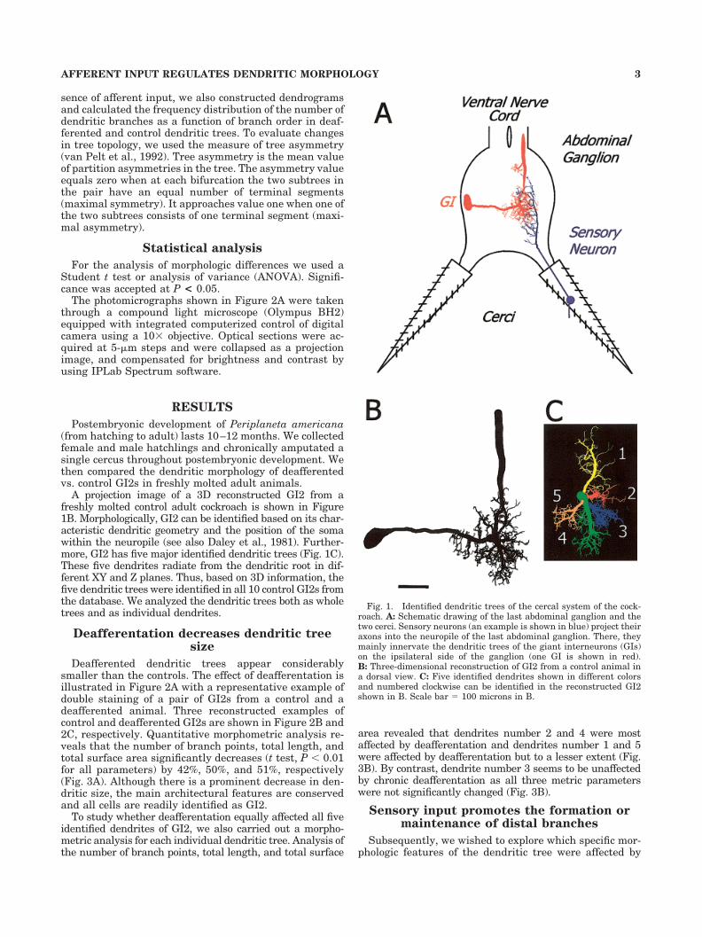
A projection image of a 3D reconstructed GI2 from afreshly molted control adult cockroach is shown in Figure1B. Morphologically, GI2 can be identified based on its char-acteristic dendritic geometry and the position of the somawithin the neuropile (see also Daley et al., 1981). Further-more, GI2 has five major identified dendritic trees (Fig. 1C).These five dendrites radiate from the dendritic root in dif-ferent XY and Z planes. Thus, based on 3D information, thefive dendritic trees were identified in all 10 control GI2s fromthe database. We analyzed the dendritic trees both as wholetrees and as individual dendrites.
Deafferentation decreases dendritic treesize
Deafferented dendritic trees appear considerablysmaller than the controls. The effect of deafferentation isillustrated in Figure 2A with a representative example ofdouble staining of a pair of GI2s from a control and adeafferented animal. Three reconstructed examples ofcontrol and deafferented GI2s are shown in Figure 2B and2C, respectively. Quantitative morphometric analysis re-veals that the number of branch points, total length, andtotal surface area significantly decreases (t test, P � 0.01for all parameters) by 42%, 50%, and 51%, respectively(Fig. 3A). Although there is a prominent decrease in den-dritic size, the main architectural features are conservedand all cells are readily identified as GI2.
To study whether deafferentation equally affected all fiveidentified dendrites of GI2, we also carried out a morpho-metric analysis for each individual dendritic tree. Analysis ofthe number of branch points, total length, and total surface
area revealed that dendrites number 2 and 4 were mostaffected by deafferentation and dendrites number 1 and 5were affected by deafferentation but to a lesser extent (Fig.3B). By contrast, dendrite number 3 seems to be unaffectedby chronic deafferentation as all three metric parameterswere not significantly changed (Fig. 3B).
Sensory input promotes the formation ormaintenance of distal branches
Subsequently, we wished to explore which specific mor-phologic features of the dendritic tree were affected by
Fig. 1. Identified dendritic trees of the cercal system of the cock-roach. A: Schematic drawing of the last abdominal ganglion and thetwo cerci. Sensory neurons (an example is shown in blue) project theiraxons into the neuropile of the last abdominal ganglion. There, theymainly innervate the dendritic trees of the giant interneurons (GIs)on the ipsilateral side of the ganglion (one GI is shown in red).B: Three-dimensional reconstruction of GI2 from a control animal ina dorsal view. C: Five identified dendrites shown in different colorsand numbered clockwise can be identified in the reconstructed GI2shown in B. Scale bar � 100 microns in B.
3AFFERENT INPUT REGULATES DENDRITIC MORPHOLOGY

Fig. 2. The architecture of dendritic trees of deafferented GI2s.A: Photomicrographs of double staining of left and right GI2s from acontrol and a deafferented animal (left and right, respectively). Arrowpoints to the deafferented dendritic tree. B: Three examples of recon-
structed dendritic trees of control GI2s. C: Three examples of recon-structed dendritic trees of deafferented GI2s. Scale bars � 100 mi-crons in A, 100 microns in B (applies to B,C).
4 A. MIZRAHI AND F. LIBERSAT

deafferentation. For this, we first aligned the trees at thecenter and analyzed their gross spatial distribution byusing Sholl analysis (Sholl, 1953). Two representative ex-amples of dendritic trees from control (brown neuron) anddeafferented (purple neuron) are shown separately (Fig.4A) and in their aligned position within a schematic Shollgrid (Fig. 4B). The maximum distance occupied by thetrees significantly decreased from 164 � 6 to 116 � 7microns (Fig. 4C; t test, P � 0.001). The maximum numberof branches was concentrated at a radius of 70 microns inthe control animals and shifted to a radius of 50 micronsin the deafferented animals. This was accompanied by adecrease in the maximum number of branch points from
56 � 4 at 70 microns in the controls to 40 � 4 at 50microns in the deafferented animals. Thus, it seems thatthe most distal branches from the dendritic root either didnot form or were pruned in the deafferented dendritictrees. However, Sholl analysis has been criticized for notdiscriminating between topologic and metrical aspects of
Fig. 4. Sholl analysis for control and deafferented dendritic trees.A: One reconstructed dendritic tree from a control animal (brown) andone from a deafferented animal (purple) shown separately. B: The twodendritic trees from A represented aligned to their axonal-dendriticjunction. In Sholl analysis, the number of branch points is countedwithin each Sholl sphere (white dashed lines). C: Sholl plot showingthe number of branch points (mean � SE) along the spheres from thecenter to the periphery. In control dendritic trees (solid line), the peaknumber of branch points is located at roughly 70 microns and thedistribution of branch points ranges from 0 to 170 microns from thecenter. In deafferented dendritic trees (dashed line), the peak numberof branch points is located at roughly 50 microns and the distributionof branch points ranges from 0 to 130 microns from the center. Scalebar � 100 microns in A.
Fig. 3. Morphometric analysis of deafferented dendritic trees.A: Morphometric analysis of full dendritic trees. Histograms of thenumber of branch points, total length, and total surface area of thedendritic tree of GI2s from control (full bars) and deafferented (emptybars) animals. B: Morphometric analysis of each of the five identifieddendritic trees of GI2. Histograms of the number of branch points,total length, and total surface area of the five identified dendritic treesof GI2 from control (full bars) and deafferented (empty bars) animals.Values represent average � SEM. Control, n � 10; deafferented, n �5. Significant differences are indicated above the bars (t test; *P �0.05, **P � 0.01).
5AFFERENT INPUT REGULATES DENDRITIC MORPHOLOGY

Fig. 5. Four models for dendritic pruning. A: A dendrogram froma control GI2 with 271 nodes. B: Branch order distribution of thedendrogram shown in A. C–J: Dendrograms and branch order distri-butions after pruning 116 nodes from the control dendritic tree in A.C: Terminal dendrites model. A dendrogram of the control dendritictree after it has been pruned at terminal branches regardless of thebranch order hierarchy. D: Branch order distribution of the dendro-gram in D (dashed line) vs. control (solid line). E: Full dendritesmodel. A dendrogram of the control dendritic tree after it has been
pruned at distinct dendrites from tip to the root. F: Branch orderdistribution of the dendrogram in E (dashed line) vs. control (solidline). G: High-order dendrites model. A dendrogram of the controldendritic tree after it has been pruned at high-order branches.H: Branch order distribution of the dendrogram in G (dashed line) vs.control (solid line). I: Distal dendrites model. A dendrogram of thecontrol dendritic tree after it has been pruned at distal branches.J: Branch order distribution of the dendrogram in I (dashed line) vs.control (solid line). Dendrogram scale � 100 microns.

tree structure (Uylings et al., 1986). Therefore, we con-structed dendrograms and further analyzed these trees byusing segment analysis, which is based on branch order.This analysis allows us to address separately some of thetopologic aspects of these trees.
The number of branches in a tree may decrease accord-ing to several different models. Figure 5 shows four dif-ferent possible ways of pruning branches from a controlneuron. The corresponding branch order distribution isshown as a graph next to each dendrogram. Thus, Figure5A and Figure 5B show a dendrogram of a control GI2from our database and its branch order distribution, re-spectively. The dendritic tree from Figure 5A has 271nodes distributed in 23 orders. Four different ways toprune this tree to 155 nodes (a constraint imposed by themorphometric analysis in Fig. 3) are shown in Figure5C–J. In the first possibility, called terminal dendrites,the control dendritic tree is pruned only at random termi-nal branches regardless of their branch order (Fig. 5C).Because terminal branches from all orders are pruned,both the peak of the curve and the maximum number ofbranches will decrease, but the maximum branch orderwill be only mildly affected (Fig. 5D). In the second possi-bility called, full dendrites, full dendritic trees (from thetip to the root) are pruned (Fig. 5E). In this case, themaximum number of branches at the peak of the curvewill decrease, whereas the maximum branch order will beonly mildly affected (Fig. 5F). In the third possibility,called high-order dendrites, high-order branches arepruned (Fig. 5G). This pruning mildly affects the peak ofthe curve but results in sharp decrease of the maximumbranch order (Fig. 5H). Finally, in the fourth possibility,called distal dendrites, most distal branches are prunedfrom the tree (Fig 5I). Although this model of pruningresembles the high-order dendrites pruning model, theformer is based on spatial distance regardless of branchorder. This method of pruning will mildly affect the peakof the curve but will result in a substantial decrease in themaximum branch order (Fig. 5J).
The plots of branch order distribution for the controland the deafferented groups are shown in Figure 6A andappear to fit the distal dendrites model. For the lowerbranch order range (2–8), the frequency of branches is notsignificantly different between both groups. In addition,the two curves peak at similar branch orders (control, 9thorder; deafferented, 8th order). Furthermore, the maxi-mum number of branches is decreased from 40 � 1 to 34 �5 but is not significantly different between the control andthe deafferented groups (Fig. 6A; t test; P � 0.05). How-ever, the maximum branch order is significantly de-creased from 21 � 0.9 orders in the controls to 16 � 1.2orders in the deafferented dendritic trees (Fig. 6A; t test;P � 0.01). Therefore, the branch order analysis stronglysupports the Sholl analysis. Taken together, the Sholl andthe branch order analyses suggest that sensory depriva-tion affects mainly the formation or the maintenance ofdistal high-order dendritic branches of the dendritic treeof GI2.
Deafferentation affects neither elongationnor branching
Dendritic development is eventually a process of elon-gation and branching (Cline, 2001). Branching of the low-order branches seems to be normal in deafferented ani-mals because the number of branch points is remarkably
similar to the controls for the initial branch orders (see leftpart of the curve in Fig. 6A). To study whether elongationis affected, we compared the segment length of the controland deafferented dendritic trees. Figure 6B shows thesegment length distribution at different branch orders forthe control (squares) and the deafferented (open squares)dendritic trees. A comparison between the two groupsacross the same branch order range (orders 2–19), re-vealed no significant differences (ANOVA, P � 0.05).These results show that the deafferented dendritic treesdeveloped normally at lower branch orders and suggeststhat, in this region of the tree, the innate capacity of theGIs to elongate and branch is not affected by afferentinput.
Finally, we analyzed the topologic structure of the con-trol and deafferented trees by calculating the tree asym-metry of the whole trees. Tree asymmetry values of thecontrol dendritic trees (0.56 � 0.04) were not significantlydifferent from the values of the deafferented dendritictrees (0.52 � 0.04) (t test; P � 0.05).
Fig. 6. Deafferentation affects high-order branching but not elon-gation of low-order branches. A: Distribution of the number ofbranches of the dendritic trees from control GI2s (squares) and thedendritic trees from deafferented GI2s (diamonds) as a function ofbranch order. The maximum number of branches is not significantlydifferent between the groups (t test; P � 0.05). The maximum branchorder is significantly lower in deafferented dendritic trees (t test; P �0.01). B: Plot of dendritic segments length as a function of branchorder for the control group (squares) and the deafferented group (opensquares). For the comparable part of the graph (orders 2–19), thecurves are not significantly different (analysis of variance; P � 0.05).
7AFFERENT INPUT REGULATES DENDRITIC MORPHOLOGY

DISCUSSION
In this work, we explored the effects of sensory inputdeprivation on the dendritic geometry of an identifiedneuron. Our results indicate that the axonal terminals ofsensory afferent neurons mostly affect development ofhigh-order distal dendritic segments, with little effect onthe lower order proximal dendritic segments. This resultsuggests that interactions between pre- and postsynapticneurons play a specific regulatory role in the formation ormaintenance of high-order distal dendritic branches of theGIs’ trees.
Role of afferent innervation on dendriticformation
At the single cell level, a detailed quantitative analysisof the effects of afferent input on dendritic tree maturationhas been performed on neurons characterized by a rela-tively simple dendritic geometry. One example is the iden-tified Mauthner neurons of developing amphibians whosedendritic tree branch only two to three orders. In theMauthner cell, deafferentation reduces, whereas supra-innervation enhances dendritic growth specifically in thedeprived region (Goodman and Model, 1988). In addition,incoming axons stimulate dendritic growth in an activityindependent manner (Goodman and Model, 1990). An-other example is the early dendritic development of hip-pocampal neurons. Dendritic branch formation is inducedby afferent innervation in vitro by means of both activity-dependent and activity-independent mechanisms (Kosselet al., 1997). Like for the Mauthner neuron, the effect isregion specific because the effect of the afferents is ob-served only on dendrites, which established contact withthe afferents. In that respect (in the case of GI2), it ispossible that cercal afferents in GI2 do not establish syn-apses with this region of the tree because identified den-drite number 3 did not change after deafferentation. Ja-cobs and Theunissen (2000) recently have shown spatialsegregation of input on the dendritic tree of a GI wherebydifferent regions of the tree receive inputs from differentsets of cercal afferents. Our results support the idea thatthere is such a functional partition of afferent input on thedendritic arbor of GI2.
In crickets, sensory deprivation induces dendriticgrowth of the medial dendrites and a concomitant de-crease of the lateral dendrites of an auditory interneuron(Hoy et al., 1985). GI2, like most GIs, has small contralat-eral dendrites branches that sprout from the link segmentin the contralateral portion of the neuropile (Daley et al.,1981). These short and mostly unbranched dendrites re-ceive subtreshold afferent input from a few columns ofhairs located on the contralateral cercus (Daley andCamhi, 1988). When the ipsilateral cercus is ablated, theinput from the contralateral cercus is enhanced (Volmanand Camhi, 1988; Volman, 1989). To examine whether themorphology of these contralateral dendrites is affected byamputation of the ipsilateral cercus, we reconstructedthese small dendrites in a few deafferented and controlGI2s. Morphometric analysis shows no obvious differencesbetween the two groups of cells. The number of contralat-eral dendrites per neuron, total length, total surface area,and total volume were not significantly different betweenthe control and experimental groups (data not shown).This finding suggests that the change in the efficacy of the
contralateral input after long-term deafferentation is notcorrelated with structural changes of the postsynapticdendritic targets.
Analysis of dendritic branching
Given that metrics and topology are different aspects oftree structure they must be analyzed separately (Uylingset al., 1986). The interpretation of Sholl analysis is lim-ited, because this analysis does not discriminate betweenthese two aspects of tree structure. In the present study,we examine separately both the metric and the topologicproperties of the dendritic trees of GIs. The branch orderanalysis complements the results of the Sholl analysis inthat it suggests that changes are indeed confined to thedistal branches of the deafferented tree rather than lowerorder branches. The deafferented dendritic tree is neithera shrunken version of the adult neuron nor does it seem tobe topologically reorganized as depicted by the similartree asymmetries.
Morphologically, dendritic trees expand by both elonga-tion of the dendritic segments and branching. Neuronalbranching can occur by formation of new branches at theterminals and/or by preterminal branching (Acebes andFerrus, 2000). Are either of these growth mechanismsaffected by the absence of afferent input? Although mod-ern imaging techniques have been used to identify modesof growth (Dailey and Smith, 1996; Wu et al., 1999), thesetechniques are difficult to implement on a long time scale.In our work, we can only speculate as to whether deaffer-entation affects elongation, bifurication, or interstitialbranching. However, our quantitative analyses indicate ahigh degree of structural similarity between controls andthe deafferented trees at lower branch orders. Thus, itseems that neither elongation nor branching is signifi-cantly affected at lower branch orders. Rather, the effectof deafferentation is confined to a specific periphery of thedendritic arbor without affecting the intrinsic mecha-nisms of formation of the core of the dendritic arbor.
Role of synaptic activity on thedevelopment of dendrites
Axonal navigation and appropriate synaptic connectioncan be precisely achieved at initial stages of developmentin an activity-independent manner. Knockout mice com-pletely lacking neurotransmitter secretion develop normallayered structures, normal fiber pathways, and morpho-logically defined synapses (Verhage et al., 2000). Thus, theinitial assembly of neuronal circuitry seems activity inde-pendent, whereas neuronal (and synaptic) maintenanceappears to be activity dependent. For instance, synapticinput is closely linked to structural stability of dendritesin the somatosensory barrel cortex (Wallace and Fox,1999; Lendvai et al., 2000). Thus, as a general rule, syn-aptic activity is critical at later stages of developmentwhere it refines and stabilizes existing connections (forreview see also Goodman and Shatz, 1993; Katz andShatz, 1996; Tessier-Lavigne and Goodman, 1996). Itseems that the cockroach central nervous system obeys asimilar rule of wiring, as functional synapses between thesensory axons and the GIs form before the onset of affer-ent activity (Blagburn et al., 1996). In addition, synapticactivity has been shown to be important in the enhance-ment of the excitability of deafferented GIs but had littleeffect on the excitability in the nondeafferented GIs (Vol-
8 A. MIZRAHI AND F. LIBERSAT

man and Camhi, 1988). This finding suggests that theabsence of synaptic activity has little consequence on thefine dendritic architecture of the GIs, whereas deafferen-tation does.
Role of environmental factors in thedevelopment of dendrites
In addition, a dimensional constraint may be induced bythe change in the volume of the neuropile. Estimation ofthe volumetric change of the neuropile of deafferentedcercal system of crickets has shown a correlation betweenthe scaling down of the neuropile and the shortening of thedendrites (Murphey et al., 1975). However, the possibilityof a causal link between changes in the dimensions of theneuropile and dendritic trees is unlikely for the followingtwo reasons. First, we have found no correlations betweenthe size of the ganglion and the size of dendritic trees ofspecific GIs (Mizrahi et al., 2000). Second, the presentstudy shows that different dendrites of GI2 are selectivelyaffected to a different extent one of which appears todevelop to full size (Fig. 3B). Given these results, thedecrease in dendritic size is unlikely to be due to shrink-age of the neuropile.
To conclude, dendritic tree maturation of the GIs isregulated by extrinsic (environmental) as well as intrinsic(genetic) factors. In the cercal system, intrinsic factorsmight govern the basic (lower order) branching pattern ofthe dendritic tree, whereas extrinsic factors might play animportant role in the formation or maintenance of thedistal higher order branches. In addition, the ability ofneurons to branch and elongate appears to be, at least, tosome extent, innate because it is not affected by deaffer-entation.
ACKNOWLEDGMENTS
We thank J. Schaeffer and A. Weisel-Eichler for criti-cally reading and improving the manuscript. These exper-iments comply with Principles of Animal Care, NIH pub-lication no. 86-23, revised in 1985, and also with thecurrent laws of the State of Israel.
LITERATURE CITED
Acebes A, Ferrus A. 2000. Cellular and molecular features of axon collat-erals and dendrites. TINS 23:557–565.
Blagburn JM, Beadle DJ. 1982. Morphology of identified cercal afferentsand giant interneurones in the hatchling cockroach Periplaneta ameri-cana. J Exp Biol 97:421–426.
Blagburn JM, Beadle DJ. 1984. Synapses between an identified giantinterneuron and a filiform hair sensory neuron in the terminal gan-glion of first instar cockroaches Periplaneta americana. J Exp Biol113:477–481.
Blagburn JM, Thompson KSJ. 1990. Specificity of filiform hair afferentsynapses onto giant interneurons in Periplaneta americana: anatomyis not a sufficient determinant. J Comp Neurol 302:255–271.
Blagburn JM, Sosa MA, Blanco RE. 1996. Specificity of identified centralsynapses in the embryonic cockroach: appropriate connections formbefore the onset of spontaneous afferent activity. J Comp Neurol 373:511–528.
Bugnion E. 1921. The growth of the antenna and cerci of the cockroach,Periplaneta americana. Bull Entomol Soc Egypt Econ Ser 6:56–66.
Camhi JM. 1984. Neuroethology. Sunderland: Sinaur Association.Chiba A, Shepherd D, Murphey RK. 1988. Synaptic rearrangement during
postembryonic development in the cricket. Science 240:901–905.Cline HT. 1999. Development of dendrites. In: Stuart G, Spruston N,
Hausser M., editors. Dendrites. New York: Oxford University Press. p35–67.
Cline HT. 2001. Dendritic arbor development and synaptogenesis. CurrOpin Neurobiol 11:118–126.
Dagan D, Volman S. 1982. Sensory basis for directional wind detection infirst instar cockroaches, Periplaneta americana. J Comp Physiol 147:471–478.
Dailey ME, Smith SJ. 1996. The dynamics of dendritic structure in devel-oping hippocampal slices. J Neurosci 16:2983–2994.
Daley DL, Camhi JM. 1988. Connectivity pattern of the cercal-to-giantinterneuron system of the American cockroach. J Neurophysiol 60:1350–1367.
Daley DL, Vardi N, Appignani B, Camhi JM. 1981. Morphology of the giantinterneurons and cercal nerve projections of the American cockroach.J Comp Neurol 196:41–52.
Deitch JS, Rubel EW. 1984. Afferent influences on brain stem auditorynuclei of the chicken: time course and specificity of dendritic atrophyfollowing deafferentation. J Comp Neurol 229:66–79.
Goodman LA, Model PG. 1988. Superinnervation enhances the dendriticbranching pattern of the Mauthner cell in the developing axolotl.J Neurosci 8:776–791.
Goodman LA, Model PG. 1990. Eliminating afferent impulse activity doesnot alter the dendritic branching of the amphibian Mauthner cell.J Neurobiol 21:283–294.
Goodman CS, Shatz CJ. 1993. Developmental mechanisms that generateprecise patterns of neuronal connectivity. Cell 72(Suppl):77–98.
Gray L, Smith Z, Rubel EW. 1982. Developmental and experimentalchanges in dendritic symmetry in n. laminaris of the chick. Brain Res244:360–364.
Hamon A, Guillet JC, Callec JJ. 1994. Patterns of monosynaptic input tothe giant interneurons 1-3 in the cercal system of the adult cockroach.J Comp Physiol 174:91–102.
Hill AAV, Edwards DH, Murphey RK. 1994. The effect of neuronal growthon synaptic integration. J Comput Neurosci 1:239–254.
Hoy RR, Nolen TG, Casaday GC. 1985. Dendritic sprouting and compen-satory synaptogenesis in an identified interneuron follow auditory de-privation in a cricket. Proc Natl Acad Sci U S A 82:7772–7776.
Jacobs GA, Nevin R. 1991. Anatomical relationships between sensoryafferent arborizations in the cricket cercal system. Anat Rec 231:563–572.
Jacobs GA, Theunissen FE. 2000. Extraction of sensory parameters from aneural map by primary sensory interneurons. J Neurosci 20:2934–2943.
Katz LC, Shatz CJ. 1996. Synaptic activity and the construction of corticalcircuits. Science 274:1133–1138.
Koch C, Segev I. 2000. The role of single neurons in information processing.Nat Neurosci 3:1171–1177.
Kossel AH, Williams CV, Schweizer M, Kater SB. 1997. Afferent innerva-tion influences the development of dendritic branches and spines viaboth activity-dependent and non-activity-dependent mechanisms.J Neurosci 17:6314–6324.
Lendvai B, Stern EA, Chen B, Svoboda K. 2000. Experience-dependentplasticity of dendritic spines in the developing rat barrel cortex in vivo.Nature 404:876–881.
Mainen ZF, Sejnowski TJ. 1996. Influence of dendritic structure on firingpattern in model neocortical neurons. Nature 382:363–366.
Mizrahi A, Ben-Ner E, Katz MJ, Kedem K, Glusman JG, Libersat F. 2000.Comparative analysis of dendritic architecture of identified neuronsusing the Hausdorff distance metric. J Comp Neurol 422:3 415–428.
Murphey RK, Mendenhall B, Palka J, Edwards JS. 1975. Deafferentationslows the growth of specific dendrites of identified giant interneurons.J Comp Neurol 159:407–418.
Rall W, Burke RE, Holmes WR, Jack JJ, Redman SJ, Segev I. 1992.Matching dendritic neuron models to experimental data. Physiol Rev72:S159–S186.
Ritzmann RE. 1984. The neural organization of cockroach escape and itsrole in context dependent orientation. In: Beer RD, Ritzmann RE,McKenna T, editors. Biological neural networks in invertebrate neuro-methology and robotics. New York: Academic Press. p 113–137.
Scott EK, Luo L. 2001. How do dendrites take their shapes? Nat Neurosci4:359–365.
Sholl DA. 1953. Dendritic organization in the neurons of the visual cortexand motor cortices of the cat. J Anat 87:387–406.
9AFFERENT INPUT REGULATES DENDRITIC MORPHOLOGY

Smith ZD, Gray L, Rubel EW. 1983. Afferent influences on brainstemauditory nuclei of the chicken: n. laminaris dendritic length followingmonaural conductive hearing loss. J Comp Neurol 220:199–205.
Tessier-Lavigne M, Goodman CS. 1996. The molecular biology of axonguidance. Science 274:1123–1133.
Uylings HB, Ruiz Marcos A, van Pelt J. 1986. The metric analysis ofthree-dimensional dendritic tree patterns: a methodological review.J Neurosci Methods 18:127–151.
van Pelt J, Uylings HB, Verwer RW, Pentney RJ, Woldenberg MJ. 1992.Tree asymmetry: a sensitive and practical measure for binary topolog-ical trees. Bull Math Biol 54:759–784.
Verhage M, Maia AS, Plomp JJ, Brussaard AB, Heeroma JH, Vermeer H,Toonen RF, Hammer RE, van den Berg TK, Missler M, Geuze HJ,
Sudhof TC. 2000. Synaptic assembly of the brain in the absence ofneurotransmitter secretion. Science 287:864–869.
Vetter P, Roth A, Hausser M. 2001. Propagation of action potential indendrites depends on dendritic morphology. J Neurophysiol 85:926–937.
Volman SF. 1989. Localization of the enhanced input to cockroach giantinterneurons after partial deafferentation. J Neurobiol 20:762–783.
Volman SF, Camhi JM. 1988. The role of afferent activity in behavioral andneuronal plasticity in an insect. J Comp Physiol 162:781–791.
Wallace H, Fox K. 1999. Local cortical interactions determine the form ofcortical plasticity. J Neurobiol 41:58–63.
Wu GY, Zou DJ, Rajan I, Cline H. 1999. Dendritic dynamics in vivo changeduring neuronal maturation. J Neurosci 19:4472–83.
10 A. MIZRAHI AND F. LIBERSAT