Embed Size (px)
Citation preview
Osaka University
Title Afferent and efferent enkephalinergic systems of the tegmentalnuclei of Gudden in the rat : an immuno-cyto chemical study
Author(s) Yamano, Mariko
Citation
Issue Date
Text Version ETD
URL http://hdl.handle.net/11094/37420
DOI
Rights
22 Brain Research, 408 ( I t~87 j 22- 30 Elsevicr
BRE 12473
Afferent and efferent enkephalinergic systems of the tegmental nuclei of Gudden in the rat" an immunocytochemical study
Mariko Yamano and Masaya Tohyama Second Department of Anatomy, Osaka University Medical School, Nakanoshima Kitaku, Osaka (Japan)
Osaka University Medical School, Nakanoshima Kitaku, Osaka (Japan)
(Accepted 19 August 1986)
Key words: L-Enkephalin; Dorsal tegmental nucleus; Ventral tegmental nucleus; Fiber connection; Mammillary body; Interpeduncular nucleus; Immunocytochemistry; Rat
We studied the afferent source of L-enkephalin-like immunoreactive (L-ENKLI) fibers in the ventral tegmental nucleus (VT) of Gudden, and efferent and afferent connections of L-ENKLI structures in the dorsal part of the dorsal tegmental nucleus (DDT) of Gudden in the rat, using immunocytochemistry combined with knife-cut and lesion experiments. The VT had a dense plexus of L-ENKLI fibers but no L-ENKLI cells. Destruction of the dorsal premammillary nucleus and medial mammillary nucleus pars media- lis, which contained a large group of L-ENKLI neurons, markedly reduced L-ENKLI fibers in the ipsilateral VT, suggesting that most of these fibers originate in these nuclei. The DDT contained a large collection of L-ENKLI neurons together with dense L-ENKLI fi- bers. Destruction of the DDT caused a contralateral marked reduction of L-ENKLI fibers in the dorsolateral part of the interpeduncu- lar nucleus (DL), suggesting that L-ENKLI neurons in the DDT project contralaterally to the DL. L-ENKLI fibers in the DDT may be of intrinsic origin.
INTRODUCTION must be first explored. Accordingly , we here studied
enkephal inergic project ions in the D T and VT in the
Since the discovery of the enkephal ins , which are rat using immunocytochemis t ry combined with knife-
two natural ly occurring pen tapep t ides with a high af- cut and lesion exper iments .
finity for the opia te receptor 13, the widespread pres-
ence of enkephal ins in the central nervous system has MATERIALS AND METHODS
been detected by rad io immunoasay and immunocy- tochemistry 7,12,t6,22,2329,3°,33,35. The dorsal (DT) and Experimental animals
ventral (VT) tegmenta l nuclei of Gudden are re la ted We used 39 albino rats (body weight, about 70 g).
to the limbic function as a relay center of the upper Seven were used in the analysis of the normal distri-
brainstem, which is connected to the pos ter ior hypo- bution of L-ENKLI structures. Of these, 3 received
thalamus and the in terpeduncular nucleus 1"2"4"5" an injection of 6/xl of colchicine (3/xg//~l) into the 8-11,15,24,25,27.31,3,L36 Immunocytochemica l studies third or fourth ventricle stereotaxically. These rats
have shown the presence of L-enkephalin-l ike immu- were kil led 24-48 h later. Colchicine inhibits axonal
noreact ive (L-ENKLI) structures in the DT and t ransport and has been used to increase the cel l-body
VT 21'28. These findings suggest that t , -ENKLI here levels of neurot ransmit ters and neuromodula tors 6'1~.
relates closely to limbic function. To elucidate the Here it was injected to make visible L-ENKLI neu-
function of L-ENKLI structures here, the fiber con- rons that would otherwise be undetectable . The re-
nections of L-ENKLI structures in the DT and VT maining rats were divided into two groups for a knife-
Correspondence: M. Yamano, Second Department of Anatomy, Osaka University Medical School, 4-3-57, Nakanoshima, Kitaku, Osaka, 530, Japan.
0006-8993/87/$03.50 O 1987 Elsevier Science Publishers B.V. (Biomedical Division)
23
cut study (17 animals) and a lesion study (15 ani- microscope after Cresyl violet staining. These ani- mals), mals were kept alive for 5-7 days after the operation
and processed for immunocytochemistry.
Knife-cut study In the brains of 17 rats, 4 kinds of transection and Preparation of tissue
two kinds of knife-cuts were made. The transections All rats were perfused intracardially with 50 ml of were just caudal to the tegmental nuclei (Fig. la), ice-cold saline followed by 200 ml of cold Zamboni's just rostral to the tegmental nuclei, just caudal to the fixative 37. The brain was removed rapidly, immersed interpeduncular nucleus (IP) (Fig. lb), just caudal to in the same fixative for 12 h, and then rinsed for 24 h the mammillary body (MB) (Fig. lc) or just rostral to in phosphate buffer containing 30% sucrose at 4 °C. the MB (Fig. ld). The sagittal cuts were just lateral Serial frontal sections 10~m thick were cut on a cryo- to the MB or just lateral to the tegmental nuclei (Fig. star. Half of the sections was immediately incubated le). The rats were kept alive for 5-7 days after the in cold phosphate-buffered saline (PBS) for 30 min, operation and then processed for immunocytochem- then processed for indirect immunofluorescence 3. istry. Rabbit antiserum diluted 1:1000 with PBS against L-
enkephalin (L-ENK) was applied to the sections, Lesion study which were then incubated for 12 h at room tempera-
The above study suggested (1) that L-ENKLI fi- ture (17 °C) in a humid atmosphere. The sections bers in the VT generally arise on the area enclosed in were rinsed in cold PBS containing 1% Triton X-100 the cut shown in Fig. l c - e and (2) that L-ENKLI neu- for 10 min and in PBS for 30 min, followed by 12 h of rons in the DDT project to the DL. Therefore, vari- incubation at room temperature with goat antirabbit ous areas located in the area enclosed by 3 kinds of IgG conjugated with fluorescein isothiocyanate knife-cuts and DDT were unilaterally destroyed in (FITC) and diluted 1:1000 with PBS containing 1% other rats using stereotaxic equipment by passage of Triton X-100 for 10 min and in cold PBS for 30 min, a DC current of 1 mA for 10 s through a monopolar and were then mounted in a PBS-glycerine mixture electrode. Damage was assessed using a bright-field (1:1). After observations, the sections were stained
with Cresyl violet for exact identification of the brain regions. The remaining sections were rinsed in PBS
d c b a and stained with Cresyl violet. The brain region and ~ ~ the extent of the damage caused by the knife-cut or
lesion were assessed on these sections.
__~ ~ Specificity of antiserum Antiserum against L-ENK was produced in re-
- " I sponse to a conjugate of 1-ethyl-o-(3,3'-dimethylami- | , , : - - ,
L".~-- t'J~~_ ,::,~i~;,~! nopropyl)carbodiimide, synthetic L-ENK (Japanese . ' :, / -~- Peptide Lab.), and bovine thyroglobulin (Sigma).
I P V T DT MB The specificity of the L-ENK antiserum was evalu- ~ ~ , ated by radioimmunoassay. Cross-reactivity was less
than 1% with methionine-enkephalin (M-ENK), less than 0.01% with dynorphin, and less than 0.01% with adrenocorticotropic hormone. There was no cross-reactivity with e, fl or 7-endorphins, or with
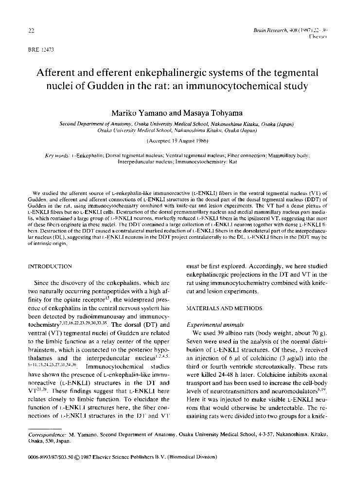
Fig. 1. Schematic drawing showing knife-cut sites in the brain. a: transection just caudal to the dorsal tegmental nucleus of substance P (SP), somatostatin (SOM), neurotensin Gudden (DT). b: transection just rostral to the ventral tegmen- (NT), luteinizing hormone-releasing hormone (LH- tal nucleus of Gudden (VT). c: transection just rostral to the in- RH), cholecystokinin-8 (CCK), or thyrotropic-re- terpeduncular nucleus (IP). d: transection just rostral to the leasing hormone (TRH). The specificity of the anti- mammillary body (MB). e: sagittal section lateral to the MB. Horizontal plane, serum was also tested histochemically, by (1) re-
24
placement of the specific serum with normal rabbit , , , , " ~ ( / / ) / '~ serum, (2) omission of the specific serum, and (3) ad- ml f--~ sorption of anti-L-ENK serum to L-ENK, M-ENK, " ~ ' J - " ) SP. NT, CCK, SOM, and LH-RH. Adsorption was at ~ . _ - 4 °C with each peptide at a final concentration of 10 5 M, followed by centrifugation at 3000 g for 30 min. / / ~T[,,. ", !i[~i~ 1 ~ 0 The supernatant was used for the later tests. Struc- tures that satisfied all the following criteria were tak- , q en to be positive for L-ENK: there was no immuno- , , staining(1) whenthespec i f icse rumwasrep lacedby ~ s c n ~ ' ~ ~ ~ normal serum or omitted or (2) when anti-L-ENK se- .._ ' 3 ~ / ~ x . . . . . rum was absorbed with L-ENK, but (3) there was no / decrease in immunostaining when sections were in-
cubated with antiserum against L-ENK adsorbed by ][)-~VT")' [~ i)i![~!/~"ii:'\ ';-" '"~ • b any of the other peptides tested. '~
RESULTS ,.
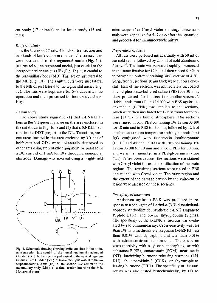
Dis tributionofL-ENKLIstructuresintheDTandVT \ ' ~ ~ / / ~ ' [ - - ' , l In the VT, a dense plexus of L-ENKLI fibers was ' ~ '" "" "'
f x
seen (Fig. 2a-c). L-ENKLI fibers were evenly distrib- . . . . ~ [ /DT ; . - , o Z..¢.- - , ~l, , / ~ " " . , . uted in this nucleus in its rostral and caudal one-third s c ~ / \ "'~" , DT ,' ,, / / (Fig. 2a, c). However, at the middle level of this nu- / c ' " cleus, the fibers tended to be concentrated in the dor- .5"~'"
solateral portion (Fig. 2b); in the ventromedial por- - - VT X ~ • If ~ ; ":~'.:! tion, fewer L-ENKLI fibers were detected. No immu- noreactive neurons were identified in this nucleus, . . ~ . ~ J ~ , , , ~ even in the rats treated with colchicine. 4 V
The DT is divided into the pars dorsalis (DDT) and ' ' . . . . . . . . . pars ventralis (VDT) based upon the cytoarchitec- ) L~ ~, ' ¢4 ~ ~', , , ! ture II. In the DDT, a large collection of L-ENKL1 ' ~ ~ I / ~ ~ . / ' ,,. ,. , ,VDI". ' . , "-. ; / ' ,a neurons and fibers was detected. Immunoreactive ' .~ ~" , --" / , . . " neurons were distributed evenly throughout the DDT (Fig. 2c-e). The VDT was devoid of L-ENKLI structures except for a small region in its rostral pole " ' ~ M~..J L / ~ (Fig. 2c). There, a small group of L-ENKLI neurons , .~, D ~ ~ 2 ~ D T ~ . . - , ~ ~ [ [ . . , ' .. together with immunoreactive fibers was found in its ." , , ' medioventral border (Fig. 2c). A group of fibers left 'LC' /a',o,l~ \ (~.~.#-'~, ', ;.
, , i u.] ,~. .- L '
this nucleus ventrolaterally and intermingled with a , 7-"~VLJ I-~-- , ', ,' . / / -=U" ' . dense plexus of L-ENKLI fibers in the VT (Fig. 2c). ... "-~" "- - ' 2 e
" ~ 2 e
Origin of the L-ENKLl fibers in the VT , / Knife-cut study. Unilateral transection of the brain
concentration of L-ENKLI fibers in the VT and dorsal part of just caudal to the VT (Fig. la) did not decrease L- the DT (DDT), and that of L-ENKLI neurons in the DDT.
Frontal plane. Arranged from rostral to caudal. LC, locus coe- Fig. 2. Schematic drawing of L-ENKLI structures in the teg- ruleus; LDT, laterodorsal tegmental nucleus; mlf, medial Ion- mental nuclei of Gudden. Large dots indicate the L-ENKLI gitudinal fasciculus; scp, superior cerebellar peduncle; VDT, cells seen in colchicine-treated rats. Small dots indicate k- ventral part of the DT, xscp, decussation of the scp; 4V, fourth ENKLI fibers in rats without colchicine treatment. Note the ventricle.
25
ENKLI fibers in the VT. Unilateral transection just lJ rostral to the VT (Fig. lb) or just caudal to the MB V (Fig. lc)resulted in the ipsilateral disappearance of n f ~ the dense collection of L-ENKLI fibers in the VT. L- k . J x /
") i ENKLI fibers in the VT were unchanged after tran- ®,P section of the brain just caudal to the ventromedial ~ ~ .- .... '.. . . . . . . . . , -o.p~~r-. e ~
hypothalamic nucleus (vmh)(Fig. ld), or after a sa- ~ M ~ " ' ~ ' i , i'" ~ . ~ / gittal cut in the brain at the level lateral to the MB a (Fig. 1el. These results suggest that L-ENKLI cells in the area surrounded by the 3 kinds of knife-cuts shown in Fig. lc -e are the main origin of L-ENKLI ri-
O bers in the VT. This area is tentatively called the "1"--'.}]~,. 0 0 / - - mammillary area in this study. ~ 0 ( 3 : i
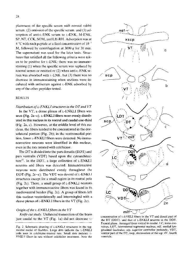
Lesion study. Fig. 3 shows the location of L-ENK- ~ pl~[~" ',, e-e 6"- ' ,~ ~ LI cells in the mammillary area in the colchicine- r"q/?o_'o:o'o
~ (-'" ~ T o o " "-'-'" °' " " - "~'~:'" O ~ '¢ treated animals. Sites that contained L-ENKLI cells ~ ' - ~ " ) Ar Uoo) {." b were the dorsal mammillary nucleus (PMD) (Figs. 3, 4a), medial mammillary nucleus pars medialis (MM)
© 0 (Fig. 3, 4a), arcuate nucleus (Ar), and ventral pre- mammillary nucleus (PMV). Density was highest in
m t _ _ the PMD and MM; there were fewer immunoreac- tive cells in the Ar, and very few in the PMV. In addi- -'~,;"',::IML/',M M e: • ", ', ) , " ~)/ tion, scattered L-ENKLI cells were seen in the neuro- ~, "~,,, : :e',ee '. '---".' .,7
\ . . . . . . . . . . , e o - e I , ' e '.-:,~/ ,- pil adjacent to these nuclei. L-ENKLI cells found in N~.:... " ~ e_ o.:.::.,~/ the PMD fused mediocaudally with those found in the MM (Fig. 3b), and no definite border between the PMD and MM could be seen in the distribution of / ~ C) L-ENKLI cells. Accordingly, in this study, these two m r 9 nuclei are called the medial mammillary complex (MMC). To decide which of the regions mentioned " ~ ' - ' ~ ' L ' I ~ ~ " o - " ~ " "'" '" I- " ' ' : ~ ' - ' ~ above is the main source of L-ENKLI fibers in the , " ' " VT, these regions were destroyed, separately. \*~. ~,M k \, ee, ' , , : i ,, '/
When the lesions were centered in the rostral ~ " . -- 3d MMC and when other regions of the mammillary
Fig. 3. Schematic drawing of L-ENKLI cells in the area sur- a rea r e m a i n e d intact (Fig. 4b), n u m b e r s of L-ENKL1 rounded by the 3 kinds of knife-cuts shown in Fig. lc, d and e in fibers decreased in the caudal part of the ipsilateral colchicine-treated rats. The location of these cells is repre- VT (Fig. 4c), but L-ENKLI fibers in the rostral part sented on the right side by filled circles. Note a large group of L-
ENKLI cells in the medial mammillary nucleus pars medialis of the VT were unchanged. When the lesion was cen- (MM) and dorsal premammillary nucleus (PMD). Ar, arcuate tered in the caudal part of the MMC and when other nucleus; f, fornix; LM, lateral mammillary nucleus; ML, medi- regions of the mammillary area remained intact, L- al mammillary nucleus pars lateralis; mt, mammillothalamic
tract; mtg, mammillotegmental tract; PMV, ventral premam- ENKLI fibers in the rostral part of the VT decreased millary nucleus. Frontal plane. Arranged from rostral to cau- markedly on the operated side, while no large de- dal. crease of L-ENKLI fibers was seen in the caudal part of the VT. In addition, when the lesion was large and involved the entire MMC, L-ENKLI fibers disap- the VT. In this case, in the mtg rostral to the lesion, peared in the entire VT on the operated side. When fibers that accumulated L-ENKLI structures ap- the lesion involved the mammillotegmental tract peared. These fibers could be traced to the MMC (mtg), L-ENKLI fibers disappeared ipsilaterally in where L-ENKLI neurons had much stronger fluores-
26
b
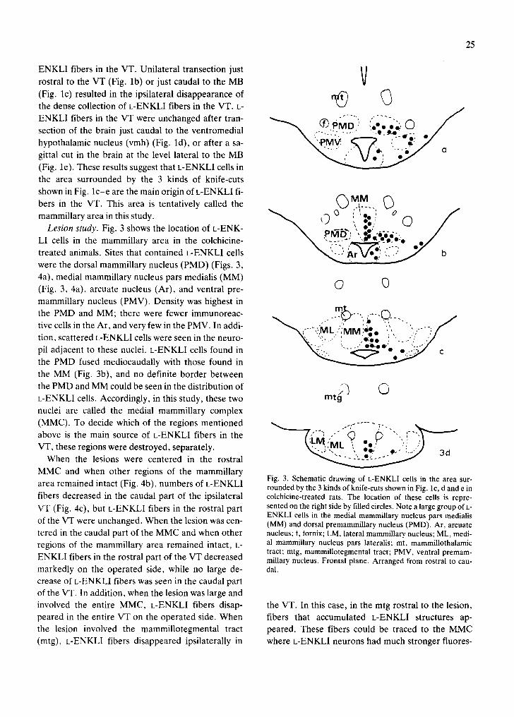
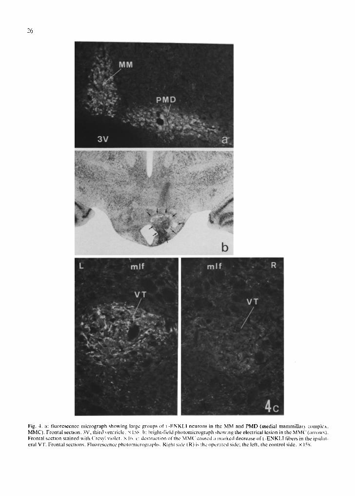
Fig. 4. a: f luorescence micrograph showing large groups of I.-ENKLI neurons in the MM and PMD (medial mammillary complex, MMC). Frontal section. 3V, third ventricle, x 158. h: bright-field photomicrograph showing the electrical lesion in the MMC (arrows). Frontal section stained with Cresyl violet. × 1~,. c: destruction of the MMC caused :l marked decrease of I.-ENKLI fibers in the ipsilat- eral VT. Frontal sections. Fluorescence photomicrographs. Righl side (R) is the operated side; the left, the control side. × 158.
27
cent intensity than on the normal side. When the le-
sions were made in other parts of the mammillary
area and the MMC remained intact, L-ENKLI fibers
decreased little in the VT.
L-ENKLI projection from DD T to the IP ~ Hemitransection of the brain rostral to the VT and
DT resulted in a contralateral disappearance of L-
ENKLI fibers in the dorsolateral part of the IP (DL),
while total transection of the brain just caudal to the
DT (Fig. la), or sagittal cut lateral to the tegmental
nuclei (Fig. le), did not decrease L-ENKLI fibers in
the IP, including the DL. These findings suggest the
possibility that L-ENKLI neurons in the DDT project
to the contralateral DL, because DDT had many L-
ENKLI neurons (Fig. 5a) and was between the two
sections mentioned above. To examine this possibili-
ty, we destroyed the DT and observed subsequent
in L-ENKLI fibers in the IP. Following de- 1 changes
struction of the DT (Fig. 5b), a marked decrease of L-
ENKLI fibers occurred in the contralateral side of !if! ~
ID the DL (Fig. 5c).
?
Origins of L-ENKLI fibers in the D D T Transection of the brain rostral to the DT and cau-
dal to the DT failed to decrease L-ENKLI fibers in
the DDT. A sagittal cut lateral to the DT caused no
effects on L-ENKLI fibers in the DDT. These find-
ings indicate that L-ENKLI neurons located in the
area surrounded by these 3 kinds of knife-cut are the main origin of L-ENKLI fibers in the DDT.
DISCUSSION
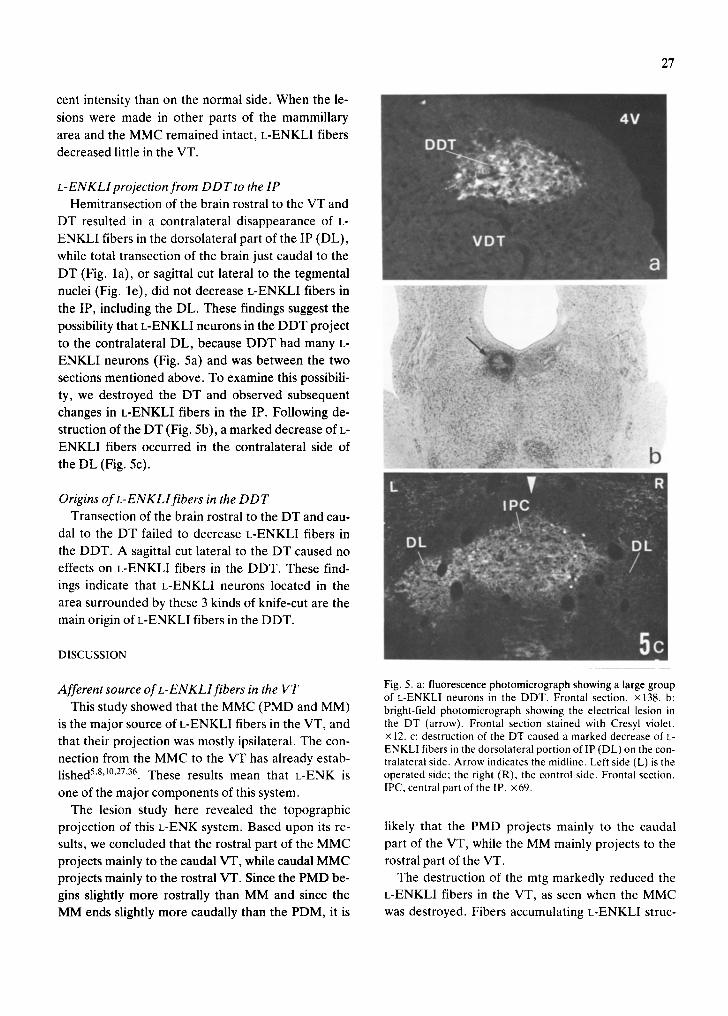
Afferent source of L-ENKLlfibers in the VT Fig. 5. a: fluorescence photomicrograph showing a large group of L-ENKLI neurons in the DDT. Frontal section, x138. b:
This study showed that the MMC (PMD and MM) bright-field photomicrograph showing the electrical lesion in is the major source of L-ENKLI fibers in the VT, and the DT (arrow). Frontal section stained with Cresyl violet. that their projection was mostly ipsilateral. The con- × 12. c: destruction of the DT caused a marked decrease of L-
ENKLI fibers in the dorsolateral portion oflP (DL) on the con- nection from the MMC to the VT has already estab- tralateral side. Arrow indicates the midline. Left side (L) is the lished 5'8't°'27'36. These results mean that L-ENK is operated side; the right (R), the control side. Frontal section. one of the major components of this system. IPC, central part of the IP. ×69.
The lesion study here revealed the topographic projection of this L-ENK system. Based upon its re- likely that the PMD projects mainly to the caudal
suits, we concluded that the rostral part of the MMC part of the VT, while the MM mainly projects to the
projects mainly to the caudal VT, while caudal MMC rostral part of the VT. projects mainly to the rostral VT. Since the PMD be- The destruction of the mtg markedly reduced the gins slightly more rostrally than MM and since the L-ENKLI fibers in the VT, as seen when the MMC
MM ends slightly more caudally than the PDM, it is was destroyed. Fibers accumulating L-ENKLI struc-
28
tures that could be traced to the MMC of the oper- ated side were seen in the mtg rostral to the lesion, in addition, L-ENKLI neurons found in the MMC of the
operated side were much more intense than those on the normal side. This may be due to the retrograde
accumulation of L-ENKLI structures in the soma of the L-ENKLI cells, caused by the transection of the mtg. These findings indicated that the axons from the L-ENKLI neurons in the MMC reached the VT via
the mtg. !
In the VT' a large c°llecti°n °f CeK-like immun°" I ~ ~ ~ M B'~'- "'"~.r'~ " ~ reactive (CCKLI) fibers was present 17'2°. These fi- "---i- ' bers arise from CCKLI neurons in the supramammil- , I P ' I
lary region and reach the VT via the mtg 1~. Thus, the ~ l J origins and their fiber trajectories are very close. .. I
However, t h e i r f u n c t i o n i n t h e V T s e e m t o b e d i f f e r - ( VT '}I ' 1 ent, because CCKLI structures in this system are DT ;-~'iII seen only during the very early postnatal stage 17, '"'; ~
while L-ENKLI structures could be identified even in N~
adult rats 21"3°. L-ENK in this system may be a neuro- E N K o
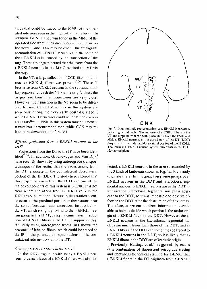
transmitter or neuromodulator, while CCK may re- Fig. 6. Diagrammatic representation of L-ENKLI innervatior, late to the development of the VT. in the tegmental nuclei. The majority of L-ENKLI fibers in the
VT are supplied from the MB, particularly from the PMD and MM. L-ENKLI neurons in the dorsal part of the DT (DDT)
Efferent projection from L-ENKLI neurons in the project to the contralateral dorsolateral portion of the IP (DL). DDT The intrinsic L-ENKLI neuron system also exists in the DDT
Projections from the DT to the IP have been iden- Horizontal plane.
tified 32'34. In addition, Groenewegen and Van Dijk 9
have recently shown, by using anterograde transport
technique of the lectin, that the axons arising from tected. L-ENKLI neurons in the area surrounded by the DT terminate in the contralateral dorsolateral the 3 kinds of knife-cuts shown in Fig. la, b, e mainly portion of the IP (DL). The study here showed that originate there. In this area, there were groups of L- this projection arises from the DDT and one of the ENKLI neurons in the DDT and laterodorsal teg-
major components of this system is L-ENK It is not mental nucleus. L-ENKLI neurons are in the DDT it- clear where the axons from L-ENKLI cells in the self and the laterodorsal tegmental nucleus is adja- DDT cross the midline. However, decussation seems cent to the DDT, so it was impossible to observe ef- to occur at the proximal portion of these axons near fects in the DDT after the destruction of these areas. the soma, because hemitransections just rostral to Therefore, at present no direct information is avail-
the VT, which is slightly rostral to the L-ENKLI neu- able to help us decide which portion is the major ori- ron group in the DDT, caused a contralateral reduc- gin of L-ENKLI fibers in the DDT. However, the L- tion of L-ENKLI fibers in the DL. In support of this, ENKLI neurons in the laterodorsal tegmental nu- the study using anterograde tracer 9 has shown the cleus are much fewer than those of the DDT, and L- presence of labeled fibers, which could be traced to ENKLI fibers in the DDT can sometimes be traced to the IP, in the paramedian raphe nucleus on the con- L-ENKLI neurons in the DDT, so it is likely that L- tralateral side just rostral to the DT. ENKLI fibers in the DDT are of intrinsic origin.
Previously, Huitinga et al. 14 suggested, by means Origin of L-ENKLlfibers in the DDT of a combination of fluorescent retrograde tracing
In the DDT, together with many L-ENKLI neu- and immunohistochemical staining for L-ENK, that rons, a dense plexus of L-ENKLI fibers was also de- L-ENKLI fibers in the DT originate from L-ENKLI
29
neurons in the IP. However, this possibility seems to (SPLI) fibers was present 2L26'es. Both SPLI and L-
be unlikely, because in our additional experiments, ENKLI structures were seen in the DDT, hut not in
destruction of IP failed to decrease L-ENKLI fibers the ventral part of the DT (VDT), so the function of
in the DDT. Thus, the discrepancy between our and the D D T and VDT is different. The fiber connections
their studies could be explained as follows: injection of these two subnuclei are different: L-ENKLI fibers
of the fluorescein dye in their study was made into the are of intrinsic origin, while SPLI fibers are supplied
dorsal tegmental area. Therefore, their injection site by SPLI neurons outside the DT.
involved various adjacent structures in addition to The fiber connections of the L-ENKLI system elu-
t h e D T . , cidated in this study are shown schematically in
In the DDT, in addition to L-ENKLI structures, a Fig. 6.
dense plexus of substance P-like irnmunoreactive
REFERENCES 13 Hughes, I., Smith, T.W., Kosterlitz, H.W., Fothergill, L.H., Morgan, B.A. and Morris, H.R., Identification of
1 Bleir, R., Retrograde transsynaptic cellular degeneration two related pentapeptides from the brain with potent opiate in mammillary and ventral tegmental nuclei following lim- agonist activity, Nature (London), 258 (1975) 577-579. bic decortication in rabbits of various ages, Brain Research, 14 Huitinga, I., Van Dijk, C.A. and Groenewegen, H.J., Sub- 15 (1969)365-393. stance P- and enkephalin-containing projections from the
2 Briggs, T.L. and Kaelber, W.W., Efferent fiber connec- interpeduncular nucleus to the dorsal tegmental region in tions of the dorsal and deep tegmental nuclei of Gudden. the rat, Neurosci. Lett., 62 (1985)311-316. An experimental study in the cat, Brain Research, 29 (1971) 15 Irle, E. and Markowitsch, H.J., Connections of the hippo- 17-29. campal formation, mammillary bodies, anterior thalamus
3 Coons, A.H., Fluorescent antibody method. In J.F. and cingulate cortex: a retrograde study using horseradish Danielli (Ed), General Cytochemical Methods, Academic peroxidase in the cat, Exp. Brain Res., 47 (1982) 79-94. Press, New York, 1958, pp. 399-422. 16 Khachaturian, H., Lewis, M. and Watson, S., Enkephalin
4 Cowan, W.M., Guillery, R.W. and Powell, T.P.S., The ori- systems in diencephalon and brainstem of the rat, J. Comp. gin of the mammillary peduncle and other hypothalamic Neurol., 220 (1983) 310-320. connections from the midbrain, J. Anat., 98 (1964) 17 Kiyama, H., Shiosaka, S., Kubota, Y., Cho, H.J., Takagi, 345-363. H., Tateishi, K., Hashimura, E., Hamaoka, T. and Tohya-
5 Cruce, J.A.F., An autoradiographic study of the descend- ma, M., Ontogeny of cholecystokinin-8-containing neuron ing connections of the mammillary nuclei of the rat, J. system of the rat: an immunohistochemical analysis. II. Comp. Neurol., 176 (1977) 631-644. Lower brainstem, Neuroscience, 10 (1983) 1341-1359.
6 Dahlstr6m, A., Effects of colchicine on transport of amine 18 Kiyama, H., Shiosaka, S., Tateishi, K., Hashimura, E., storage granules in sympathetic nerve of the rat, Eur. J. Hamaoka, T. and Tohyama, M., Cholecystokinin-8-1ike Pharmacol., 5 (1968) 111-113. immunoreactive neuron pathway from the supramammil-
7 Finley, J.C.W., Maderdrut, J.L. and Petrusz, P., The im- lary region to the ventral tegmental nucleus of Gudden of munocytochemical localization of enkephalin in the central the rat, Brain Research, 304 (1984) 397-400. nervous system of the rat, J. Comp. Neurol., 198 (1981) 19 Kreutzberg, G., Neuronal dynamics and flow. IV. Block- 541-565. age of intra-axonal enzyme transport by colchicine, Proc.
8 Fry, W.J. and Cowan, W.M., A study of retrograde cell de- Natl. Acad. Sci. U.S.A., 62 (1969) 722-728. generation in the lateral mammillary nucleus of the cat, 20 Kubota, Y., Inagaki, S., Shiosaka, S., Cho, H.J., Tateishi, with special reference to the role of axonal branching in the K., Hashimura, E., Hamaoka, T. and Tohyama, M., The preservation of the cell, J. Comp. Neurol., 144 ( 1 9 7 2 ) distribution of cholecystokinin octapeptide-like structures 1-24. in the lower brainstem of the rat: an immunohistochemical
9 Groenewegen, H.J. and Van Dijk, C.A., Efferent connec- analysis, Neuroscience, 9 (1983) 587-604. tions of the dorsal tegmental region in the rat, studied by 21 Matsuzaki, T., Shiosaka, S., Inagaki, S., Sakanaka, M., means of anterograde transport of the lectin Phaseolus vul- Takatsuki, K., Takagi, H., Senba, E., Kawai, Y. and To- garis-leucoagglutinin (PHA-L), Brain Research, 304 (1984) hyama, M., Distribution of neuropeptides in the dorsal 367-371. pontine tegmental area of the rat, Cell. Mol. Biol., 27
10 Guillery, R.W., Degeneration in the hypothalamic connec- (1981) 499-508. tions of the albino rat, J. Anat., 91 (1957) 91-115. 22 Miller, R., Chang, K.-J., Cooper, B. and Cuatrecasas, P.,
11 Hayakawa, T. and Zyo, K., Afferent connections of Gud- Radioimmunoassay and characterization of enkephalins in den's tegmental nuclei in the rabbit, J. Comp. Neurol., 235 the rat tissue, J. Biol. Chem., 253 (1978) 531-538. (1985) 169-181. 23 Miller, R.J. and Pickel, V.M., The distribution and func-
12 H6kfelt, T., Elde, R., Johansson, O., Terenius, L. and tions of the enkephalins, J. Histochem. Cytochem., 28 Stein, L., The distribution of enkephalin immunoreactive (1980) 903-917. cell bodies in the rat central nervous system, Neurosci. 24 Morest, D.K., Connections of the dorsal tegmental nucleus Lett., 5 (1977) 25-32. in rat and rabbit, J. Anat., 95 (1961) 229-246.
30
25 Nauta, W.J.H., Hippocampal projections and related neu- grade and retrograde axonal transport of horseradish pcl- tonal pathways to the mid-brain in the cat, Brain, 81 (1958) oxidase in the connections of the mammillary mlclei in the 319-340. cat, Brain Research, 85 (1979) 321-324.
26 Nomura, H., Shiosaka, S., Inagaki, S., Ishimoto, I., Senba, 32 Shibata, H. and Suzuki, T., Efferent projections ¢)[ the in- E., Sakanaka, M., Takatsuki, K., Matsuzaki, T. Kubota, terpeduncular complex in the rat, with special reference to Y., Saito, H., Takase, S., Kogure, K. and Tohyama, M., its subnuclei: a retrograde horseradish peroxidase study, Distribution of substance P-like immunoreactivity in the Brain Research, 296 (1984) 345-349. lower brainstem of the human fetus: an immunohistochemi- 33 Simantov, R., Kuhar, M.J., Uhl, G.R. and Snyder, S .H , cal study, Brain Research, 252 (1982) 315-325. Opiate peptide enkephalin: immunohistochemical mapping
27 Petrovicky, P., Note on the connections of Gudden's teg- in rat central nervous system, Proc. Natl. Acad. Sci. mental nuclei. I. Efferent ascending connections in the U.S.A.. 74(1976)2167-2171. mammillary peduncle, Acta Anat. (Basel), 86 (1973) 34 Smaha, L.A. and Kaeelber, W.W., Efferent fiber projec- 165-190. tions of the habenula and the interpeduncular nucleus. An
28 Pickel, V.M., Sumal, K.K., Reis, D.J., Miller, R.J. and experimental study in the opossum and cat, Brain Research, Hervonen, A., Immunocytochemical localization of en- 16(t973) 291-308. kephalin and substance P in the dorsal tegmental nuclei in 35 Uhl, G.R., Goodman, R.R., Kuhar, M.J., Childers, S.R. human fetal brain, J. Comp. Neurol., 193 (1980) 805-814. and Snyder, S.H., Immunohistochemical mapping of en-
29 Sar, M., Stumpf, W.E., Miller, R.J., Chang, K.-J. and kephalin containing cell bodies, fibers and nerve terminals Cuatrecasas, P., Immunohistochemical localization of en- in the brainstem of the rat, Brain Research. 16 (1979) kephalin in rat brain and spinal cord, J. Comp. Neurol., 182 75-94. (1978) 17-38. 36 Veazey, R.B., Amaral, D.G. and Cowan, W.M., The mor-
30 Senba, E., Shiosaka, S., Hara, Y., Inagaki, S., Kawai, Y., phology and connections of the posterior hypothalamus in Takatsuki, K., Sakanaka, M., Iida, H., Takagi, H., Mina- the cynomolgus monkey (Macaca fascicularis). II. Efferent gawa, H. and Tohyama, M., Ontogeny of the leucine-en- connections, J. Comp. Neurol., 207 (1982)135-156. kephalin neuron system of the rat: immunohistochemical 37 Zamboni, L. and De Martino, C., Buffered picric acid: a analysis. I. Lower brainstem, J. Comp. Neurol., 205 (1982) new rapid fixative for electron microscopy, J. (:ell. Biol., 35 341-359. (1967) 148A.
31 Serlock, D.A. and Raisman, G., A comparison of antero-