Embed Size (px)
Citation preview
Afferent and Efferent Connections of theCaudolateral Neostriatum in the Pigeon
(Columba livia): A Retro- andAnterograde Pathway Tracing Study
SVEN KRONER* AND ONUR GUNTURKUN
AE Biopsychologie, Fakultat fur Psychologie, Ruhr-Universitat Bochum,44780 Bochum, Germany
ABSTRACTThe avian caudolateral neostriatum (NCL) was first identified on the basis of its dense
dopaminergic innervation. This fact and data from lesion studies have led to the notion thatNCL might be the avian equivalent of prefrontal cortex (PFC). A key feature of the PFC is theability to integrate information from all modalities needed for the generation of motor plans.By using antero- and retrograde pathway tracing techniques, we investigated the organiza-tion of sensory afferents to the NCL and the connections with limbic and somatomotor centersin the basal ganglia and archistriatum. Data from all tracing experiments were comparedwith the distribution of tyrosine-hydroxylase (TH)-immunoreactive fibers, serving as amarker of dopaminergic innervation. The results show that NCL is reciprocally connectedwith the secondary sensory areas of all modalities and with at least two parasensory areas.Retrograde tracing also demonstrated further afferents from the deep layers of the Wulst andfrom the frontolateral neostriatum as well as the sources of thalamic input. Efferents of NCLproject onto parts of the avian basal ganglia considered to serve somatomotor or limbicfunctions. Projections to the archistriatum are mainly directed to the somatomotor part of theintermediate archistriatum. In addition, cells in caudal NCL were found to be connected withthe ventral and posterior archistriatum, which are considered avian equivalents of mamma-lian amygdala. All afferents and projection neurons were confined to the plexus of densest THinnervation. Our results show that the NCL is positioned to amalgamate information from allmodalities and to exert control over limbic and somatomotor areas. This organization mightcomprise the neural basis for such complex behaviours as working memory or spatialorientation. J. Comp. Neurol. 407:228–260, 1999. r 1999 Wiley-Liss, Inc.
Indexing terms: parasensory cortex; sensorimotor integration; tyrosine hydroxylase;
immunocytochemistry; basal ganglia; prefrontal cortex
The neostriatum caudolaterale (NCL) of the pigeon hasbeen compared with the mammalian prefrontal cortex(PFC), due to its dense dopaminergic innervation (Divac etal., 1985, 1994; Divac and Mogensen, 1985; Waldmann andGunturkun, 1993; Wynne and Gunturkun, 1995), and thebehavioral deficits in working memory (Mogensen andDivac, 1982, 1993; Gagliardo et al., 1996, 1997; Gun-turkun, 1997), reversal learning (Hartmann and Gun-turkun, 1998), and go/no-go tasks (Gunturkun, 1997) thatfollow its ablation. Working memory tasks selectivelytarget the control of reactions depending on past experi-ences and the ongoing stream of sensory information thatis currently being experienced, which in mammals is a keyfeature of the PFC (Goldman-Rakic, 1987; Fuster, 1989). Aclear prerequisite for a structure in nonmammalian spe-
cies that can subserve such functions and may thus beregarded as an analogue of the PFC is the ability tointegrate all available sensory information and to exertinfluence over motor and limbic structures.
A number of previous studies have demonstrated affer-ent input to the lateral neostriatum from visual (Ritchie,1979; Shimizu et al., 1995), auditory (Wild et al., 1993) and
Grant sponsor: Sonderforschungsbereich 509 NEUROVISION derDeutschen Forschungsgemeinschaft.
*Correspondence to: Sven Kroner, AE Biopsychologie, Fakultat furPsychologie, Ruhr-Universitat Bochum, 44780 Bochum, Germany.E-mail: [email protected]
Received 19 June 1998; Revised 12 November 1998; Accepted 1 December1998
THE JOURNAL OF COMPARATIVE NEUROLOGY 407:228–260 (1999)
r 1999 WILEY-LISS, INC.
somatosensory (Shimizu et al., 1995) areas, as well as aprojection from the multimodal thalamic nucleus dorsolat-eralis posterior (DLP; Waldmann and Gunturkun, 1993;Leutgeb et al., 1996). Based on cytoarchitectonic andhodological data, Rehkamper and Zilles (1991) have pro-posed that the complete posterior neostriatum, includingits caudolateral aspect, might represent an area of multi-modal integration. Furthermore, in a recent retrogradelabeling study focused on the NCL (Leutgeb et al., 1996), ithas been shown that the NCL is reached by afferents fromall major secondary sensory areas within the pigeon brain.Until now, however, a detailed study of the terminationpattern of these afferents and a possible functional segre-gation within the pigeon’s NCL is still lacking. Retrogradetracing data from a very recent study in chicks (Metzger etal., 1998) revealed distinct and nonoverlapping termina-tion zones for the trigeminal, tectofugal, and auditorysystems within the NCL. Because retrograde tracing inpigeons (Leutgeb et al., 1996) suggested a substantiallydifferent organization of the NCL with largely overlappingsensory compartments, this report describes the afferentand efferent connections of the NCL as studied by retro-and anterograde pathway tracing and compares it with thedistribution of tyrosine hydroxylase-immunoreactive fibers.
MATERIALS AND METHODS
Neuroanatomical pathwaytracing experiments
Fifty-seven adult pigeons (Columba livia) from localstock provided the data presented here. Treatment of
animals conformed to NIH guidelines and specifications ofthe German Animal Welfare Act. Accordingly, prior tosurgery, the animals were deeply anesthetized with 0.33–0.4 ml Equithesin per 100 g body weight. Animals receivedpressure injections of either the sensitive antero- andretrograde tracer cholera toxin, subunit b (CTb, 1% indistilled water; List Labs, Campbell, CA) or biotynilateddextran amines as anterograde tracer (BDA, 10,000 molecu-lar weight form, 10% in sodium phosphate buffer, pH 7.3;Molecular Probes, Leiden, The Netherlands). Tracer wasdelivered through glass micropipettes (tip diameter 15–20µm) attached to a nanoliter injector (World PrecisionInstruments, Sarasota, FL). Because previous studies(Leutgeb et al., 1996; Metzger et al., 1998) and our owndata have indicated that the majority of connections of theNCL are restricted to the ipsilateral hemisphere, mostanimals received bilateral injections to minimize the num-ber of pigeons used (Table 1). Stereotaxic coordinates forthe injections were determined by using the atlas ofKarten and Hodos (1967). The afferent sources of the NCLwere determined by injections of CTb (30 –54 nl) intomedial and lateral parts of the NCL along its rostrocaudalextent, as well as into the directly adjacent neostriatumdorsale (Nd, Bonke et al., 1979; Wild et al., 1993; Wild,1994). We included Nd in our analysis because the patternof catecholaminergic innervation suggests that Nd is con-tinuous with NCL (Fig. 1) and might constitute its audi-tory subcomponent. To study the areal pattern of afferentinput to the NCL, injections of CTb (9–80 nl) and BDA(40–100 nl) were made into most of the telencephalicregions which by means of retrograde transport had been
AC nucleus accumbensAi archistriatum intermedium, pars centraleAidd archistriatum intermedium, pars dorsodorsaleAidv archistriatum intermedium, pars dorsoventraleAL ansa lenticularisAm archistriatum medialeAp archistriatum posteriorAPH area parahippocampalisAv archistriatum intermedium, pars ventraleAVT area ventralis tegmentalisBas nucleus basalisBNST bed nucleus of the stria terminalisCDL corticoidea dorsolateralisCPi cortex piriformisDIP nucleus dorsointermedius posterior thalamiDIVA nucleus dorsalis intermedius ventralis anteriorDLL nucleus dorsolateralis anterior thalami, pars lateralisDLM nucleus dorsolateralis anterior thalami, pars medialisDLP nucleus dorsolateralis posteriorDMA nucleus dorsomedialis anterior thalamiDMP nucleus dorsomedialis posterior thalamiE ectostriatumEp ectostriatal beltFPL fasciculus prosencephali lateralisGLd dorsolateral geniculate nucleiHA hyperstriatum accessoriumHD hyperstriatum dorsaleHIS hyperstriatum intercalatus superiorHOM tractus occipitomesencephalicus, pars hypothalamiHp hippocampusHV hyperstriatum ventraleHVdv hyperstriatum ventrale dorso-ventraleHVvv hyperstriatum ventrale ventroventraleICo nucleus intercollicularisIHA nucleus intercalatus of the hyperstriatum accessoriumIMHV intermediate and medial part of the hyperstriatum ventraleL1 field L1L2 field L2
Abbreviations
L3 field L3LFS lamina frontalis superiorLH lamina hyperstriaticaLHy nucleus lateralis hypothalamiLPO lobus parolfactoriusNCL neostriatum caudolateraleNCm neostriatum caudomedialeNd neostriatum dorsaleNFL neostriatum frontolateraleNFT neostriatum fronto-trigeminaleNI neostriatum intermediumNIL neostriatum intermedium lateraleNIM neostriatum intermedium medialisNIMl neostriatum intermedium medialis, pars lateraleNIMm neostriatum intermedium medialis, pars medialeOM tractus occipitomesencephalicusOv nucleus ovoidalisPA paleostriatum augmentatumPP paleostriatum primitivumPT nucleus pretectalisRt nucleus rotundusSAC stratum album centraleSCI stratum cellulare internumSGC stratum griseum centraleSGP substantia grisea et fibrosa periventricularisSPC nucleus tractus septomesencephaliciSPL nucleus spiriformis lateralisSRt nucleus subrotundusT nucleus triangularisTn nucleus taeniaeTO optic tectumTPc nucleus tegmenti pedunculopontinus, pars compactaTPO area temporo-parieto-occcipitalisTSM tractus septomesencephalicusVa valleculaVIA ventrointermediate area of the thalamusVIP ventrointermediate area of the posterior nuclei
CONNECTIONS OF THE AVIAN NEOSTRIATUM CAUDOLATERALE 229
shown to constitute afferent sources of the NCL. Efferentconnections of the NCL to motor and limbic structureswere studied by placing injections into the somatomotorand limbic parts of the archistriatum (Zeier and Karten,1971) as well as into parts of the avian basal ganglia thatare comparable to parts of mammalian striatum.
Immunohistochemical procedures
Survival times were 3 days for CTb and 4–6 days forBDA. Fifteen minutes prior to perfusion, the animalswere injected with 1,000 IU heparin and deeply anaes-thetized with 0.4–0.5 ml Equithesin per 100 g body weight.Pigeons were then perfused transcardially with 200 ml0.9% saline (40°C) followed by 1,000 ml of 4% para-formaldehyde in 0.12 M phosphate buffer (PB; 4°C, pH7.4). After perfusion, brains were dissected and postfixedin the same fixative to which 30% sucrose was added, andthen transferred to 30% sucrose in phosphate buffercontaining 0.9% NaCl (PBS; pH 7.4) for approximately18 hours at 4°C. Brains were cut in frontal slices of 40 µmon a freezing microtome and collected in PBS containing0.01% NaN3 as a preservative. Representative sectionswere then processed for the avidin-biotin-conjugate tech-nique (ABC).
Immunohistochemical labelingfor Cholera Toxin b
Endogeneous peroxidases were blocked by preincubat-ing slices in a solution of 0.5% H2O2. Slices were washed
and for immunohistochemistry of CTb free-floating sec-tions were incubated overnight at 4°C in anti-CTb fromgoat (Jackson, West Grove, PA; 1:20,000) in PBS contain-ing 0.3% Triton X-100 (Sigma, Deisenhofen, Germany).The following steps were carried out at room temperature,separated by three washes in PBS of 10 minutes each.After washing, slices were first incubated for 1 hour inbiotinylated donkey anti-goat (Jackson,1:500 in 0.3% Tri-ton X-100 PBS) for 1 hour and then in the avidin-biotincomplex (ABC Elite, Vector Labs, Burlingame, CA; 1:100in PBS with 0.3% Triton X-100) for 1 hour. Washes in PBSwere followed by two additional washes in 0.12 M acetatebuffer (pH 6). Staining was achieved by the 3,3’-diamino-benzidine (DAB) technique with heavy metal amplification(modified from Adams, 1981) by adding H8N2NiO8S2 (2.5 g/100 ml), NH4Cl, and CoCl2 (both 40 mg/100 ml). After 15minutes of preincubation, the reaction was catalyzed witha solution of 0.5% H2O2. The reaction was stopped byrinsing the tissue in 0.12 M acetate buffer and PBS. Sliceswere then mounted, dehydrated, and coverslipped.
Labeling for BDA
Visualization of BDA was identical to that of CTb, exceptthat primary and secondary antibodies could be omittedand slices were directly incubated in ABC, followed by thestaining procedure described above. Selected series la-beled for BDA or CTb were counterstained with cresylviolet.
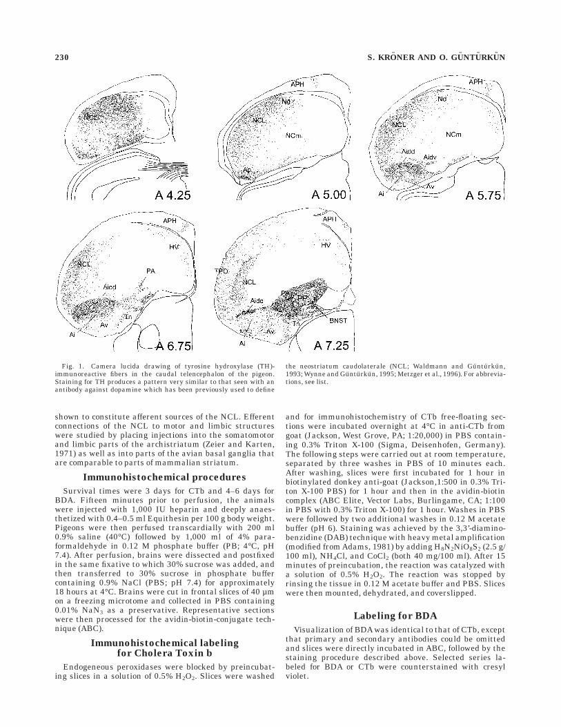
Fig. 1. Camera lucida drawing of tyrosine hydroxylase (TH)-immunoreactive fibers in the caudal telencephalon of the pigeon.Staining for TH produces a pattern very similar to that seen with anantibody against dopamine which has been previously used to define
the neostriatum caudolaterale (NCL; Waldmann and Gunturkun,1993; Wynne and Gunturkun, 1995; Metzger et al., 1996). For abbrevia-tions, see list.
230 S. KRONER AND O. GUNTURKUN
Immunohistochemical labelingfor tyrosine hydroxylase
To compare the distribution of afferent fibers and retro-gradely labeled neurons within NCL with the distributionof putative dopaminergic fibers, several sections wereprocessed for tyrosine hydroxylase (TH). Therefore, addi-tional sections stained for CTb or BDA were either double-labeled with TH, or occasionally, sections adjacent to thesewere single-labeled for TH. The same basic procedure asfor immunohistochemistry of CTb was applied for labelingof TH: After blocking of endogeneous peroxidases andthorough washing, free-floating sections were incubatedovernight at 4°C in monoclonal mouse anti-TH (Boeh-ringer, Mannheim, Germany) diluted 1:200 in PBS contain-ing 0.3% Triton X. Slices were then incubated at roomtemperature in biotinylated rabbit anti-mouse (Chemicon,Temecula, CA; 1:200 in 0.3% Triton PBS) for 1 hour andfinally in ABC for 1 hour. Staining was achieved using theDAB-technique as described for CTb. In those cases thatwere double-labeled, enhancement of the DAB staining bynickel ammonium sulfate was ommitted, resulting in alight brown signal for TH which could be clearly distin-guished from the black reaction product of the tracers.
RESULTS
Retrograde tracing of afferent sourcesof the NCL
Our injections of CTb into medial and lateral parts ofNCL generally replicated the results of an earlier study(Leutgeb et al., 1996). All injections were confined to NCLas defined by TH and dopamine (DA)-like immunoreactiv-ity (Waldmann and Gunturkun, 1993; Wynne and Gun-turkun, 1995; present study), and extended from thecaudal tip of the telencephalon at approximately anterior(A) 4.25 to rostral A 7.00.
Telencephalic afferents to NCL. Areas that werefound to project to the NCL included the medial aspect ofthe hyperstriatum accessorium (HA) throughout its visual(Karten et al., 1973; Shimizu et al., 1995) and somatosen-sory part (Delius and Benetto, 1972; Wild, 1987b; Funke,1989; but see Deng and Wang, 1992, 1993), the ectostriatalbelt (Ep) surrounding the ectostriatum dorsally and later-ally (Karten and Hodos, 1970) and the adjoining lateralneostriatum, the anterior neostriatum overlying thenucleus basalis (neostriatum fronto-trigeminale, NFT, ofWild et al., 1985), and field L1 and L3 of the auditory fieldL complex. A large number of cells were labeled in themedial part of the intermediate neostriatum (NIM ofVeenman et al., 1995b) and the overlying intermediate andmedial parts of the hyperstriatum ventrale (HV): part ofthese neurons probably correspond to the previously de-scribed parasensory area in the intermediate neostriatum,situated between the rostral pole of field L and thecaudomedial border of the visual ectostriatum, that re-ceives multimodal input via the DLP (Gamlin and Cohen,1986; Wild, 1987a, 1994; Funke, 1989; Korzeniewska andGunturkun, 1990). These cells were predominantly la-beled if injections were made into more rostral and medial(including Nd) parts of the NCL. After injections intocaudal NCL, a distinct cell group occupying the remainingmedialmost aspect of the NIM between approximately A8.50 and A 12.50 was labeled. Cells within this area alsoextended across the lamina hyperstriatica (LH) into theintermediate and medial parts of the HV. These two cell
groups roughly followed the cytoarchitectonic borders ofthe regions Ne 9 or Ne 8 and Ne 3, respectively, asdescribed by Rehkamper et al. (1985). For the reason ofsimplicity, we will refer to them as the medial part of theNIM (NIMm) and the lateral part of the NIM (NIMl),respectively.
In the caudomedial neostriatum (NCm), scattered cellswere found dorsal to the lamina medullaris dorsalis (LMD).Another band of cells stretched along the border betweenthe hyperstriatum dorsale (HD) and the dorsal division ofthe ventral hyperstriatum (HVdv). These cells could befound in most cases, irrespective of the injection sitewithin NCL. In cases that were centered more medially orcaudally within NCL, this band of cells also extended intoa small triangular-shaped zone within the vallecula. Fi-nally, in several cases a number of cells were observed inthe frontolateral part of the neostriatum (NFL). Injectionsof CTb into this area did indeed confirm a reciprocalconnection with the ventrolateral part of the NCL (datanot shown). The large majority of fibers from this part ofthe anterior neostriatum, however, terminated within thearea corticoidea dorsolateralis (CDL) and the area temporo-parieto-occcipitalis (TPO) overlying the NCL. Therefore,we cannot fully exclude that part of the labeling withinNFL was attributable to tracer spread into CDL.
In addition to labeling in sensory areas, retrogradetransport revealed a number of labeled neurons in thedorsal and ventral parts of the intermediate archistria-tum. Cells in the ventral archistriatum were the onlytelencephalic connection found to project bilaterally ontothe NCL. In contrast to Leutgeb et al. (1996), we neverobserved any labeling within the nucleus taeniae (Tn).Furthermore, a few cells within the ventral striatum justdorsal to the fasciculus prosencephali lateralis (FPL) werealso found to project to the NCL.
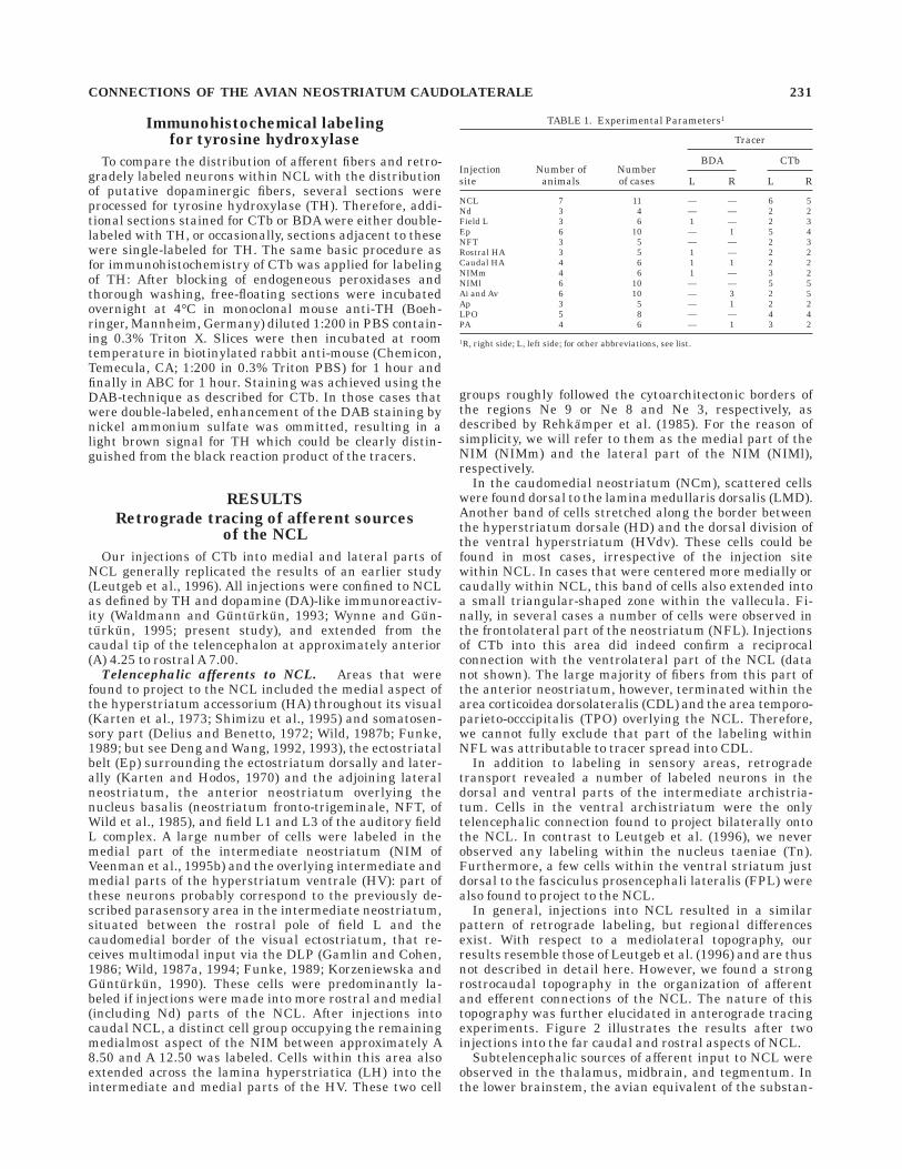
In general, injections into NCL resulted in a similarpattern of retrograde labeling, but regional differencesexist. With respect to a mediolateral topography, ourresults resemble those of Leutgeb et al. (1996) and are thusnot described in detail here. However, we found a strongrostrocaudal topography in the organization of afferentand efferent connections of the NCL. The nature of thistopography was further elucidated in anterograde tracingexperiments. Figure 2 illustrates the results after twoinjections into the far caudal and rostral aspects of NCL.
Subtelencephalic sources of afferent input to NCL wereobserved in the thalamus, midbrain, and tegmentum. Inthe lower brainstem, the avian equivalent of the substan-
TABLE 1. Experimental Parameters1
Injectionsite
Number ofanimals
Numberof cases
Tracer
BDA CTb
L R L R
NCL 7 11 — — 6 5Nd 3 4 — — 2 2Field L 3 6 1 — 2 3Ep 6 10 — 1 5 4NFT 3 5 — — 2 3Rostral HA 3 5 1 — 2 2Caudal HA 4 6 1 1 2 2NIMm 4 6 1 — 3 2NIMl 6 10 — — 5 5Ai and Av 6 10 — 3 2 5Ap 3 5 — 1 2 2LPO 5 8 — — 4 4PA 4 6 — 1 3 2
1R, right side; L, left side; for other abbreviations, see list.
CONNECTIONS OF THE AVIAN NEOSTRIATUM CAUDOLATERALE 231
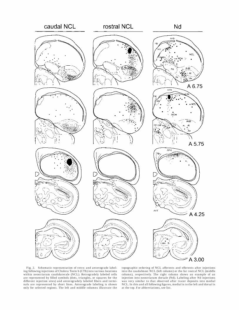
Fig. 2. Schematic representation of retro- and anterograde label-ing following injections of Cholera Toxin b (CTb) into various locationswithin neostriatum caudolaterale (NCL). Retrogradely labeled cellsare represented by filled symbols (dots, triangles, or squares for thedifferent injection sites) and anterogradely labeled fibers and termi-nals are represented by short lines. Anterograde labeling is shownonly for selected regions. The left and middle columns illustrate the
topographic ordering of NCL afferents and efferents after injectionsinto the caudalmost NCL (left column) or the far rostral NCL (middlecolumn), respectively. The right column shows an example of aninjection into neostriatum dorsale (Nd). Labeling after Nd injectionswas very similar to that observed after tracer deposits into medialNCL. In this and all following figures, medial is to the left and dorsal isat the top. For abbreviations, see list.
Figure 2 (Continued)
CONNECTIONS OF THE AVIAN NEOSTRIATUM CAUDOLATERALE 233
tia nigra, the nucleus tegmenti pedunculopontinus, parscompacta (TPc), the locus coeruleus (LoC), and the areaventralis tegmentalis (AVT) were labeled. These cell groupshave been shown to be immunoreactive for TH and dopa-mine and contain the sources of dopaminergic afferents tothe telencephalon (Waldmann and Gunturkun, 1993;Metzger et al., 1996; present study). The projection fromthe LoC and some cells in the formatio reticularis wasbilaterally organized. In the thalamus, retrogradely la-beled cells were mainly found in the nucleus subrotundus(SRt) and the DLP. Yet, we also found a projection from then. dorsointermedius posterior thalami (DIP) and a band ofcells that seemed to intersperse between DLP and DIP andalso extended ventral to the latter two. Therefore, wetermed these cells collectively the ventrointermediate areaof the posterior nuclei (VIP). Figure 3 shows examples ofretrogradely labeled neurons from sensory forebrain areas(Fig. 3A–F) and the sources of thalamic input to NCLfollowing injections of CTb into NCL.
Anterograde labeling of efferents of the NCL. Thepattern of anterograde labeling resulting from injectionsinto NCL was studied within the basal ganglia and thearchistriatum. Fibers showing terminal-like varicositieswere found throughout most parts of the archistriatum.For the description of archistriatal subdivisions, we adoptedthe terminology of Wynne and Gunturkun (1995). Injec-tions into the very caudal aspect of NCL predominantlylabeled fibers in parts of the central (Ai) and ventral parts(Av) of the intermediate archistriatum and in some casesalso in the posterior archistriatum (Ap). Injections intomore rostral NCL resulted in terminal labeling that wasconfined to the dorsal and central parts of the intermediatearchistriatum. After injections into the caudal NCL, weobserved terminal-like labeling in the medial lobus parol-factorius (LPO) and the ventral striatum, i.e., the nucleusaccumbens (AC) and the bed nucleus of the stria termina-lis (BNST), which was not attributable to tracer spreadinto the piriform cortex (CPi) or the caudal archistriatum.Injections into rostral NCL, on the other hand, resulted indense terminal labeling in the lateral aspects of LPO andlarge parts of the paleostriatum augmentatum (PA). Label-ing in the PA was most abundant in its caudal extent. Bothtermination patterns within the archistriatum and thebasal ganglia thus suggest that the anterior NCL may beconnected to areas concerned with somatomotor functions,i.e., the dorsal and lateral striatum (Veenman et al.,1995a,b), as well as the dorsal and central archistriatum(Zeier and Karten, 1971), whereas the caudal aspect ofNCL might project to limbic parts of the striatum (Veen-man et al., 1995b) and the presumed avian homologue ofthe amygdala, e.g., the ventral and caudal archistriatum(Zeier and Karten, 1971; Dubbeldam et al., 1997). Figure 4shows examples of anterograde labeling in the basalganglia and the archistriatum following injections of CTbinto NCL.
Labeling after Nd injections. Labeling after CTbinjections into Nd largely paralled the results after medialNCL injections. A large number of retrogradely labeled cellbodies and fibers could be observed throughout the NIM,which seemed to cluster in two distinct cell groups as alsoobserved after injections into the NCL. Strong labelingwas also present in the caudomedial neostriatum andareas L1 and L3 of the field L complex. Furthermore, cellswere found in the medial HV and along the border of HVand HD, the caudal and rostral aspects of HA, the val-leculla (Va), and the NFL. Finally, somata and terminal-
like labeling could be observed in the ventromedial archi-striatum and lateral parts of the NCL. Terminal-likelabeling in the basal ganglia after Nd injections wasrestricted to the medial and central parts of the caudal PA.In the thalamus, labeling was most prominent in the SRtand DLP. A number of cells was also found within the shellof the nucleus ovoidalis (Ov) and the nucleus semilunarisparovoidalis. In the midbrain AVT, TPc and LoC containedlabeled cell bodies. The results following an injection intoNd are summarized schematically in Figure 2 (rightcolumn).
Anterograde tracing experiments
Injections into the ectostriatal belt. After injectionsinto the dorsal and lateral ectostriatal belt and the adjoin-ing neostriatum intermedium (NI) underneath the exter-nal pallium (Veenman et al., 1995b), a massive projectionof labeled fibers was observed to move caudally throughthe NI and to innervate the anterior and lateral NCL. Thisprojection seemed to be topographically organized andextended in the case of caudal injections posteriorly toabout A 5.00. In addition to terminals, a number ofretrogradely labeled cell bodies also were found, indicatinga reciprocal connection between the NCL and the ectostria-tal belt region. As shown in Figure 5, a massive reciprocalconnection of the Ep exists with the deeper hyperstriatallayers, especially with the lateral part of HV. Cells withinHV and the NI or NFL, respectively, were distributed up tofar rostrally within the hemisphere. Relatively few cellsand fibers were found within the outer rind of the pallium,i.e., the lateral NI (NIL), TPO, and CDL, the larger portionof them remaining underneath the border that separatesthe NI and anterior NCL from the overlying externalpallium. Furthermore, in the dorsal and ventral aspects ofthe rostral archistriatum, small clusters of cells werelabeled, as well as a number of somata in the paleostria-tum primitivum (PP). Within the ectostriatal core, labeledcells were found in a narrow area below the injection site.In the thalamus, large numbers of neurons were labeled innucleus rotundus which probably are due to tracer spreadinto the ectostriatal core.
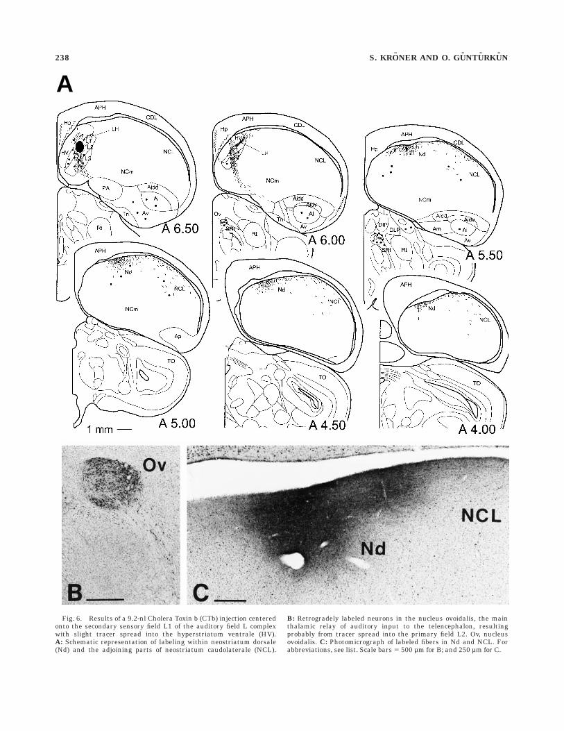
Injections into field L. In accordance with previousreports (Bonke et al., 1979; Wild et al., 1993), tracerdeposits into the auditory field L complex which weremainly centered on the secondary sensory field L1 lead tomassive terminal-like labeling in Nd. Yet, injections of CTbrevealed a diffuse projection that also extended furtherlaterally in caudal NCL (Fig. 6). In the medioventralaspect of NCL also a small number of retrogradely labeledcells could be observed. Field L injections also resulted inretrograde labeling of neurons throughout the HA, NIM,and HV, as well as in ventral and intermediate archistria-tum. Diencephalic neurons were labeled within shell andcore regions of n. ovoidalis and the n. semilunaris parovoid-alis.
Injections into the hyperstriatum accessorium. Af-ter injections into the Wulst that were centered on themedial aspect of the dorsal hyperstriatum accessorium,fibers were found to travel through the lamina frontalissuperior (LFS) and further laterally along the border thatseparates the anterior NCL from the overlying LPO andCDL. Fibers showing terminal-like varicosities remainedin close apposition to the ventricle as they moved furthercaudally through NCL. Neurons that project back to theinjected site generally followed the distribution of terminat-ing fibers but extended somewhat ventrally to these.
234 S. KRONER AND O. GUNTURKUN
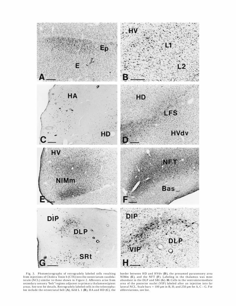
Fig. 3. Photomicrographs of retrogradely labeled cells resultingfrom injections of Cholera Toxin b (CTb) into the neostriatum caudola-terale (NCL) similar to those shown in Figure 2. Afferents arise fromsecondary sensory ‘‘belt’’ regions adjacent to primary thalamorecipientareas. See text for details. Retrogradely labeled cells in the telencepha-lon include the ectostriatal belt (A), field L 1 (B), HA and HD (C), the
border between HD and HVdv (D), the presumed parasensory areaNIMm (E), and the NFT (F). Labeling in the thalamus was mostabundant in the DLP and SRt (G). H: Cells in the ventrointermediatearea of the posterior nuclei (VIP) labeled after an injection into farlateral NCL. Scale bars 5 100 µm in B, H; and 250 µm for A, C – G. Forabbreviations, see list.
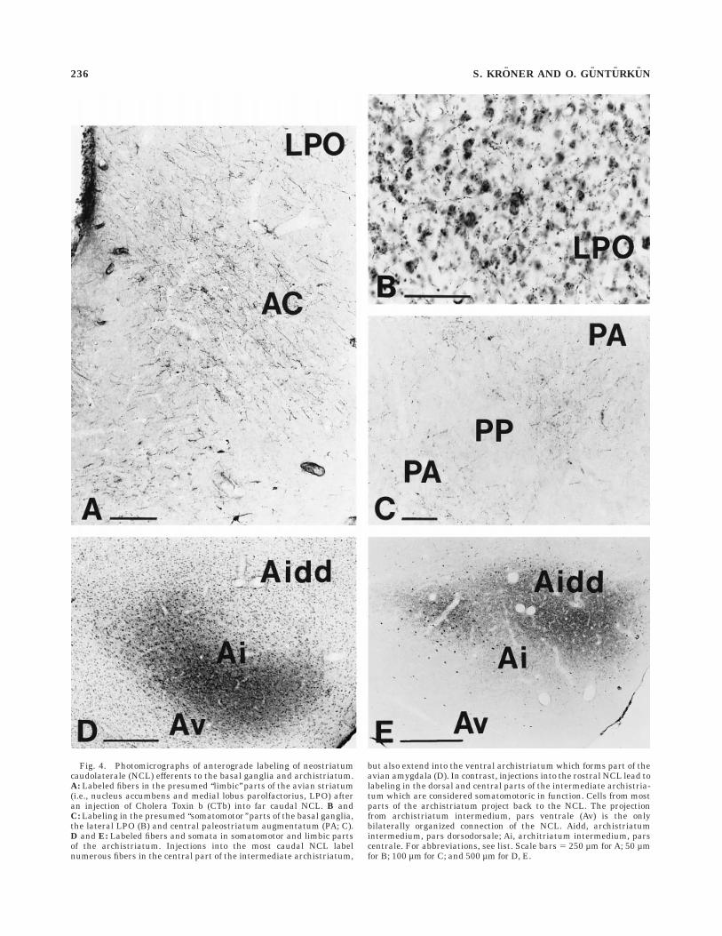
Fig. 4. Photomicrographs of anterograde labeling of neostriatumcaudolaterale (NCL) efferents to the basal ganglia and archistriatum.A: Labeled fibers in the presumed ‘‘limbic’’ parts of the avian striatum(i.e., nucleus accumbens and medial lobus parolfactorius, LPO) afteran injection of Cholera Toxin b (CTb) into far caudal NCL. B andC: Labeling in the presumed ‘‘somatomotor’’ parts of the basal ganglia,the lateral LPO (B) and central paleostriatum augmentatum (PA; C).D and E: Labeled fibers and somata in somatomotor and limbic partsof the archistriatum. Injections into the most caudal NCL labelnumerous fibers in the central part of the intermediate archistriatum,
but also extend into the ventral archistriatum which forms part of theavian amygdala (D). In contrast, injections into the rostral NCL lead tolabeling in the dorsal and central parts of the intermediate archistria-tum which are considered somatomotoric in function. Cells from mostparts of the archistriatum project back to the NCL. The projectionfrom archistriatum intermedium, pars ventrale (Av) is the onlybilaterally organized connection of the NCL. Aidd, archistriatumintermedium, pars dorsodorsale; Ai, architriatum intermedium, parscentrale. For abbreviations, see list. Scale bars 5 250 µm for A; 50 µmfor B; 100 µm for C; and 500 µm for D, E.
236 S. KRONER AND O. GUNTURKUN
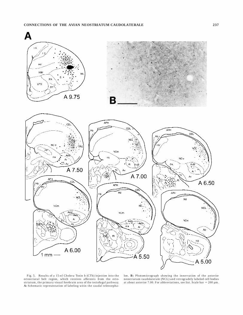
Fig. 5. Results of a 15-nl Cholera Toxin b (CTb) injection into theectostriatal belt region, which receives afferents from the ecto-striatum, the primary visual forebrain area of the tectofugal pathway.A: Schematic representation of labeling witin the caudal telencepha-
lon. B: Photomicrograph showing the innervation of the anteriorneostriatum caudolaterale (NCL) and retrogradely labeled cell bodiesat about anterior 7.00. For abbreviations, see list. Scale bar 5 200 µm.
CONNECTIONS OF THE AVIAN NEOSTRIATUM CAUDOLATERALE 237
A
Fig. 6. Results of a 9.2-nl Cholera Toxin b (CTb) injection centeredonto the secondary sensory field L1 of the auditory field L complexwith slight tracer spread into the hyperstriatum ventrale (HV).A: Schematic representation of labeling within neostriatum dorsale(Nd) and the adjoining parts of neostriatum caudolaterale (NCL).
B: Retrogradely labeled neurons in the nucleus ovoidalis, the mainthalamic relay of auditory input to the telencephalon, resultingprobably from tracer spread into the primary field L2. Ov, nucleusovoidalis. C: Photomicrograph of labeled fibers in Nd and NCL. Forabbreviations, see list. Scale bars 5 500 µm for B; and 250 µm for C.
238 S. KRONER AND O. GUNTURKUN
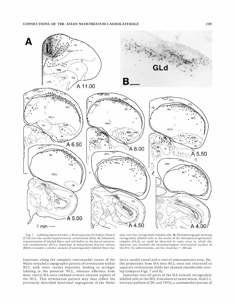
Injections along the complete rostrocaudal extent of theWulst revealed a topographic pattern of termination withinNCL with more caudal injections leading to strongerlabeling in the posterior NCL, whereas afferents frommost rostral HA were confined to more anterior aspects ofthe NCL. This termination pattern may thus reflect thepreviously described functional segregation of the Wulst
into a caudal visual and a rostral somatosensory area. Yet,the projections from HA onto NCL were not restricted toseparate termination fields but showed considerable over-lap (compare Figs. 7 and 8).
Injections into all parts of the HA evinced retrogradelylabeled cells in the HD, frontolateral neostriatum, field L1,external pallium (CDL and TPO), a caudomedial portion of
Fig. 7. Labeling observed after a 40-nl injection of Cholera Toxin b(CTb) into the caudal hyperstriatum accessorium (HA). A: Schematicrepresentation of labeled fibers and cell bodies in the dorsal neostria-tum caudolaterale (NCL). Injections of biotynilated dextran amines(BDA) revealed a similar amount of anterogradely labeled fibers but
only very few retrogradely labeled cells. B: Photomicrograph showingretrogradely labeled cells in the nuclei of the dorsolateral geniculatecomplex (GLd), as could be observed in some cases in which theinjection site involved the thalamorecipient intercalated nucleus ofthe HA. For abbreviations, see list. Scale bar 5 200 µm.
CONNECTIONS OF THE AVIAN NEOSTRIATUM CAUDOLATERALE 239
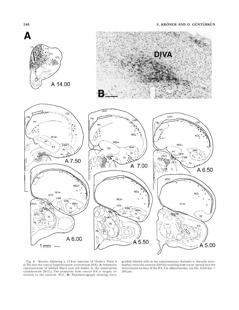
Fig. 8. Results following a 13.8-nl injection of Cholera Toxin b(CTb) into the rostral hyperstriatum accessorium (HA). A: Schematicrepresentation of labeled fibers and cell bodies in the neostriatumcaudolaterale (NCL). The projection from rostral HA is largely re-stricted to the anterior NCL. B: Photomicrograph showing retro-
gradely labeled cells in the somatosensory thalamic n. dorsalis inter-medius ventralis anterior (DIVA) resulting from tracer spread into theintercalated nucleus of the HA. For abbreviations, see list. Scale bar 5200 µm.
240 S. KRONER AND O. GUNTURKUN
the area parahippocampalis (APH), and the ventral andcentral archistriatum. Injections into rostral HA addition-ally labeled cells in the NIM. In several cases in whichtracer spread occurred into the thalamorecipient granularlayer of the intercalated nucleus of the HA (IHA), retro-gradely labeled cells were observed in the thalamic relaystations of the thalamofugal visual and somatosensorysystem, respectively: deposits of tracer into the caudalWulst evinced cells in the dorsal aspect of the lateralgeniculate nuclei (GLd; Karten et al., 1973; Gunturkun etal., 1993). Injections into the rostral HA, on the otherhand, predominatly labeled cells in the somatosensorynucleus dorsalis intermedius ventralis anterior (DIVA;Wild, 1987b; Funke, 1989; Medina et al., 1997).
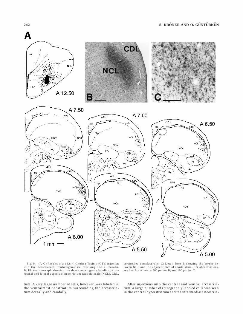
Injections into the neostriatum frontotrigeminale.
After injections into the NFT dorsal to the nucleus basalis(Bas), fibers gathered in the tractus fronto-archistriatalisand passed caudally until they terminated within thelateral part of the anterior archistriatum and the rostraland ventral part of NCL. As can be seen in Figure 9, theterminal field of this projection occupied a large part of theanterior and lateral NCL and extended caudally up toabout A 5.00. Within this dense terminal field, a compar-atively small number of neurons were found to projectback to the injection site. In those cases that showed no orminimal tracer spread into Bas, retrogradely labeled fibersand cells within Bas were restricted to the area directlyventral to the injection site. Furthermore, cells werelabeled in the HV dorsal to the injection site, the anteriorarchistriatum, along the tractus fronto-archistriatalis, andin medial HA.
Injections into the neostriatum intermedium, pars
medialis. Injections into the intermediate neostriatumattempted to evaluate whether two separate projectionsfrom this area onto NCL exist as indicated by the resultsfrom our retrograde tracing experiments reported above.Tracer deposits were thus aimed at the terminal field ofthe DLP just medially and caudally to the ectostriatum,termed NIMl here, and the medialmost aspect of NI, theNIMm, respectively.
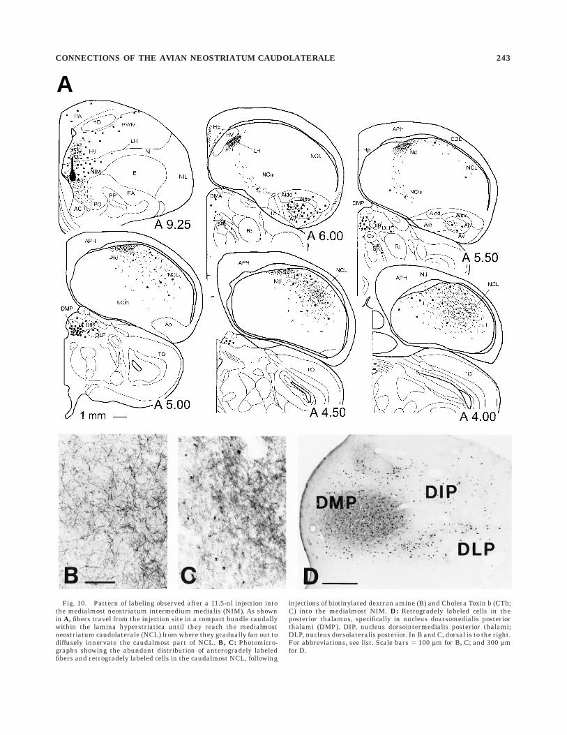
Injections into NIMm consistently produced dense termi-nal labeling and a considerable number of retrogradelylabeled cell bodies in the most caudal aspect of NCL. Fiberbundles left the injection site laterally and travelledcaudally within the LH. They reached the caudal neostria-tum at the rostral end of Nd where they made massiveterminations. The remainder of labeled fibers coursedlaterally and caudally within the periventricular roof ofNCL. On their way towards the caudal pole of the hemi-sphere, they gradually fanned out towards the midlineuntil they occupied a large proportion of the hemisphere(Fig. 10).
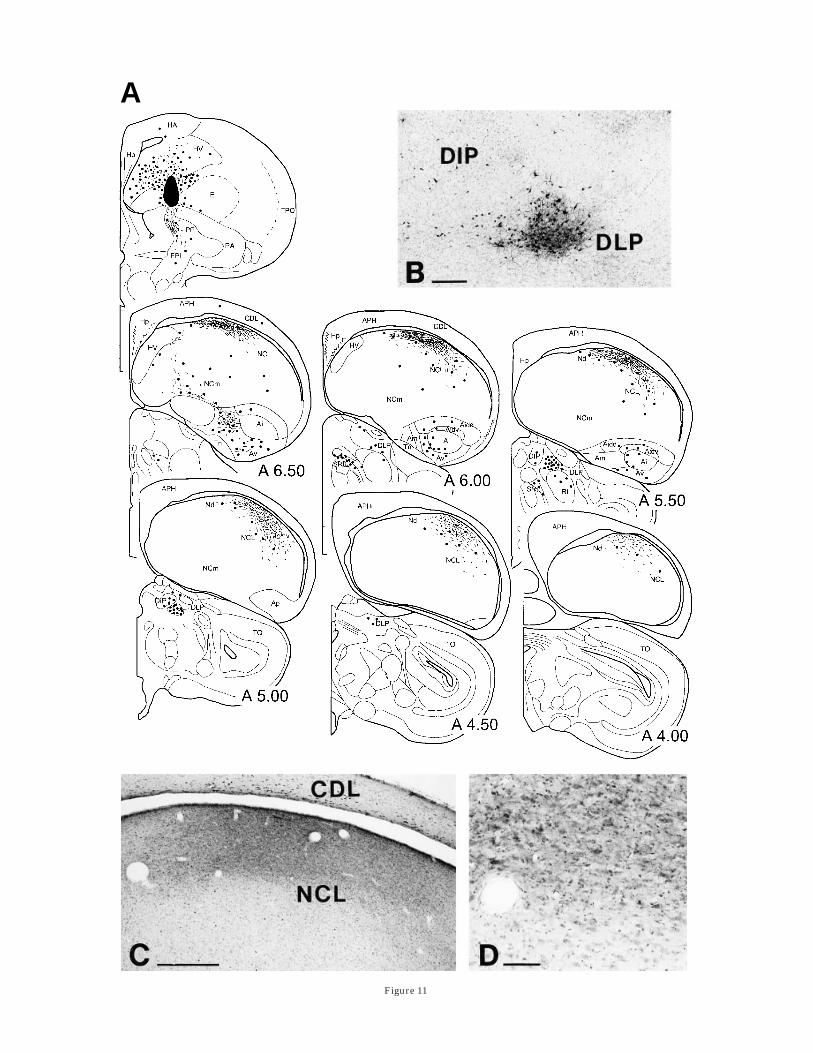
Small injections that were entirely confined to NIMlresulted in anterograde labeling that was largely re-stricted to Nd and the dorsal roof of the medial NCL.Labeling in these cases was virtually identical to that seenafter field L injections (compare Fig. 6). However, labelingwithin the NCL varied considerably with the amount oftracer used. Larger NIMl injections that also extended intoone of the adjacent areas, HV, NIMm, or Ep, led to a muchhigher number of labeled fibers and somata within NCL,whereas at the same time, the terminal field also expandedfurther lateral and medial (Fig. 11). However, the distribu-tion of these fibers did not resemble the terminal fieldsresulting from injections into NIMm or Ep. In addition, allof these cases showed a similar labeling in the thalamus
that was distinct from the pattern observed after NIMminjections (below).
Deposits of CTb into the medialmost NI yielded numer-ous neurons within the medial HV, dorsal to the injectionsite, HA, HD, in Ai and Av. Retrogradely labeled cells fromAv were also found on the contralateral side. Massiveanterograde labeling was seen in medial LPO. Injectionsinto lateral NIM resulted in a similar pattern of labelingwhich included the somatosensory and visual Wulst, me-dial HV dorsal to the injection site, as well as Av and Ai. Inaddition, cells were labeled in field L1 and L3, as well as inlarge aspects of the caudomedial neostriatum. In thesecases, labeled fibers were seen in the PA ventral to theinjection site, but not within LPO. Labeling in the thala-mus further strengthened the notion of two distinct subar-eas within NIM: injections into the NIMl confirmed theprojection from DLP (Kitt and Brauth, 1982; Gamlin andCohen, 1986; Wild, 1994) and in addition labeled cells inSRt, the shell of Ov and DIP. In contrast, after tracerdeposits into the medialmost NI with slight spread into theoverlying HV, but not into PA or LPO, a large number ofretrogradely cells were found in the medial part of thedorsal thalamus, namely the n. dorsomedialis posteriorthalami (DMP) and DIP, and only very few within DLP.
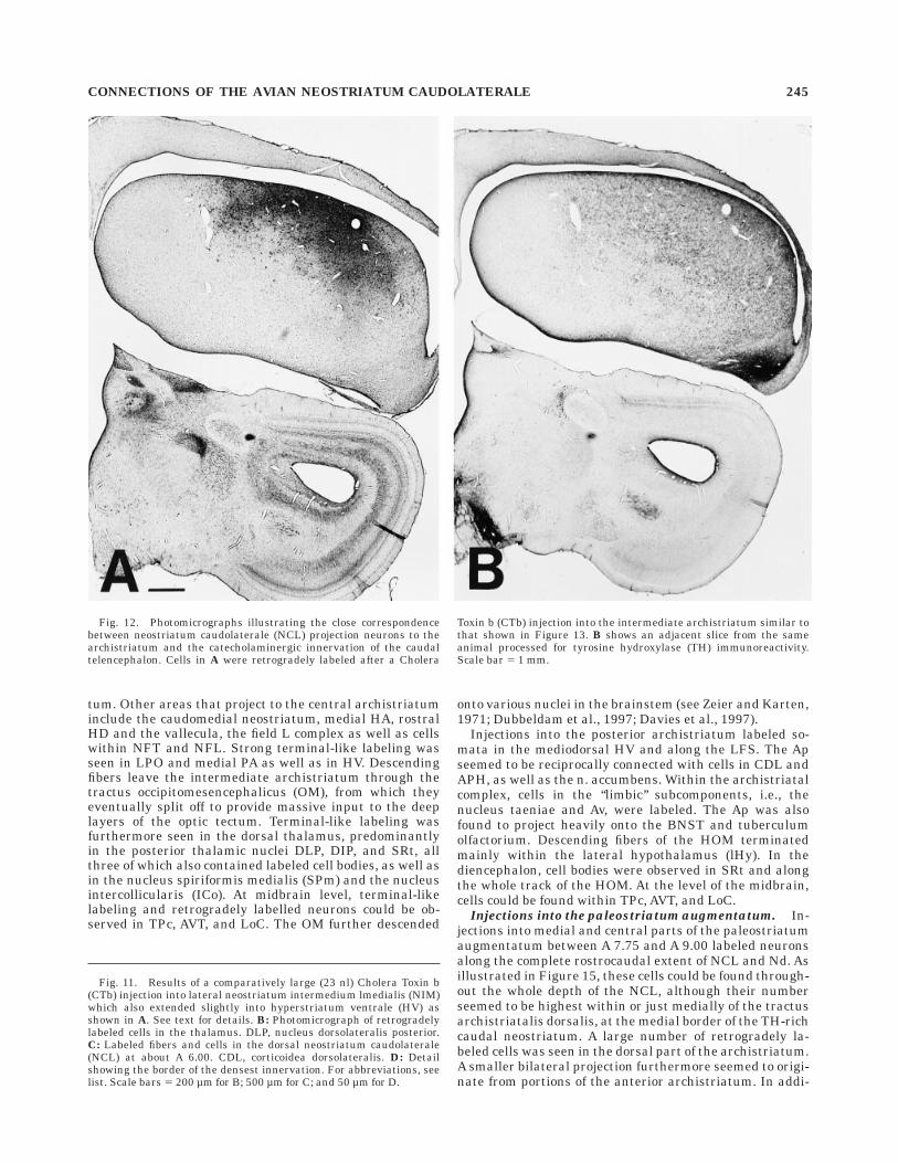
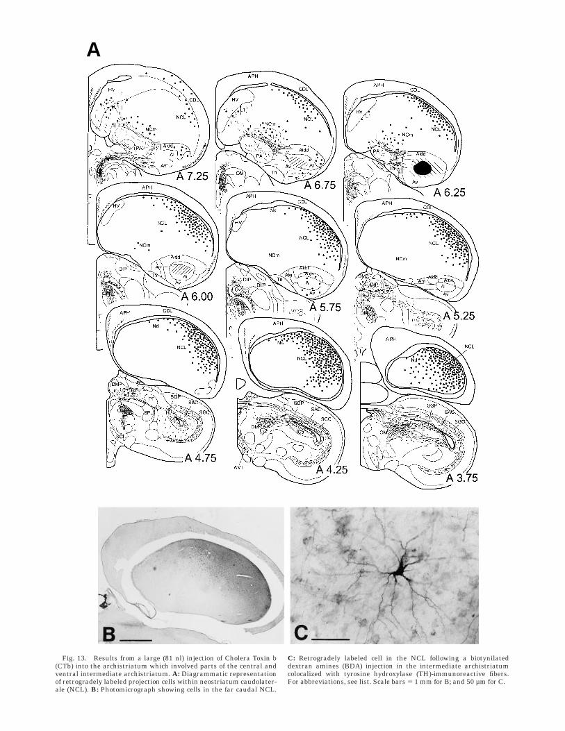
Injections into the archistriatum. One of the mostintriguing results of our retrograde labeling experimentswas the extremely high number of cells seen in the NCLthat project to the archistriatum, and the close matchbetween the distribution of these cells and the distributionof TH-immunoreactive fibers (Fig. 12). After injections ofCTb into the anterior two-thirds of the archistriatum thatinvolved the central and ventral parts of the intermediatearchistriatum, cells along the whole rostrocaudal extent ofthe NCL were labeled (Fig. 13). These cells clusteredunderneath the dorsal roof of the NCL and graduallyfanned out towards the medial neostriatum. This distribu-tion thus follows closely the original description of theNCL based on the distribution of dopaminergic and cate-cholaminergic fibers (Waldmann and Gunturkun, 1993;Wynne and Gunturkun, 1995; Metzger et al., 1996; presentstudy) and it thus seems that all parts of the NCL,including Nd, project upon the archistriatum.
According to the injection site within the archistriatum,this projection showed an anterior to posterior gradientwith more rostral archistriatal injections labeling rela-tively more cells in anterior NCL. Yet, in general, thehighest absolute number of cells was seen in the caudal-most aspect of NCL where labeled cells extend far mediallyand ventrally. There might exist a further parcellationwith respect to dorsal and ventral subdivisions of thearchistriatum as suggested from the anterograde tracingexperiments, yet with our injections which always in-volved various subdivisions of the intermediate archistria-tum, we could not confirm this.
A different pattern was seen after injections into theposterior archistriatum which is the source of the tractusoccipitomesencephalicus, pars hypothalami (HOM) andwhich together with parts of the ventromedial archistria-tum has been suggested to constitute the avian homologueof the amygdala (Zeier and Karten, 1971; Dubbeldam etal., 1997; Davies et al., 1997). In these cases, a smallnumer of retrogradely labeled cells could be observed inthe periventricular rim of the NCL and Nd (Fig. 14). Theirnumber increased towards the caudal tip of the hemi-sphere, supporting the notion that the most caudal NCLprojects to the limbic/amygdaloid part of the archistria-
CONNECTIONS OF THE AVIAN NEOSTRIATUM CAUDOLATERALE 241
tum. A very large number of cells, however, was labeled inthe ventralmost neostriatum surrounding the archistria-tum dorsally and caudally.
After injections into the central and ventral archistria-tum, a large number of retrogradely labeled cells was seenin the ventral hyperstriatum and the intermediate neostria-
Fig. 9. (A–C) Results of a 13.8-nl Cholera Toxin b (CTb) injectioninto the neostriatum frontotrigeminale overlying the n. basalis.B: Photomicrograph showing the dense anterograde labeling in therostral and lateral aspects of neostriatum caudolaterale (NCL). CDL,
corticoidea dorsolateralis. C: Detail from B showing the border be-tween NCL and the adjacent medial neostriatum. For abbreviations,see list. Scale bars 5 500 µm for B; and 100 µm for C.
242 S. KRONER AND O. GUNTURKUN
A
Fig. 10. Pattern of labeling observed after a 11.5-nl injection intothe medialmost neostriatum intermedium medialis (NIM). As shownin A, fibers travel from the injection site in a compact bundle caudallywithin the lamina hyperstriatica until they reach the medialmostneostriatum caudolaterale (NCL) from where they gradually fan out todiffusely innervate the caudalmost part of NCL. B, C: Photomicro-graphs showing the abundant distribution of anterogradely labeledfibers and retrogradely labeled cells in the caudalmost NCL, following
injections of biotinylated dextran amine (B) and Cholera Toxin b (CTb;C) into the medialmost NIM. D: Retrogradely labeled cells in theposterior thalamus, specifically in nucleus doarsomedialis posteriorthalami (DMP). DIP, nucleus dorsointermedialis posterior thalami;DLP, nucleus dorsolateralis posterior. In B and C, dorsal is to the right.For abbreviations, see list. Scale bars 5 100 µm for B, C; and 300 µmfor D.
CONNECTIONS OF THE AVIAN NEOSTRIATUM CAUDOLATERALE 243
A
Figure 11
tum. Other areas that project to the central archistriatuminclude the caudomedial neostriatum, medial HA, rostralHD and the vallecula, the field L complex as well as cellswithin NFT and NFL. Strong terminal-like labeling wasseen in LPO and medial PA as well as in HV. Descendingfibers leave the intermediate archistriatum through thetractus occipitomesencephalicus (OM), from which theyeventually split off to provide massive input to the deeplayers of the optic tectum. Terminal-like labeling wasfurthermore seen in the dorsal thalamus, predominantlyin the posterior thalamic nuclei DLP, DIP, and SRt, allthree of which also contained labeled cell bodies, as well asin the nucleus spiriformis medialis (SPm) and the nucleusintercollicularis (ICo). At midbrain level, terminal-likelabeling and retrogradely labelled neurons could be ob-served in TPc, AVT, and LoC. The OM further descended
onto various nuclei in the brainstem (see Zeier and Karten,1971; Dubbeldam et al., 1997; Davies et al., 1997).
Injections into the posterior archistriatum labeled so-mata in the mediodorsal HV and along the LFS. The Apseemed to be reciprocally connected with cells in CDL andAPH, as well as the n. accumbens. Within the archistriatalcomplex, cells in the ‘‘limbic’’ subcomponents, i.e., thenucleus taeniae and Av, were labeled. The Ap was alsofound to project heavily onto the BNST and tuberculumolfactorium. Descending fibers of the HOM terminatedmainly within the lateral hypothalamus (lHy). In thediencephalon, cell bodies were observed in SRt and alongthe whole track of the HOM. At the level of the midbrain,cells could be found within TPc, AVT, and LoC.
Injections into the paleostriatum augmentatum. In-jections into medial and central parts of the paleostriatumaugmentatum between A 7.75 and A 9.00 labeled neuronsalong the complete rostrocaudal extent of NCL and Nd. Asillustrated in Figure 15, these cells could be found through-out the whole depth of the NCL, although their numberseemed to be highest within or just medially of the tractusarchistriatalis dorsalis, at the medial border of the TH-richcaudal neostriatum. A large number of retrogradely la-beled cells was seen in the dorsal part of the archistriatum.A smaller bilateral projection furthermore seemed to origi-nate from portions of the anterior archistriatum. In addi-
Fig. 11. Results of a comparatively large (23 nl) Cholera Toxin b(CTb) injection into lateral neostriatum intermedium lmedialis (NIM)which also extended slightly into hyperstriatum ventrale (HV) asshown in A. See text for details. B: Photomicrograph of retrogradelylabeled cells in the thalamus. DLP, nucleus dorsolateralis posterior.C: Labeled fibers and cells in the dorsal neostriatum caudolaterale(NCL) at about A 6.00. CDL, corticoidea dorsolateralis. D: Detailshowing the border of the densest innervation. For abbreviations, seelist. Scale bars 5 200 µm for B; 500 µm for C; and 50 µm for D.
Fig. 12. Photomicrographs illustrating the close correspondencebetween neostriatum caudolaterale (NCL) projection neurons to thearchistriatum and the catecholaminergic innervation of the caudaltelencephalon. Cells in A were retrogradely labeled after a Cholera
Toxin b (CTb) injection into the intermediate archistriatum similar tothat shown in Figure 13. B shows an adjacent slice from the sameanimal processed for tyrosine hydroxylase (TH) immunoreactivity.Scale bar 5 1 mm.
CONNECTIONS OF THE AVIAN NEOSTRIATUM CAUDOLATERALE 245
A
Fig. 13. Results from a large (81 nl) injection of Cholera Toxin b(CTb) into the archistriatum which involved parts of the central andventral intermediate archistriatum. A: Diagrammatic representationof retrogradely labeled projection cells within neostriatum caudolater-ale (NCL). B: Photomicrograph showing cells in the far caudal NCL.
C: Retrogradely labeled cell in the NCL following a biotynilateddextran amines (BDA) injection in the intermediate archistriatumcolocalized with tyrosine hydroxylase (TH)-immunoreactive fibers.For abbreviations, see list. Scale bars 5 1 mm for B; and 50 µm for C.
tion, our injections into PA labeled cells in HV, lateral HD,and the caudal pallium (e.g., NIL, TPO, and CDL). A fewcells were also seen along the needletrack in NI, thecaudomedial HV, and the NCm. In the thalamus, cellswere labeled within the lateral ‘‘somatic’’ area of the dorsalthalamic zone, namely DIP, VIP, and to a lesser extent inDLP, SRt, n. suprarotundus (SpRt), and nuclei of the ansalenticularis. Sometimes marked neurons were also ob-served within the ventrointermediate area of the thalamus(VIA) and n. rotundus (Rt) as well as n. triangularis (T).Although labeling within VIA was most likely due to tracerspread into the dorsal pallidum, i.e., PP (Medina andReiner, 1997), labeling in Rt and T derived from tracerspread into the ectostriatum (Benowitz and Karten, 1976).At the level of the midbrain, AVT and TPc also contained anumber of labeled cells.
Injections into the medial lobus parolfactorius. La-beling within NCL after deposits of CTb into medial LPObetween A 9.50 and A 11.50 was very similar to thatobserved after PA injections. Again, retrogradely labeled
neurons were distributed diffusely along the medioventraland rostrocaudal extension of NCL, yet cells were predomi-nantly located along the medial border (Fig. 16). This wasalso true for two cases in which the injection accidentallywas centered onto lateral LPO and PA. Regarding apossible rostrocaudal topography of NCL projections to thebasal ganglia, we found a relatively larger number ofretrogradely labeled neurons projecting to the LPO incaudal NCL, whereas neurons projecting to the PA tendedto be located more rostrally and medially. However, asnoted above, these projections largely overlapped. Label-ing in other areas of the telencephalon included the NIMand HV dorsal to the injection site, medial HA and HD, andthe area prehippocampalis (APrH). Many cells were alsoseen in TPO, CDL, and up to far rostral levels within NFL,but only few in NIL. In the archistriatum, large parts ofthe Av and Ai were labeled. The projection from the Avseemed to be bilaterally organized. We found that in thethalamus, CTb injections which were centered on medialLPO yielded numerous neurons within n. dorsomedialis
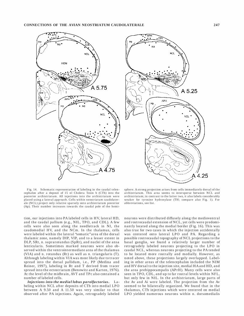
Fig. 14. Schematic representation of labeling in the caudal telen-cephalon after a deposit of 15 nl Cholera Toxin b (CTb) into theposterior archistriatum. All injections into the archistriatum wereplaced using a lateral approach. Cells within neostriatum caudolater-ale (NCL) project only relative sparsely onto archistriatum posterior(Ap). Their number increases towards the caudal pole of the hemi-
sphere. A strong projection arises from cells immediately dorsal of thearchistriatum. This area seems to intersperse between NCL andarchistriatum; in contrast to the latter two, it also labels considerablyweaker for tyrosine hydroxylase (TH; compare also Fig. 1). Forabbreviations, see list.
CONNECTIONS OF THE AVIAN NEOSTRIATUM CAUDOLATERALE 247
A
Figure 15
anterior thalami (DMA) and DMP, and to a lesser extentalso in the n. subhabenularis (SHL) and DIP. In themesencephalon, cells were found in AVT, TPc, and LoC.
DISCUSSION
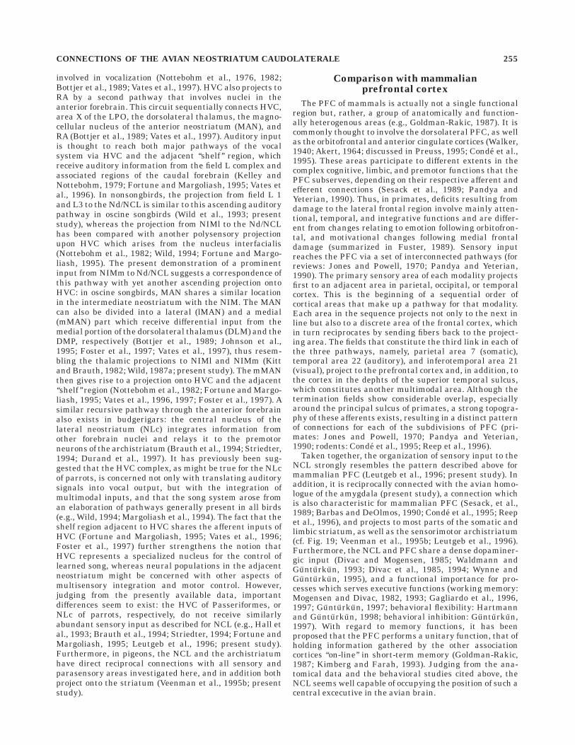
The main results of this study can be summarized asfollows: the NCL has reciprocal connections with thesecondary sensory areas of all modalities, as well with atleast two parasensory areas in the NIM and the deeplayers of HV. The afferents from these areas have diffuseand largely overlapping termination fields within NCL(see Fig. 17). These connections are restricted to the plexusof the densest innervation by catecholaminergic and pre-sumably dopaminergic fibers as demonstrated here by THimmunoreactivity. Furthermore, all parts of the so definedNCL project onto the basal ganglia and the archistriatum.The importance of these findings for sensorimotor integra-tion in the avian telencephalon and their possible rel-evance for the evolution of cortex-equivalent structureswill be discussed separately in detail below.
Sensory integration within NCL
All five secondary sensory areas were found to project toNCL. The organization of these pathways conforms with ageneral pattern of sensory processing in the avian telen-cephalon (summarized in Veenman et al., 1995b): theprimary receptive fields of subtelencephalic input relaythe information to adjacent secondary sensory structures,which in turn project to areas of the external pallium. Inthe case of the tectofugal visual pathway, the ectostriatumis the primary sensory structure (Kondo, 1933; Benowitzand Karten, 1976). From there, intratelencephalic projec-tions lead to the ectostriatal belt (Karten and Hodos, 1970;Watanabe et al., 1985). The present study demonstrates inaccordance with Leutgeb et al. (1996) that the NCL is thetertiary telencephalic component of the tectofugal com-plex. This pattern also holds for the other sensory systems.Caudal IHA and the caudolateral component of HD are theprimary sensory areas of the thalamofugal visual pathway(Karten et al., 1973), from where projections lead to HA(Shimizu et al., 1995), which by itself projects to NCL(Shimizu et al., 1995; Leutgeb et al., 1996). More rostrally,somatosensory thalamic fibers terminate in HD/hyperstria-tum intercalatus superior (HIS) and IHA (Delius andBennetto, 1972; Funke, 1989; Wild, 1997) which in turnproject to rostral HA (Wild, 1987b). Field L2 is the primarytelencephalic area of the auditory system (Wild et al.,1993), which projects to L1 and L3, from where efferentslead to Nd/NCL (Bonke et al., 1979; Wild et al., 1993;Leutgeb et al., 1996). N. basalis is the primary telence-
phalic area of the trigeminal system (Schall et al., 1986),which projects to NFT (Wild et al., 1985; Wild and Far-abaugh, 1996), from where efferents lead to NCL (Wild etal., 1985; Schall et al., 1986; Wild and Farabaugh, 1996).Thus, the NCL is the tertiary telencephalic component ofall sensory modalities examined in the present study. Dueto this multimodal input and the broad overlap of termina-tions, the NCL is a true associative forebrain structure.Additionally, parts of the caudal neostriatum may alsohave access to olfactory information: after injections intothe posterior archistriatum, we found a large number ofcells in the ventralmost neostriatum overlying the archis-triatum. This region has been suggested to be reciprocallyconnected with the avian piriform cortex and olfactorybulb, which, as their mammalian counterparts, processolfactory information (Reiner and Karten, 1985; Haberlyand Bower, 1989; Bingman et al., 1994). In concert withthe CPi and limbic archistriatum, this area might thus beconcerned with viscerolimbic functions. However, from ourcombined immunocytochemical and pathway tracing data,it seems that this part of the NC is not a genuinesubdivision of the NCL, although it might be ventrallycontinuous with it. Olfactory information can reach theNCL, nevertheless, via its connections with the HD. Inaddition to afferents from the visual dorsolateral thalamus(Karten et al., 1973; Gunturkun and Karten, 1991; Gun-turkun et al., 1993) and nonspecific input from the medio-dorsal thalamus (Bagnoli and Burkhalter, 1983), the HD isalso reciprocally connected with the hippocampal forma-tion and the CPi (Casini et al., 1986; Bingman et al., 1994;Shimizu et al., 1995).
The pattern of afferents within NCL might provide cluesto the functional architecture of the pigeon’s nervoussystem. The only two pathways which show no or littleoverlap within NCL are those from the auditory and thetrigeminal systems. Indeed, conditioning paradigms haveshown that pigeons have severe constraints to associateauditory signals with food reward in an appetitive learn-ing paradigm in which pecks on a key serve as an operant(LoLordo and Furrow, 1976; Delius and Emmerton, 1978).The same animals are rapidly able to associate in aclassical conditioning paradigm the very same auditorysignals with a mild shock applied to the body (Delius andEmmerton, 1978). Delius and Emmerton (1978) specu-lated that a granivorous animal such as a pigeon is simplynot in need of associating acoustic cues with food objectsbecause grains are mostly silent. However, grains can bequite noisy during pecking due to the direct auditoryfeedback generated by the impact of the beak on thesubstrate. Indeed, Delius (1985) could show that pigeonsare easily able to learn an auditory tone discrimination inan appetitive paradigm if the acoustic signals are gener-ated by the pecks. Yet, delaying the acoustic feedback bymore than 0.5 seconds abolishes learning. Because the n.basalis not only receives trigeminal projections but alsosome auditory afferents from the lemniscal nuclei (Deliuset al., 1979; Schall et al., 1986; Arends and Zeigler, 1986;Wild and Farabaugh, 1996), it is likely that the specializedauditory-trigeminal associations occurring during peckingare directly processed within the basalis system. Indeed,lesions of the n. basalis eliminate the ability to discrimi-nate acoustic signals generated by the animals’ own pecks(Schall and Delius, 1991). Thus, it is conceivable that theseparation of auditory and trigeminal afferents withinNCL is related to the severe constraints of pigeons inassociating acoustic and trigeminal stimuli.
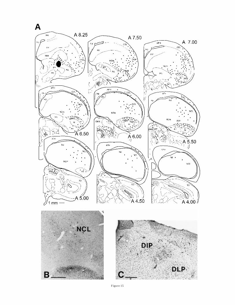
Fig. 15. Results following an injection of 28 nl Cholera Toxin b(CTb) into the ‘‘somatic’’ central paleostriatum augmentatum (PA).A: Schematic representation of retrogradely labeled cells in the caudaltelencephalon. The most abundant labeling can be seen in the dorsalpart of the archistriatum and the rostral neostriatum caudolaterale(NCL). See text for details. B: Photomicrograph illustrating thelocation of projection neurons at the medial border of the NCL andwithin the archistriatum intermedium, pars dorsodorsale (Aidd).C: Photomicrograph of labeling in the posterior dorsal thalamus. Inaddition to labeled neurons within nucleus dorsointermedialis poste-rior thalami (DIP), cells could often be observed in the ventrointerme-diate area of the posterior nuclei (VIP), nucleus dorsolateralis poste-rior (DLP), and nucleus subrotundus (SRt). For abbreviations, see list.Scale bars 5 500 µm for B; and 200 µm for C.
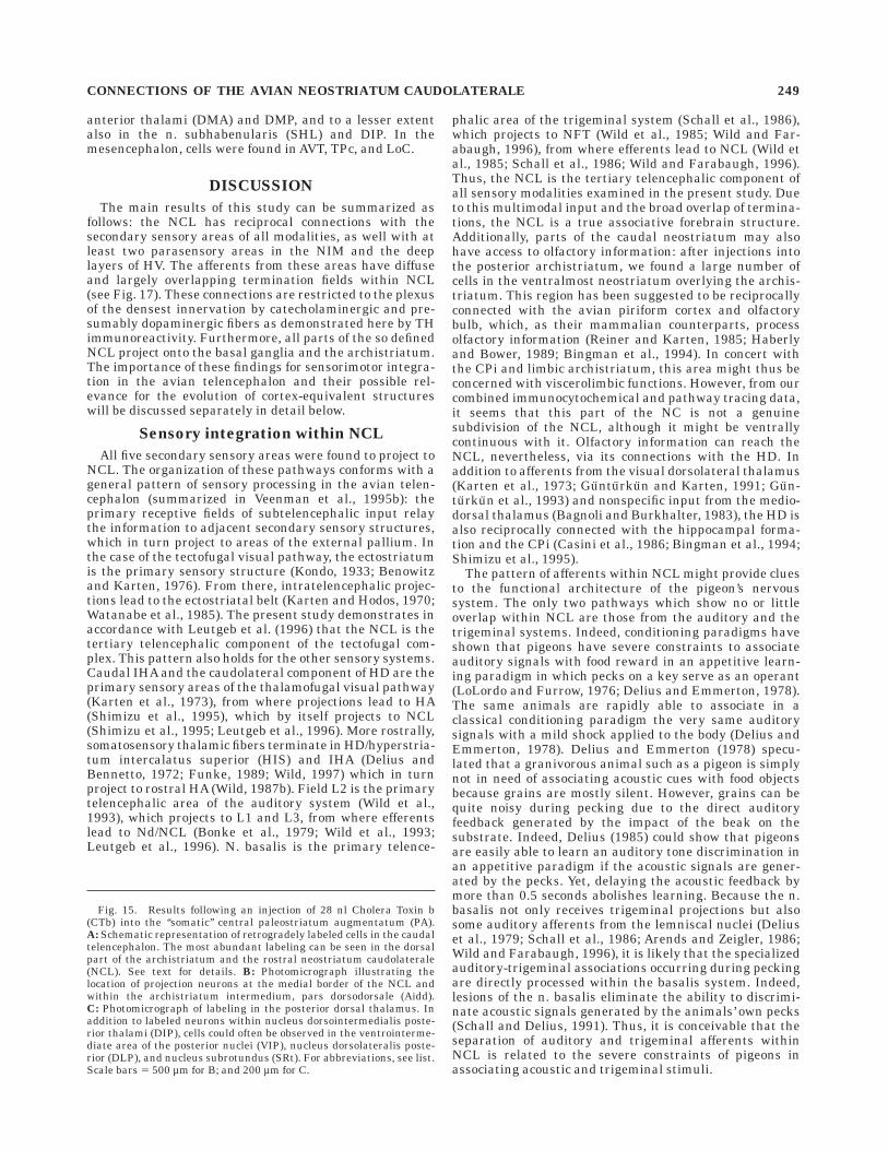
CONNECTIONS OF THE AVIAN NEOSTRIATUM CAUDOLATERALE 249
A
Figure 16
250 S. KRONER AND O. GUNTURKUN
Both the tectofugal and the thalamofugal systems projectto NCL, but their common area of termination seemslimited. This fact might be related to differences in visualfield representations between the thalamo- and the tectofu-gal pathway. The retina of pigeons has two areas ofenhanced vision (Galifret, 1968; Binggeli and Paule, 1969).One is the central fovea which looks into the monocularlateral field. The other is the so-called ‘‘red field’’ in thedorsotemporal retina with which the animal views stimuliin the inferior frontal visual field (Gunturkun, 1998). Therepresentation of the red field within the GLd of thethalamofugal pathway is extremly sparse, whereas thecentral fovea is heavily represented (Remy and Gun-turkun, 1991). Thus, the thalamofugal pathway mainlyprocesses stimuli from the lateral rather than the frontalvisual field. Accordingly, thalamofugal lesions produceminor deficits when tested with frontal stimuli, but severedeficits when tested with lateral stimuli (Gunturkun andHahmann, 1998). Contrarily, there is evidence that tectofu-gal rotundus lesions impair frontal acuity while leavinglateral acuity intact (Gunturkun and Hahmann, 1998).This in turn may be due to the fact that ventral tectal cells,which represent the lower frontal visual field, projectheavily onto rotundus, whereas the contribution of dorsaltectal cells to the tectofugal pathway is limited (Hellmannand Gunturkun, 1997). Thus, thalamo- and tectofugalvisual pathways in pigeons seem to differentially repre-sent lateral and frontal vision, respectively. Their largelycomplementary representation within NCL might thus berelated to their differential representation of the visualfield. Despite the small overlap of the tecto- and thethalamofugal visual fields within NCL, these domainsextensively overlap with the trigeminal and the auditoryprojections, respectively. The large common territory of thetectofugal visual and the trigeminal system might berelated to the specialization of the tectofugal pathway tothe lower and frontal visual field (Hellmann and Gun-turkun, 1997; Gunturkun and Hahmann, 1998), whichwithin the egocentric space overlaps with trigeminal in-puts from the beak. Thus, a common sensory focus of bothsystems could create a need for common coding whichmight be accomplished by extensive areas of terminaloverlap in an associative structure such as NCL. The largeoverlap between thalamofugal visual and auditory do-mains might be similarly interpreted. As outlined above,the thalamofugal pathway in pigeons is oriented laterally(Remy and Gunturkun, 1991; Gunturkun and Hahmann,1998). Like most other birds, pigeons fixate distant objectslaterally with their central fovea (Blough, 1971; Martinoyaet al., 1981; Bischof, 1988), because their lateral monocu-
lar acuity is about twice as high as their frontal monocularone (Hahmann and Gunturkun, 1993; Gunturkun andHahmann, 1994). Thus, the thalamofugal visual system ismore related to distant visual objects and might thereforeneed common processing with audition, which also is adistance sense.
Connections of the NCL with the NIMand ventral hyperstriatum
Our retrograde and anterograde tracing experimentshave suggested the existence of two different, albeit con-tinuous, sources of afferents from NIM to NCL. Whereasthe NIMl projection innervates the Nd region and thelaterally adjacent parts of the NCL, afferents from NIMmterminate abundantly throughout the caudalmost NCL.Both areas receive differential input from the dorsalthalamus (Kitt and Brauth, 1982; Gamlin and Cohen,1986; Wild, 1987a, 1994; Metzger et al., 1996; presentstudy) and differ in their cytoarchitectonic characteristics(Rehkamper et al., 1985). The NIMl has been shown toreceive visual and somatosensory information from telen-cephalic areas (Funke, 1989; Wild, 1987b, 1994; Shimizuet al., 1995; present study). Because we found retrogradelylabeled cells in L1 and L3 after injections into NIMl, thisstructure probably also integrates auditory information.In addition, polysensory input reaches the NIMl via DLP(Kitt and Brauth, 1982; Gamlin and Cohen, 1986; Korz-eniewska and Gunturkun, 1990; present study).
The NIMm, as defined here, resembles in its locationand connectivity the mediorostral neostriatum/hyperstria-tum ventrale (MNH) of the chick, which has been exten-sively studied in the context of imprinting (e.g., Metzger etal., 1996; Gruss and Braun, 1996; Bredenkotter andBraun, 1997; for review: Scheich, 1987). Injections intoNCL which labeled cells in NIMm also always producedcells in the overlying HV, suggesting a similar translami-nar organization of this area in the pigeon, as is character-istic for the MNH. Data on the connections of either theMNH or NIMm with telencephalic regions are very limited(Metzger et al., 1996). Our results indicate that the NIMm,like the NIMl, might also receive input from the visual andsomatosensory areas of the Wulst. Thalamic input toNIMm originates mainly in the DMP and to a much lesserextent in the DMA, a situation which is reversed for theMNH of chicks (Kitt and Brauth, 1982; Wild, 1987a;Metzger et al., 1996; present study). The sensory proper-ties of these afferents are not entirely clear, but the nucleiof the dorsomedial thalamus receive input from the hypo-thalamus and tractus solitarius (Berk and Butler, 1981;Berk and Hawkin, 1985; Arends et al., 1988; Wild et al.,1990) and in turn project to the viscerolimbic striatum andCPi (Kitt and Brauth, 1982; Wild, 1987a; Bingman et al.,1994; Veenman et al., 1995a, 1997; Metzger et al., 1996).Furthermore, the NIMm and to a lesser extent also theNCL of pigeons display strong immunoreactivity for thecalcium-binding protein parvalbumin (S. Kroner, unpub-lished observations), as has previously been shown for theMNH of chicks (Braun et al., 1991a). Parvalbumin isassociated with fast spiking gamma aminobutyric acid(GABA)ergic neurons (DeFelipe et al., 1989; Kawaguchiand Kubota, 1993), and in songbirds it is characteristic ofnuclei displaying high metabolic activity, notably all nucleiof the vocal motor system (Braun et al., 1985, 1991b). Asoutlined above, both areas of the NIM appear capable ofmultisensory processing. This draws attention to ourobservation that their afferents terminate predominantly
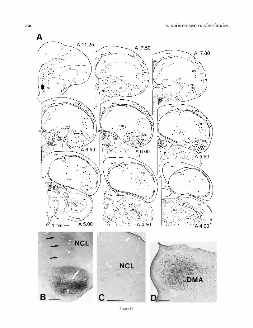
Fig. 16. Projections to the limbic medial striatum. A: Results of a22-nl injection of Cholera Toxin b (CTb) into the medial lobusparolfactorius (LPO) with no obvious tracer spread into nucleusaccumbens (AC) or lateral LPO. Labeling in the caudal telencephalonwas complementary to that observed after injections into the somaticparts of the basal ganglia. Most abundant labeling was seen in theventral part of the intermediate archistriatum and the caudal neostria-tum caudolaterale (NCL). B: Photomicrograph showing the location ofNCL projection neurons (arrows) at the medial border of the tractusarchistriatalis dorsalis as well as in architriatum intermedium, parscentrale (Ai) and archistriatum intermedium, pars ventrale (Av) in aslice double-labeled for tyrosine hydroxylase (TH). C: Retrogradelylabeled cells in the posterior neostriatum cluster at the medial extentof the NCL. D: Photomicrograph of retrogradely labeled thalamicneurons within nucleus dorsomedialis anterior thalami (DMA). Forabbreviations, see list. Scale bars 5 500 µm for B, C; 200 µm for D.
CONNECTIONS OF THE AVIAN NEOSTRIATUM CAUDOLATERALE 251
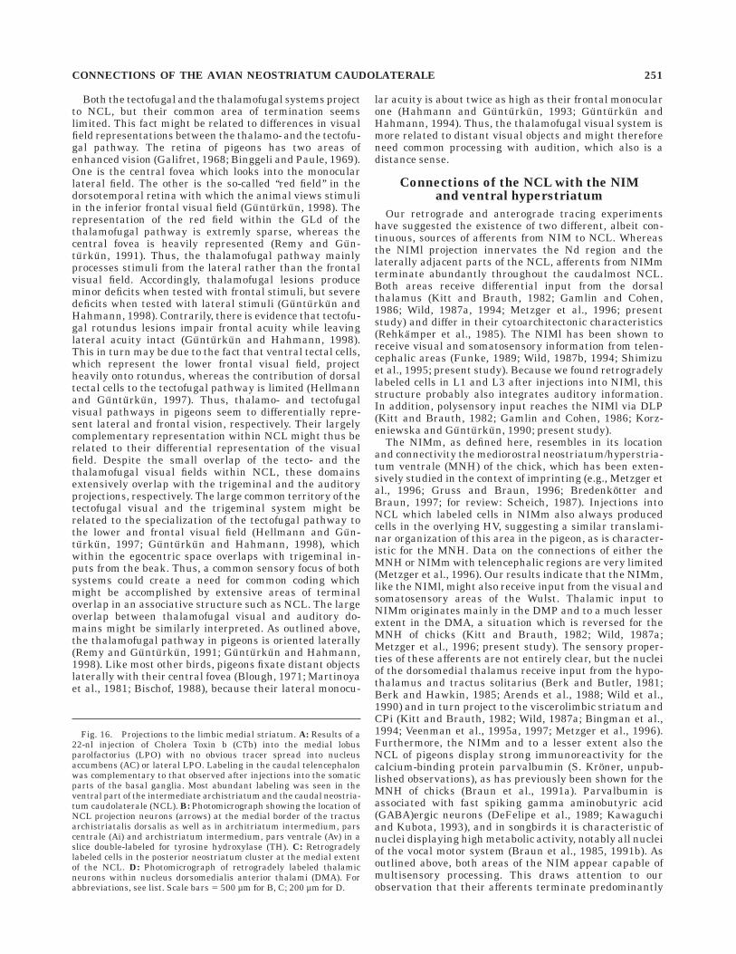
Fig. 17. Schematic representation of terminal fields within neostriatum caudolaterale (NCL) showingthe large overlap of afferents from secondary sensory areas. Note that the afferents from the presumedparasensory areas within neostriatum intermedium medialis (NIM), which occupy large parts of thecaudalmost NCL are not included in this figure. See text for details.
in those aspects of the NCL which receive relatively littleinput from secondary sensory regions (compare Fig. 17).Although it is possible that other telencephalic areaswhich send projections to NCL (e.g., HA or NFT) are alsoparasensory in nature (Schall et al., 1986; Deng and Wang,1992, 1993; Wild and Farabaugh, 1996), the nuclei withinNIM clearly are a step progressed in an assumed hierarchyof stimulus processing, as they, like the NCL, receive theirafferents from secondary sensory structures. It thus seemsthat parasensory inputs from NIM to the NCL complementthe pattern of multimodal integration.
It should be noted that our injections into NCL alsolabeled cells in the HV which, based on their location,resemble the intermediate and medial part of the hyper-striatum ventrale (IMHV), another area which in the chickhas been extensively studied in the context of imprintingand memory formation (Bradley et al., 1985; Davies et al.,1988; review by Horn, 1998). Although we did not targetthese cells in the anterograde tracing experiments, resultsfrom the study of Shimizu et al. (1995) suggest thatafferents from the HV also terminate abundantly through-out the caudalmost neostriatum (Shimizu et al., 1995).
Projections to the archistriatum
We found a continous band of cells in the NCL whichproject in a topographically ordered manner onto mostparts of the intermediate archistriatum. In addition, theposterior archistriatum receives a weak projection fromcells in the caudal- and dorsalmost aspects of the NCL. Theconnections with the intermediate archistriatum are recip-rocally organized (Leutgeb et al., 1996; Davies et al., 1997;Metzger et al., 1998; present study) and bilaterally project-ing cells in the Av provide a means of interhemisphericcomparison (Wild and Farabaugh, 1996; Metzger et al.,1998; present study). Functionally, the avian archistria-tum has generally been divided into two main subdivisions(Zeier and Karten, 1971; Davies et al., 1997; Dubbeldam etal., 1997). These are a somatic sensorimotor part which inthe pigeon comprises parts of the anterior and intermedi-ate archistriatum (Aa, Ai, Aidd, Aidv), and a viscerolimbicdivision that includes the posterior and medial archistria-tum, as well as the ventral parts of the intermediatearchistriatum (Ap, Am, Av). The sensorimotor archistria-tum receives widespread afferent projections from higherorder sensory areas of the telencephalon (e.g., Ritchie,1979; Wild et al., 1985, 1993; Shimizu et al., 1995) and isinvolved in high-level motor control (Zeier, 1971; Knudsenet al., 1995) and memory function (Knudsen and Knudsen,1996). Furthermore, via the OM, the archistriatum projectsto premotor areas of the brainstem (e.g., reticular forma-tion and lateral pontine nuclei) in all birds (Zeier andKarten, 1971; Wild et al., 1985, 1993; Davies et al., 1997;Dubbeldam et al., 1997; present study) and to some specificgroups of brainstem motoneurons involved in respirationand vocalization in songbirds (Nottebohm et al., 1976;Wild, 1993). The limbic portion of the archistriatum, on theother hand, is considered homologous to the mammalianamygdala (Zeier and Karten, 1971; Davies et al., 1997;Dubbeldam et al., 1997). It seems to be crucially involvedin such viscerolimbic functions as agonistic behavior andhomeostasis. (e.g., Cohen, 1975; Ramirez and Delius, 1979;Lowndes and Davies, 1995). The archistriatum’s func-tional segregation is reflected in the extratelencephalicprojections via the OM or HOM, respectively (Zeier andKarten, 1971; Davies et al., 1997; Dubbeldam et al., 1997),and in its connections with limbic or somatic striatum
(Veenman et al., 1995b; present study). Whereas the mainoutput from the NCL might be directed to the sensorimotorpart of the archistriatum (Leutgeb et al., 1996), especiallythe caudalmost NCL also appears to be capable of modulat-ing the amygdaloid division of the archistriatum (presentresults). The very large number of neurons from NCL thatproject to the archistriatum, and the close correspondencebetween the position of these output neurons and thedensest catecholaminergic innervation, underline the im-portance of this projection for an understanding of thefunctions of the NCL. In addition, we believe that theprojection from the caudal neostriatum to the archistria-tum might serve as a criterion for the delineation of NCL.Given the fact that injections into the archistriatumlabeled a continuous band of cells that extended from theventrolateral NCL up to Nd, we think it reasonable toconsider Nd the ‘‘auditory subcomponent’’ of NCL. Thisview is also supported by the facts that the Nd sharesafferents with the adjacent NCL from areas other than thefield L complex, shows the same dense dopaminergicinnervation, and shows a similar organization of efferentconnections other than those to the archistriatum.
Projections to the basal ganglia
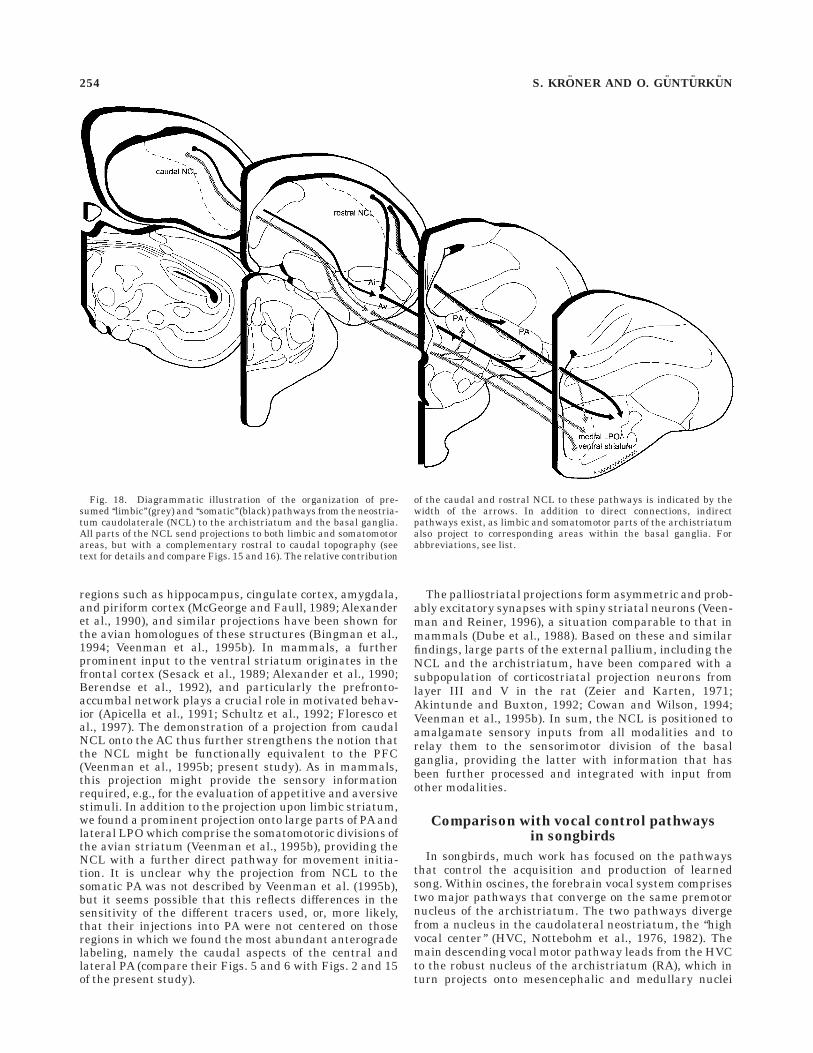
The connectivity, neurotransmitter content, and cytoar-chitecture of the avian basal ganglia are highly similar tothat in mammals (Karten and Dubbeldam, 1973; Reiner etal., 1984; Veenman and Reiner, 1994; Veenman et al.,1995b). In mammals, corticostriatal inputs from associa-tion, sensorimotor, and limbic cortices project in a segre-gated manner onto three distinct striatal regions referredto as associative, sensorimotor, and limbic striatal territo-ries (Parent, 1990; Parent and Hazrati, 1995). Thesecorticostriatal inputs provide the basal ganglia with extero-ceptive sensory information as to the location of objects inspace, interoceptive sensory information on relative bodyposition in space, and neural feedback and feedforwardinformation on ongoing and impending body movements(McGeorge and Faull, 1989; Alexander et al., 1990; Romoet al., 1992; Schultz et al., 1992; Aldridge and Berridge,1998). Based on its palliostriatal connections, a similarfunctional segregation has recently been suggested for theavian striatum (Veenman et al., 1995b): ventral striatalstructures such as the AC, the BNST, and olfactory tu-bercle constitute the ‘‘limbic’’ parts of the avian striatum.These ‘‘limbic’’ parts also include the medial LPO andlateralmost PA. The remaining dorsal striatal parts (lat-eral LPO, medial PA), on the other hand, are consideredsensorimotor in nature. We show here that all parts of theNCL project to the ventral and dorsal aspects of thestriatum. Our combined retro- and anterograde tracingexperiments suggest a rostrocaudal topography of theseprojections, with caudal parts of NCL projecting predomi-nantly onto limbic medial LPO, and more rostral parts ofNCL projecting to lateral LPO and mainly medial PA (Fig.18). The projection onto the limbic striatum had alreadybeen indicated by the study of Veenman et al. (1995b), butas their injections also involved the CPi and Ap (see theirFig. 18 and Discussion), which also project heavily onto thelimbic parts of the avian basal ganglia (Bingman et al.,1994; Veenman et al., 1995b; present study), until now theexistence of this connection from the NCL was not entirelyclear. In addition to this direct pathway, the NCL is alsocapable of influencing the ventral basal ganglia via itsconnection with the limbic archistriatum. The ventralstriatum of mammals receives input from limbic cortical
CONNECTIONS OF THE AVIAN NEOSTRIATUM CAUDOLATERALE 253
regions such as hippocampus, cingulate cortex, amygdala,and piriform cortex (McGeorge and Faull, 1989; Alexanderet al., 1990), and similar projections have been shown forthe avian homologues of these structures (Bingman et al.,1994; Veenman et al., 1995b). In mammals, a furtherprominent input to the ventral striatum originates in thefrontal cortex (Sesack et al., 1989; Alexander et al., 1990;Berendse et al., 1992), and particularly the prefronto-accumbal network plays a crucial role in motivated behav-ior (Apicella et al., 1991; Schultz et al., 1992; Floresco etal., 1997). The demonstration of a projection from caudalNCL onto the AC thus further strengthens the notion thatthe NCL might be functionally equivalent to the PFC(Veenman et al., 1995b; present study). As in mammals,this projection might provide the sensory informationrequired, e.g., for the evaluation of appetitive and aversivestimuli. In addition to the projection upon limbic striatum,we found a prominent projection onto large parts of PA andlateral LPO which comprise the somatomotoric divisions ofthe avian striatum (Veenman et al., 1995b), providing theNCL with a further direct pathway for movement initia-tion. It is unclear why the projection from NCL to thesomatic PA was not described by Veenman et al. (1995b),but it seems possible that this reflects differences in thesensitivity of the different tracers used, or, more likely,that their injections into PA were not centered on thoseregions in which we found the most abundant anterogradelabeling, namely the caudal aspects of the central andlateral PA (compare their Figs. 5 and 6 with Figs. 2 and 15of the present study).
The palliostriatal projections form asymmetric and prob-ably excitatory synapses with spiny striatal neurons (Veen-man and Reiner, 1996), a situation comparable to that inmammals (Dube et al., 1988). Based on these and similarfindings, large parts of the external pallium, including theNCL and the archistriatum, have been compared with asubpopulation of corticostriatal projection neurons fromlayer III and V in the rat (Zeier and Karten, 1971;Akintunde and Buxton, 1992; Cowan and Wilson, 1994;Veenman et al., 1995b). In sum, the NCL is positioned toamalgamate sensory inputs from all modalities and torelay them to the sensorimotor division of the basalganglia, providing the latter with information that hasbeen further processed and integrated with input fromother modalities.
Comparison with vocal control pathwaysin songbirds
In songbirds, much work has focused on the pathwaysthat control the acquisition and production of learnedsong. Within oscines, the forebrain vocal system comprisestwo major pathways that converge on the same premotornucleus of the archistriatum. The two pathways divergefrom a nucleus in the caudolateral neostriatum, the ‘‘highvocal center’’ (HVC, Nottebohm et al., 1976, 1982). Themain descending vocal motor pathway leads from the HVCto the robust nucleus of the archistriatum (RA), which inturn projects onto mesencephalic and medullary nuclei
Fig. 18. Diagrammatic illustration of the organization of pre-sumed ‘‘limbic’’ (grey) and ‘‘somatic’’ (black) pathways from the neostria-tum caudolaterale (NCL) to the archistriatum and the basal ganglia.All parts of the NCL send projections to both limbic and somatomotorareas, but with a complementary rostral to caudal topography (seetext for details and compare Figs. 15 and 16). The relative contribution
of the caudal and rostral NCL to these pathways is indicated by thewidth of the arrows. In addition to direct connections, indirectpathways exist, as limbic and somatomotor parts of the archistriatumalso project to corresponding areas within the basal ganglia. Forabbreviations, see list.
254 S. KRONER AND O. GUNTURKUN
involved in vocalization (Nottebohm et al., 1976, 1982;Bottjer et al., 1989; Vates et al., 1997). HVC also projects toRA by a second pathway that involves nuclei in theanterior forebrain. This circuit sequentially connects HVC,area X of the LPO, the dorsolateral thalamus, the magno-cellular nucleus of the anterior neostriatum (MAN), andRA (Bottjer et al., 1989; Vates et al., 1997). Auditory inputis thought to reach both major pathways of the vocalsystem via HVC and the adjacent ‘‘shelf ’’ region, whichreceive auditory information from the field L complex andassociated regions of the caudal forebrain (Kelley andNottebohm, 1979; Fortune and Margoliash, 1995; Vates etal., 1996). In nonsongbirds, the projection from field L 1and L3 to the Nd/NCL is similar to this ascending auditorypathway in oscine songbirds (Wild et al., 1993; presentstudy), whereas the projection from NIMl to the Nd/NCLhas been compared with another polysensory projectionupon HVC which arises from the nucleus interfacialis(Nottebohm et al., 1982; Wild, 1994; Fortune and Margo-liash, 1995). The present demonstration of a prominentinput from NIMm to Nd/NCL suggests a correspondence ofthis pathway with yet another ascending projection ontoHVC: in oscine songbirds, MAN shares a similar locationin the intermediate neostriatum with the NIM. The MANcan also be divided into a lateral (lMAN) and a medial(mMAN) part which receive differential input from themedial portion of the dorsolateral thalamus (DLM) and theDMP, respectively (Bottjer et al., 1989; Johnson et al.,1995; Foster et al., 1997; Vates et al., 1997), thus resem-bling the thalamic projections to NIMl and NIMm (Kittand Brauth, 1982; Wild, 1987a; present study). The mMANthen gives rise to a projection onto HVC and the adjacent‘‘shelf ’’ region (Nottebohm et al., 1982; Fortune and Margo-liash, 1995; Vates et al., 1996, 1997; Foster et al., 1997). Asimilar recursive pathway through the anterior forebrainalso exists in budgerigars: the central nucleus of thelateral neostriatum (NLc) integrates information fromother forebrain nuclei and relays it to the premotorneurons of the archistriatum (Brauth et al., 1994; Striedter,1994; Durand et al., 1997). It has previously been sug-gested that the HVC complex, as might be true for the NLcof parrots, is concerned not only with translating auditorysignals into vocal output, but with the integration ofmultimodal inputs, and that the song system arose froman elaboration of pathways generally present in all birds(e.g., Wild, 1994; Margoliash et al., 1994). The fact that theshelf region adjacent to HVC shares the afferent inputs ofHVC (Fortune and Margoliash, 1995; Vates et al., 1996;Foster et al., 1997) further strengthens the notion thatHVC represents a specialized nucleus for the control oflearned song, whereas neural populations in the adjacentneostriatum might be concerned with other aspects ofmultisensory integration and motor control. However,judging from the presently available data, importantdifferences seem to exist: the HVC of Passeriformes, orNLc of parrots, respectively, do not receive similarlyabundant sensory input as described for NCL (e.g., Hall etal., 1993; Brauth et al., 1994; Striedter, 1994; Fortune andMargoliash, 1995; Leutgeb et al., 1996; present study).Furthermore, in pigeons, the NCL and the archistriatumhave direct reciprocal connections with all sensory andparasensory areas investigated here, and in addition bothproject onto the striatum (Veenman et al., 1995b; presentstudy).
Comparison with mammalianprefrontal cortex
The PFC of mammals is actually not a single functionalregion but, rather, a group of anatomically and function-ally heterogenous areas (e.g., Goldman-Rakic, 1987). It iscommonly thought to involve the dorsolateral PFC, as wellas the orbitofrontal and anterior cingulate cortices (Walker,1940; Akert, 1964; discussed in Preuss, 1995; Conde et al.,1995). These areas participate to different extents in thecomplex cognitive, limbic, and premotor functions that thePFC subserves, depending on their respective afferent andefferent connections (Sesack et al., 1989; Pandya andYeterian, 1990). Thus, in primates, deficits resulting fromdamage to the lateral frontal region involve mainly atten-tional, temporal, and integrative functions and are differ-ent from changes relating to emotion following orbitofron-tal, and motivational changes following medial frontaldamage (summarized in Fuster, 1989). Sensory inputreaches the PFC via a set of interconnected pathways (forreviews: Jones and Powell, 1970; Pandya and Yeterian,1990). The primary sensory area of each modality projectsfirst to an adjacent area in parietal, occipital, or temporalcortex. This is the beginning of a sequential order ofcortical areas that make up a pathway for that modality.Each area in the sequence projects not only to the next inline but also to a discrete area of the frontal cortex, whichin turn reciprocates by sending fibers back to the project-ing area. The fields that constitute the third link in each ofthe three pathways, namely, parietal area 7 (somatic),temporal area 22 (auditory), and inferotemporal area 21(visual), project to the prefrontal cortex and, in addition, tothe cortex in the dephts of the superior temporal sulcus,which constitutes another multimodal area. Although thetermination fields show considerable overlap, especiallyaround the principal sulcus of primates, a strong topogra-phy of these afferents exists, resulting in a distinct patternof connections for each of the subdivisions of PFC (pri-mates: Jones and Powell, 1970; Pandya and Yeterian,1990; rodents: Conde et al., 1995; Reep et al., 1996).
Taken together, the organization of sensory input to theNCL strongly resembles the pattern described above formammalian PFC (Leutgeb et al., 1996; present study). Inaddition, it is reciprocally connected with the avian homo-logue of the amygdala (present study), a connection whichis also characteristic for mammalian PFC (Sesack, et al.,1989; Barbas and DeOlmos, 1990; Conde et al., 1995; Reepet al., 1996), and projects to most parts of the somatic andlimbic striatum, as well as the sensorimotor archistriatum(cf. Fig. 19; Veenman et al., 1995b; Leutgeb et al., 1996).Furthermore, the NCL and PFC share a dense dopaminer-gic input (Divac and Mogensen, 1985; Waldmann andGunturkun, 1993; Divac et al., 1985, 1994; Wynne andGunturkun, 1995), and a functional importance for pro-cesses which serves executive functions (working memory:Mogensen and Divac, 1982, 1993; Gagliardo et al., 1996,1997; Gunturkun, 1997; behavioral flexibility: Hartmannand Gunturkun, 1998; behavioral inhibition: Gunturkun,1997). With regard to memory functions, it has beenproposed that the PFC performs a unitary function, that ofholding information gathered by the other associationcortices ‘‘on-line’’ in short-term memory (Goldman-Rakic,1987; Kimberg and Farah, 1993). Judging from the ana-tomical data and the behavioral studies cited above, theNCL seems well capable of occupying the position of such acentral excecutive in the avian brain.
CONNECTIONS OF THE AVIAN NEOSTRIATUM CAUDOLATERALE 255
However, at least one important difference hamperscomparisons between NCL and PFC: the afferents fromthe mediodorsal (MD) nucleus of the thalamus serve as acommon hallmark for the delination of the PFC acrossmammalian species (Rose and Woolsey, 1948; Akert, 1964;Preuss, 1995). Thalamic afferents to the NCL arise mainlyfrom DLP and surrounding structures. DLP, however, isvery likely not the avian version of mammalian MD. Basedon its afferents and electrophysiological properties, DLPwas suggested to be equivalent to the posterior complex ofnuclei and especially its suprageniculate component (Kor-zeniewska, 1987; Korzeniewska and Gunturkun, 1990).Like the posterior nuclei, DLP is dominated by afferentsfrom the vestibular nuclei (mammals: Mickle and Ades,1954; birds: Wild, 1988; Korzeniewska and Gunturkun,1990), dorsal column nuclei (mammals: Feldman andKruger, 1980; Berkley et al., 1986; birds: Funke, 1989;Wild, 1989; Korzeniewska and Gunturkun, 1990), superiorcolliculus (mammals: Hicks et al., 1986; Katoh andBenedek, 1995; birds: Gamlin and Cohen, 1986; Korze-niewska and Gunturkun, 1990), and reticular formation(mammals: Hicks et al., 1986; birds: Korzeniewska andGunturkun, 1990). Amygdalar or hypothalamic afferentsto mammalian posterior thalamic nuclei (Jones, 1985) oravian DLP (Korzeniewska and Gunturkun, 1990) have notbeen reported. Contrary to this pattern, mammalian MD is
reported to receive, among others, afferents from theamygdaloid complex, diagonal band of Broca, and differenthypothalamic and preoptic structures (Siegel et al., 1977;Sapawi and Divac, 1978; Velayos and Reinoso-Suarez,1982; Irle et al., 1984; Russchen et al., 1987; Cornwall andPhillipson, 1988; Groenewegen, 1988). Based on this evi-dence, any general connectional similarity between MDand DLP could be denied. This view results from acomparison between DLP and the whole MD. If, however,DLP is compared with only lateral MD (rats: paralamellarMD portion, Cornwall and Phillipson, 1988; Groenewegen,1988; cats: intermediate and lateral MD component, Ve-layos and Reinoso-Suarez, 1982; monkeys: parvocellularand multiform lateral MD portion, Russchen et al., 1987),the picture changes. The lateral MD portion does notreceive afferents from the amygdala but is reached byinputs from vestibular nuclei, superior colliculus, andreticular formation. It thus resembles the pigeon’s DLP inmany aspects. It is the lateral portion of MD in cats andmonkeys which heavily projects onto the dorsolateralcomponent of prefrontal cortex (Markowitsch and Pritzel,1979; Giguere and Goldman-Rakic, 1988; Ray and Price,1993), which is known to be of prime importance in delaytasks. The paralamellar MD in rats is innervated by deeplayers of superior colliculus and projects to medial precen-tral cortex (Groenewegen, 1988), an area which has been
Fig. 19. Diagrammatic summary of the circuitry in which the NCLparticipates as it emerges from this study. The neostriatum caudolater-ale (NCL) is reciprocally connected with the secondary sensory of allmodalities as well as at least two parasensory areas. It projects to theavian equivalents of the amygdala (Ap, Av) and premotor cortex (Ai),
as well as limbic and somatic parts of the avian striatum, which takentogether by means of their long descending projections, exert controlover limbic and motoric centers in the diencephalon, midbrain, andbrainstem. For abbreviations, see list.
256 S. KRONER AND O. GUNTURKUN
compared with the frontal eye field in the monkey (Reep,1984), a caudal portion of prefrontal cortex (Fuster, 1989;Pandya and Yeterian, 1990). But despite these similaritiesbetween lateral MD of mammals and avian DLP, impor-tant differences also exist. For example, the pigeon’s DLPreceives a prominent innervation by the cuneatus-graciliscomplex and consequently the majority of neurons in thisstructure respond to somatosensory stimuli either as uni-,bi-, or multimodal units (Korzeniewska and Gunturkun,1990; Wild, 1994), which is clearly different from theanatomical and physiological pattern found in lateral MD.Thus, at the present state of knowledge, the initial sugges-tion of Korzeniewska and Gunturkun (1990) that the DLPis equivalent to the mammalian posterior complex ofnuclei and not to MD still seems to be the most validconcept. Nevertheless, the pigeon’s DLP might serve func-tions similar to MD, because DLP lesions were shown todisrupt working memory tasks (Gunturkun, 1997). Veen-man et al. (1997) have suggested that DLP might behomologous to the intralaminar nuclei in mammals. In ourview, this seems not very likely because the mammalianintralaminar nuclei receive extensive afferents from me-dial hypothalamus, preoptic area, lateral septum, bednucleus of the stria terminalis, ventral subiculum, perirhi-nal cortex, and amygdala (Price, 1995). Additionally, theyare characterized by a dense cholinergic input (Tohyamaand Takatsuji, 1998), and their lesion effects are wellcharacterized (Orem et al., 1973). A comparable patterndoes not exist for DLP (Korzeniewska and Gunturkun,1990; Gunturkun, 1997; Veenman et al., 1997). The sugges-tion of Miceli and Reperant (1985) that the avian SRt,which also projects onto NCL, is an intralaminar structurethus seems to be more likely, given the hodological dataprovided by these authors.
Recently, Metzger et al. (1996, 1998) suggested that theMNH of chicks might constitute the avian PFC. Thishypothesis is based on the observation that MNH ischaracterized by a medium density of dopaminergic affer-ents (Metzger et al., 1996), a high density of dopaminocep-tive neurons (Schnabel et al., 1997), and afferents from theDMA (Metzger et al., 1996). If avian DMA should provehomologous to mammalian MD (Veenman et al., 1997), thehypothesis of Metzger et al. (1996, 1998) would obviouslygain weight. Yet, as discussed above, data on the connec-tions of DMA and MNH are limited. However, the DMA isknown to project not only onto MNH, but also to theaccumbens (Veenman et al., 1997), medial LPO (Szekely etal., 1994), and piriform cortex (Bingman et al., 1994),projections which have not been shown for mammalianMD (Groenewegen, 1988; Price, 1995). Given these contra-dictions in the DMA-MD comparison and the completelack of functional data on DMA, clearly more informationis needed to clarify the comparison between avian MNHand mammalian PFC. These discussions on the organiza-tion of avian associative structures and mammalian PFCdemonstrate that simple equivalencies between avian andmammalian prefrontal systems will not hold, even if twoentities resemble each other with regard to their anatomi-cal organization and function. However, difficulties inestablishing homologous areas are not specific to compari-sons between vertebrate classes, but also apply to intra-class comparisons, as reflected in the recent discussionabout the criteria that delineate prefrontal cortex in rats(Conde et al., 1995; Preuss, 1995).
ACKNOWLEDGMENTS
Grant sponsor: Sonderforschungsbereich 509 NEURO-VISION der Deutschen Forschungsgemeinschaft to O.G.
LITERATURE CITED
Adams JC. 1981. Heavy metal intensification of DAB-based HRP reactionproduct. J Histochem Cytochem 29:775.
Akert K. 1964. Comparative anatomy of frontal cortex and thalamofrontalconnections. In: Warren JM, Akert K, editors. The frontal granularcortex and behavior. New York: McGraw-Hill. p 372–396.
Akintunde A, Buxton DF. 1992. Origins and collateralization of corticospi-nal, corticopontine, corticorubral and corticostriatal tracts: a multipleretrograde fluorescent tracing study. Brain Res 586:208–218.
Aldridge JW, Berridge KC. 1998. Coding of serial order by neostriatalneurons: a ‘‘natural action’’ approach to movement sequence. J Neurosci18:2777–2787.
Alexander GE, Crutcher MD, DeLong MR. 1990. Basal ganglia-thalamocor-tical circuits: parallel substrates for motor, oculomotor, ‘‘prefrontal’’ and‘‘limbic’’ functions. In: Uylings HBM, Van Eden CG, de Bruin JPC,Corner MA, Feenstra MGP, editors. The prefrontal cortex: its structure,function, and pathology. Prog Brain Res 85:119–146.
Apicella P, Ljungberg T, Scarnati E, Schultz W. 1991. Responses to rewardin monkey dorsal and ventral striatum. Exp Brain Res 85:491–500.
Arends JJA, Zeigler HP. 1986. Anatomical identification of auditory path-way from nucleus of lateral lemniscular system to frontal telencephalonof pigeon. Brain Res 398:375–381.
Arends JJA, Wild JM, Zeigler HP. 1988. Projections of the nucleus of thetractus solitarius in the pigeon (Columba livia). J Comp Neurol278:405–429.
Bagnoli P, Burkhalter A. 1983. Organization of the afferent projections tothe Wulst in the pigeon. J Comp Neurol 214:103–113.
Barbas H, DeOlmos J. 1990. Projections from the amygdala to basoventraland mediodorsal prefrontal regions in the rhesus monkey. J CompNeurol 300:549–571
Benowitz LI, Karten HJ. 1976. Organization of the tectofugal visualpathway in the pigeon: a retrograde transport study. J Comp Neurol167:503–520.
Berendse HW, Galis-de Graaf Y, Groenewegen HJ. 1992. Topographicalorganization and relationship with ventral striatal compartments ofprefrontal corticostriatal projections in the rat. J Comp Neurol 316:314–347.
Berk ML, Butler AB. 1981. Efferent projections of the medial preopticnucleus and medial hypothalamus in the pigeon. J Comp Neurol203:379–399.
Berk ML, Hawkin RF. 1985. Ascending projections of the mammillaryregion in the pigeon: emphasis on telencephalic connections. J CompNeurol 239:330–340.
Berkley KJ, Budell RJ, Blomquist A, Bull M. 1986. Output systems ofdorsal column nuclei in cat. Brain Res Bull 11:199–225.
Binggeli RL, Paule WJ. 1969. The pigeon retina: quantitative aspects of theoptic nerve and ganglion cell layer. J Comp Neurol 137:1–18.
Bingman VP, Casini G, Nocjar C, Jones TJ. 1994. Connections of thepiriform cortex in homing pigeons (Columba livia) studies with FastBlue and WGA-HRP. Brain Behav Evol 43:206–218.
Bischof HJ. 1988. The visual field and visually guided behavior in the zebrafinch (Taeniopygia guttata). J Comp Physiol 163:329–337.
Blough PM. 1971. The visual acuity of the pigeon for distant targets. J ExpAnal Behav 15:57–67.
Bonke BA, Bonke D, Scheich H. 1979. Connectivity in the auditoryforebrain nuclei in the Guinea fowl (Numida meleagris). Cell Tiss Res200:101–121.
Bottjer SW, Halsema KA, Brown SA, Miesner EA. 1989. Axonal connectionsof a forebrain nucleus involved with vocal learning in zebra finches. JComp Neurol 279:312–326.
Bradley P, Davies DC, Horn G. 1985. Connections of the hyperstriatumventrale of the domestic chick (Gallus domesticus). J Anat 140:577–589.
Braun K, Scheich H, Schachner M, Heizmann CW. 1985. Distribution ofparvalbumin, cytochrome oxidase activity and 14C-2-deoxyglucose up-take in the brain of the zebra finch. I. Auditory and vocal motor system.Cell Tiss Res 240:101–115.
Braun K Scheich H, Braun S, Rogers JH, Heizmann CW. 1991a. Parvalbu-min-, calretinin- and calbindin-D28k-immunoreactivity and GABA in a
CONNECTIONS OF THE AVIAN NEOSTRIATUM CAUDOLATERALE 257
forebrain region involved in auditory filial imprinting. Brain Res539:31–44.
Braun K, Scheich H, Heizmann CW, Hunziker W. 1991b. Parvalbumin andcalbindin-D28K immunoreactivity as developmental markers of audi-tory and vocal motor nuclei of the zebra finch. Neuroscience 40:853–869.
Brauth SE, Heaton JT, Durand SE, Liang W, Hall WS. 1994. Functionalanatomy of forebrain auditory pathways in the budgerigar (Melopsitta-cus undulatus). Brain Behav Evol 44:210–233.
Bredenkotter M, Braun K. 1997. Changes of neuronal responsiveness in themediorostral neostriatum/hyperstriatum after auditory filial imprint-ing in the domestic chick. Neuroscience 76:355–365.
Casini G, Bingman VP, Bagnoli P. 1986. Connections of the pigeondorsomedial forebrain studied with WGA-HRP and 3H-proline. J CompNeurol 245:454–470.
Cohen DH. 1975. Involvement of the avian amygdalar homologue (archi-striatum posterior and mediale) in defensively conditioned heart ratechange. J Comp Neurol 160:13–35.
Conde F, Maire-Lepoivre E, Audinat E, Crepel F. 1995. Afferent connectionsof the medial frontal cortex of the rat. II. Cortical and subcorticalafferents. J Comp Neurol 352:567–593.
Cornwall J, Phillipson OT. 1988. Afferent projections to the dorsal thalamusof the rat as shown by retrograde lectin transport — I. The mediodorsalnucleus. Neuroscience 24:1035–1049.
Cowan RL, Wilson CJ. 1994. Spontaneous firing patterns and axonalprojections of single corticostriatal neurons in the rat medial agranularcortex. J Neurophysiol 71:17–32.
Davies DC, Taylor DA, Johnson MH. 1988. The effects of hyperstriatallesions on one-trial passive-avoidance learning in the chick. J Neurosci8:4662–4666.
Davies DC, Csillag A, Szekely AD, Kabai, P. 1997. Efferent connections ofthe domestic chick archistriatum: a phaseolus lectin anterograde trac-ing study. J Comp Neurol 389:679–693.
DeFelipe J, Hendry SHC, Jones EG. 1989. Visualization of chandelier cellaxons by parvalbumin immunoreactivity in monkey cerebral cortex.Proc Nat Acad Sci USA 86:2093–2097.
Delius JD. 1985. The peck of the pigeon: free for all. In: Lowe CF, RichelleM, Blackman DE, Bradshaw CM, editors. Behaviour analysis andcontemporary psychology. London: Lawrence Erlbaum.
Delius JD, Bennetto K. 1972. Cutaneous sensory projections to avianforebrain. Brain Res 37:205–221.
Delius JD, Emmerton J. 1978. Stimulus-dependent asymmetry in classicaland instrumental discrimination learning by pigeons, Psychol Rec28:425–434.
Delius JD, Runge TE, Oeckinghaus H. 1979. Short-latency auditoryprojection to frontal telencephalon of pigeon. Exp Neurol 63:594–609.
Deng C, Wang B. 1992. Overlap of somatic and visual response areas in theWulst of pigeon. Brain Res 582:320–322.
Deng C, Wang B. 1993. Convergence of somatic and visual afferentimpulses in the Wulst of pigeon. Exp Brain Res 96:287–290.
Divac I, Mogensen J. 1985. The prefrontal ‘‘cortex’’ in the pigeon—catecholamine histofluorescence. Neuroscience 15:677–682.
Divac I, Mogensen J, Bjorklund A. 1985. The prefrontal ‘‘cortex’’ in thepigeon. Biochemical evidence. Brain Res 332:365–368.
Divac I, Thibault J, Skageberg G, Palacios M, Dietl MM. 1994. Dopaminer-gic innervation of the brain in pigeons. The presumed ‘‘prefrontalcortex’’. Acta Neurobiol Exp 54:227–234.
Dubbeldam JL, den Boer-Visser AM, Bout RG. 1997. Organization andefferent connections of the archistriatum of the mallard, Anas platyrhyn-chos L.: an anterograde and retrograde tracing study. J Comp Neurol388:632–657.
Dube L, Smith AD, Bolam JP. 1988. Identification of synaptic terminals ofthalamic or cortical origin in contact with distinct medium-size spinyneurons in the rat neostriatum. J Comp Neurol 267:455–471.
Durand SE, Heaton JT, Amateau SK, Brauth SE. 1997. Vocal controlpathways through the anterior forebrain of a parrot (Melopsittacusundulatus). J Comp Neurol 377:179–206.
Feldman SG, Kruger L. 1980. Axonal transport study of ascending projec-tion of medial lemniscal neurons in rat. J Comp Neurol 192:427–454.
Floresco SB, Seamans JK, Phillips AG. 1997. Selective roles for hippocam-pal, prefrontal cortical, and ventral striatal circuits in radial-arm mazetasks with or without a delay. J Neurosci 17:1880–1890.
Fortune ES, Margoliash D. 1995. Parallel pathways and convergence ontoHVc and adjacent neostriatum of adult zebra finches (Taeniopygiaguttata). J Comp Neurol 360:413–441.
Foster EF, Mehta RP, Bottjer SW. 1997. Axonal connections of the medialmagnocellular nucleus of the anterior neostriatum in zebra finches. JComp Neurol 382:364–381.
Funke K. 1989. Somatosensory areas in the telencephalon of the pigeon II:spinal pathways and afferent connections. Exp Brain Res 76:620–638.
Fuster JM. 1989. The prefrontal cortex. Anatomy, physiology, and neuropsy-chology of the frontal lobe. 2nd. ed. New York: Raven Press.
Gagliardo A, Bonadonna F, Divac I. 1996. Behavioural effects of ablations ofthe presumed ‘‘prefrontal cortex’’ or the corticoid in pigeons. BehavBrain Res 78:155–162.
Gagliardo A, Mazzotto M, Divac I. 1997. Memory of radial maze behavior inpigeons after ablations of the presumed equivalent of mammalianprefrontal cortex. Behav Neurosci 111:955–962.
Galifret Y. 1968. Les diverses aires fonctionelles de la retine di pigeon. ZZellforsch Mikrosk Anat 86:535–545.
Gamlin PDR, Cohen DH. 1986. A second ascending visual pathway from theoptic tectum to the telencephalon in the pigeon (Columba livia). J CompNeurol 250:296–310.
Giguere M, Goldman-Rakic PS. 1988. Mediodorsal nucleus: areal, laminarand tangential distribution of afferents and efferents in the frontal lobeof rhesus monkeys. J Comp Neurol 277:195–213.
Goldman-Rakic PS. 1987. Circuitry of primate prefrontal cortex and theregulation of behavior by representational memory. In: Plum F, Mount-castle VB, editors. Handbook of physiology, vol 5; higher corticalfunction. Washington, DC: American Physiology Society. p 373–417.
Groenewegen. HJ. 1988. Organization of the afferent connections of themediodorsal thalamic nucleus in the rat, related to the mediodorsal-prefrontal topography. Neuroscience 24:379–431.
Gruss M, Braun K. 1996. Stimulus-evoked increase of glutamate in themediorostral neostriatum/hyperstriatum ventrale of domestic chickafter auditory filial imprinting: an in vivo microdialysis study. JNeurochem 66:1167–1173.
Gunturkun O. 1997. Cognitive impairments after lesions of the neostria-tum caudolaterale and its thalamic afferent in pigeons: Functionalequivalencies to the mammalian prefrontal system? J. Brain Res1:133–144.
Gunturkun O. 1998. Sensory physiology: vision. In: Whittow GC, editor.Sturkie’s avian physiology. Orlando: Academic Press. p 1–19.
Gunturkun O, Hahmann U. 1994. Cerebral asymmetries and visual acuityin pigeons. Behav Brain Res 60:171–175.
Gunturkun O, Hahmann U. 1999. Functional subdivisions of the ascendingvisual pathways in the pigeon. Behav Brain Res 98:193–201.
Gunturkun O, Karten HJ. 1991. An immunocytochemical analysis of thelateral geniculate complex in the pigeon (Columba livia). J CompNeurol 314:721–749.
Gunturkun O, Miceli D, Watanabe M. 1993.Anatomy of the avian thalamofu-gal pathway. In: Zeigler HP, Bischof H-J, editors.Vision, brain andbehaviour in birds. Cambridge: MIT. p 115–135.
Haberly LB, Bower JM. 1989. Olfactory cortex: model circuit for study ofassociative memory? Trends Neurosci 12:258–264.
Hahmann U, Gunturkun O. 1993. The visual acuity for the lateral visualfield in the pigeon (Columba livia). Vision Res 33:1659–1664.
Hall WS, Cohen PL, Brauth SE. 1993. Auditory projections to the anteriortelencephalon in the budgerigar (Melopsittacus undulatus). BrainBehav Evol 41:97–116.
Hartmann B, Gunturkun O. 1998. Selective deficits in reversal learningafter neostriatum caudolaterale lesions in pigeons—possible behavioralequivalencies to the mammalina prefrontal system. Behav Brain Res96:125–133.
Hellmann B, Gunturkun O.1997. Two ’visual worlds’ in the pigeon: segre-gated space representations in the ascending visual projections. In:Elsner N, Wassle H, editors. Gottingen Neurobiology Report 1997.Stuttgart: George Thieme. p 533.
Hicks TP, Stark CA, Fletcher WA. 1986. Origins of afferents to visualsuprageniculate nucleus of the cat. J Comp Neurol 246:544–554.
Horn G. 1998. Visual imprinting and the neural mechanisms of recognitionmemory. Trends Neurosci 21:300–305.
Irle E, Markowitsch HJ, Streicher M. 1984. Cortical and subcortical,including sensory-related, afferents to the thalamic mediodorsal nucleusof the cat. J Hirnforsch 25:29–51.
Johnson F, Sablan MM, Bottjer SW. 1995. Topographic organization of aforebrain pathway involved with vocal learning in zebra finches. JComp Neurol 358:260–278.
Jones EG. 1985. The thalamus. New York: Plenum Press.Jones EG, Powell TPS. 1970. An anatomical study of converging sensory
pathways within the cerebral cortex of the monkey. Brain 93:793–820.
258 S. KRONER AND O. GUNTURKUN
Karten HJ, Dubbeldam JL. 1973. The organization and projections of thepaleostriatal complex in the pigeon (Columba livia). J Comp Neurol148:61–90.
Karten HJ, Hodos W. 1967. Stereotaxic atlas of the brain of the pigeon(Columba livia). Baltimore:John Hopkins Press.
Karten HJ, Hodos W. 1970. Telencephalic projections of the nucleusrotundus in the pigeon (Columba livia). J Comp Neurol 140:35–52.
Karten HJ, Hodos W, Nauta WHJ, Revzin AM. 1973. Neural connections ofthe ‘‘visual Wulst’’ of the avian telencephalon. Experimental studies inthe pigeon (Columba livia) and the owl (Speotyto cunicularia). J CompNeurol 150:253–277.
Katoh YY, Benedek G. 1995. Organization of the colliculo-suprageniculatepathway in the cat: a wheat germ agglutinin-horseradish peroxidasestudy. J Comp Neurol 352:381–397.
Kawaguchi Y, Kubota Y. 1993. Correlation of physiological subgroupings ofnonpyramidal cells with parvalbumin- and calbindin D28k-immunore-active neurons in layer V of rat frontal cortex. J Neurophysiol 70:387–396.
Kelley DB, Nottebohm F. 1979. Projections of a telencephalic auditorynucleus-field L-in the canary. J Comp Neurol 183:455–469.
Kimberg DY, Farah MJ. 1993. A unified account of cognitive impairmentsfollowing frontal lobe damage: the role of working memory in complex,organized behavior. J Exp Psychol Gen 122:411–428.
Kitt CA, Brauth SE. 1982. A paleostriatal-thalamic-telencephalic path inpigeons. Neuroscience 7:2735–2751.
Knudsen EI, Cohen YE, Masino T. 1995. Characterization of a forebraingaze field in the archistriatum of the barn owl: microstmulation andanatomical connections. J Neurosci 15:5139–5151.
Knudsen EI, Knudsen PF. 1996. Disruption of auditory spatial workingmemory by inactivation of the forebrain archistriatum in barn owls.Nature 383:428–431.
Kondo T. 1933. Uber die Verbindung des Sehhugels mit dem Streifenhugelbeim Huhn. OkayamaJZ 45:797–817.
Korzeniewska E. 1987. Multisensory convergence in the thalamus of thepigeon (Columba livia). Neurosci Lett 80:55–60.
Korzeniewska E, Gunturkun O. 1990. Sensory properties and afferents ofthe n. dorsolateralis posterior thalami (DLP) of the pigeon. J CompNeurol 292:457–479.
Leutgeb S, Husband S, Riters LV, Shimizu T, Bingman VP. 1996. Telence-phalic afferents to the caudolateral neostriatum of the pigeon. BrainRes 730:173–181.
LoLordo VM, Furrow DR. 1976. Control by the auditory or the visualelement of a compound stimulus: effects of feedback. J Exp Anal Behav25:251–256.
Lowndes M, Davies DC. 1995. The effect of archistriatal lesions on ‘openfield’ and fear/avoidance behaviour in the domestic chick. Behav BrainRes 722:25–32.
Margoliash D, Fortune ES, Sutter ML, Yu AC, Wren-Hardin BD, Dave A.1994. Distributed representation in the song system of oscines: evolu-tionary implications and functional consequences. Brain Behav Evol44:247–264.
Markowitsch HJ, Pritzel M. 1979. The prefrontal cortex: projection area ofthe thalamic mediodorsal nucleus? Physiol Psychol 7:1–6.
Martinoya C, Rey J, Bloch S. 1981. Limits of the pigeon’s binocular field anddirection for best binocular viewing. Vis Res 21:1197–1200.
McGeorge AJ, Faull RL. 1989. The organization of the projection from thecerebral cortex to the striatum in the rat. Neuroscience 29:503–537.
Medina L, Reiner A. 1997. The efferent projections of the dorsal and ventralpallidal parts of the pigeon basal ganglia, studied with biotinylateddextran amine. Neuroscience 81:773–802.
Medina L, Veenman CL, Reiner A. 1997. Evidence for a possible aviandorsal thalamic region comparable to the mammalian ventral anterior,ventral lateral, and oral ventroposterolateral nuclei. J Comp Neurol384:86–108.
Metzger M, Jiang S, Wang J, Braun K. 1996. Organization of the dopamin-ergic innervation of forebrain areas relevant to learning: A combinedimmunohistochemical/retrograde tracing study in the domestic chick. JComp Neurol 376:1–27.
Metzger M, Jiang S, Braun K. 1998. Organization of the dorsocaudalneostriatal complex: a retrograde and anterograde tracing study in thedomestic chick with special emphasis on pathways relevant to imprint-ing. J Comp Neurol 395:380–404.
Miceli D, Reperant J. 1985. Telencephalic afferent projections from thediencephalon and brainstem in the pigeon. A retrograde multiple-labelfluorescent study. Exp Biol 44:71–99.
Mickle WA, Ades HW. 1954. Rostral projection pathway of the vestibularsystem. Am J Physiol 176:243–246.
Mogensen J, Divac I. 1982. The prefrontal ‘‘cortex’’ in the pigeon: Behavioralevidence. Brain Behav Evol 21:60–66.
Mogensen J, Divac I. 1993. Behavioural effects of ablation of the pigeon-equivalent of the mammalian prefrontal cortex. Behav Brain Res55:101–107.
Nottebohm F, Stokes TM, Leonard CM. 1976. Central control of song in thecanary, Serinus canarius. J Comp Neurol 165:457–486.
Nottebohm F, Kelley DB, Paton JA. 1982. Connections of vocal controlnuclei in the canary telencephalon. J Comp Neurol 207:344–357.
Orem J, Schlag-Rey M, Schlag J. 1973. Unilateral visual neglect andthalamic intralaminar lesions in the cat. Exp Neurol 40:784–797.
Pandya DN, Yeterian EH. 1990. Prefrontal cortex in relation to othercortical areas in rhesus monkey: architecture and connections. In:Uylings HBM, Van Eden CG, de Bruin JPC, Corner MA, Feenstra MGP,editors. The prefrontal cortex: its structure, function, and pathology.Prog Brain Res 85:63–94.
Parent A. 1990. Extrinsic connections of the basal ganglia. Trends Neurosci13:254–258.
Parent A, Hazrati LN. 1995. Functional anatomy of the basal ganglia. I.The cortico-basal ganglia-thalamo-cortical loop. Brain Res Brain ResRev 20:91–127.
Preuss TM. 1995. Do rats have prefrontal cortex? The Rose-Woolsey-Akertprogram reconsidered. J Cogn Neurosci 7:1–24.
Price JL. 1995. Thalamus. In: Paxinos G, editor. The rat nervous system.2nd ed. San Diego: Academic Press. p 629–648.
Ramirez JM, Delius JD. 1979. Aggressive behavior of pigeons: suppressionby archistriatal lesions. Aggress Behav 5:3–17.
Ray JP, Price JI. 1993. The organization of projections from the mediodorsalnucleus of the thalamus to orbital and medial prefrontal cortex inmacaque monkeys. J Comp Neurol 337:1–31.
Reep RL. 1984. Relationship between prefrontal and limbic cortex: acomparative anatomical review. Brain Behav Evol 25:5–80.
Reep RL, Corwin JV, King V. 1996. Neuronal connections of orbital cortex inrats: topography of cortical and thalamic afferents. Exp Brain Res111:215–232.
Rehkamper G, Zilles K. 1991. Parallel evolution in mammalian and avianbrains: comparative cytoarchitectonic and cytochemical analysis. CellTiss Res 263:3–28.
Rehkamper G, Zilles K, Schleicher A. 1985. A quantitative approach tocytoarchitectonics. X. The areal pattern of the neostriatum in thedomestic pigeon, Columba livia f.d. A cyto- and myeloarchitectonicalstudy. Anat Embryol 171:345–355.
Reiner A, Karten HJ. 1985. Comparison of olfactory bulb projections inpigeons and turtles. Brain Behav Evol 27:11–27.
Reiner A, Davis BM, Brecha NC, Karten HJ. 1984. The distribution ofenkephalinlike immunoreactivity in the telencephalon of the adult anddeveloping domestic chicken. J Comp Neurol 228:245–262.
Remy M, Gunturkun O. 1991. Retinal afferents of the tectum opticum andthe nucleus opticus principalis thalami in the pigeon. J Comp Neurol305:57–70.
Ritchie TLC. 1979. Intratelencephalic visual connections and their relation-ship to the archistriatum in the pigeon (Columba livia). Ph.D. Thesis,University of Virginia, Charlotsville.
Romo R, Scarnati E, Schultz W. 1992. Role of primate basal ganglia andfrontal cortex in the internal generation of movements. II. Movement-related activity in the anterior striatum. Exp Brain Res 91:385–395.
Rose JE, Woolsey CN. 1948. The orbitofrontal cortex and ist connectionswith the mediodorsal nucleus in rabbit, sheep and cat. Res Publ AssocRes Nerv Ment Dis 27:210–232.
Russchen FT, Amaral DG, Price JL. 1987. The afferent input to themagnocellular division of the mediodorsal thalamic nucleus in themonkey, macaca fascicularis. J Comp Neurol 256:175–210.
Sapawi RR, Divac I. 1978. Connections of the mediodorsal nucleus of thethalamus in the tree shrew. I. afferent connections. Neurosci Lett7:183–189.
Schall U, Delius JD. 1991. Grasping in the pigeon: control through soundand vibration feedback mediated by the Nucleus basalis. Physiol Behav50:983–988.
Schall U, Gunturkun O, Delius JD 1986. Sensory projections to the nucleusbasalis prosencephali of the pigeon. Cell Tiss Res 245:539–546.
Scheich H. 1987. Neural correlates of auditory filial imprinting. J CompPhysiol 161:605–619.
CONNECTIONS OF THE AVIAN NEOSTRIATUM CAUDOLATERALE 259
Schnabel R, Metzger M, Jiang S, Hemmings HC, Greengard P, Braun K.1997. Localization of dopamine D1 receptors and dopaminoceptiveneurons in the chicks forebrain. J Comp Neurol 388:146–168.
Schultz W, Apicella P, Scarnati E, Ljungberg T. 1992. Neuronal activity inmonkey ventral striatum related to the expectation of reward. JNeurosci 12:4595–4610.
Sesack SR, Deutch AY, Roth RH, Bunney BS. 1989. Topographical organiza-tion of the efferent projections of the medial prefrontal cortex in the rat:an anterograde tract-tracing study with Phaseolus vulgaris Leucoagglu-tinin. J Comp Neurol 290:213–242.
Shimizu T, Cox K, Karten HJ. 1995. Intratelencephalic projections of thevisual wulst in pigeons (Columba livia). J Comp Neurol 359:551–572.
Siegel A, Fukushima T, Meibach R, Burke L, Edinger H, Weiner S. 1977.The origin of the afferent supply to the mediodorsal thalamic nucleus:enhancemet of HRP transport by selective lesions. Brain Res 135:11–23.
Striedter GF. 1994. The vocal control pathways in budgerigars differ fromthose in songbirds. J Comp Neurol 343:35–56.
Szekely AD, Boxer MI, Stewart MG, Csillag A. 1994. Connectivity of thelobus parolfactorius of the domestic chicken (Gallus domesticus): ananterograde and retrograde pathway tracing study, J. Comp Neurol348:374–393.
Tohyama M, Takatsuji K. 1998. Atlas of neuroactive substances and theirreceptors in the rat. Oxford: Oxford University Press.
Vates GE, Broome BM, Mello CV, Nottebohm F. 1996. Auditory pathways ofcaudal telencephalon and their relation to the song system of adultmale zebra finches. J Comp Neurol 366:613–642.
Vates GE, Vicario DS, Nottebohm, F. 1997. Reafferent thalamo- ‘‘cortical’’loops in the song system of oscine songbirds. J Comp Neurol 380:275–290.
Veenman CL, Reiner A. 1994. The distribution of GABA-containing peri-karya, fibers, and terminals in the forebrain and midbrain of pigeons,with particular reference to the basal ganglia and its projection targets.J Comp Neurol 339:209–250.
Veenman CL, Reiner A. 1996. Ultrastructrual morphology of synapsesformed by corticostriatal terminals in the avian striatum. Brain Res707:1–12.
Veenman CL, Karle EJ, Anderson KD, Reiner A. 1995a. Thalamostriatalprojection neurons in birds utilize LANT6 and neurotensin: a light andelectron microscopic double-labeling study. J Chem Neuroanat 9:1–16.
Veenman CL, Wild JM, Reiner, A. 1995b. Organization of the avian‘‘corticostriatal’’ projection system: A retrograde and anterograde path-way tracing study in pigeons. J Comp Neurol 354:87–126.
Veenman CL, Medina L, Reiner A. 1997. Avian homologue of mammalianintralaminar, mediodorsal and midline thalamic nuclei: immunohisto-chemical and hodological evidence. Brain Behav Evol 49:78–98.
Velayos JL, Reinoso-Suarez F. 1982. Topographic organization of thebrainstem afferents to the mediodorsal thalamic nucleus. J CompNeurol 206:17–27.
Waldmann C, Gunturkun O. 1993. The dopaminergic innervation of thepigeon caudolateral forebrain: Immunocytochemical evidence for a‘‘prefrontal cortex’’ in birds? Brain Res 600:225–234.
Walker AE. 1940. A cytoarchitectural study of the prefrontal area of themacaque monkey. J Comp Neurol 73:59–86.
Watanabe M, Ito H, Ikushima M. 1985. Cytoarchitecture and ultrastruc-ture of the avian ectostriatum: afferent terminals from the dorsaltelencephalon and some nuclei in the thalamus. J Comp Neurol236:241–257.
Wild JM. 1987a. Thalamic projections to the paleostriatum and neostria-tum in the pigeon (Columba livia). Neuroscience 20:305–327.
Wild JM. 1987b. The avian somatosensory system: connections of regions ofbody representation in the forebrain of the pigeon. Brain Res 412:205–223.
Wild JM. 1988. Vestibular projections to the thalamus of the pigeon: ananatomical study. J Comp Neurol 271:451–460.
Wild JM. 1989. Avian somatosensory system: II. ascending projections ofthe dorsal column and external cuneate nuclei in the pigeon. J CompNeurol 287:1–18.
Wild JM. 1993. Descending projections of the songbird nucleus robustusarchistriatalis. J Comp Neurol 338:225–241.
Wild JM. 1994. Visual and somatosensory inputs to the avian song systemvia nucleus uvaeformis (Uva) and a comparison with the projections of asimilar thalamic nucleus in a nonsongbird (Columba livia). J CompNeurol 349:512–535.
Wild JM. 1997. The avian somatosensory system: the pathway from wing toWulst in a passerine (Chloris Chloris). Brain Res 759:122–134.
Wild JM, Farabaugh SM. 1996. Organization of afferent and efferentprojections of the nucleus basalis prosencephali in a passerine, Taenio-pygia guttata. J Comp Neurol 365:306–328.
Wild JM., Arends JJA, Zeigler HP. 1985. Telencephalic connections of thetrigeminal system in the pigeon (Columba livia): A trigeminal sensori-motor circuit. J Comp Neurol 234:441–464.
Wild JM, Arends JJA, Zeigler HP. 1990. Projections of the parabrachialnucleus in the pigeon (Columba livia). J Comp Neurol 293:499–523.
Wild JM, Karten HJ, Frost BJ. 1993. Connections of the auditory forebrainin the pigeon (Columba livia). J Comp Neurol 337:32–62.
Wynne B, Gunturkun O. 1995. Dopaminergic innervation of the telencepha-lon of the pigeon (Columba livia): a study with antibodies againsttyrosine hydroxylase and dopamine. J Comp Neurol 57:446–464.
Zeier H. 1971. Archistriatal lesions and response inhibition in the pigeon.Brain Res 31:327–339.
Zeier H, Karten HJ. 1971. The archistriatum of the pigeon: organization ofafferent and efferent connections. Brain Res 31:313–326.
260 S. KRONER AND O. GUNTURKUN