Embed Size (px)
Citation preview
AequorinAequorinThe Blue The Blue
Fluorescent ProteinFluorescent ProteinSarah McPeakSarah McPeakBiochemistry 310Biochemistry 310Dr. Dick LuraDr. Dick LuraDecember 7, 2004December 7, 2004
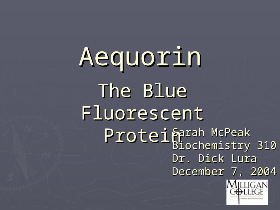
BioluminescenceBioluminescence The release of “cold light” from an The release of “cold light” from an
organism.organism.Caused by the oxidation of a Caused by the oxidation of a
substance within the organism.substance within the organism.Biological chemical reaction that Biological chemical reaction that
induces an excited state then induces an excited state then radiativly decays producing light.radiativly decays producing light. A A A*A*A + hvA + hv
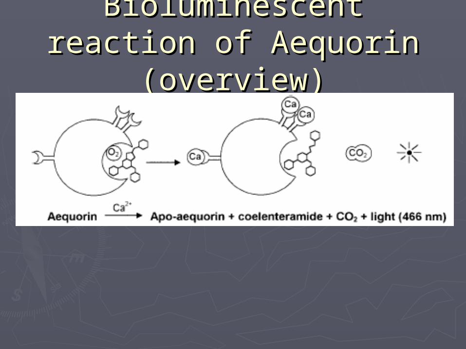
Bioluminescent reaction of Bioluminescent reaction of Aequorin (overview)Aequorin (overview)
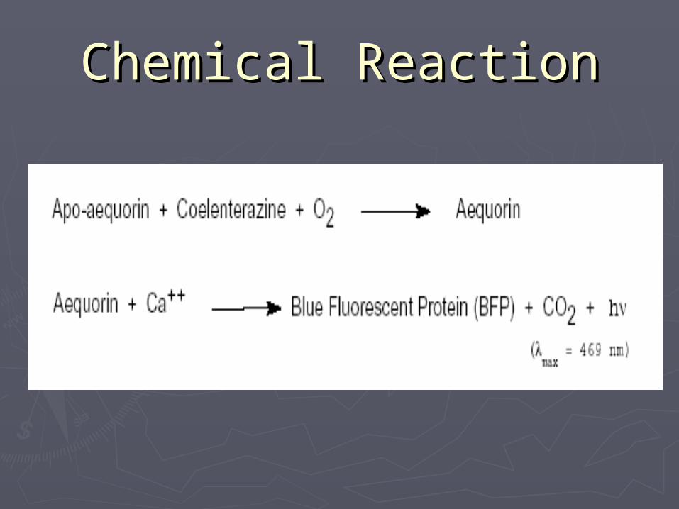
Chemical ReactionChemical Reaction

UsesUsesUsed as a marker in plants.Used as a marker in plants.
Studied for comparison to Studied for comparison to other photoproteinsother photoproteins
Used as a detector for CaUsed as a detector for Ca2+2+
Pre-crystal StructurePre-crystal Structure Used Circular Dichroism Spectra to Used Circular Dichroism Spectra to
look at portions of the secondary look at portions of the secondary structure and the effects point structure and the effects point mutations had on it.mutations had on it.
Concluded that position 71 and 84 Concluded that position 71 and 84 were likely candidate for the were likely candidate for the coelenterazine docking cite coelenterazine docking cite
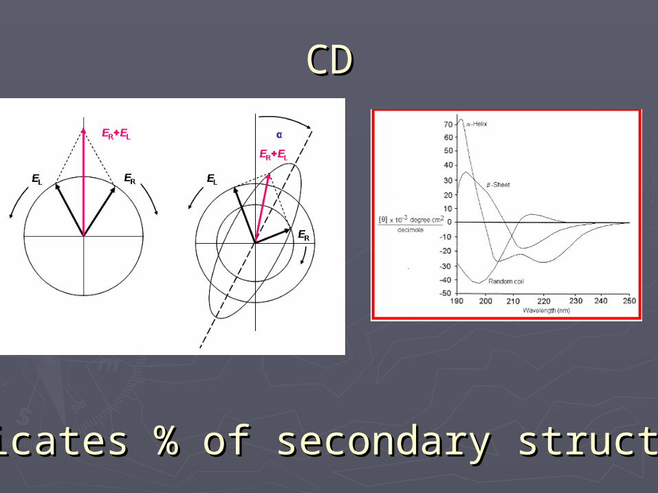
CDCD
Indicates % of secondary structuresIndicates % of secondary structures
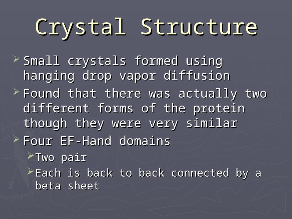
Crystal StructureCrystal Structure Small crystals formed using hanging Small crystals formed using hanging
drop vapor diffusiondrop vapor diffusion Found that there was actually two Found that there was actually two
different forms of the protein though different forms of the protein though they were very similarthey were very similar
Four EF-Hand domainsFour EF-Hand domainsTwo pairTwo pairEach is back to back connected by a beta Each is back to back connected by a beta
sheetsheet

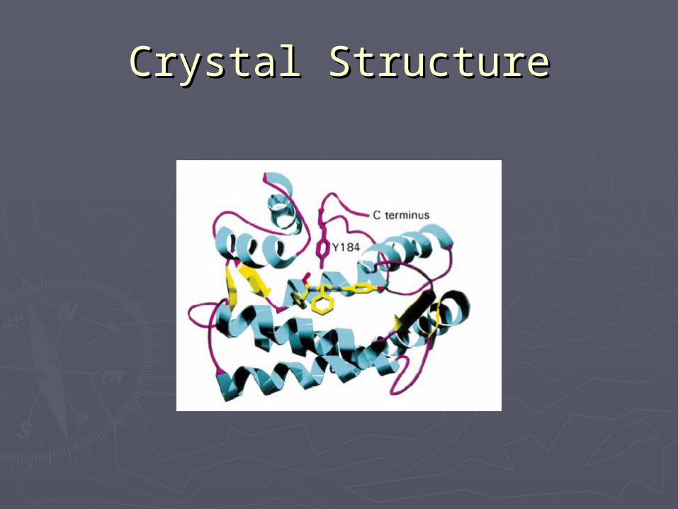
Crystal StructureCrystal Structure Cups are hydrophobicCups are hydrophobic
Coelenterazine binds thereCoelenterazine binds there Peroxide on the Coelenterazine is Peroxide on the Coelenterazine is
stabilized by hydrogen bondingstabilized by hydrogen bondingTyr 184Tyr 184
The one EF-hand that does not accept The one EF-hand that does not accept a calcium may serve as a backbonea calcium may serve as a backbone
Explanation of Rnx From Explanation of Rnx From Crystal StructureCrystal Structure
““Displacement of the helices flanking Displacement of the helices flanking the C-terminal loop would be expected the C-terminal loop would be expected to disrupt the hydrogen-bonding to disrupt the hydrogen-bonding network of the loop, resulting in a network of the loop, resulting in a relocation of the side chain of Tyr 184. relocation of the side chain of Tyr 184. This, in turn, would disrupt the hydrogen This, in turn, would disrupt the hydrogen bounds to His 169 and the peroxide. No bounds to His 169 and the peroxide. No longer stabilized, the peroxide would be longer stabilized, the peroxide would be free to attack the adjacent carbonylic C3 free to attack the adjacent carbonylic C3 to initiate the light-emitting reaction.”8to initiate the light-emitting reaction.”8
Crystal StructureCrystal Structure
PropertiesProperties Gives off Blue light (466)Gives off Blue light (466) Heat ResistantHeat Resistant
Not when coupled physically with Not when coupled physically with GFPGFP
2.1 power relationship between [Ca2.1 power relationship between [Ca2+2+] ] and luminescent intensityand luminescent intensity10 fold increase = ~100 fold 10 fold increase = ~100 fold
increaseincrease
ProductionProduction Harvested from jellyfish Harvested from jellyfish Aequorea VictoriaAequorea Victoria
2 tons of jellyfish = 125 mg protein2 tons of jellyfish = 125 mg proteinEasily contaminated and can be harmful Easily contaminated and can be harmful
to the organismsto the organisms Clone using Clone using Escherichia coliEscherichia coli
After purified Charged with After purified Charged with coelenterazinecoelenterazine
Safe to useSafe to useIn vivo for small cellsIn vivo for small cellsLarger cells can be microinjected Larger cells can be microinjected
Aequorin in NatureAequorin in Nature
The luminescence from the The luminescence from the aequorin excites GFP causing the aequorin excites GFP causing the green glow of the jellyfishgreen glow of the jellyfish
Coelenterazine is a substrate that Coelenterazine is a substrate that is needed for the rnx to workis needed for the rnx to workThis is essential part of the jellyfish’s This is essential part of the jellyfish’s
dietdietMonterey bay aquarium study (MBA)Monterey bay aquarium study (MBA)

Monterey Bay AquariumMonterey Bay Aquarium
Used jelly fish that had no diet consumption Used jelly fish that had no diet consumption of coelenterazineof coelenterazine Upon stimulation no light foundUpon stimulation no light found Fluorescence microscopy indicated GFP was Fluorescence microscopy indicated GFP was
presentpresent Small amounts of jellyfish were soaked in the Small amounts of jellyfish were soaked in the
coelenterazine produced blue glowcoelenterazine produced blue glow Allows to consume other jellyfishAllows to consume other jellyfish
GlowedGlowed Given CoelenterazineGiven Coelenterazine
GlowedGlowed

CaCa2+2+ indicator indicator Able to monitor different portions of the Able to monitor different portions of the
cell with specific mutants of the aequorincell with specific mutants of the aequorin Used quite frequently in plant studiesUsed quite frequently in plant studies
Used to indicate stress factorsUsed to indicate stress factorsCalcium ion concentration increases Calcium ion concentration increases as stresses are addedas stresses are added
Used as a marker to observe the Used as a marker to observe the tobacco mosaic virus on tobacco tobacco mosaic virus on tobacco plantsplants
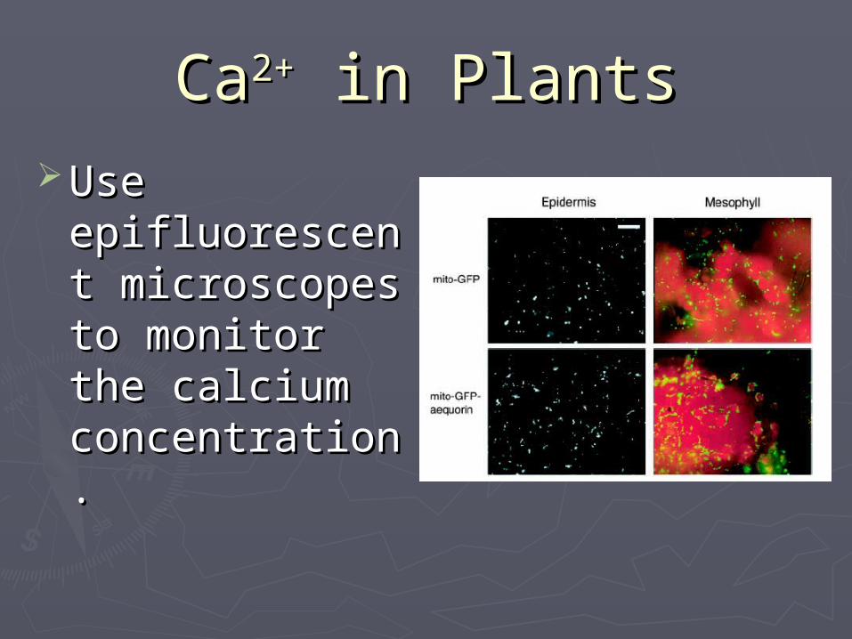
CaCa2+2+ in Plants in Plants Use Use
epifluorescent epifluorescent microscopes to microscopes to monitor the monitor the calcium calcium concentration.concentration.
ConsConsResponds Slowly to rapid Responds Slowly to rapid
change in Calciumchange in CalciumMgMg2+2+ effects luminescent out effects luminescent out
putputProduces low light outputProduces low light output
Need special instrumentationNeed special instrumentationi.e. CCD Camerai.e. CCD Camera
ProsPros Very low background because there is Very low background because there is
not excitation source beamnot excitation source beam High detection rangeHigh detection range Photo damage is not an issuePhoto damage is not an issue
Can be recharged with coelenterazineCan be recharged with coelenterazine Chance for future studies looks brightChance for future studies looks bright
Continually mutating to fit specific needsContinually mutating to fit specific needs

AcknowledgementsAcknowledgements
Michael RoachMichael Roach Dan McCauleyDan McCauley University of Virginia LibraryUniversity of Virginia Library Dr. Dick LuraDr. Dick Lura

SourcesSources► 1. 1. Aequorin: A Bioluminescent Ca2+ Indicator.Aequorin: A Bioluminescent Ca2+ Indicator. Molecular Probes. 30 Nov. 2004. Molecular Probes. 30 Nov. 2004.
<www.probes.com/handbook/print/1905.html><www.probes.com/handbook/print/1905.html>► 2. Varkova, S. V. et. al. “Obelin from the Bioluminescent Marine Hydroid 2. Varkova, S. V. et. al. “Obelin from the Bioluminescent Marine Hydroid Obelia geniculataObelia geniculata: Cloning, Expression, and : Cloning, Expression, and
Comparison of some Properties with Those of Other Ca2+ -Regulated Photoproteins.” Comparison of some Properties with Those of Other Ca2+ -Regulated Photoproteins.” BiochemistryBiochemistry. 41 (2002): 2227-2236.. 41 (2002): 2227-2236.► 3. Pichler, A., Prior, J. L., Piwnica-Worms, D. “Imaging Reversal of Multidrug Resistance in Living Mice with Bioluminescence: 3. Pichler, A., Prior, J. L., Piwnica-Worms, D. “Imaging Reversal of Multidrug Resistance in Living Mice with Bioluminescence:
MDR1 P-glycoprotein Transports Coelenterazine.” MDR1 P-glycoprotein Transports Coelenterazine.” PNSA.PNSA. 101.6 (2004) 1702-1707. 101.6 (2004) 1702-1707.► 4. Inouye, S. “Blue Fluorescent Protein from the Calcium-sensitive Photoprotein Aequorin is a Heat Resistant Enzyme, 4. Inouye, S. “Blue Fluorescent Protein from the Calcium-sensitive Photoprotein Aequorin is a Heat Resistant Enzyme,
Catalyzing the Oxidation of Coelenterazine.” Catalyzing the Oxidation of Coelenterazine.” Federation of European Biochemical Societies. Federation of European Biochemical Societies. 557 (2004): 105-110. 557 (2004): 105-110.► 5. Lu Deng, Svetlana V., et. al. “Crystal Structure of a Ca2+ -discharged Photoprotein.” 5. Lu Deng, Svetlana V., et. al. “Crystal Structure of a Ca2+ -discharged Photoprotein.” The Journal of Biological ChemistryThe Journal of Biological Chemistry. .
279.23 (2004) 33647-33652.279.23 (2004) 33647-33652.► 6. Vysotski, E. S., Lee, J. “Ca2+ -Regulated Photoproteins: Structural Insight into the Bioluminescence Mechanism. 6. Vysotski, E. S., Lee, J. “Ca2+ -Regulated Photoproteins: Structural Insight into the Bioluminescence Mechanism. Accounts Accounts
of chemical Research.of chemical Research. 37.6 (2004): 405-415. 37.6 (2004): 405-415.► 7. Creton, R., Dreiling, J. A., Jaffe, L. F. “Calcium Imaging with Chemiluminescence.” 7. Creton, R., Dreiling, J. A., Jaffe, L. F. “Calcium Imaging with Chemiluminescence.” Microscopy Research and Technique.Microscopy Research and Technique. 46 46
(1999): 390-397.(1999): 390-397.► 8. Head, J. F., Inouye, S., Teranishl, K., Shimomura, O. “The Crystal Structure of the Photoprotein aequorin at 2.3 A 8. Head, J. F., Inouye, S., Teranishl, K., Shimomura, O. “The Crystal Structure of the Photoprotein aequorin at 2.3 A
Resoulution.” Resoulution.” NatureNature. 405 (2000): 372-376.. 405 (2000): 372-376.► 9. Lewis, J. C., Lopez-Moya, J.J. Daunert, S. “Bioluminescence and Secondary Structure Properties of Aequorin Mutants 9. Lewis, J. C., Lopez-Moya, J.J. Daunert, S. “Bioluminescence and Secondary Structure Properties of Aequorin Mutants
Produced for Site-Specific Conjugation and Immobilization.” Produced for Site-Specific Conjugation and Immobilization.” Bioconjugate Chemistry. Bioconjugate Chemistry. 11 (2000): 65-70.11 (2000): 65-70.► 10. 10. Circular Dichroism Spectroscopy.Circular Dichroism Spectroscopy. Dec. 3 2004 <www.-structure.llnl.gov/cd/cdtutorial.htm> Dec. 3 2004 <www.-structure.llnl.gov/cd/cdtutorial.htm>► 11. Haddock, S. H. D., Rivers, T. J., Robinson, F. H. “Can Coelenterates Make Coelenterazine? Dietary Requirement for 11. Haddock, S. H. D., Rivers, T. J., Robinson, F. H. “Can Coelenterates Make Coelenterazine? Dietary Requirement for
Luciferin in Cnidarian Bioluminescence.” Luciferin in Cnidarian Bioluminescence.” PNSA.PNSA. 98.20 (2001): 11148-11151. 98.20 (2001): 11148-11151.► 12. Mithofer, A., Mazars, C. “Equorin-Based Measurements of Intracellular Ca2+ -Signatures in Plant Cells.” 12. Mithofer, A., Mazars, C. “Equorin-Based Measurements of Intracellular Ca2+ -Signatures in Plant Cells.” Biological Biological
Procedures Online.Procedures Online. 4.1 (2002): 105-118. <www.biologicalprocedures.com/bpo/arts/1/40/ 4.1 (2002): 105-118. <www.biologicalprocedures.com/bpo/arts/1/40/► m40.pdf>m40.pdf>► 13. Diveki, A., Salanki, K., Balazs, E. “Limited Utility of Blue Fluorescent Protein (BFP) in Monitoring Plant Virus Movement.” 13. Diveki, A., Salanki, K., Balazs, E. “Limited Utility of Blue Fluorescent Protein (BFP) in Monitoring Plant Virus Movement.”
Biochimie.Biochimie. 84 (2002): 997-1002. 84 (2002): 997-1002.► 14. Logan, D. C., Knight, M. R. “Mitochondrial and Cytosolic Calcium Dynamics are Differentially Regulated in Plants.” 14. Logan, D. C., Knight, M. R. “Mitochondrial and Cytosolic Calcium Dynamics are Differentially Regulated in Plants.” Plant Plant
Physiology.Physiology. 133 (2003): 21-24. 133 (2003): 21-24.