Embed Size (px)
Citation preview
Available online at www.pelagiaresearchlibrary.com
Pelagia Research Library
Asian Journal of Plant Science and Research, 2018, 8(1):34-39
ISSN : 2249-7412CODEN (USA): AJPSKY
Pelagia Research Library34
Adventitious Root Derived Callus Culture and Anthraquinone Production in Gynochthodes umbellata (L.) Razafim. & B. bremer (Rubiaceae)
Arun R Pillai and Gangaprasad A* Department of Botany, University of Kerala, Kariyavattom, Thiruvananthapuram, Kerala, India
ABSTRACT
The exploitation of Gynacthodes umbellata roots for anthraquinone extraction increased along with the demand for the compound and has led to the depletion of its natural habitats. As an alternative strategy for the compound production tissue culture techniques were utilized. Callus culture from in vitro derived roots of the plant using different concentrations of the auxins IAA IBA 2,4-D and NAA followed by anthraquinone extraction and its quantification was done spectrophotometrically using alizarine as a standard IAA, IBA and NAA showed promotory effect on callusing and anthraquinone production while 2,4 D expressed an inhibitory role. 2 mg/l IAA proved to be the best auxin concentration for anthraquinone production from in vitro derived callus culture of the plant. The study revealed an efficient strategy as well as the best hormone concentration for this medicinally and industrially important natural dye production from the plant which can be further utilized for the enhanced production of the compound thereby conserving the plant in its natural habitat..
Keywords: G. umbellata, Tissue culture, Callus, Anthraquinone, UV visible spectrophotometry
INTRODUCTION
It has been ages since plants have been utilized for a large number of chemicals including pharmaceuticals, agricultural chemicals, flavoring agents, fragrance, sweeteners and natural dyes. Anthraquinones are such chemicals naturally occurring in selected groups of flowering plants, bacteria, fungi, lichen and are widely used as a colouring pigment in food, drugs, hair dyes, cosmetics and textiles [1,2]. In addition anthraquinones have been reported to exhibit antibacterial, antitumor, antifungal, antituberculosis, antioxidant and immunomodulatory properties [3-6]. Also anthraquinones are an important class of dye used in dyeing textiles and are the second largest group of natural dyes used in textile industry since it seems to be environmentally safe [7]. Anthraquinones are found in flowering plant families like Verbanaceae, Bignoniacae, Scrophulariaceae and Rubiaceae. Among them, members of the Rubiaceae are the chief source of anthraquinone derivatives which accumulated in its roots, leaves and fruit of the species especially belonging to Rubioideae subfamily, for which they were shown to be a useful chemotaxonomic marker [8].G. umbellata commonly known as Neyvalli or Kudalchuruki in Malayalam is a woody climber with bright orange fruits and is distributed in all regions of Kerala. Traditionally G. umbellta has been used for treating intestinal disorders, dysentery and diarrhea, the presence of anthraquinones is responsible for its therapeutic properties [9-11]. The roots and woody stem of Gynacthodes umbellata (Syn. Morinda umbellata) belonging to the family Rubiaceae is known to contain anthraquinones and is identified as a potential source for the compound extraction in large scale which has led to overexploitation of its natural habitat [10,11].In the present investigation an alternative strategy for the production of these compounds through tissue culture method was attempted. Plant tissue and cell culture system has emerged as a safe and ecofriendly alternative system for the production and accumulation of important bioactive compounds. There are reports on the in vitro multiplication and quantification of anthraquinone from nodal explants of G. umbellate [11]. Since the roots of G. umbellata has been reported to contain a substantial source of anthraquinones, in the present investigation, in vitro derived adventitious roots of G. umbellata was used as initial explants for the enhanced production of anthraquinone. We intend to find the best hormone concentration for its large scale production from the culture. The compound was extracted using alcohol and quantified using UV visible spectrophotometry.
Pillai et al Asian J. Plant Sci. Res., 2018, 8(1):34-39
Pelagia Research Library35
MATERIALS AND METHOD
Initiation of in vitro shoot culture
Nodal segments collected from young top cutting of G. umbellata plant growing naturally in the University of Kerala, Kariyavattom campus was used for culture initiation and maintenance in Murashige and Skoog (MS) [12] medium containing 3% sucrose and 2 mg/l IBA solidified with 0.8% of agar. This is the standardized medium for the multiplication of G .umbellata developed in our laboratory. The nodal segments (4th node from the tip) collected from young branches were defoliated and washed thoroughly in running tap water using labolene (Glaxo India Ltd., Mumbai) detergent for 30 min followed by rinsed in distilled water. These explants were surface sterilized using 0.1% (w/v) HgCl2 for 6 min followed by several rinse in sterile distilled water. Nodal explants were inoculated into MS medium containing 3% sucrose and 2 mg/l BA for culture initiation and multiplication. The culture were subcultured every 4 weeks for maintain the multiplication. These cultures were maintained in a culture room at 25 ± 2°C under 12 h photoperiod at 50 µmol m-2 s-1 irradiance provided by cool white fluorescent tubes (Philips, India) and 60-65% relative humidity.
Root initiation
Individual micro shoots (3-4 cm) with three to four leaves were harvested from the culture after the second subculture last for 30 days each were harvested and transferred to ½ strength MS medium supplemented with 0.2 mg/l IAA for adventitious root initiation. This is the standardized protocol for the production of maximum number of roots [11].
Callus induction
Forty five ay old adventitious roots raised in presence of 0.2 mg/l IAA in half strength MS medium was used as the explant source for the induction of callus and quantification of anthraquinone content. The 45 day old roots from the micro shoots were harvested and washed in sterile distilled water to remove the traces of agar particle and approximately 10-15 mg of roots per culture tube were cultured on MS medium supplemented with 3% sucrose, 0.8% agar and various individual concentrations of (0.5-5 mg/l) different plant growth regulators viz. IAA, IBA, NAA and 2,4-D (Sigma Chemicals Co Bangalore, India) in culture tubes (25 × 150 mm). The pH of the medium was adjusted to 5.8 before autoclaving in an autoclave at 1.08 kg cm2 for 18 min. After 45 days, the callus raised in different media composition was harvested for taking total biomass of the callus (fresh weight and dry weight).The dried callus was further used for anthraquinone content. The cultures were incubated at 25 ± 2°C under a relative humidity of 50-60% and 12 h photoperiod at a photon flux density of 50 to 60 µEm2s from day light fluorescent tubes (Philips India Ltd., Mumbai) adjusted 30 cm high from the culture tubes.
Observation and statistical analysis
All experiments were conducted using completely randomized block design for shoot initiation, shoot multiplication, initiation of roots and initiation of callus and each treatment consist of 8 replicates and repeated thrice. Data are presented as percentage and average ± SD.
Quantification of anthraquinone in the callus
Analysis of anthraquinones was done according to the method described by Hagendoorn [13]. The fresh callus were taken and extracted twice with 80% ethanol for 45 min in a boiling water bath at 80°C (KEMI Co. Ltd., Mumbai, India). The ethanolic extracts were collected by centrifuging the tubes at 1500 rpm for 10 min and the supernatant was collected. The absorption was determined at 434 nm on UV visible spectrophotometer (UV-1700, Shimadzu, Japan) and anthraquinones was estimated using the standard graph of alizarin as pure standard (Fluka Analytical, US).
RESULTS
In vitro shooting
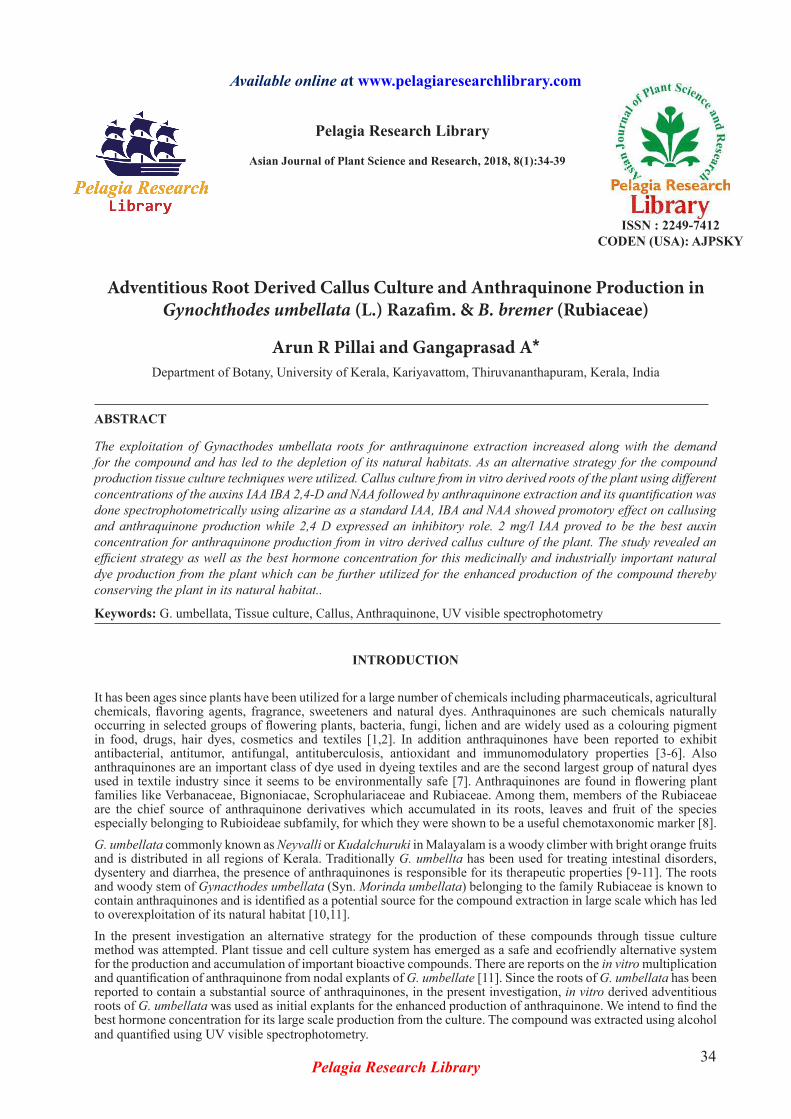
Nodal explants collected from wild growing plants were cultured on MS medium supplemented with 2 mg/l BA and 0.8% agar according to the standardized protocol developed in our lab. Culture initiation was noticed within 10 days after inoculation (Figure 1a). After 30 days, the proliferated micro shoots were obtained which is used for raising roots (Figure 1b).
Rooting
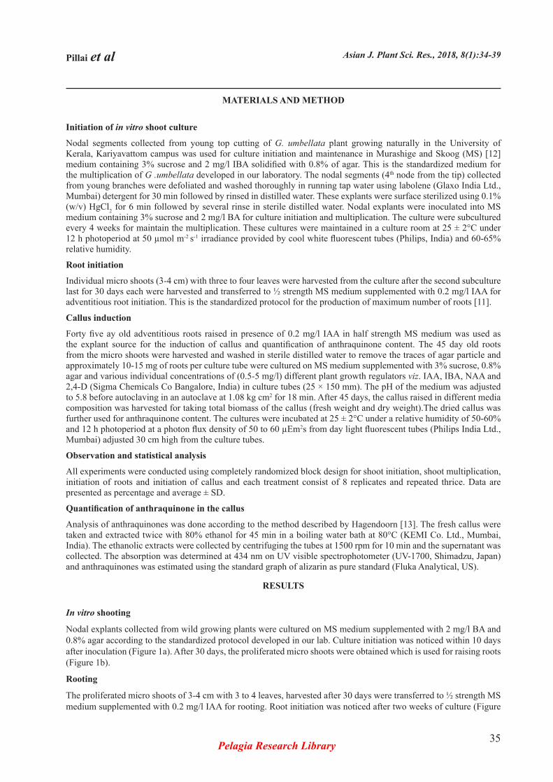
The proliferated micro shoots of 3-4 cm with 3 to 4 leaves, harvested after 30 days were transferred to ½ strength MS medium supplemented with 0.2 mg/l IAA for rooting. Root initiation was noticed after two weeks of culture (Figure
Pillai et al Asian J. Plant Sci. Res., 2018, 8(1):34-39
Pelagia Research Library36
2a) and fully developed roots were obtained after 45 days of culture. An average of 17.2 roots/shoot was obtained in half strength MS medium containing 0.2 mg/l IAA (Figure 2b).
Callusing
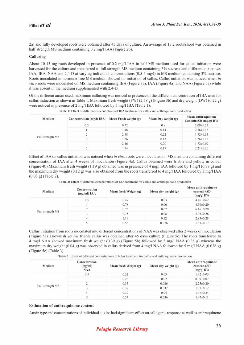
About 10-15 mg roots developed in presence of 0.2 mg/l IAA in half MS medium used for callus initiation were harvested for the culture and transferred to full strength MS medium containing 3% sucrose and different auxins viz. IAA, IBA, NAA and 2,4-D at varying individual concentrations (0.5-5 mg/l) in MS medium containing 3% sucrose. Roots inoculated in hormone free MS medium showed no initiation of callus. Callus initiation was noticed when in vitro roots were inoculated on MS medium containing IBA (Figure 3a), IAA (Figure 4a) and NAA (Figure 5a) while it was absent in the medium supplemented with 2,4-D.
Of the different auxin used, maximum callusing was noticed in presence of the different concentration of IBA used for callus induction as shown in Table 1. Maximum fresh weight (FW) (2.58 g) (Figure 3b) and dry weight (DW) (0.22 g) were noticed in presence of 2 mg/l IBA followed by 5 mg/l IBA (Table 1).
Medium Concentration (mg/l) IBA Mean Fresh weight (g) Mean Dry weight (g) Mean anthraquinone Content±SD (mg/g) DW
Full strength MS
0.5 0.72 0.8 2.09±0.231 1.48 0.14 2.56±0.182 2.58 0.22 1.72±0.333 1.26 0.13 1.26±0.134 2.16 0.20 1.72±0.095 1.74 0.17 2.21±0.20
Table 1: Effect of different concentrations of IBA treatment for callus and anthraquinone production
Effect of IAA on callus initiation was noticed when in vitro roots were inoculated on MS medium containing different concentration of IAA after 4 weeks of inoculation (Figure 4a). Callus obtained were friable and yellow in colour (Figure 4b).Maximum fresh weight (1.19 g) obtained was in presence of 4 mg/l IAA followed by 1 mg/l (0.78 g) and the maximum dry weight (0.12 g) was also obtained from the roots transferred to 4 mg/l IAA followed by 3 mg/l IAA (0.08 g) (Table 2).
Medium Concentration(mg/ml) IAA Mean fresh Weight (g) Mean dry weight (g)
Mean anthraquinone content ±SD(mg/g) DW
Full strength MS
0.5 0.47 0.03 4.46±0.621 0.78 0.06 4.50±0.202 0.73 0.07 6.16±0.793 0.75 0.08 2.95±0.284 1.19 0.12 2.85±0.285 0.53 0.076 1.83±0.17
Table 2: Effect of different concentrations of IAA treatment for callus and anthraquinone production
Callus initiation from roots inoculated into different concentrations of NAA was observed after 2 weeks of inoculation (Figure 5a). Brownish yellow friable callus was obtained after 45 days culture (Figure 5c).The roots transferred to 4 mg/l NAA showed maximum fresh weight (0.39 g) (Figure 5b) followed by 3 mg/l NAA (0.38 g) whereas the maximum dry weight (0.04 g) was observed in callus derived from 4 mg/l NAA followed by 5 mg/l NAA (0.036 g) (Figure 5c) (Table 3).
MediumConcentration
(mg/ml)NAA
Mean fresh Weight (g) Mean dry weight (g)Mean anthraquinone
content ±SD(mg/g) DW
Full strength MS
0.5 0.32 0.03 1.42±0.051 0.26 0.02 0.99±0.072 0.35 0.026 2.25±0.203 0.38 0.032 1.37±0.124 0.39 0.04 1.47±0.245 0.37 0.036 1.47±0.11
Table 3: Effect of different concentrations of NAA treatment for callus and anthraquinone production
Estimation of anthraquinone content
Auxin type and concentrations of individual auxins had significant effect on callogenic response as well as anthraquinone
Pillai et al Asian J. Plant Sci. Res., 2018, 8(1):34-39
Pelagia Research Library37
Figure 1: Culture initiation was noticed within 10 days after inoculation and 30 days
Figure 2: Root initiation was noticed after two weeks of culture and fully developed roots were obtained after 45 days of culture
Figure 4: Callus initiation was noticed when in vitro roots were inoculated on MS medium containing IAA
Figure 5: Callus initiation was noticed when in vitro roots were inoculated on MS medium containing NAA
Figure 3: Callus initiation was noticed when in vitro roots were inoculated on MS medium containing IBA
Pillai et al Asian J. Plant Sci. Res., 2018, 8(1):34-39
Pelagia Research Library38
production. The anthraquinone content in callus was estimated through UV using alizarin as the standard. Among the different auxins used for callus induction from in vitro derived roots, maximum anthraquinone content 6.16 ± 0.79 mg/g was observed in 2 mg/l IAA as shown in table 2 and minimum anthraquinone content 0.99 ± 0.07 mg/g DW was detected in 1 mg/l NAA (Table 3). Maximum anthraquinone content observed in the medium supplemented with different concentration on NAA was 2.25 ± 0.20 mg/g DW in the callus derived from 2 mg/l NAA followed by 1.47 ± 0.24 mg/g DW in the callus derived from medium supplemented with 4 mg/l NAA. Among the different concentrations of IBA tested, 1 mg/l IBA resulted in the production of maximum 2.56 ± 0.18 mg/g DW anthraquinone followed by 2.21 ± 0.20 mg/g DW anthraquinone in the presence of 5 mg/l IBA (Table 1) .
DISCUSSION
Plant growth regulators play an important role in the induction and repression of biosynthetic pathway leading to secondary metabolites. Result of the present investigation suggests that callus growth and anthraquinone synthesis is regulated in a different ways by the type and concentration of auxin in adventitious root derived callus cultures of G. umbellata. AQ synthesis is regulated in a different way by the type and concentration of auxins [14]. Callus, which is an unorganized mass of tissue, can be developed in vitro for the study of phytochemicals in the plant parts. Among the four auxins IBA, IAA, NAA and 2,4-D used for callus culture, IAA resulted in the production of maximum amount of callus from in vitro derived roots while callusing in presence of 2,4-D was absent. Maximum anthraquinone content was observed from callus derived from MS medium supplemented with 2 mg/l IAA which is a promising candidate for future studies. Earlier workers also reported the promotory effect of IAA, IBA and NAA on callusing [15]. In Morinda citrifolia, NAA, IBA promoted the production of anthraquinone while 2,4-D is least effective in adventitious derived callus and suspension culture as reported by Sreeranjini and Siril [16]. In contrary to this, in vitro leaf derived callus of Gynacthodes umbellata produced maximum yellow coloured friable callus and anthraquinone content in presence of 2,4-D [11].
CONCLUSION
The present investigation demonstrates that 2 mg/l IAA is the best auxin concentration for callus induction for maximum anthraquinone production from in vitro derived root explant of G. umbellata. The callus developed from such in vitro derived root explants can be used for the enhanced production of the industrially and medicinally important natural dye anthraquinone. This method proved to be an alternate strategy for the production of this secondary metabolite from the plant which can reduce the over exploitation of its roots from its natural habitat.
CONFLICT OF INTEREST STATEMENT
We declare that we have no conflict of interest.
ACKNOWLDGEMENT
The authors are thankful to the Head, Department of Botany, University of Kerala for providing facilities for doing the research works.
REFERENCES
[1] Brown JP, Brown RJ. Mutagenesis by 9,10-anthraquinone derivatives and related compounds in Salmonella typhimurium. Mutat Res, 1976, 40(3): 203-224.
[2] Mori H, Yoshimi N, Iwata H, Mori Y, Hara A, et al. Carcinogenicity of naturally occurring 1-hydroxyanthraquinone in rats: Induction of large bowel, liver and stomach neoplasms. Carcinogenesis, 1990, 11(5): 799-802.
[3] Koumaglo K, Gbeassor M, Nikabu O, De Souza C, Werner W. Effects of three compounds extracted from Morinda lucida on Plasmodium falciparum. Planta Medica, 1992, 58(6): 533-534.
[4] Chang P, Chen C. Isolation and characterization of antitumor anthraquinones from Morinda umbellata. Chin Pharm J, 1995, 47: 347-353.
[5] Ismail NH, Ali AM, Aimi N, Kitajima M, Takayama H, et al. Anthraquinones from Morinda elliptica. Phytochemistry, 1997, 45(8): 1723-1725.
Pillai et al Asian J. Plant Sci. Res., 2018, 8(1):34-39
Pelagia Research Library39
[6] Wang MY, West BJ, Jensen CJ, Nowicki D, Su C, et al. Morinda citrifolia (Noni): A literature review and recent advances in Noni research. Acta Pharmacol Sin, 2002, 23(12): 1127-1141.
[7] Siddiqui MF, Andleeb S, Ali N, Ghumro PB, Ahmed S. Biotreatment of anthraquinone dye Drimarene Blue K 2 RL. Afr J Environ Sci Technol, 2010, 4(1): 45-50.
[8] Young A, Boyle T, Brown T. The population genetic consequences of habitat fragmentation for plants. Trends Ecol Evol, 1996, 11(10): 413-418.
[9] Van Rheede HA. Hortus indicus malabaricus. 1688.
[10] Vijayaraghavan G. Comprehensive medicinal plants. Plant Monographs Alphabetically, 2011, 4.
[11] Anjusha S, Gangaprasad A. Callus culture and in vitro production of anthraquinone in Gynochthodes umbellata (L.) Razafim. & B. bremer (Rubiaceae). Ind Crops Prod, 2017, 95: 608-614.
[12] Classic Murashige T, Skoog F. A revised medium for rapid growth and bioassays with tobacco tissue cultures. Physiol Plant, 1962, 15: 473-497.
[13] Hagendoorn MJ, van der Plas LH, Segers GJ. Accumulation of anthraquinones in Morinda citrifolia cell suspensions. Plant Cell Tissue Organ Cult, 1994, 38(2): 227-234.
[14] Han YS, Van der Heijden R, Verpoorte R. Biosynthesis of anthraquinones in cell cultures of the Rubiaceae. Plant Cell Tissue Organ Cult, 2001, 67(3): 201-220.
[15] Gaspar T, Kevers C, Penel C, Greppin H, Reid DM, et al. Plant hormones and plant growth regulators in plant tissue culture. In Vitro Cell Dev Biol Plant, 1996, 32(4): 272-289.
[16] Sreeranjini S, Siril EA. Production of anthraquinones from adventitious root derived callus and suspension cultures of Morinda citrifolia L. in response to auxins, cytokinins and sucrose levels. Asian J Plant Sci Res, 2013, 3: 131-138.