Embed Size (px)
Citation preview

Advances in Testicular Cancer
Dr Elaine DunwoodieConsultant Medical Oncologist
Leeds Teaching Hospitals NHS Trust
Wednesday 18th September 2019
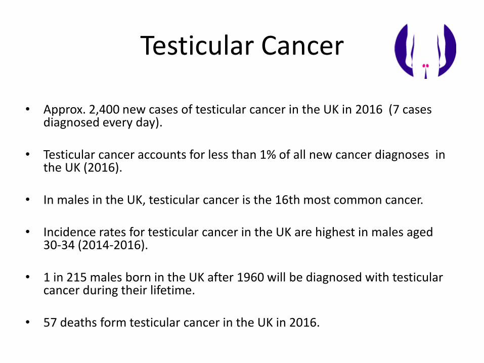
Testicular Cancer
• Approx. 2,400 new cases of testicular cancer in the UK in 2016 (7 cases diagnosed every day).
• Testicular cancer accounts for less than 1% of all new cancer diagnoses in the UK (2016).
• In males in the UK, testicular cancer is the 16th most common cancer.
• Incidence rates for testicular cancer in the UK are highest in males aged 30-34 (2014-2016).
• 1 in 215 males born in the UK after 1960 will be diagnosed with testicular cancer during their lifetime.
• 57 deaths form testicular cancer in the UK in 2016.

Testicular cancer age-standardised incidence rates, males, UK, 1993 to 2106

Testicular Cancer (C62): 1971-2011Age-Standardised Ten-Year Net Survival, England and Wales
Poor prognostic GCT remains accountable for the greatest average number of years of life lost of any adolescent or adult malignancy
Seer database, 2007

Concepts
• Confidence in management• Reduce burden of treatment in early stage disease
– TE19/TE24/BEP111
• Better understanding biology (molecular markers) & biological differences (sites/age/gender)– Immunohistochemistry of primordial germ cells, potential
targets selective inhibition– Epigenetics, e.g. decreased methylation– Age – GCNIS– Female
• 75-110 cases per year in UK, usually <20yrs old• Relapsed disease poorer outcomes than males

Concepts
• Better understanding inheritance & biological effects– No germ-line mutation identified
• familial clustering – weak predispositions, shared in utero and postnatal risk factors, coincidental somatic mutations
– Testicular dysgenesis• Hypospadias, cryptorchidism, poor semen quality, testicular
germ cell tumour
• Global co-operation in rarer presentations, poor prognosis, salvage.

Current Management
• ESMO Consensus Conference November 2016, published Annals Oncology 2018.
• Current practice
• Recent studies
• Areas of active interest
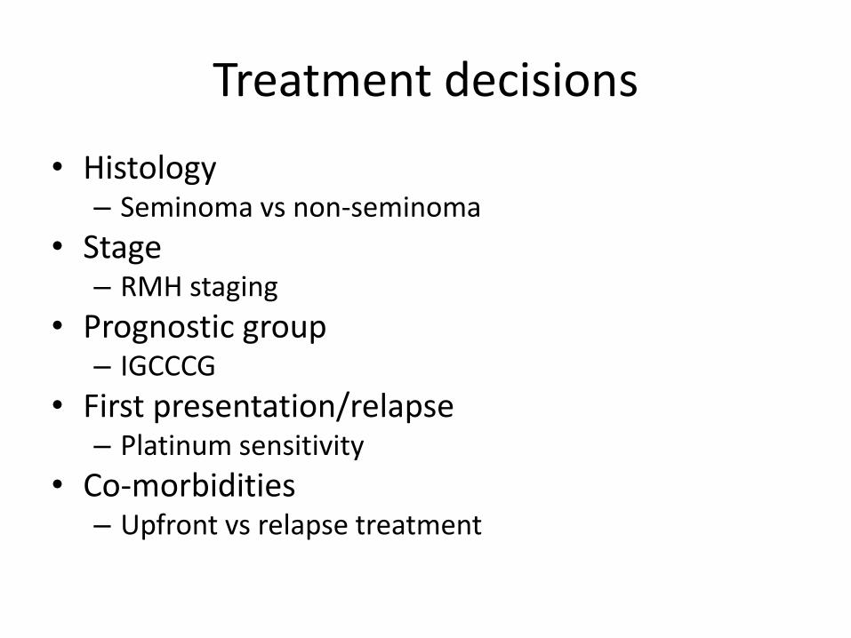
Treatment decisions
• Histology– Seminoma vs non-seminoma
• Stage– RMH staging
• Prognostic group– IGCCCG
• First presentation/relapse– Platinum sensitivity
• Co-morbidities– Upfront vs relapse treatment

British TTP&R WHO
Seminoma
Germ cell neoplasia in situ
Seminoma
Spermatocytic seminoma Spermatocytic tumour
Non-seminomatous GCT
Malignant teratoma differentiated
Malignant teratoma intermediate
Malignant teratoma undifferentiated
Yolk sac tumour
Malignant teratoma trophoblastic
Teratoma
Teratoma
Embryonal carcinoma
Yolk sac tumour
Choriocarcinoma
Histology – WHO update 2016
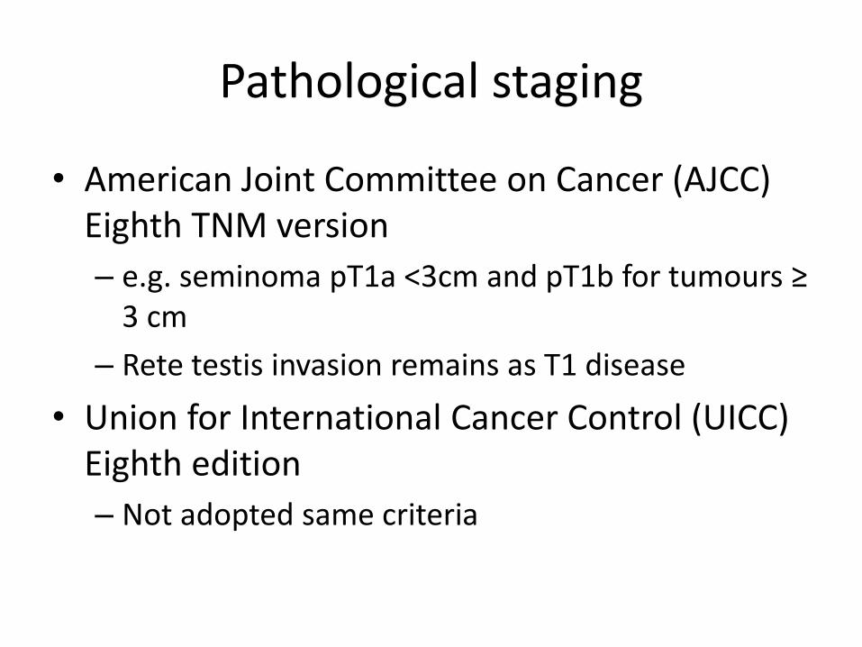
Pathological staging
• American Joint Committee on Cancer (AJCC) Eighth TNM version
– e.g. seminoma pT1a <3cm and pT1b for tumours ≥ 3 cm
– Rete testis invasion remains as T1 disease
• Union for International Cancer Control (UICC) Eighth edition
– Not adopted same criteria
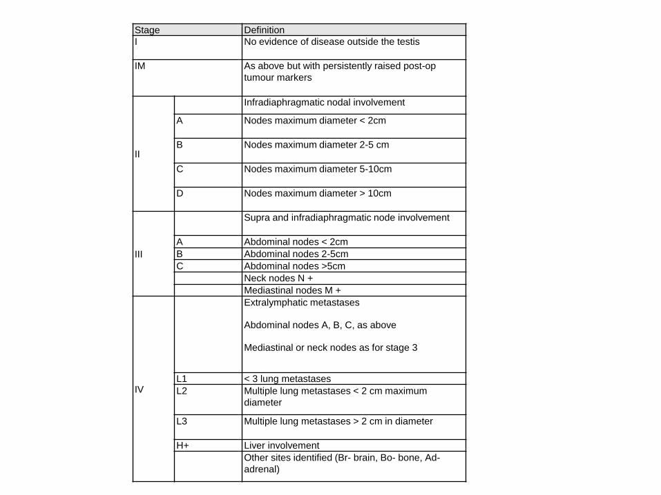
Stage Definition
I No evidence of disease outside the testis
IM As above but with persistently raised post-op
tumour markers
II
Infradiaphragmatic nodal involvement
A Nodes maximum diameter < 2cm
B Nodes maximum diameter 2-5 cm
C Nodes maximum diameter 5-10cm
D Nodes maximum diameter > 10cm
III
Supra and infradiaphragmatic node involvement
A Abdominal nodes < 2cm
B Abdominal nodes 2-5cm
C Abdominal nodes >5cm
Neck nodes N +
Mediastinal nodes M +
IV
Extralymphatic metastases
Abdominal nodes A, B, C, as above
Mediastinal or neck nodes as for stage 3
L1 < 3 lung metastases
L2 Multiple lung metastases < 2 cm maximum
diameter
L3 Multiple lung metastases > 2 cm in diameter
H+ Liver involvement
Other sites identified (Br- brain, Bo- bone, Ad-
adrenal)
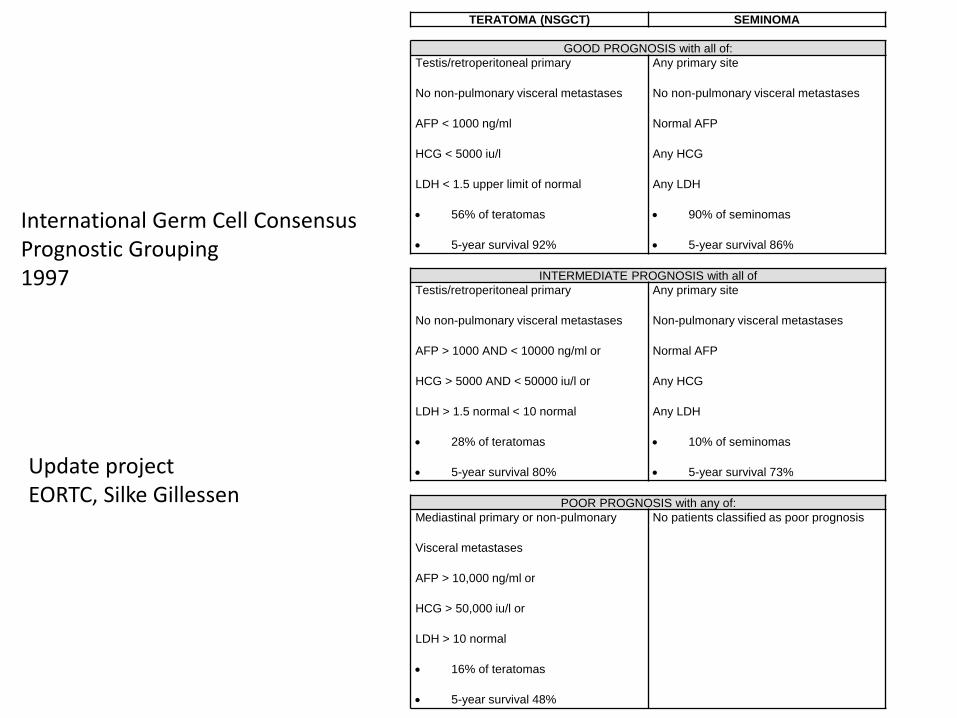
TERATOMA (NSGCT) SEMINOMA
GOOD PROGNOSIS with all of:
Testis/retroperitoneal primary
No non-pulmonary visceral metastases
AFP < 1000 ng/ml
HCG < 5000 iu/l
LDH < 1.5 upper limit of normal
56% of teratomas
5-year survival 92%
Any primary site
No non-pulmonary visceral metastases
Normal AFP
Any HCG
Any LDH
90% of seminomas
5-year survival 86%
INTERMEDIATE PROGNOSIS with all of
Testis/retroperitoneal primary
No non-pulmonary visceral metastases
AFP > 1000 AND < 10000 ng/ml or
HCG > 5000 AND < 50000 iu/l or
LDH > 1.5 normal < 10 normal
28% of teratomas
5-year survival 80%
Any primary site
Non-pulmonary visceral metastases
Normal AFP
Any HCG
Any LDH
10% of seminomas
5-year survival 73%
POOR PROGNOSIS with any of:
Mediastinal primary or non-pulmonary
Visceral metastases
AFP > 10,000 ng/ml or
HCG > 50,000 iu/l or
LDH > 10 normal
16% of teratomas
5-year survival 48%
No patients classified as poor prognosis
International Germ Cell ConsensusPrognostic Grouping1997
Update projectEORTC, Silke Gillessen
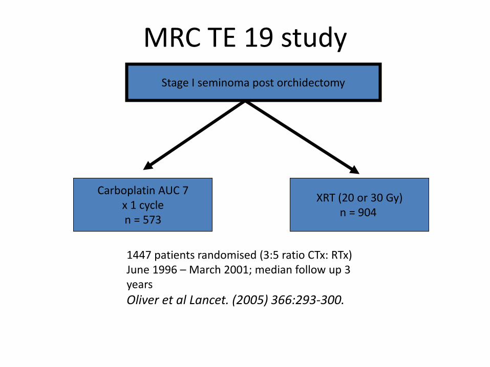
MRC TE 19 study
Stage I seminoma post orchidectomy
Carboplatin AUC 7x 1 cyclen = 573
XRT (20 or 30 Gy)n = 904
1447 patients randomised (3:5 ratio CTx: RTx) June 1996 – March 2001; median follow up 3 years
Oliver et al Lancet. (2005) 366:293-300.

TE 19RFS
rates
RTx
(n=904)
Chemotherapy
(n=573)
2 yrs 96.7 % 97.7 %
3 yrs 95.9 % 94.8 %
Relapses RTx Chemotherapy
Number 32 27
Pattern Pelvis, neck,
mediastinum
abdominal
nodes

TE 19 - Long Term Toxicity
New primary cancers
• Chemotherapy – 2 GCT, 3 other
• RTx – 10 GCT, 4 other
Fertility
• Effect of carboplatin unknown in this setting

TE24
• Stage 1 seminoma, randomised• Aim
– Observe potential to reduce radiation exposure during surveillance without adversely affecting outcome
• Rationale– Unknown risk of single cycle carboplatin AUC 7– Mx stratified (Warde criteria)– RMH surveillance 7 CT scans over 5 years (8-20
elsewhere)• Risk of secondary malignancy from single chest/abdo/pelvis
CT is 1 in 2000, calculated at 1 in 300 if 7 CTsBerrington de Gonzalez & Darby, Lancet 2004
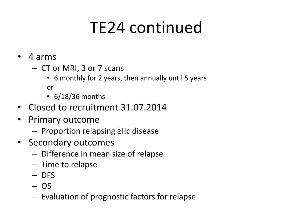
TE24 continued
• 4 arms– CT or MRI, 3 or 7 scans
• 6 monthly for 2 years, then annually until 5 yearsor• 6/18/36 months
• Closed to recruitment 31.07.2014• Primary outcome
– Proportion relapsing ≥IIc disease
• Secondary outcomes– Difference in mean size of relapse– Time to relapse– DFS– OS– Evaluation of prognostic factors for relapse
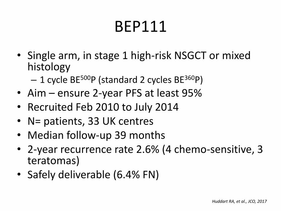
BEP111
• Single arm, in stage 1 high-risk NSGCT or mixed histology– 1 cycle BE500P (standard 2 cycles BE360P)
• Aim – ensure 2-year PFS at least 95%• Recruited Feb 2010 to July 2014• N= patients, 33 UK centres• Median follow-up 39 months• 2-year recurrence rate 2.6% (4 chemo-sensitive, 3
teratomas)• Safely deliverable (6.4% FN)
Huddart RA, et al., JCO, 2017

Relapses after adjuvant BEP stage 1 NSGCT
• Retrospective analysis, global cancer group• N = 51, 18 centres, 11 countries• 5-year PFS 64%, OS 78%• Outcomes worse c/f chemo naïve patients with metastatic
disease, better than patients relapsed after chemo for metastatic disease.
• Late relapses:– 29% relapsed > 3 years after adjuvant treatment– Latest relapse >25 years– Assoc with statistically significant increased risk death GC cancer– Subsequent relapses – 29% after 1st relapse
• Diversity in treatment strategy– Consider 3-4# 3 drug regimen, exchange bleomycin for
ifosfmaide
Stefanie Fischer, ASCO GU Feb 2019

Accelerated BEP
• Previous strategies to improve 1st line chemo tested in phase II and III trials have included;– alternative or additional chemo drugs– more complex multi-drug regimens– high dose chemo with stem cell support– Examples;
• Induction C-BOP/BEP (2005, 2011), GAMEC (2007), high dose 1st line (2007), BOP/VIP-B (1998), paclitaxel BEP (2012), POM B/ACE (1997), cis/cyclo/dox (2008)
• None improved cure rates, all more toxic than BEP
• 2014 phase II, ANZUP (n=45), UK trial n=16 – 1st line int/poor prog
• Strategy successful other cancer sites (OS in NHL x2, early breast cancer, SCLC x2)– E.g. DLBCL CHOP 3-weekly since 1970s, accelerated to 2-weekly,
addition of rituximab

UK P3BEP
• Aligned with ANZUP P3BEP
• 20 UK sites, target recruitment 150
• phase III for pts with intermediate/poor prognosis GCT of ovary/testis/retroperitoneum/mediastinum
• 1:1 randomisation, standard 3-weekly vs 2-weekly BEP
• Primary aim – PFS
• Secondary aims – OS, delivery dose intensity, treatment preference, QoL, adverse events.

TIGER trial• Optimising outcome of salvage treatment in relapsed or refractory germ
cell tumours.• Patients with relapsed GCTs
– 30% relapse, 20% ultimately die of GCT– Standard treatment
• VIP – CR 50%, long-term PFS 25% (unselected patients)*• TIP – n=46, phase II, selected patients, CR 70%, PFS 73% at 7 years**
• 1st line salvage high-dose chemo regimens– Durable remission in 30-60%– Sequential vs single high dose chemo – trial closed due to unacceptable
toxicity in single high dose arm***– Only 1 RCT c/f conventional chemo (VIP/VeIP) vs high-dose carbo-PEC as 1st
salvage tx, but used single rather than sequential high-dose (n=280, no diff OS or PFS)****
– Matched paired analysis conventional chemo vs high-dose, approx 10% benefit 2-year PFS and OS#
– Single-centre trials e.g. phase I/II, n=107, TI-CE 3 cycles, selected pts with poor prog features, durable remission approx. 50% at 5-years##
– Retrospective study, n=1594, 1st line salvage chemo high-dose vs conventional, 2-year PFS 50vs 28%, p<0.001, 5-year OS 53% vs 41%, p<0.001.###
*Motzer et al, Cancer, 1991. **Kondagunta et al., JCO, 2005. ***Lorch et al., JCO 2007. **** Pico et al., ann Onc, 2005. #Beyer et al., JCO, 1996. ##Feldman et al., JCO, 2010. ###Lorch et al., JCO, 2011.

TIGER
• International randomised phase III trial– Conventional TIP vs 2# mobilising paclitaxel/ifosfamide
followed by 3# high dose carbo-etop (TI-CE)
• Aims– Primary – OS– Secondary – PFS/toxicity (incl. mortality)/evaluate
prognostic scoring system (incl. TM decline rate)/QoL/tumour biology
• Leeds– 7 patients screened– 1 in follow-up, 1 left trial, 1 declined, 4 not eligible.

Other areas of activity• ABC trial (SWENOTECA)
– “High-risk” stage 1 seminoma adjuvant chemo– 1# carboplatin vs 1# BEP
• hCG-positive seminoma– Use of existing datasets to evaluate if hCG elevations in seminoma impact
on prognosis, and if hCG level correlates with outcome (Christoph Seidel, Denmark)
• Residual tumour resection after 1st line chemo in metastatic seminoma– Use of international datasets to analyse outcomes of patients with
residual masses after 1st line chemo (Baciarello, France)
• Post chemo residual tumours outside the retroperitoneum– Use of international datasets to analyse surgical approach, histology,
prognostic impact of residual sites outside the peritoneum (Fankhauser, Switzerland)
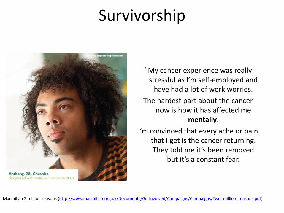
Survivorship
‘ My cancer experience was really stressful as I’m self-employed and
have had a lot of work worries.
The hardest part about the cancer now is how it has affected me
mentally.
I’m convinced that every ache or pain that I get is the cancer returning. They told me it’s been removed
but it’s a constant fear.
Macmillan 2 million reasons (http://www.macmillan.org.uk/Documents/GetInvolved/Campaigns/Campaigns/Two_million_reasons.pdf)
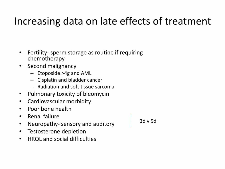
Increasing data on late effects of treatment
• Fertility- sperm storage as routine if requiring chemotherapy
• Second malignancy– Etoposide >4g and AML– Cisplatin and bladder cancer– Radiation and soft tissue sarcoma
• Pulmonary toxicity of bleomycin• Cardiovascular morbidity• Poor bone health• Renal failure• Neuropathy- sensory and auditory• Testosterone depletion• HRQL and social difficulties
3d v 5d

Community Shared Surveillance
Self-report statusNurse/Consultant Discussion QTool
Time Post-treatment
Yr 1 Yr2 Yr 3 Yr4 Yr5 Yr6 Yr7 Yr8 Yr9 Yr10
X-ray
Cancer Centre GP/District hospital Cancer Centre
Tumour Markers (AFP, hCG, LDH, etc)
Cancer Centre GP/District hospital Cancer Centre
CT scanCancer Centre
D, K, L%
A1, A2, A3, B, C, F %
E, G, H, J, M %
Chase results and upload onto shared systems (i.e. PPM, Tracker) by Service
Coordinator
Chase results and upload onto shared systems (i.e. PPM, Tracker) by Service
Coordinator
CTs run and uploaded onto shared systems by local radiology staff. Result
discussed in OPA as usual.
• Results interpreted by local treatment team
Patient recalled to Centre if problems arisePatient seen in Centre once/year or when
offered CT resultsElse – Okay Letter
• Q-Tool replaces face to face symptom self-report discussion in a standardised
comprehensive manner
SJUH Local GCT based onEAU & EGCCCG follow-up guidelines
IGCCCG risk groupsRMH Staging
NICE Guidelines
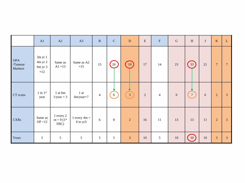
A1 A2 A3 B C D E F G H J K L
OPA
/Tumour
Markers
3m yr 1
4m yr 2
6m yr 3
=12
Same as
A1 =13
Same as A2
=1515 26 10 17 14 23 33 21 7 7
CT scans1 in 1st
year
1 at 6m
1/year = 3
1 at
6m/year=74 6 3 2 4 6 7 6 2 3
CXRsSame as
OP =12
1 every 2
m = 9 (1st
FBC)
1 every 4m =
6 to yr36 8 2 16 11 13 13 11 2 3
Years 5 5 5 5 5 3 10 5 10 10 10 3 3
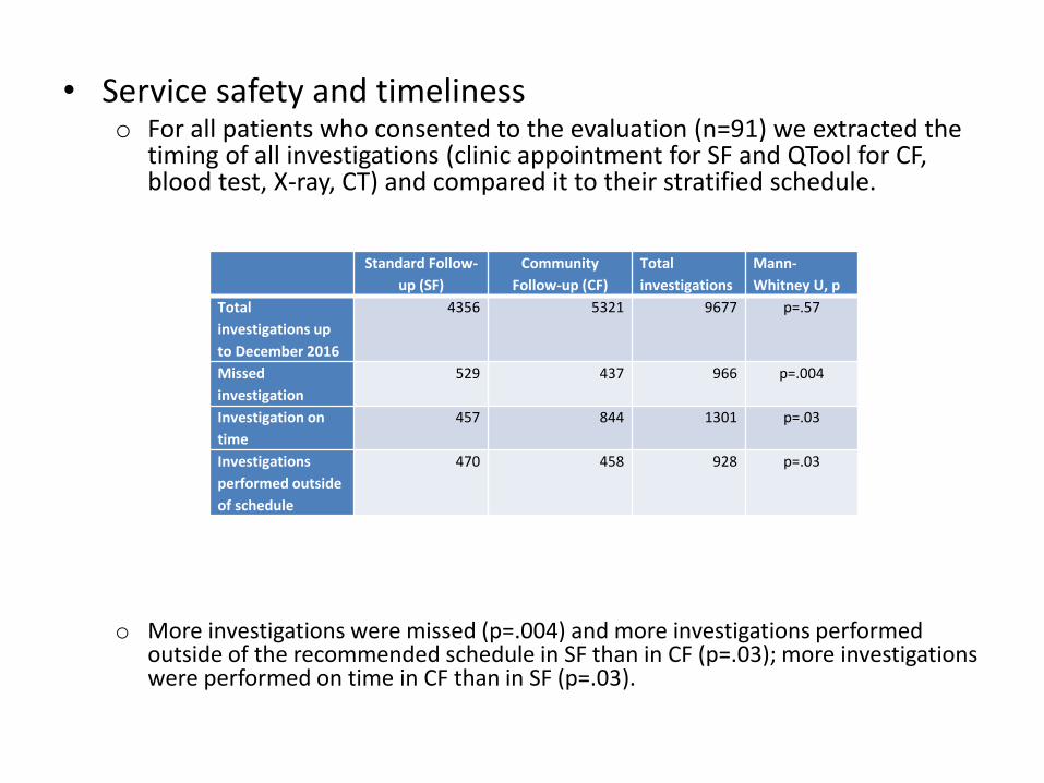
• Service safety and timelinesso For all patients who consented to the evaluation (n=91) we extracted the
timing of all investigations (clinic appointment for SF and QTool for CF, blood test, X-ray, CT) and compared it to their stratified schedule.
o More investigations were missed (p=.004) and more investigations performed outside of the recommended schedule in SF than in CF (p=.03); more investigations were performed on time in CF than in SF (p=.03).
Standard Follow-
up (SF)
Community
Follow-up (CF)
Total
investigations
Mann-
Whitney U, p
Total
investigations up
to December 2016
4356 5321 9677 p=.57
Missed
investigation
529 437 966 p=.004
Investigation on
time
457 844 1301 p=.03
Investigations
performed outside
of schedule
470 458 928 p=.03

SF Patients approached
N=52
Declined participationN=8 (15.3%)
Consented N=44
(84.6%)
Did not return questionnaire N=7 (15.9%)
Completed evaluation N=37 (84%)
27 always in SF10 switched from
CF
CF Patients approached N=199
Via post/email N=166
In clinics N=33
Incorrect address on record N=22
(13.2%)
Consented N=62 (31%)
Did not return questionnaire N=8
(12.9%)
Completed evaluation N=54
(87%)27 always in CF
27 switched from SF
Comparison on investigation timeliness, safety, acceptability
Evaluating acceptability - Recruitment
Abbreviations – SF, standard follow-up; CF, community follow-up
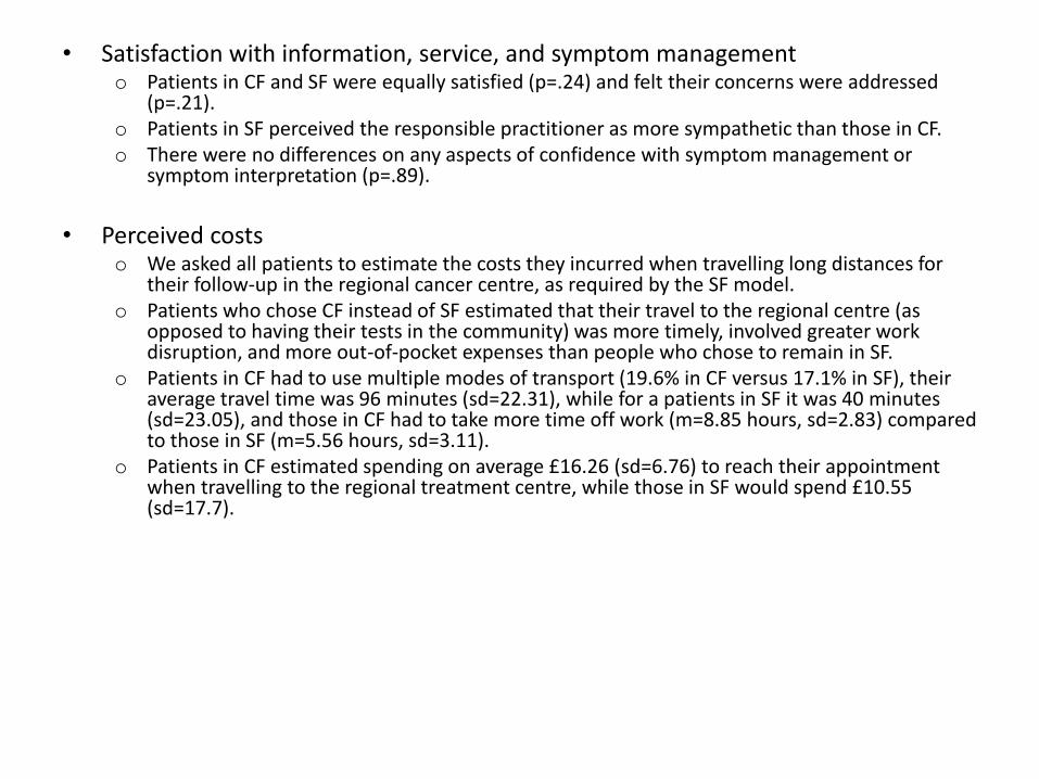
• Satisfaction with information, service, and symptom managemento Patients in CF and SF were equally satisfied (p=.24) and felt their concerns were addressed
(p=.21). o Patients in SF perceived the responsible practitioner as more sympathetic than those in CF. o There were no differences on any aspects of confidence with symptom management or
symptom interpretation (p=.89).
• Perceived costso We asked all patients to estimate the costs they incurred when travelling long distances for
their follow-up in the regional cancer centre, as required by the SF model. o Patients who chose CF instead of SF estimated that their travel to the regional centre (as
opposed to having their tests in the community) was more timely, involved greater work disruption, and more out-of-pocket expenses than people who chose to remain in SF.
o Patients in CF had to use multiple modes of transport (19.6% in CF versus 17.1% in SF), their average travel time was 96 minutes (sd=22.31), while for a patients in SF it was 40 minutes (sd=23.05), and those in CF had to take more time off work (m=8.85 hours, sd=2.83) compared to those in SF (m=5.56 hours, sd=3.11).
o Patients in CF estimated spending on average £16.26 (sd=6.76) to reach their appointment when travelling to the regional treatment centre, while those in SF would spend £10.55 (sd=17.7).
Summary
• Background• Concepts• Current practice• Recent trials/research
– TE19, TE24, BEP111, relapsed stage 1 NSGCT, UK3 PBEP, TIGER
• Areas of activity– Trials, data collection– Survivorship– Community follow-up