Embed Size (px)
Citation preview
0123456789();:
DNA- binding transcription factors (TFs) represent one of the most essential classes of proteins in the eukaryotic proteome1. By binding to specific DNA sites and con-trolling transcriptional output of genes in close spatial proximity, TFs play foundational roles in the regulation of virtually all of a cell’s genome2. TFs dictate the identity and fate of individual cells in multicellular organisms by differentially regulating the common genetic code, and are responsible for rapidly coordinating responses to internal and external stimuli by serving as end points in cell signalling networks3,4. It is estimated that there are at least 1,600 TFs in the human genome, around 19% of which have been associated with a disease phenotype1. Concordantly, given their central importance to bio-logy, TFs are frequent drivers of disease and represent tantalizing therapeutic targets3,5,6.
The significant potential of direct TF modulators was best encapsulated almost two decades ago by James Darnell in the context of anticancer therapeutics5. He highlighted how TFs, more so than upstream signalling proteins such as GPCRs or kinases, have the capac-ity for highly specific disease modulation given their foundational role in selective gene regulation. That is, a hypothetical inhibitor of a dysregulated TF could limit toxicity while increasing efficacy by only inhibiting tran-scriptional programmes driven by that TF, without the collateral damage sometimes associated with inhibit-ing signalling proteins that are involved with multiple signalling networks unrelated to the disease7,8. Because individual TFs typically only regulate a limited set of gene targets that are governed by their DNA- binding
specificity, such an inhibitor is also likely to be less prone to compensatory resistance mechanisms common to other pharmacological modalities such as tyrosine kinase inhibitors9. This exceptional potential of ther-apeutically modulating TF action is illustrated by the enduring success of myriad nuclear receptor- targeting drugs, which represent the standard of care across several different disease areas10.
Despite the broad therapeutic promise of TF mod-ulators, there are major roadblocks associated with TFs as a target class that have impeded countless attempts at drugging TFs outside the nuclear receptor family. Consequently, of the roughly 300 TFs that have been associated with a disease phenotype, only a handful have been successfully targeted by small molecules1. A fun-damental challenge is that most TFs are predominantly intrinsically disordered and lack classical well- formed small- molecule binding pockets11. TFs function pri-marily by forming highly dynamic protein–DNA interactions and protein–protein interactions (PPIs), and consequently the most critical functional sites also represent exceptionally challenging regions to directly target with small molecules. Beyond just the basic dif-ficulties of TF ligand development, the regulation and function of individual TF domains is often highly com-plex or poorly understood, obfuscating the domains that would actually be fruitful to modulate. This, combined with continually emerging evidence that challenges our fundamental understanding of gene regulation and TF mechanisms of action12,13, makes TFs some of the thorniest targets in the proteome.
Advances in targeting ‘undruggable’ transcription factors with small moleculesMatthew J. Henley 1,2,3 ✉ and Angela N. Koehler 1,2,3 ✉
Abstract | Transcription factors (TFs) represent key biological players in diseases including cancer, autoimmunity, diabetes and cardiovascular disease. However, outside nuclear receptors, TFs have traditionally been considered ‘undruggable’ by small- molecule ligands due to significant structural disorder and lack of defined small- molecule binding pockets. Renewed interest in the field has been ignited by significant progress in chemical biology approaches to ligand discovery and optimization, especially the advent of targeted protein degradation approaches, along with increasing appreciation of the critical role a limited number of collaborators play in the regulation of key TF effector genes. Here, we review current understanding of TF- mediated gene regulation, discuss successful targeting strategies and highlight ongoing challenges and emerging approaches to address them.
1David H. Koch Institute for Integrative Cancer Research, Massachusetts Institute of Technology, Cambridge, MA, USA.2The Broad Institute of MIT and Harvard, Cambridge, MA, USA.3Department of Biological Engineering, Massachusetts Institute of Technology, Cambridge, MA, USA.
✉e- mail: [email protected]; [email protected]
https://doi.org/10.1038/ s41573-021-00199-0
REVIEwS
Nature reviews | Drug Discovery
0123456789();:
This Review synthesizes current understanding of TF function and gene regulation with emerging pharmaco-logical approaches that can or could be used to drug this target class. We discuss the basic mechanisms by which TFs participate in gene regulation and drive myriad dis-eases, and then evaluate key lessons from successful and promising examples of TF modulator development. We close by highlighting technologies that could facilitate progress in drugging even the most recalcitrant TFs and reflect on how emerging medicinal chemistry, biophys-ics and chemical biology approaches could be adapted to address the unique challenges associated with TFs.
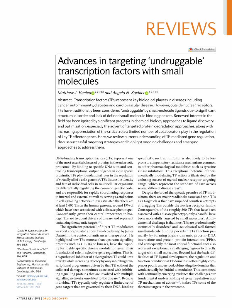
Functional domains of TFsThe key role of a TF is to recruit transcriptional regula-tory machinery to specific genomic loci to regulate gene expression14. A minimal TF is thus defined by just the presence of two key elements: a DNA- binding domain (DBD) that recognizes specific DNA sequences, and an effector domain that recruits members of transcrip-tional activation or repression machinery14 (Fig. 1). TFs that act as transcriptional activators use a transactivation domain to recruit chromatin remodelling enzymes, his-tone modifying enzymes, transcriptional co- activators and/or many general TFs to increase the accessibility of target genes, epigenetically mark them as active, and recruit and activate RNA polymerase II (Pol II)12,14–18. Conversely, TFs that behave as transcriptional repres-sors use a transrepression domain to recruit chromatin remodelling and epigenetic enzymes to decrease the accessibility of target genes and mark them as inactive17. In some cases, prototypical transactivation domains can have repressive functions that are controlled by the
presence and/or activity of co- repressors19. By this basic mechanism of recruitment, TFs act as the directors of transcriptional output of the genome and play key roles across wide- ranging cellular processes18.
The structural and biophysical mechanisms by which key TF regulatory domains function have been a sub-ject of intense study for decades. The determinants of DBD specificity for DNA sequence in vitro have been extensively dissected with advances in high- throughput binding assays and determination of numerous DBD structures, in both the presence and absence of DNA20. However, significant challenges in predicting func-tional TF binding sites in the genome remain, which is complicated by the complex three- dimensional chro-matin architecture and an abundance of non- specific TF binding sites that can compete with TF binding to scarcer specific TF binding sites21.
Conversely, the basic mechanisms by which effec-tor domains function are much less defined. Although there are certainly some instances of well- studied and functionally important PPIs made by single effector domains22–24, the generalizability of these examples to the class as a whole has not been possible25–27. For example, although several structures of transactivation domains bound to co- activators such as CBP/p300 (reF.24) have been proposed, these structures do not explain the repeated observation that roughly 1% of any random sequence of amino acids — with the only commonal-ity being the preponderance of acidic and hydrophobic residues — stitched to a DBD can function as transac-tivation domains25,27. Thus, the general mechanisms by which effector domains actually effectuate recruitment are still under considerable debate. Current universal models of effector domain function hypothesize that they form non- specific dynamic PPIs with transcrip-tional machinery as well as phase- separating with dis-ordered regions of co- activators/co- repressors to form transcriptional condensates13,28–30, although in some indi-vidual cases there is evidence that other mechanisms are more consistent with experimental data31,32. Put together, there are many remaining questions about the mecha-nisms by which the two key TF domains function that may have drastic implications for the success of various targeting strategies.
As well as the two class- defining TF functional domains, many TFs contain additional layers of regu-lation that add further complexity to their function and regulation (Fig. 1). For example, the STAT family of TFs contain a SH2 domain that controls homodimerization or heterodimerization with other STAT TFs, and thereby regulates the TF localization to the nucleus33. Nuclear receptors, by far the most druggable family of TFs, con-tain a ligand- binding domain (LBD) that typically acts in cooperation with a prototypical disordered transactiva-tion domain to recruit transcriptional machinery when bound to a small- molecule ligand34. Other TFs such as the basic helix–loop–helix family require dimerization with other family members to form competent DBDs35. These diverse regulatory domains and mechanisms have historically served as the most promising entry points for medicinal chemists to develop effective TF modulators6.
DNA-binding domain• Sequence-specific recognition
Regulatory domain(optional)• Dimerization• Nuclear transport• Autoinhibition
Effector domain• Recruitment
Collaborators
Chromatin remodellingenzymes
Co-activators/co-repressors
General transcriptionalmachinery
Fig. 1 | Anatomy of a TF. All transcription factors (TFs) contain two general protein domains: a DNA- binding domain (DBD) that binds to specific DNA regulatory sequences, and an effector domain that recruits various transcriptional ‘collaborators’ to regulate chromatin accessibility and transcriptional output. Many TFs also contain one or more regulatory domains, which typically serve to regulate TF localization and functional activity.
Non- specific TF binding sitesSequences of DNA that do not contain the consensus sequence for a transcription factor (TF) DNA- binding domain (DBD). Most DBDs have low affinity for non- specific sites, but because of the exceptionally high ratio of non- specific to specific sites, TFs often spend significant time at non- specific sites.
Specific TF binding sitesSequences of DNA that contain the consensus sequence for a transcription factor (TF) DNA- binding domain.
Transcriptional condensatesLiquid–liquid phase- separated droplets containing transcription factors, co- activators, rNA polymerase ii (Pol ii) and other transcriptional machinery.
www.nature.com/nrd
R e v i e w s
0123456789();:
Gene regulation by TFsA key lesson emerging over recent years is that eukar-yotic gene regulation is an exceptionally complex and dynamic process that is often counter- intuitive and facil-itates many surprising behaviours12,30,36,37. Whether a TF functions at a specific binding site depends not only on the thermodynamic stability of the TF–DNA complex but on a number of interoperating factors, including multidimensional DNA/chromatin architecture, the cooperative action of other TFs and co- activators at nearby or overlapping sites and the kinetics of the TF binding to DNA itself13,21,38–40. Here, we focus on recent insights into the mechanisms that regulate the strength of TF- driven transcriptional activation.
Influence of genome structure on TF action. For dec-ades, it has been understood that the organization of the genome, across several dimensions, is a key determinant of whether a gene is turned on or off2. TFs control the expression of most genes by binding to promoter and/or enhancer regions of DNA18. Promoters are characterized by their inclusion of a transcription start site (TSS) and a TATA- box/Inr DNA sequence, the latter of which ena-bles assembly of the pre- initiation complex and subsequent activation of RNA Pol II2. Enhancers, conversely, do not contain a functional TSS and can be located up to sev-eral kilobases away from a TSS. Enhancers instead bind to TFs and activate transcription when placed in close spatial proximity to a promoter41. This reliance on three- dimensional proximity for enhancer function has many remarkable consequences, chiefly that many enhancers only function at genes located at long genomic distances (>1 kb) instead of at closer genomic loci2. Although mechanisms of transcriptional activation at promot-ers have been extensively characterized — down to the structures of the pre- initiation complex at different steps of the activation process42–44 — understanding how genes are regulated by enhancer regions is still an area of intense study and emerging therapeutically relevant insights.
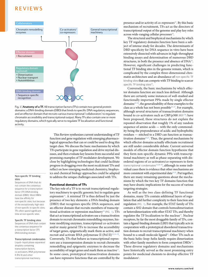
A breakthrough in understanding how enhancers are placed into proximity of the target genes has been the identification of chromatin neighbourhoods or topo-logically associated domains (TADs)38,45–47. TADs are essentially extruded chromatin loops that are bound by the proteins cohesin and CTCF, and enable cells to dictate the three- dimensional structure of specific regions of the genome38 (Fig. 2a). TADs are frequently conserved within cell types and are thought to place key cell- identity genes under the control of multiple enhan cers to maintain robust expression48. Accordingly, TADs can be restructured upon differentiation of pro-genitor cells as a mechanism to remodel cell- identity transcriptional programmes46. Remarkably, not all genes within a TAD are necessarily dependent on the TAD for function, suggesting additional complexities to genomic structure that could be relevant for selective therapeutic targeting of genes within TADs49.
A particularly noteworthy advance in the basic biology of gene regulation has been the discovery and characterization of super- enhancers50,51 (Fig. 2a). Super- enhancers are defined as extended clusters of enhancers with particularly elevated levels of bound TFs and co- activators as well as epigenetic marks associated with active transcription (for example, H3K27Ac). Due to their high sustained levels of transcriptional activ-ity, super- enhancers often act in concert with TADs to control expression of key cell- identity genes48,50,51. For example, in development, super- enhancers have been observed as regulators of core regulatory TFs that con-trol the process and timeline of cell differentiation52. Super- enhancers are also inactivated or repurposed over the course of development to initiate changes in cell characteristics; in cancer, these mechanisms enable malignancies to use super- enhancers to drive oncogenic transcriptional programmes50,51,53.
Dynamics of TF action. A crucial fact that underlies our current understanding of eukaryotic transcrip-tional regulation is that it is a highly dynamic and
Pre- initiation complexA large complex comprising general transcription factors, Mediator and other proteins that position and activate rNA polymerase ii (Pol ii) at the transcription start site.
b
Cohesin
Super-enhancer
CTCF
TF1
TF2
PIC
RNA Pol II
Co-activator
a
PromoterEnhancerGene
Fig. 2 | overview of the modern model of the transcriptional activation process. a | Depiction of a topologically associated domain (TAD) bound by cohesin and CTCF containing a super- enhancer that controls a gene. b | Zoomed view of the phase- separation model of transcriptional activation, where transcription factors (TFs) and co- activators form transcriptional condensates spanning the enhancer and promoter. PIC, pre- initiation complex; RNA Pol II, RNA polymerase II.
Nature reviews | Drug Discovery
R e v i e w s
0123456789();:
out- of- equilibrium process12. This has led to several surprising revelations about in vivo mechanisms of TF and co- regulator function that interact intimately with emerging insights about genome structure.
The remarkable dynamics of TF binding to target sites in vivo has several surprising consequences. The classical view of TF–DNA interactions is that TFs reside on DNA for long periods of time to carry out their function, but modern in vivo imaging studies have esti-mated that the lifetimes of TFs with target DNA sites can be as short as a few seconds12,40,54,55. This dynamism is thought to serve as a regulatory mechanism to keep TFs from being trapped at non- specific DNA sites for extended time periods. For example, the relative frac-tion of binding to specific sites over non- specific sites of some nuclear receptors only marginally increases when activated, but fast turnover and extended lifetimes at specific sites facilitate a rapid and significant increase in transcriptional output at target genes40.
Dynamic TF–DNA binding can also lead to surpris-ing modes of TF cooperativity. For example, whereas cooperative activity of multiple TFs at a promoter or enhancer is classically thought to be enacted by different TFs binding to adjacent sites and stabilizing each other’s binding, rapid TF binding and unbinding has also been found to lead to cooperativity from different TFs bind-ing the same site12,36. The low lifetimes and long periods of time between binding of each TF enable unimpeded exchange between different TFs, acting to keep chro-matin in an open conformation and to recruit distinct members of the transcriptional apparatus.
Other key examples of unexpected behaviour in transcription have been observed during characteri-zation of super- enhancer- driven transcriptional acti-vation. Strikingly, super- enhancers display marked increases in both the inter- reliance and the binding and unbinding dynamics of TFs and co- activators over typical enhancers51,56. The amplified cooperativity at super- enhancers causes them to be especially sensitive to slight changes in TF and co- regulator composition. This can result, for example, in inhibitors of general co- activators such as BRD4 displaying exquisite selectiv-ity for super- enhancer- driven transcription56. Inhibitors of general transcriptional regulatory enzymes have even been observed to copy the phenotypes of removing core regulatory TFs in some cell types57–59. On the other hand, the action of transcriptional co- repressors at super- enhancers can be paradoxically critical for main-taining maximum transcriptional output, exposing a highly dynamic steady state of chromatin accessibility and TF/co- regulator binding needed for super- enhancer activity60–62.
Liquid–liquid phase separation (LLPS) has emerged as a popular biophysical framework to rationalize the exceptionally cooperative and dynamic behaviour of TFs and co- regulators at super- enhancers13,30,37 (Fig. 2b). Significant levels of TF binding at super- enhancers is hypothesized to create high local concentrations of co- activators and other members of the transcrip-tional machinery; at a critical concentration, these TFs and other cofactors form phase- separated ‘tran-scriptional condensates’ spanning the super- enhancer
and the TSS13,30. Consistent with this notion, multiple experimental efforts have demonstrated that TFs and co- activators form highly concentrated puncta at active super- enhancer sites in vivo13,30,37,63. Corresponding in vitro experiments using purified TFs and co- activators have shown that low- complexity intrinsically disor-dered regions (IDRs) in TFs, co- activators and gen-eral transcriptional machinery (including RNA Pol II) have the capacity to co- condense into phase- separated droplets13,37,64,65.
To many, LLPS serves as an exceedingly useful frame-work to rationalize otherwise puzzling transcriptional phenomena. For example, IDRs are highly enriched in TFs and co- regulators, but because these regions are disregarded in standard structure–function paradigms, it was previously challenging to understand how they could participate in transcriptional regulation. Within an LLPS framework, it is theoretically possible to iden-tify functions and mechanisms of TF IDRs by simply considering their physicochemical properties, concen-tration and the landscape of DNA- binding sites at a given enhancer, which together dictate their ability to participate in transcriptional condensates66. Transitions into and out of a condensate by a single protein can, consequently, be facilitated by post- translational modifications that change IDR properties64, and the formation and dissolution of individual condensates can be regulated by fluctuations in composition of the proteins and RNA that are active during the pro-cess of transcription65. Examples where applications of LLPS frameworks to the dissection of IDR functional mechanisms have given potential answers to other-wise perplexing experimental observations include rationalizations of transcriptional bursting65, RNA Pol II promoter release64, enhancer–promoter contact restrictions67 and the extraordinary sequence diversity of functional transactivation domains13. Phase transi-tions have also been implicated in distinct mechanisms of gene regulation outside transcriptional activation, and are thought to play roles in the function of splicing condensates64,68, repressive Polycomb repressor com-plex (PRC) bodies69 and heterochromatin/euchromatin transitions70,71.
It is also worth noting that the existence of transcrip-tional condensates is still somewhat contentious, due to the challenge associated with unequivocally demon-strating that puncta containing TFs and transcriptional machinery in vivo indeed constitute a separated liquid phase72,73. Specifically, there have been concerns raised that much of the experimental data are phenomeno-logical, and that other mechanisms could underlie the same observations32. Moreover, the difficulty of stud-ying transcriptional condensates is higher than for other well- characterized examples of LLPS due to the highly dynamic and localized nature of transcriptional activation. Nonetheless, there is broad agreement that the formation of dynamic concentrated hubs of TFs and transcriptional apparatus plays a critical role in transcription, especially at super- enhancers30,37.
Altogether, the often- surprising outcomes of the three- dimensional and dynamic nature of transcrip-tion strongly indicate that many general assumptions
Cooperativityin transcription, a phenomenon where binding of one transcription factor and/or co- regulator at a regulatory element enhances the binding of other factors, and vice versa.
Core regulatory TFs(Also known as master TFs). Self- regulated transcription factors (TFs) that control cell identity and fate.
www.nature.com/nrd
R e v i e w s
0123456789();:
about TF action developed from decades of mecha-nistic studies in simplified systems have significant potential to be misleading12. Concordantly, TF mech-anism of action may be highly variable from cell type to cell type, gene to gene and binding site to binding site. When selecting possible targets to affect the func-tion of a given TF, including individual domains of the TF itself or its co- regulatory binding partners, unbi-ased functional data are therefore critical for effective decision- making.
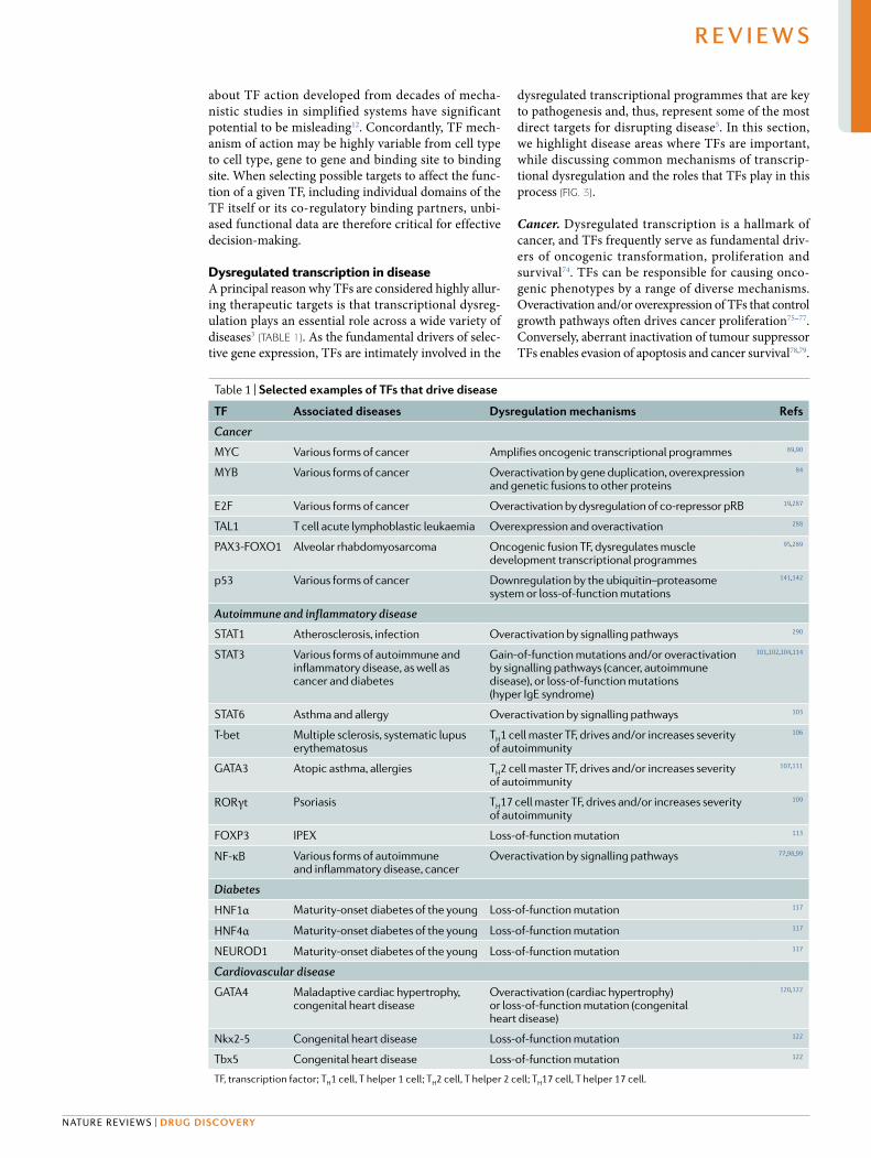
Dysregulated transcription in diseaseA principal reason why TFs are considered highly allur-ing therapeutic targets is that transcriptional dysreg-ulation plays an essential role across a wide variety of diseases3 (TABLe 1). As the fundamental drivers of selec-tive gene expression, TFs are intimately involved in the
dysregulated transcriptional programmes that are key to pathogenesis and, thus, represent some of the most direct targets for disrupting disease5. In this section, we highlight disease areas where TFs are important, while discussing common mechanisms of transcrip-tional dysregulation and the roles that TFs play in this process (Fig. 3).
Cancer. Dysregulated transcription is a hallmark of cancer, and TFs frequently serve as fundamental driv-ers of oncogenic transformation, proliferation and survival74. TFs can be responsible for causing onco-genic phenotypes by a range of diverse mechanisms. Overactivation and/or overexpression of TFs that control growth pathways often drives cancer proliferation75–77. Conversely, aberrant inactivation of tumour suppressor TFs enables evasion of apoptosis and cancer survival78,79.
Table 1 | selected examples of TFs that drive disease
TF Associated diseases Dysregulation mechanisms refs
Cancer
MYC Various forms of cancer Amplifies oncogenic transcriptional programmes 89,90
MYB Various forms of cancer Overactivation by gene duplication, overexpression and genetic fusions to other proteins
84
E2F Various forms of cancer Overactivation by dysregulation of co- repressor pRB 19,287
TAL1 T cell acute lymphoblastic leukaemia Overexpression and overactivation 288
PAX3- FOXO1 Alveolar rhabdomyosarcoma Oncogenic fusion TF, dysregulates muscle development transcriptional programmes
95,289
p53 Various forms of cancer Downregulation by the ubiquitin–proteasome system or loss- of- function mutations
141,142
Autoimmune and inflammatory disease
STAT1 Atherosclerosis, infection Overactivation by signalling pathways 290
STAT3 Various forms of autoimmune and inflammatory disease, as well as cancer and diabetes
Gain- of- function mutations and/or overactivation by signalling pathways (cancer, autoimmune disease), or loss- of- function mutations (hyper IgE syndrome)
101,102,104,114
STAT6 Asthma and allergy Overactivation by signalling pathways 103
T- bet Multiple sclerosis, systematic lupus erythematosus
TH1 cell master TF, drives and/or increases severity of autoimmunity
106
GATA3 Atopic asthma, allergies TH2 cell master TF, drives and/or increases severity of autoimmunity
107,111
RORγt Psoriasis TH17 cell master TF, drives and/or increases severity of autoimmunity
109
FOXP3 IPEX Loss- of- function mutation 113
NF- κB Various forms of autoimmune and inflammatory disease, cancer
Overactivation by signalling pathways 77,98,99
Diabetes
HNF1α Maturity- onset diabetes of the young Loss- of- function mutation 117
HNF4α Maturity- onset diabetes of the young Loss- of- function mutation 117
NEUROD1 Maturity- onset diabetes of the young Loss- of- function mutation 117
Cardiovascular disease
GATA4 Maladaptive cardiac hypertrophy, congenital heart disease
Overactivation (cardiac hypertrophy) or loss- of- function mutation (congenital heart disease)
120,122
Nkx2-5 Congenital heart disease Loss- of- function mutation 122
Tbx5 Congenital heart disease Loss- of- function mutation 122
TF, transcription factor; TH1 cell, T helper 1 cell; TH2 cell, T helper 2 cell; TH17 cell, T helper 17 cell.
Nature reviews | Drug Discovery
R e v i e w s
0123456789();:
Genetic fusion events that generate fusion TFs are a common cause of paediatric cancers, and typically dys-regulate developmental transcriptional programmes to initiate transformation and drive proliferation80. Oncogenic viruses are known to initiate transformation via a combination of the activity of viral TFs in addition to other viral proteins that co- opt or dysregulate cellular TFs and transcriptional co- regulators81. There are also some cases, for example in certain gliomas82, where TFs drive oncogenic phenotypes simply by rewiring their transcriptional programmes to regulate a different set of effector genes83.
The TF MYB serves as an excellent example of a sin-gle oncogenic TF that can act by several of the mech-anisms outlined above84. MYB is intimately involved in a variety of cancers including leukaemia (especially acute myeloid leukaemia (AML)), adenoid cystic car-cinoma, colorectal cancer and breast cancer, where it generally drives oncogenesis by becoming overactivated. Most commonly, gene duplications and overexpression of MYB lead to overactivation of MYB target genes, but MYB can also become overactivated by genetic trans-locations (for example, MYB- NFIB) that fuse it to other proteins, typically eliminating the MYB transrepression domain in the process83. In other cases, genetic changes can generate new MYB binding sites that enhance other oncogenic drivers. For example, in some forms of T cell acute lymphoblastic leukaemias, novel MYB binding sites can form in the enhancer for the driver TF TAL1 and increase its expression85.
The central role of TFs in driving oncogenesis fre-quently leads to reliance of malignancies on the activ-ity of individual TFs74. A classic example of a TF that
exhibits this ‘oncogenic addiction’ behaviour across a variety of cancers is MYC. MYC is a TF in the basic helix–loop–helix family that, along with its binding part-ner MAX, binds to the widespread E- box sequences at promoters and enhancers across the genome86. MYC primarily functions by recruiting transcriptional elon-gation machinery to enhancers to increase transcrip-tional output87,88. MYC is one of the most frequently overexpressed oncogenes and is thought to act as a gen-eral transcriptional ‘amplifier’ to drive a wide variety of oncogenic transcriptional programmes across diverse cancer types3,89,90. In vivo experiments using genetic knockdown of MYC and expression of dominant neg-ative MYC variants have shown that several distinct cancers are addicted to MYC’s amplification activity, rapidly dying or differentiating into normal cell types upon MYC inhibition91–93. Similarly, TFs as a class rep-resent a large fraction of hits in cancer genetic depen-dency databases such as DepMap94, supporting the idea that oncogenic addiction to TFs is a shared vulnerabil-ity across myriad cancers. Thus, there is exceptionally high potential for targeting TF activity as a therapeutic strategy for cancer.
In transcription, TFs do not function alone: the fundamental role of TFs is to recruit the requisite machinery to do the work required for transcriptional regulation14. Accordingly, much of the apparatus that facilitates TF- driven activation/repression can also be critical for maintaining oncogenic transcriptional programmes. This is especially relevant when consid-ering the important roles that super- enhancers have been shown to play in cancer, given the heightened levels of cooperativity between TFs and cofactors at
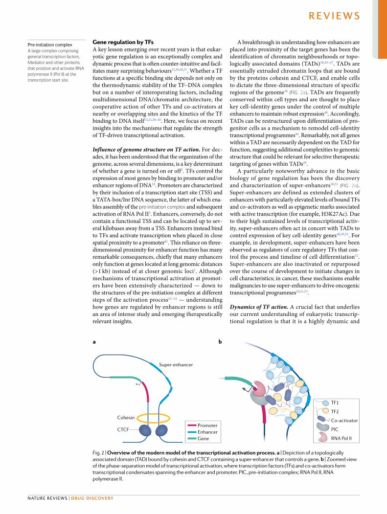
↑ Transcription
Upregulation• Amplification• Gain of function• Pathway overactivation
Downregulation• Loss of function• Overactivation of repressors
Changes in target genes• Chromatin architecture shifts• Gene translocations• Fusion transcription factors
Normal transcription
↓ Transcription
Transcriptionof new gene
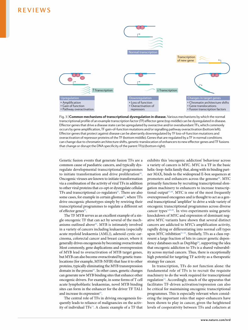
Fig. 3 | common mechanisms of transcriptional dysregulation in disease. Various mechanisms by which the normal transcriptional profile of an example transcription factor (TF) effector gene (top middle) can be dysregulated in disease. Effector genes that drive a disease state can be upregulated by overactive and/or overabundant TFs, which commonly occurs by gene amplification, TF gain- of- function mutations and/or signalling pathway overactivation (bottom left). Effector genes that protect against disease can be aberrantly downregulated by TF loss- of- function mutations and overactivation of repressor proteins of the TF (bottom middle). Genes that are regulated by a TF in normal conditions can change due to chromatin architecture shifts, genetic translocation of enhancers to new effector genes and TF fusions that change or disrupt the DNA specificity of the parent TF(s) (bottom right).
www.nature.com/nrd
R e v i e w s
0123456789();:
these regulatory elements. As discussed previously, in normal cells, super- enhancers often form around key cell- identity genes50,51,53; it has similarly been observed that malignancies frequently generate or repurpose super- enhancers around key oncogenic identity and effector genes53,95. Super- enhancers thereby grant cancer cells increased and sustained activation of these genes, and are consequently key to maintaining an undiffer-entiated cell state and enabling rapid and continuous growth.
Because of the exceptionally cooperative nature of super- enhancer function, super- enhancers are fre-quently dependent on the action of select members of the transcriptional apparatus (for example, chromatin readers, histone modifying enzymes, transcriptional kinases) in addition to TFs56–60,95,96. Without even one of these co- regulators and/or its associated enzymatic activity, super- enhancers can be rapidly depleted of TFs, active chromatin marks and the transcriptional appa-ratus. Unique super- enhancers can also have distinct cofactor dependency profiles, which can enable selec-tive inhibition of super- enhancer- driven oncogenic transcription57,59. For example, inhibitors of general tran-scriptional enzymes such as CDK9 have been shown to display strikingly selective inhibition of the oncogenic transcription programmes of androgen receptor ∆LBD splice variants by this mechanism59. Inhibiting the activ-ity of TF collaborators at super- enhancers therefore has significant therapeutic potential for treating cancer and serves as an alternative to targeting the oncogenic TF itself, especially in cases where the TF proves recalcitrant to small- molecule discovery efforts.
Autoimmune/inflammatory disease. TFs are com-mon end points of signalling pathways that medi-ate the immune response to infection or injury76,77. Consequently, dysregulation of TFs involved in immune response plays a significant role in the pathogenesis of autoimmune and inflammatory diseases. For example, the TF NF- κB is a master regulator of both innate and adaptive immunity: among other functions, it controls both the expression of pro- inflammatory cytokines in macrophages as well as the activation and differentiation of naive CD4+ T helper cells77. Several diverse signalling pathways regulate activation of NF- κB and its transit to the nucleus. Overactivation of NF- κB activity is strongly linked to myriad inflammatory and/or autoimmune dis-eases, such as rheumatoid arthritis, inflammatory bowel disease and multiple sclerosis77,97–100.
Directly downstream of NF- κB in mediating immune response lies the STAT family of TFs, which regulate the expression of cytokine- inducible genes such as interferons76. Individual STAT family mem-bers are similarly implicated in numerous autoimmune and inflammatory diseases. Overactivation of STAT activity is, in general, linked to autoimmune disease, for example activating mutations of family member STAT3 have been linked to early- onset type 1 diabetes, Crohn’s disease, psoriasis and multiple sclerosis101,102. Similarly, overactivation of STAT6 is known to play sig-nificant roles in allergy and asthma103. Inactivation of STAT family members, on the other hand, often leads to
immunodeficiency and predisposition to various types of infection76. For example, inactivating mutations in STAT3 underlie many cases of hyper IgE syndrome104.
T cells are intimately involved in the development, progression and severity of myriad autoimmune diseases105. Although the molecular mechanisms by which individual T cell types affect autoimmunity are quite complex, the master TFs that define T cell identity are thought to serve as general orchestrators of many autoimmune diseases. Overactivation/overexpression of master TFs in T helper 1 (TH1) cells (T- bet), TH2 cells (GATA3) and TH17 cells (RORγt), for example, is linked to several T cell- driven autoimmune diseases such as multiple sclerosis, systematic lupus erythema-tosus, atopic asthma and psoriasis106–110. T cell master TFs even, in some cases, protect against autoimmune disease; for example, overexpression of the master TF GATA3 (TH2 cells) displayed reduced symptom severity of multiple sclerosis in murine models111. Similarly, the master TF FOXP3 of regulatory T cells is known to drive the immuno suppressive effects of regulatory T cells and its expression is correlated to decreased severity of auto-immune disease112. Inactivation of FOXP3, on the other hand, is highly deleterious and can lead to X- linked congenital immunodeficiency syndromes113.
Altogether, immune response and T cell master regulatory TFs make enticing targets for the numerous diseases caused by aberrant immune responses, espe-cially given their rich regulatory networks that provide several possible intervention points76,77,114. We also note that many of these TFs often have a direct relationship to cancer as well, where dysregulated immune response TFs have been shown to play critical roles in enabling transformation, invasion and metastasis33,77.
Diabetes. Diabetes mellitus, characterized by an inabil-ity to properly secrete or utilize insulin, is, in general, a polygenic disease linked to changes in several genes simultaneously115. However, there are some forms of monogenic diabetes that have been directly linked to mutations in single TFs. For example, as previously men-tioned, activating STAT3 mutations have been linked to early- onset type 1 diabetes101. A significant form of monogenic diabetes is maturity- onset diabetes of the young (MODY), which accounts for around 2% of all diabetes cases in patients younger than 20 years old116. Five of the six genes that have been directly linked to MODY are TFs, which include the hepatic nuclear fac-tors HNF1α, HNF1β and HNF4α, the insulin promoter factor IPF1 and NEUROD1 (reF.117). In all cases, loss- of- function mutations lead to MODY. Interestingly, the vast majority of MODY cases are caused by mutations in one of the three hepatic nuclear factors that are pri-marily associated with liver function and previously had no obvious connection to β- cells117. Conversely, only a small fraction of MODY cases are caused by mutations in IPF1 or NEUROD1, even though they both directly regulate insulin expression. HNF1α, HNF1β and HNF4α are also known to cooperate directly to regulate target gene expression118, and thus possible therapeutic strate-gies may include the development of agonists against one of these TFs to restore overall function.
Chromatin readersProteins, such as bromodomains, that bind to post- translationally modified histones.
Nature reviews | Drug Discovery
R e v i e w s

0123456789();:
Cardiovascular disease. Cardiovascular disease, like dia-betes, is a group of diseases that are linked to multiple interrelated genetic risk factors and are not necessarily driven by single proteins. However, TFs are critical to the development and maintenance of the cardiovascular sys-tem, and thus can play significant roles in certain forms of cardiovascular disease119,120. Congenital heart defects, for example, are commonly linked to loss- of- function mutations in master TFs that control development of the cardiovascular system119,121,122. Core regulatory car-diovascular TFs such as GATA, HAND, MEF2 and SRF also play critical roles in directing the response of the cardio vascular system to stress, and overactivation of these TFs to stimuli such as pressure and volume over-load can lead to maladaptive cardiac hypertrophy120. Therapeutic modulation of key cardiovascular TFs therefore has the potential for treating several forms of cardiovascular disease.
Advances in targeting TFsModulation of TFs by small molecules is an alluring therapeutic objective given their importance across numerous diseases3,5. However, outside nuclear hormone receptors10, few drugs or even well- validated chemical probes are known to directly target TFs. Further, many consider TFs to be predominantly ‘undruggable’ because they have significant structural disorder and lack clas-sical small- molecule binding pockets5,6. The basic mecha nisms by which TFs function also contribute to this image: most known effector domains are disor-dered when unbound to partner proteins11, and whereas DBDs are typically more structured, DNA- binding sur-faces tend to be highly charged and convex in shape6. Together, these qualities can make TFs hostile enviro-nments for the development of potent and selective drug- like small molecules. Perhaps unsurprisingly, many molecules that have been reported as direct TF inhibi-tors have questionable structural properties and poorly defined mechanisms of action123,124.
However, advances in structural characterization, basic biological insights and ligand design strategies have enabled the identification of several examples of drugs and high- quality chemical probes that target TFs. Below, we review the lessons learned from examples of successful TF targeting and discuss how these insights can be applied to currently unliganded TFs.
Modulating TFs with ligand- binding domains. One of the most successful areas of drug discovery, in general, has been targeting TFs containing well- folded LBDs, namely nuclear hormone receptors. Although the spe-cifics of targeting this class of proteins have been exten-sively discussed in the literature10, here we highlight key concepts and lessons learned from decades of drug discovery with nuclear receptors.
Similar to numerous other TFs, nuclear receptors contain a DBD and a prototypical intrinsically dis-ordered transactivation domain (known as activat-ing function 1 (AF1))125. The class- defining feature of nuclear receptors is a well- folded LBD that acts as a second tunable effector domain (AF2)125. Binding of specific signalling molecules (for example, hormones
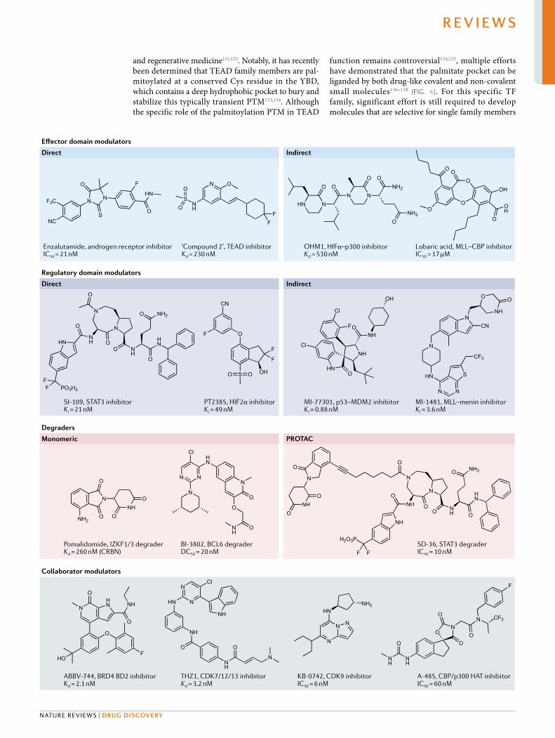
such as oestrogen, androgen and glucocorticoids) to a LBD typically leads to activation of the TF by a variety of mechanisms including localization to the nucleus, homo- oligomerization or hetero- oligomerization and recruitment of co- activators10,125. Dysregulation of nuclear receptors is a feature of several cancers and other diseases, and LBDs can serve as intrinsi-cally ligandable control points for modulating tran-scriptional activity. Accordingly, the drug discovery community has exploited this fact to develop many nuclear receptor drugs and chemical probes (for exam-ple, the FDA- approved androgen receptor antagonist enzalutamide) (Fig. 4).
One important realization from these efforts is that protein conformational flexibility underlies many of the regulatory mechanisms controlled by LBDs10,126. Ligand binding to the LBD typically activates the receptor by exposing a hydrophobic co- activator binding groove as well as, in some cases, enhancing binding to nuclear localization factors and other nuclear receptor mole-cules. Practically speaking, this has enabled multiple forms of modulation to be pursued (that is, agonism, antagonism and inverse agonism) for individual recep-tors, giving drug discovery efforts a wealth of approaches to modulate aberrant transcriptional programmes. Conformation flexibility is a common, if not central, feature of TFs11, which suggests that tuning of TF activ-ity by controlling conformation may be achievable for TFs as a class.
Although nuclear receptors are by far the most drug-gable TF family, several challenges still hamper efforts to target all family members. For example, there are many orphan receptors where the endogenous ligands are unknown or where the apparent LBD does not have a ligand binding pocket127. Further, in some diseases, such as castration- resistant prostate cancer, there can be expression of functional receptor splice variants that lack the LBD (∆LBD), rendering LBD- targeting drugs ineffective128,129. Thus, due to the lack of well- defined and functional small- molecule binding pockets, the challenges associated with orphan receptors and ∆LBD receptor variants are more in line with the challenges of targeting other classes of TFs.
Finally, it is also becoming increasingly recognized that nuclear receptors are not the only class of TFs that contain effector domains that bind to small- molecule ligands. One noteworthy example is the TEAD family of TFs, which use a folded Yap- binding domain (YBD) to recruit co- activators in a mechanism remini-scent of the LBDs of nuclear receptors130. TEAD TFs are an end point of the Hippo signalling pathway, and are thus attractive therapeutic targets for cancer
Fig. 4 | examples of molecules that target TFs by various mechanisms. Affinity (Kd/Ki), inhibitory activity (IC50) or degradation activity (DC50) are included as reported in the literature. For indirect inhibitors of transcription factor (TF) protein–protein interactions (PPIs), the molecular target is in bold. DC50, half- maximal degradation concentration; HAT, histone acetyltransferase; IC50, half- maximal inhibitory concentration; Kd, dissociation constant; Ki, inhibitory constant; PROTAC, proteolysis targeting chimaera.
▶
www.nature.com/nrd
R e v i e w s
0123456789();:
and regenerative medicine131,132. Notably, it has recently been determined that TEAD family members are pal-mitoylated at a conserved Cys residue in the YBD, which contains a deep hydrophobic pocket to bury and stabilize this typically transient PTM133,134. Although the specific role of the palmitoylation PTM in TEAD
function remains controversial134,135, multiple efforts have demonstrated that the palmitate pocket can be liganded by both drug- like covalent and non- covalent small molecules136–138 (Fig. 4). For this specific TF family, significant effort is still required to develop molecules that are selective for single family members
Collaborator modulators
PROTAC
Degraders
Monomeric
Indirect
Regulatory domain modulators
Direct
Indirect
O
N
S
N
O
HN
NC
F3C
F N O
FF
NH
SO
O
HN N
O ON N
O ONH2
NH2O
OH
O
OH
O
O
OO
O
HN
O
NH
NH2O
O
N
N
ONH
O
O
FF PO3H2
HN
CN
F
OH
F
F
O
O OS
NH
OHN
Cl
OH
NHOF
Cl
N
HN
CF3
N N
S
CN
O O
NHN
ONH
O
N
O
ONH2
N
O
O
NH
N
N N
Cl
O
HN
NH
NH2O
HN
OO
ONH
O
O
N
O
H2O3P
F F
NH
ONH O
N
N
NH
O
HN
O
N
HO
O
FO
NH
N
N
NH
Cl
HN
NH
O
N
NH2HN
N
NN
NH
NH
ON
O
F
O
OO
CF3N
Effector domain modulators
Direct
Enzalutamide, androgen receptor inhibitorIC50
= 21 nM‘Compound 2’, TEAD inhibitorKd
= 230 nM
ABBV-744, BRD4 BD2 inhibitorKd
= 2.1 nMKB-0742, CDK9 inhibitorIC50
= 6 nMA-485, CBP/p300 HAT inhibitorIC50
= 60 nMTHZ1, CDK7/12/13 inhibitorKd
= 3.2 nM
OHM1, HIFα–p300 inhibitorKd
= 530 nMLobaric acid, MLL–CBP inhibitorIC50
= 17 μM
SI-109, STAT3 inhibitorKi
= 21 nM
SD-36, STAT3 degraderIC50
= 10 nMBI-3802, BCL6 degraderDC50
= 20 nMPomalidomide, IZKF1/3 degraderKd
= 260 nM (CRBN)
PT2385, HIF2α inhibitorKi
= 49 nMMI-77301, p53–MDM2 inhibitorKi
= 0.88 nMMI-1481, MLL–menin inhibitorKi
= 3.6 nM
Nature reviews | Drug Discovery
R e v i e w s
0123456789();:
(due to significant risks of on- target toxicity of pan- TEAD inhibitors)132,139. However, the results from these initial efforts indicate exciting potential for using folded effector domains with conserved small- molecule pockets as handles for expanding the druggability of TFs.
Inhibiting TF protein–protein complexes. PPIs are a key feature of TF regulation and function. Most TFs are tightly regulated by PPIs with regulatory proteins in the cytosol and/or nucleus, and similarly the basic mecha-nism of TF function (via effector domains) is to form PPIs with members of the transcriptional apparatus. Consequently, inhibiting specific TF PPIs is a valuable means for modulating TF transcriptional activity.
Inhibition of the p53–MDM2 interaction by small molecules serves as an excellent illustration for the poten-tial of drugging TF PPIs140. In cancer, the PPI between the TF p53 — the ‘guardian of the genome’ — and the ubiquitin E3 ligase MDM2 often functions as a mecha-nism for cancer cells to evade apoptosis by downregu-lating p53 levels via the ubiquitin–proteasome system141,142.
Whereas it would be exceptionally challenging to develop a small molecule that binds to p53 and stabilizes it from this mechanism of proteasomal degradation, as p53 is a highly disordered TF143, an alternative approach is to develop antagonists of p53 binding to MDM2. The p53 binding site of MDM2 is, fortunately, highly drug-gable: it is a relatively small and well- defined hydro-phobic pocket, which has enabled several highly potent peptide and small- molecule antagonists (for example, spiro- oxindole MI-77301) (Fig. 4) of this interaction to be developed144,145. These inhibitors have been shown to effectively induce apoptotic cell death in a variety of cancers by increasing p53 levels, and several are being investigated in current clinical trials140 (TABLe 2). Given that many other important TFs are known to be regu-lated by specific E3 ligases, such as the hypoxia- inducible TF HIF1α by the Von Hippel–Lindau E3 ligase146,147, there is significant potential for this approach with TFs that are aberrantly downregulated in disease.
In addition to targeting E3 ligases, developing inhib-itors of other TF regulatory machinery is frequently
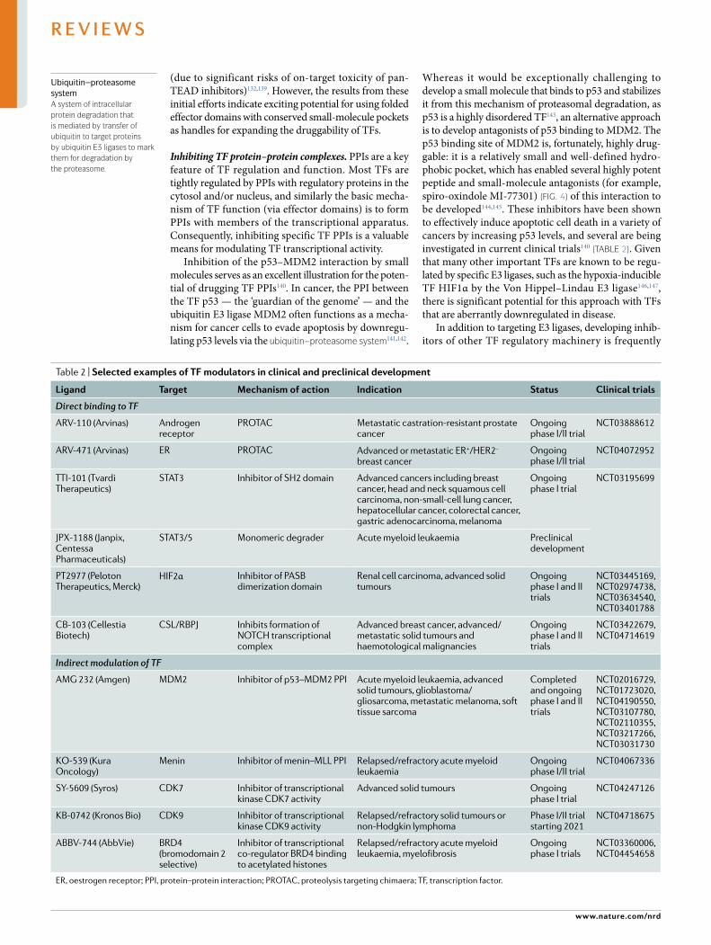
Table 2 | selected examples of TF modulators in clinical and preclinical development
Ligand Target Mechanism of action indication status clinical trials
Direct binding to TF
ARV-110 (Arvinas) Androgen receptor
PROTAC Metastatic castration- resistant prostate cancer
Ongoing phase I/II trial
NCT03888612
ARV-471 (Arvinas) ER PROTAC Advanced or metastatic ER+/HER2– breast cancer
Ongoing phase I/II trial
NCT04072952
TTI-101 (Tvardi Therapeutics)
STAT3 Inhibitor of SH2 domain Advanced cancers including breast cancer, head and neck squamous cell carcinoma, non- small- cell lung cancer, hepatocellular cancer, colorectal cancer, gastric adenocarcinoma, melanoma
Ongoing phase I trial
NCT03195699
JPX-1188 (Janpix, Centessa Pharmaceuticals)
STAT3/5 Monomeric degrader Acute myeloid leukaemia Preclinical development
PT2977 (Peloton Therapeutics, Merck)
HIF2α Inhibitor of PASB dimerization domain
Renal cell carcinoma, advanced solid tumours
Ongoing phase I and II trials
NCT03445169, NCT02974738, NCT03634540, NCT03401788
CB-103 (Cellestia Biotech)
CSL/RBPJ Inhibits formation of NOTCH transcriptional complex
Advanced breast cancer, advanced/metastatic solid tumours and haemotological malignancies
Ongoing phase I and II trials
NCT03422679, NCT04714619
Indirect modulation of TF
AMG 232 (Amgen) MDM2 Inhibitor of p53–MDM2 PPI Acute myeloid leukaemia, advanced solid tumours, glioblastoma/gliosarcoma, metastatic melanoma, soft tissue sarcoma
Completed and ongoing phase I and II trials
NCT02016729, NCT01723020, NCT04190550, NCT03107780, NCT02110355, NCT03217266, NCT03031730
KO-539 (Kura Oncology)
Menin Inhibitor of menin–MLL PPI Relapsed/refractory acute myeloid leukaemia
Ongoing phase I/II trial
NCT04067336
SY-5609 (Syros) CDK7 Inhibitor of transcriptional kinase CDK7 activity
Advanced solid tumours Ongoing phase I trial
NCT04247126
KB-0742 (Kronos Bio) CDK9 Inhibitor of transcriptional kinase CDK9 activity
Relapsed/refractory solid tumours or non- Hodgkin lymphoma
Phase I/II trial starting 2021
NCT04718675
ABBV-744 (AbbVie) BRD4 (bromodomain 2 selective)
Inhibitor of transcriptional co- regulator BRD4 binding to acetylated histones
Relapsed/refractory acute myeloid leukaemia, myelofibrosis
Ongoing phase I trials
NCT03360006, NCT04454658
ER, oestrogen receptor; PPI, protein–protein interaction; PROTAC, proteolysis targeting chimaera; TF, transcription factor.
Ubiquitin–proteasome systemA system of intracellular protein degradation that is mediated by transfer of ubiquitin to target proteins by ubiquitin e3 ligases to mark them for degradation by the proteasome.
www.nature.com/nrd
R e v i e w s
0123456789();:
considered a promising avenue for TF modulation. For example, there is significant interest in developing inhib-itors of deubiquitinating enzymes, which act in direct opposition to ubiquitin E3 ligases. Deubiquitinating enzyme inhibitors thus have the potential to destabilize overactive TFs that evade the ubiquitin–proteasome system6,148. The function of latent cytoplasmic TFs is often tightly regulated by PPIs with cytosolic repressors and import proteins5, and as the methodology for drugging PPIs has advanced, these targets have become exciting avenues for developing TF modulators. Multiple modu-lators of the TF NF- κB, for example, have been developed by targeting PPIs involved in its activation pathways149–151.
TFs also commonly require stable PPIs to become transcriptionally active. A prominent example of this is the STAT family of TFs, which generally require homodimerization or heterodimerization with other STAT proteins to translocate to the nucleus and acti-vate transcription33. STAT dimerization is intrinsically regulated by a SH2 domain and a tyrosine residue that is phosphorylated by JAK kinases upon cytokine recep-tor stimulation; the SH2 domain of one STAT molecule binds to the phosphotyrosine of the other, and vice versa. Antagonists of the phosphotyrosine–SH2 inter-action therefore represent a means for direct inhibition of STAT activity. Several efforts have demonstrated the ligandability of the SH2 domain, which has led to the development of potent chemical probes derived from phosphotyrosine mimetics as well as other non- peptidic scaffolds152 (Fig. 4). Although no STAT inhib-itor has yet successfully advanced through clinical trials, several inhibitors are in varying stages of clinical and preclinical development33 (TABLe 2).
There are several other noteworthy examples where blocking stable PPIs required for TF activity has shown significant promise. The hypoxia- inducible TF HIF2α is a well- validated target for renal cell carcinoma, and potent inhibitors — including the clinical candidates PT2385 and PT2977 (Fig. 4; TABLe 2) — have been devel-oped that block the dimerization of HIF2α with its obligatory cofactor ARNT by targeting the HIF2α PASB heterodimerization domain153–157. A similar approach has also been applied to the oncogenic TF MLL and various MLL fusion proteins, which are common driv-ers of AML. Molecules that bind the MLL cofactor menin and inhibit its association with MLL effectively abrogate oncogenic MLL transcriptional activity in cell and animal models of AML158–162 (example structure in Fig. 4). Clinical trials are currently underway to inves-tigate MLL–menin inhibitors as treatments for refrac-tory and relapsed AML (for example, KO-539) (TABLe 2). Finally, dysregulated NOTCH signalling is implicated in a wide variety of cancers163, and it was recently shown that NOTCH transcriptional activity can be effectively abrogated by a small molecule (now clinical candi-date CB-103) (TABLe 2) that binds to the TF CSL/RBPJ and inhibits association with the NOTCH intracellu-lar domain8. Importantly, preclinical data highlight a therapeutic advantage of directly targeting CSL/RBPJ, as the gastrointestinal toxicity commonly associated with upstream NOTCH inhibitors is not observed with CB-103 (reF.8).
Critical TF PPIs can also be modulated by stabilizing or destabilizing repressed forms of the TF in the nucleus. For example, the fusion protein CBFβ–SMMHC drives some forms of AML by homodimerizing and seques-tering the RUNX1 TF from target DNA sites164, and effective dimeric inhibitors have been developed to selectively inhibit CBFβ–SMMHC dimers and restore active RUNX1 (reF.165). Conversely, it has recently been shown that inhibition of MYC activity can be achieved by stabilizing transcriptionally incompetent homodi-mers of its requisite binding partner MAX with small molecules, leaving MYC unable to form a functional DBD and causing it to be rapidly degraded166.
In addition to stable PPIs with cofactors, TF tran-scriptional activity is largely dictated by recruiting co- activators to specific genomic loci, making TF–co- activator PPIs intriguing targets for controlling TF activity. Whereas many co- activators function as gen-eral transcriptional hubs, which could raise doubts about the level of selectivity achievable with this strategy, many co- activators such as CBP/p300 or Mediator con-tain multiple distinct and usually well- folded activator binding domains (ABDs) that recognize specific sub-sets of TFs via their transactivation domains15,24. Thus, targeting individual ABDs may be an effective avenue for selective inhibition of TF activity. Major challenges with this approach, however, are that these PPIs tend to be considerably more dynamic and transient than PPIs between TFs and cofactors or regulatory proteins, and the functional binding surfaces of the ABDs are rela-tively large and shallow. However, advances in peptid-omimetic strategies167,168 and increasing data indicating the highly allosteric nature of ABDs169,170 have enabled some progress against these targets. For example, mod-erately potent oligooxopiperazine α- helix mimetics have been developed for the TAZ1 domain of the co- activator CBP/p300 (reF.167) (Fig. 4), and natural products such as lobaric acid (Fig. 4) have been discovered to allosterically inhibit the CBP/p300 KIX domain171,172. Although out-side the scope of this review, peptide- based strategies have also shown promise for targeting TF–co- activator PPIs173,174. The future will hopefully see the continued development of more potent and selective chemical probes of TF–co- activator PPIs.
Modulating stability with molecular glues and mono-meric degraders. One exciting avenue for targeting TFs that is currently making clinical impact is the develop-ment of molecular glues and/or monomeric degraders that directly control TF stability. Molecular glues func-tion by inducing non- native PPIs between proteins and have been described in the literature for decades, but until recently were thought of as rare quirks of natural products175. However, it has been increasingly observed that molecular glues are a relatively frequent mechanism of action for natural and synthetic bioactive molecules175. A watershed moment in the field was the discovery that the clinically approved thalidomide- based antican-cer immunomodulatory imide drugs (IMiDs) (Fig. 4) function by inducing non- native PPIs between Ikaros zinc- finger (IKZF) TFs and the E3 ligase CRBN, leading to degradation of IKZFs by the ubiquitin–proteasome
Molecular gluesSmall molecules that directly mediate a non- native protein–protein interaction.
Nature reviews | Drug Discovery
R e v i e w s
0123456789();:
system176–180. Critically, IMiDs overcome the challenge of directly binding to a disordered TF by, instead, bind-ing first to CRBN and then stabilizing an interface that recognizes a conserved IKZF loop180,181. Other TF mole-cular glues have been reported and include the manu-mycin family of polyketides, which form a covalently linked PPI between p53 and the E3 ligase UBR7 (reF.182). Interestingly, this neo- interaction between an E3 ligase and a potential substrate results in stabilization and activation of p53, rather than degradation.
The discovery of TF- targeting molecular glues has also overlapped with the enthusiasm about developing monomeric degraders — molecules that directly bind a protein and induce its degradation — as a novel strategy for directly modulating TF activity183. The possibility of developing effective monomeric degraders became apparent after the realization that the FDA- approved oestrogen receptor (ER) antagonist fulvestrant works by binding to the receptor LBD and inducing proteo-lytic degradation184,185. This mechanism of action lends considerable therapeutic benefits over traditional ER antagonists because of the increased and prolonged efficacy associated with elimination of ER from the cell. In addition, the catalytic elimination of ER by mono-meric degraders can enable the ability to overcome resistance mutations that would decrease the occupancy of traditional LBD modulators. Because monomeric degraders rely on direct binding to TFs, development has largely focused on selective degraders of highly ligand-able nuclear receptors (that is, androgen and oestrogen receptors)183, although there are select examples of monomeric degraders of other TFs186,187.
Interestingly, it is possible that many monomeric degraders work by molecular glue- type mechanisms. During the development of inhibitors that block
homodimerization of the BTB effector domain of the TF BCL-6, it was discovered that the most potent inhibitors actually functioned as monomeric degraders that dras-tically decreased the protein levels of BCL-6 (reFS186,187). Detailed mechanistic work showed that one of these monomeric degraders (BI-3802) (Fig. 4) functioned by a molecular glue- type mechanism, where it induces self- association of BCL-6 into polymeric fibrils that are subsequently degraded by the ubiquitin–proteasome machinery188. Thus, there is probably significantly more to discover about the possible ways that molecular glues and monomeric degraders function, which could likely enable these approaches to take on more TFs.
An issue that hampers the development of molecular glues and monomeric degraders is that it is still highly challenging to rationally design or discover molecules that work by these mechanisms. Indeed, most examples of molecular glues and monomeric degraders were orig-inally discovered by phenotypic assays or developed as traditional antagonists183. Significant effort is underway to mechanistically dissect more of these molecules and to develop targeted approaches to discover additional glues or degraders, which together will enable more rational approaches to be developed188–191.
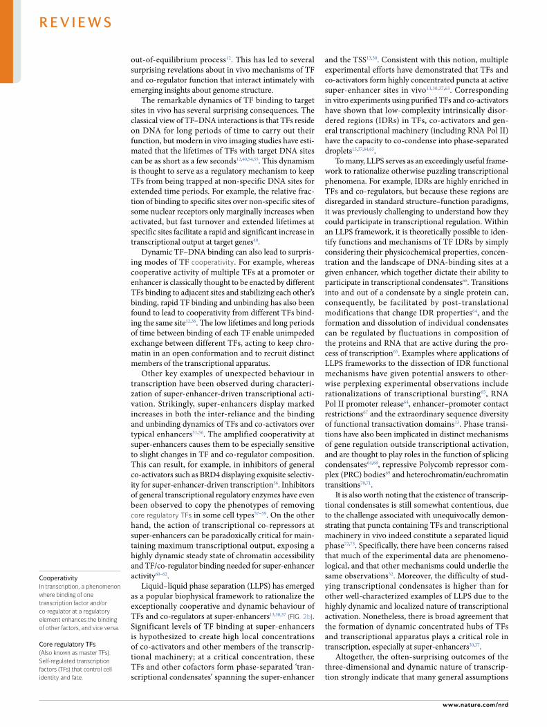
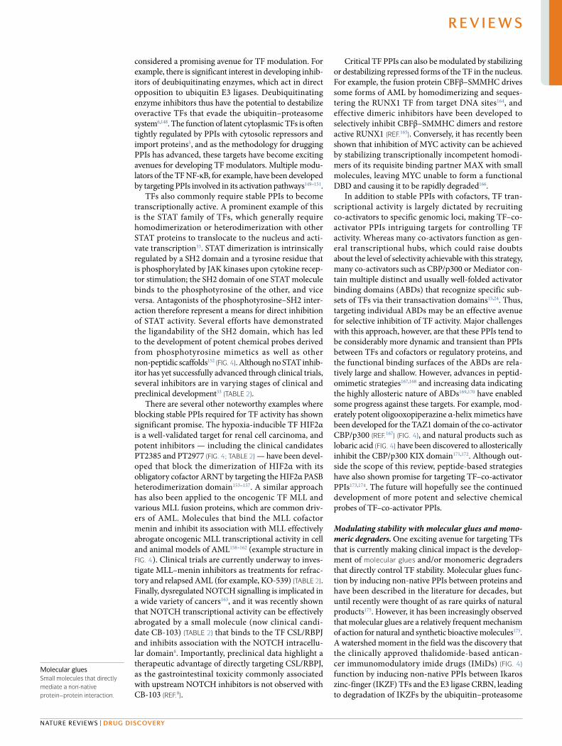
Degrading TFs with proteolysis targeting chimaeras. PROTACs offer a more rational way to design TF degraders192–194. PROTACs are bivalent molecules com-prising a ligand that binds to a target protein linked to a ligand that recruits a ubiquitin E3 ligase (Fig. 5). Similar to the IMiD drugs, PROTACs function by inducing a neo- interaction between the target protein and an E3 ligase that leads to degradation of the target protein by the ubiquitin–proteasome system. However, in contrast to molecular glue and monomeric degraders, PROTACs are modular and, in theory, any ligand that binds to a target protein has the potential to be turned into a degrader.
PROTACs have several advantages over standard occupancy- based antagonists that make them an espe-cially enticing modality for targeting TFs. Because TFs frequently contain several distinct protein domains of varying functional activity, antagonists of single domains are often not sufficient to effectively inhibit TF activity195. By degrading a target protein, a PROTAC inhibits all of its functions rather than just the activity of a single domain196–199. PROTACs also gain several bene-fits from their catalytic mechanism of action, including an increased durability of response from removal of the target protein and lowered susceptibility to resistance mutations that decrease affinity of the target ligand200–204. Further, families of TFs frequently contain a substantial degree of sequence conservation that makes the opti-mization of target selectivity for traditional inhibitors a challenging task. Because PROTAC selectivity is not governed by molecular recognition alone but by a com-bination of ternary complex formation, protein turnover rates and availability of target Lys residues for ubiqui-tylation, even highly promiscuous ligands have been shown to make surprisingly selective degraders205–209. Together, several of the traditional challenges associated with developing TF modulators have the potential to be overcome by PROTAC degraders.
Various mechanisms
Proteasomaldegradation
BindsE3 ligase
Bindstarget
Linker
a
b
PROTAC
Target
TargetUb
UbUb
Monomericdegrader
E3 ligase
Fig. 5 | overview of monomeric and ProTAc-based targeted protein degradation strategies. a | Monomeric degraders function by binding the target of interest and inducing its degradation through a variety of mechanisms (for example, inducing neo- protein–protein interactions with ubiquitin E3 ligases in the case of immunomodulatory imide drugs (IMiDs))178. b | Proteolysis targeting chimaera (PROTAC) degraders co- opt the ubiquitin–proteasome system by linking a binder of the target of interest (red) to a ligand for a ubiquitin E3 ligase (purple), which induces catalytic ubiquitylation and degradation of the target protein.
www.nature.com/nrd
R e v i e w s
0123456789();:
Widespread recognition of the promise of targeting TFs with PROTACs is illustrated by the fact that the first PROTAC degraders entering into clinical trials target the androgen and oestrogen nuclear receptors210 (ARV-110 and ARV-471) (TABLe 2). Despite concerns about the pharmacokinetic/pharmacodynamic properties of large PROTAC molecules211, preliminary data have indi-cated that these molecules can still be effective even in otherwise refractory tumours. Several other PROTACs for TFs and TF co- regulators have also been developed that show additional benefits over occupancy probes, including degraders of STAT3 (reF.195), MDM2 (reF.212) and BRD4 (reFS213,214) (for example, the highly effective STAT3 PROTAC SD-36) (Fig. 4).
Blocking aberrant transcriptional activity via TF collaborators. One rather surprising insight from recent studies of TF action is that chemical perturba-tion of individual general transcriptional cofactors such as transcriptional kinases, epigenetic proteins and co- activators can be highly specific to cell- identity transcriptional programmes. This has been linked to the fact that cell- identity genes are frequently con-trolled by super- enhancers, which are typically more sensitive to inhibition of general cofactors over typical enhancers due to their extraordinarily cooperative nature51,53,56,96. For example, it has been shown that inhib-ition of chromatin readers (BRD4)56,95,96,215,216, transcrip-tional kinases (CDK7, CDK9, CDK12)58,59,217, histone modifying enzymes (histone deacetylases (HDACs), histone acetyltransferases (HATs) and so on)60,61,218 and chromatin remodellers (BAF, CHD4)62,219 can be quite spe-cific to super- enhancer gene expression. A high degree of selectivity for specific super- enhancer- driven tran-scriptional programmes has even been observed for the CDK9 inhibitor KB-0742 and the BRD4 bromodomain 2 (BD2) inhibitor ABBV-744 (reFS59,215) (Fig. 4), which is likely caused by variation in the cofactor composition of individual super- enhancers based on differing recruit-ment preferences of individual TFs. Therapeutic win-dows for general transcriptional cofactor inhibitors may therefore be unexpectedly large. Together, this indicates that targeting TF collaborators is a promising avenue to selectively inhibit dysregulated transcription in diseases driven by super- enhancers, such as cancer.
Ongoing challenges, emerging solutionsAlthough there have been many successes in developing TF modulators over the past two decades, there are still several obstacles in the way of targeting more members of this protein class. Below, we outline current hurdles for targeting undrugged TFs and new strategies and technologies that have the potential to overcome these barriers.
Directly targeting TFs without traditionally ligandable domains. The vast majority of well- validated and potent small- molecule modulators that directly bind TFs tar-get those that contain either a small- molecule binding domain (nuclear receptors, TEADs)10,130 or a structured dimerization domain (STATs, HIF2α, BCL-6)79,152,153,186. In the case of the IMiD molecular glues, the primary
binding site of the molecule resides in a well- defined pocket in the non- TF partner176,179. Successfully targeting TFs has therefore relied heavily on the presence of well- defined protein folds with ligandable pockets. However, a shared feature of TFs is that they contain a significant degree of structural disorder11, and outside the cellular context many TFs are predicted to be almost entirely intrinsically disordered. This is a significant impedi-ment for modern targeted drug discovery strategies that heavily rely upon structural information to guide ligand optimization, such as fragment- based drug design220,221.
Recent advances in our basic biophysical under-standing of intrinsically disordered proteins (IDPs) along with the development of experimental techniques to characterize IDP structure hold significant promise for overcoming the many challenges imposed by TF disor-der. For example, despite their historical portrayal as floppy noodles completely devoid of three- dimensional structure28, IDPs display a significant degree of struc-tural variation. Depending on the amino acid sequence, IDP structures vary from extended dynamic chains, to condensed but disordered globules, all the way to struc-tural ensembles containing transient but well- defined folds222–227. Many disordered TFs, especially in the lat-ter category, may therefore contain transiently formed pockets that could be exploited to develop ligands.
Realization of this goal of developing ligands for dis-ordered TFs will require the adoption of experimental techniques that are highly suited to IDP characterization. Standard structural techniques such as X- ray crystallo-graphy and cryo- electron microscopy unfortunately fare exceptionally poorly at resolving disordered regions. On the other hand, the combined use of NMr spectroscopy and molecular dynamics approaches has been shown to be highly suited for characterizing IDPs at the atomic level, and thus could serve as excellent tools to identify and characterize transient pockets and their interactions with ligands228–231. Additional approaches with these tech-niques for characterizing IDP–ligand interactions will be very important, as unoptimized screening hits for IDPs will likely bind in a highly dynamic manner that makes successful ligand optimization challenging123,232,233.
Targeted protein degradation. The majority of reported TF PROTACs are derived from ligands developed to tar-get either nuclear receptor LBDs or TF protein–protein interfaces195,234–237. This reliance on previously devel-oped ligands results in a strong bias of TF PROTAC development towards already druggable TFs. There is therefore significant untapped potential of this technol-ogy for targeting currently intractable TFs, and several aspects of PROTAC design and mechanism may actually make them especially suited for challenging TFs over traditional occupancy- based modulators.
A sometimes underappreciated fact about PROTACs is that they obviate the requirement for occupancy- based ligands to not only bind to a protein but also modulate its function. That is, a small- molecule binder can tar-get any region of a protein — even functionally inac-tive domains — and still be able to serve as an effective PROTAC because the E3 recruiting module produces the functional effect197,199,238. Furthermore, large- scale
Histone deacetylases(HDACs). enzymes that remove acetyl groups from acetylated Lys residues in histones. generally associated with closed chromatin conformation and transcriptional repression.
Histone acetyltransferases(HATs). enzymes that transfer acetyl groups to the ε- amino group of Lys residues in histones. generally associated with open chromatin conformation and increased transcription.
Chromatin remodellersProtein complexes with a common ATPase domain that use ATP hydrolysis to move, reposition or eject nucleosomes.
Intrinsically disordered proteins(iDPs; also known as intrinsically disordered regions (iDrs)). Proteins or regions of proteins that do not adopt a well- defined structure. Often characterized as ‘ensembles’ of many unrelated protein conformations, although many iDPs/iDrs can transiently form more defined structures.
NMR spectroscopyA structural technique that utilizes the quantum- mechanical properties of nuclear spins in a magnetic field. Used in structural biology to determine protein structure, as well as to characterize conformational dynamics across a wide range of timescales (general range of picoseconds to hours/days).
Molecular dynamicsA computational technique that is used to characterize the structure and conformational dynamics of proteins by simulating the interactions between all the atoms of a protein and its surrounding solvent over time.
Occupancy- based modulatorsModulators of protein function in drug discovery where the overall change in protein activity from drug treatment is determined by the concentration of the drug in the cell and its affinity for its target. For complete inhibition of activity by an inhibitor, the drug must reach concentrations several times above its dissociation constant (Kd).
Nature reviews | Drug Discovery
R e v i e w s
0123456789();:
proteomics studies with kinase degraders have shown that even relatively weak binders of the target protein can still make highly potent PROTACs200,205,209. Together, these characteristics have the potential to overcome the major challenges and trade- offs associated with design-ing potent and functional modulators of TFs. Ligand development efforts for PROTACs can therefore focus on optimizing lead binders without the additional con-straints of activity and/or affinity thresholds necessary to be viable for use as therapeutics or chemical probes. A recent example that highlights the potential of this approach is the development of effective PROTACs directly targeting the ‘undruggable’ androgen receptor ∆LBD splice variant ArV7 from ligands that moderately inhibit the androgen receptor DBD239,240. Together with potential advances in developing ligands of dynamic and disordered regions, the PROTAC approach could open up a significant portion of TF target space.
However, several challenges still stand in the way of expanding the repertoire of TF PROTACs241. Not all E3 ligases can effectively degrade a given protein, and there are currently only a handful of E3 ligase recruit-ing ligands to choose from213,214,242,243. Current efforts are underway to add additional E3 ligases to the PROTAC toolset. An especially exciting preliminary development is the discovery of covalent E3 binders that irreversibly convert an E3 ligase into a destruction complex tar-geted towards a specific protein244–248. Additionally, it is exceptionally challenging to optimize non- functional small- molecule binders of TFs for use as PROTAC han-dles, as there are very few quantitative direct binding assays that can be used in the likely essential context of the cell249. Finally, there is considerable need to develop workflows to rapidly assess which ligands can be turned into effective PROTACs to minimize the time spent opti-mizing scaffolds that are not well suited for development into PROTACs250.
Binding- focused screening. Binding- focused screening strategies251 are increasingly being recognized as highly promising methods for the discovery of TF modula-tors. These approaches are especially relevant to the development of PROTAC strategies for TFs, which can use even non- functional binders as the foundation for effective degraders. Two technologies have demon-strated significant potential for targeting TFs: small- molecule microarray (SMM) and covalent screening approaches252–254.
The SMM screening approach utilizes libraries of small molecules covalently anchored to glass slides as an extremely rapid, versatile and functionally agnostic way to screen for small molecule–protein interactions252,253. Critically, interactions between putative small- molecule binders and a protein target can be detected by fluo-rescent antibodies specific to the protein or an added epitope tag. This enables screens to be run against not only purified proteins but also specific proteins in highly complex environments such as cellular lysates252. The latter can even be exploited to screen endogenous proteins from disease- relevant cell lines. Ligands for challenging TFs such as ETV1, SHP and MAX have been discovered using SMM screening, supporting
the potential of this technique for directly targeting TFs166,255,256. Furthermore, SMM screens using cellular lysates have the potential to identify ligands of critical TF binding partners and collaborators. This was recently illustrated by the discovery of a selective CDK9 inhibitor from a lysate screen against the ‘undruggable’ androgen receptor splice variant ARV7 (reF.59). SMM screening is thus an exceptionally useful approach for drugging TFs and their collaborators, that does not require significant protein engineering or prior knowledge to implement effectively. However, one potential drawback to this approach is that it often requires significant effort to dissect the mechanism of action of candidate molecules identified by SMM screens.
Covalent screening approaches use libraries of mole-cules with reactive warheads — most often Cys- reactive moieties such as acrylamides or chloroacetamides — to screen for covalent binding of molecules to a target protein254,257,258. Mass spectrometry is typically used to identify covalent binders, enabling significant versatil-ity for this approach. For example, although it is most common to screen covalent libraries against purified proteins, it is also possible to screen for covalent bind-ing in live cells so long as the target of interest is detect-able by the mass spectrometry technique used254,257. Excitement for this area of drug discovery, in general, has been increasing since the discovery of covalent ligands and clinical candidates for the ‘undruggable’ oncogenic KRAS- G12C mutant259–261, and many con-sider it an approach well suited for challenging targets such as TFs. Supporting this idea, unbiased screening of Cys- reactive small- molecule libraries in living cells has identified previously unknown ligandable sites in many undrugged TFs254. The recent identification of a cova-lent MYC inhibitor that selectively binds to a MYC IDR suggests that covalent approaches may even have the potential to overcome the traditional challenges associ-ated with targeting heavily disordered proteins262. There is thus significant potential for covalent approaches to open further TF target space. However, it bears noting that target identification is an absolute critical step of covalent inhibitor development because of the possibility of off- target reactivity driven by the warhead, and it is still challenging to conduct targeted screens in cells with low- abundance or unstable proteins.
Finally, one of the most popular binding- focused screening approaches in recent years has been DNA- encoded libraries (DELs), which are massive libraries of small molecules that are tagged with individ-ual DNA ‘barcodes’263–265. Because DELs are generated combinatorially and individual molecules are directly attached to a DNA record of all chemical transforma-tions performed on the scaffold, it is possible to screen mixtures of billions of compounds at once by using PCR amplification to detect binders. This approach is there-fore especially appealing for TFs, which are typically characterized by very low hit rates in traditional screens. New approaches to synthesizing DELs are even being developed to significantly increase the chemical diversity of molecules in individual screening sets, which is histor-ically a weakness of DELs266,267. However, this approach has not yet realized significant success with TFs. A key
ARV7A splice variant of the androgen receptor that lacks the ligand- binding domain.
www.nature.com/nrd
R e v i e w s
0123456789();:
challenge is that DEL screening with TFs is currently relegated to purified protein screens because the req-uisite DNA tag interferes with cellular penetration. TFs that adopt structure only in the presence of binding partners will thus be more difficult to screen effectively. Further, screening full- length TFs by this approach also poses a challenge because non- specific binding of DBDs to the DNA barcodes can interfere with the results. Nonetheless, the DEL approach has significant potential for generating chemical matter against difficult targets, and its use for TFs should be encouraged.
Target engagement and selectivity. Key to the develop-ment of targeted therapeutics and chemical probes, in general, is being able to tell that a molecule produces its phenotypic effects via engagement with the intended tar-get and not by any number of off- target mechanisms268. Many ‘undruggable’ TFs are especially challenging in this regard due to difficulties with obtaining high- quality structural, biophysical binding and cellular engagement data123,255,262. Unsurprisingly, many putative TF inhib-itors contain obvious pan- assay interference scaffolds such as rhodanines, quinones and/or other liabilities that could have been readily deprioritized or ruled out with the use of pan- assay interference scaffold filters or reliable biophysical and cellular validation assays269–271. Furthermore, because inhibition of general transcrip-tional cofactors can, in some cases, phenocopy TF inhibition profiles59,62,215, it is essential to obtain unequiv-ocal evidence that a molecule functions through direct engagement with a TF.
Several technologies have evolved in recent years to take on the challenges of determining target engage-ment and selectivity for small- molecule ligands. Direct binding approaches have been an area of sig-nificant impact with the development of cellular ther-mal shift assays (CETSAs) and photoaffinity labelling approaches249,272–274. Proteome- wide CETSAs essentially measure shifts in the melting temperature of proteins in the cell upon treatment with a ligand, which indi-cates likely molecular targets. Photoaffinity labelling approaches make use of photolabels attached to a ligand to form a covalent bond between the ligand and target protein(s) when treated with UV light. Both of these techniques can be used in living cells, giving an unprec-edented look at the global target profile of a ligand. However, both techniques have drawbacks to consider. Whereas the CETSA is one of the most straightfor-ward and practical techniques to perform, it is prone to false positives because perturbation of PPIs can also cause thermal shifts in the binding partners of the true target262,275. Given that TFs spend much of their time in complex with other proteins, this must be considered when evaluating any CETSA- based TF engagement data. On the other hand, photoaffinity labelling can give une-quivocal evidence of direct binding because the photo-label (typically a diazirine) forms a strong covalent bond to the molecular target(s), but the technique requires modification of the ligand to add the photoaffinity label. In addition, photoaffinity labelling approaches have the potential to miss ‘true’ targets due to uneven amino acid reactivity preferences of the photolabel276.
For PROTACs and covalent ligands, cellular target identification is generally rather straightforward because of the ability to use proteomics approaches. PROTACs function by degrading their targets, and therefore proteomics can be directly used to identify targets that are downregulated by PROTAC treatment205,209. However, a significant drawback is that if the degrader is slow- acting, then it may be challenging to determine whether it is degraded directly by the PROTAC or is indirectly downregulated by other means209. For Cys covalent ligands in particular, specific proteomics tech-nologies have been developed to enable proteome- wide screens of free Cys residues. The most widely used tech-nique is isotopic tandem orthogonal proteolysis- enabled activity- based protein profiling (isoTOP ABPP), which assays all reduced Cys residues that can react with a non- selective iodoacetamide ABPP probe254,262,277,278. Targets of Cys- reactive ligands can thereby be identified by a reduction in labelling by the ABPP probe upon pre-treatment by the ligand. One remaining challenge with this approach is that the ABPP probe cannot currently detect all reduced Cys residues in the proteome and the technique can be especially difficult to implement with unstable or low- abundance proteins262.
Functional target identification approaches have also become an area of significant innovation that bear con-sideration for use in any TF ligand discovery campaign. Classically, the gold standard of target identification is to test ligands against cell lines bearing point muta-tions of the putative target that inhibit molecule binding without affecting function, because this directly relates binding of a ligand to its functional effects279. However, this approach can be quite challenging for TFs outside nuclear receptors because the ligand binding sites are often challenging to identify and characterize, making generation of effective inhibitory mutations difficult. Significant potential for rapid target identification has recently been demonstrated by unbiased CRISPR dependency screening, to identify targets that confer effectiveness to a ligand280,281. However, for TFs, results from knockdown- based screens must be taken with caution because cells that are addicted to the target TF’s activity may not tolerate its knockdown.
Together, the target engagement and identification approaches described in this section represent imperfect but invaluable tools for TF drug discovery campaigns. Given the historical challenges associated with develop-ing selective TF modulators, use of one or ideally several of these approaches should be considered essential for the development of any new TF- targeting ligand.
Targeting phase- separated condensates. The emergence of data linking transcriptional activation to the forma-tion of phase- separated transcriptional condensates13,37 naturally invokes questions about how the physico-chemical properties of condensates could influence the action of TF- targeting ligands. Indeed, in vitro studies have indicated that drugs and drug candidates that tar-get TF and/or TF co- regulators often have significant preferences for entering transcriptional condensates over other types of condensates, even in the absence of the ligand binding site in the target protein282. It is therefore
Nature reviews | Drug Discovery
R e v i e w s
0123456789();:
pertinent to evaluate the physicochemical properties associated with partitioning into condensates, which could have a significant effect on a ligand’s ability to reach its target. Moreover, it will be extremely important to gain an understanding of the circumstances where entering a condensate is required for drug action. For certain approaches, it may even be advantageous to engage a TF outside transcriptional condensates; for example, a PROTAC that preferentially partitions into transcriptional condensates may have difficulties reach-ing an E3 ligase outside the condensate. At present, there are many more questions than answers regarding the relationship between condensates and drug action, but future insights may have significant impact on the ways we approach TF- targeting and ligand design.
OutlookThe field of targeting TFs has come a long way since James Darnell penned his eponymous call to arms almost two decades ago5. Indeed, some of the TFs he nominated as appealing targets (for example, STAT TFs) have seen significant drug discovery efforts, with promising molecules in preclinical development or entering clinical trials (TABLe 2). Yet there are still many more TFs that have not been successfully targeted. In the near term, innovations in identifying and target-ing TF collaborators and the continued development of TF PPI inhibitors (especially for TF regulatory domains) appear to have the most significant translational potential to deliver additional TF modulators and drugs.
However, even with these more tried and true approaches, there are still major challenges associated with targeting TFs as a class that must be addressed over the longer term. Even for well- studied TFs, there are still many mysteries about the molecular determi-nants of function that produce roadblocks to rationally devise ways to effectively drug them. For understudied TFs, this challenge is made orders of magnitude more difficult. Therefore, it is essential to adopt or develop approaches that suit the specific challenges of this target class to enable continued success in this field.
One excellent example of a set of related approaches that appear to be highly suited to TF modulation are proximity- inducing bifunctional molecules such as PROTACs. The induced proximity strategy, by essen-tially splitting ligand binding and function, can negate the need to identify and target functionally relevant surfaces of TFs. Although this approach has been rel-egated to developing PROTACs for already druggable TFs, it can and should be used for currently intractable TFs. Looking towards the future, the addition of novel induced proximity approaches to the chemical biology toolbox, for example phosphorylation- inducing chimae-ric small molecules283, may even enable these approaches to have more fine- tuned control over TF action.
Additionally, the pervasive level of intrinsic disorder in TFs is still a significant hurdle to developing potent ligands. Unbiased binding- based approaches can help to uncover the ligandable sites of TFs, especially when cou-pled with extensive biophysical dissection to determine the binding modes of promising ligands. Indeed, there are several promising hints that some IDRs may indeed be ligandable. For example, drug- like direct binders of the disordered TFs ETV1 (reF.255), EWS–FLI1 (reF.284) and MYC262, along with binders of disordered AF1 regions of nuclear receptors285,286, have been reported in the litera-ture. Unfortunately, in some of these reported cases, the functional link between the ligand- produced phenotype and IDR binding has not been fully elucidated, under-scoring the need for rigorous target engagement and identification studies on all putative TF binders.
Although once thought of as almost entirely undrug-gable, it has become clear that, with the right set of tools and sustained effort, many TFs can be successfully tar-geted. With several promising strategies emerging, the future holds exhilarating potential for drug discoverers to access new areas of the TF target class. Moreover, given the foundational role TFs play in numerous dis-eases, the benefits to patients from drugging TFs cannot be overstated.
Published online xx xx xxxx
1. Lambert, S. A. et al. The human transcription factors. Cell 172, 650–665 (2018).
2. Vernimmen, D. & Bickmore, W. A. The hierarchy of transcriptional activation: from enhancer to promoter. Trends Genet. 31, 696–708 (2015).
3. Lee, T. I. & Young, R. A. Transcriptional regulation and its misregulation in disease. Cell 152, 1237–1251 (2013). This review is a valuable reference for understanding the mechanisms by which transcription is dysregulated in disease.
4. Brivanlou, A. H. & Darnell, J. E. Signal transduction and the control of gene expression. Science 295, 813–818 (2002).
5. Darnell, J. E. Transcription factors as targets for cancer therapy. Nat. Rev. Cancer 2, 740–749 (2002).
6. Bushweller, J. H. Targeting transcription factors in cancer — from undruggable to reality. Nat. Rev. Cancer 19, 611–624 (2019).
7. Shah, D. R., Shah, R. R. & Morganroth, J. Tyrosine kinase inhibitors: their on- target toxicities as potential indicators of efficacy. Drug Saf. 36, 413–426 (2013).
8. Lehal, R. et al. Pharmacological disruption of the Notch transcription factor complex. Proc. Natl Acad. Sci. USA 117, 16292–16301 (2020). This paper demonstrates pharmacological advantages for directly targeting TFs over upstream signalling proteins and is one of the few studies that use functional target identification
methods to support an on- target mechanism for an ‘undruggable’ TF inhibitor.
9. Lovly, C. M. & Shaw, A. T. Molecular pathways: resistance to kinase inhibitors and implications for therapeutic strategies. Clin. Cancer Res. 20, 2249–2256 (2014).
10. Gronemeyer, H., Gustafsson, J.-Å. & Laudet, V. Principles for modulation of the nuclear receptor superfamily. Nat. Rev. Drug Discov. 3, 950–964 (2004).
11. Liu, J. et al. Intrinsic disorder in transcription factors. Biochemistry 45, 6873–6888 (2006).
12. Voss, T. C. & Hager, G. L. Dynamic regulation of transcriptional states by chromatin and transcription factors. Nat. Rev. Genet. 15, 69–81 (2014). This article presents a comprehensive review of spatial and temporal mechanisms of transcriptional regulation, and highlights the importance of studying transcription from the perspective of single TF molecules.
13. Boija, A. et al. Transcription factors activate genes through the phase- separation capacity of their activation domains. Cell 175, 1842–1855.e16 (2018).
14. Ptashne, M. & Gann, A. Transcriptional activation by recruitment. Nature 386, 569–577 (1997).
15. Allen, B. L. & Taatjes, D. J. The Mediator complex: a central integrator of transcription. Nat. Rev. Mol. Cell Biol. 16, 155–166 (2015).
16. Centore, R. C., Sandoval, G. J., Soares, L. M. M., Kadoch, C. & Chan, H. M. Mammalian SWI/SNF chromatin remodeling complexes: emerging mechanisms and therapeutic strategies. Trends Genet. 36, 936–950 (2020).
17. Lee, T. I. & Young, R. A. Transcription of eukaryotic protein- coding genes. Annu. Rev. Genet. 34, 77–137 (2000).
18. Spitz, F. & Furlong, E. E. M. Transcription factors: from enhancer binding to developmental control. Nat. Rev. Genet. 13, 613–626 (2012).
19. Giacinti, C. & Giordano, A. RB and cell cycle progression. Oncogene 25, 5220–5227 (2006).
20. Rohs, R. et al. Origins of specificity in protein–DNA recognition. Annu. Rev. Biochem. 79, 233–269 (2010).
21. Goldstein, I. & Hager, G. L. Dynamic enhancer function in the chromatin context: dynamic mechanism for enhancer activation. Wiley Interdiscip. Rev. Syst. Biol. Med. 10, e1390 (2018).
22. Dames, S. A., Martinez- Yamout, M., De Guzman, R. N., Dyson, H. J. & Wright, P. E. Structural basis for Hif-1/CBP recognition in the cellular hypoxic response. Proc. Natl Acad. Sci. USA 99, 5271–5276 (2002).
23. Zor, T., De Guzman, R. N., Dyson, H. J. & Wright, P. E. Solution structure of the KIX domain of CBP bound to the transactivation domain of c- Myb. J. Mol. Biol. 337, 521–534 (2004).
www.nature.com/nrd
R e v i e w s
0123456789();:
24. Dyson, H. J. & Wright, P. E. Role of intrinsic protein disorder in the function and interactions of the transcriptional coactivators CREB- binding protein (CBP) and p300. J. Biol. Chem. 291, 6714–6722 (2016).
25. Ma, J. & Ptashne, M. A new class of yeast transcriptional activators. Cell 51, 113–119 (1987).
26. Warfield, L., Tuttle, L. M., Pacheco, D., Klevit, R. E. & Hahn, S. A sequence- specific transcription activator motif and powerful synthetic variants that bind Mediator using a fuzzy protein interface. Proc. Natl Acad. Sci. USA 111, E3506–E3513 (2014).
27. Staller, M. V. et al. A high- throughput mutational scan of an intrinsically disordered acidic transcriptional activation domain. Cell Syst. 6, 444–455.e6 (2018).
28. Sigler, P. B. Acid blobs and negative noodles. Nature 333, 210–212 (1988).
29. Tuttle, L. M. et al. Gcn4-mediator specificity is mediated by a large and dynamic fuzzy protein–protein complex. Cell Rep. 22, 3251–3264 (2018).
30. Chong, S. et al. Imaging dynamic and selective low-complexity domain interactions that control gene transcription. Science 361, eaar2555 (2018).
31. Henley, M. J. et al. Unexpected specificity within dynamic transcriptional protein–protein complexes. Proc. Natl Acad. Sci. USA 117, 27346–27353 (2020).
32. McSwiggen, D. T. et al. Evidence for DNA- mediated nuclear compartmentalization distinct from phase separation. eLife 8, e47098 (2019).
33. Loh, C.-Y. et al. Signal transducer and activator of transcription (STATs) proteins in cancer and inflammation: functions and therapeutic implication. Front. Oncol. 9, 48 (2019).
34. McEwan, I. J. in The Nuclear Receptor Superfamily Vol. 1443 (ed. McEwan, I. J.) 3–9 (Humana, 2016).
35. Jones, S. An overview of the basic helix–loop–helix proteins. Genome Biol. 5, 226 (2004).
36. Voss, T. C. et al. Dynamic exchange at regulatory elements during chromatin remodeling underlies assisted loading mechanism. Cell 146, 544–554 (2011).
37. Sabari, B. R. et al. Coactivator condensation at super-enhancers links phase separation and gene control. Science 361, eaar3958 (2018). This study represents the first demonstration that super- enhancers can concentrate transcriptional apparatus into phase- separated condensates.
38. Szabo, Q., Bantignies, F. & Cavalli, G. Principles of genome folding into topologically associating domains. Sci. Adv. 5, eaaw1668 (2019).
39. Beagan, J. A. & Phillips- Cremins, J. E. On the existence and functionality of topologically associating domains. Nat. Genet. 52, 8–16 (2020).
40. Paakinaho, V. et al. Single- molecule analysis of steroid receptor and cofactor action in living cells. Nat. Commun. 8, 15896 (2017).
41. Schoenfelder, S. & Fraser, P. Long- range enhancer–promoter contacts in gene expression control. Nat. Rev. Genet. 20, 437–455 (2019).
42. He, Y., Fang, J., Taatjes, D. J. & Nogales, E. Structural visualization of key steps in human transcription initiation. Nature 495, 481–486 (2013).
43. He, Y. et al. Near- atomic resolution visualization of human transcription promoter opening. Nature 533, 359–365 (2016).
44. Schilbach, S. et al. Structures of transcription pre-initiation complex with TFIIH and mediator. Nature 551, 204–209 (2017).
45. Bickmore, W. A. The spatial organization of the human genome. Annu. Rev. Genomics Hum. Genet. 14, 67–84 (2013).
46. Bonev, B. & Cavalli, G. Organization and function of the 3D genome. Nat. Rev. Genet. 17, 661–678 (2016).
47. Hnisz, D., Day, D. S. & Young, R. A. Insulated neighborhoods: structural and functional units of mammalian gene control. Cell 167, 1188–1200 (2016).
48. Dowen, J. M. et al. Control of cell identity genes occurs in insulated neighborhoods in mammalian chromosomes. Cell 159, 374–387 (2014).
49. Ghavi- Helm, Y. et al. Highly rearranged chromosomes reveal uncoupling between genome topology and gene expression. Nat. Genet. 51, 1272–1282 (2019).
50. Hnisz, D. et al. Super- enhancers in the control of cell identity and disease. Cell 155, 934–947 (2013).
51. Whyte, W. A. et al. Master transcription factors and mediator establish super- enhancers at key cell identity genes. Cell 153, 307–319 (2013).
52. Saint- André, V. et al. Models of human core transcriptional regulatory circuitries. Genome Res. 26, 385–396 (2016).
53. Hnisz, D. et al. Convergence of developmental and oncogenic signaling pathways at transcriptional super- enhancers. Mol. Cell 58, 362–370 (2015).
54. McNally, J. G. The glucocorticoid receptor: rapid exchange with regulatory sites in living cells. Science 287, 1262–1265 (2000).
55. Hager, G. L., McNally, J. G. & Misteli, T. Transcription dynamics. Mol. Cell 35, 741–753 (2009).
56. Lovén, J. et al. Selective inhibition of tumor oncogenes by disruption of super- enhancers. Cell 153, 320–334 (2013). This study is the first to show that super- enhancers can be more sensitive to inhibition of general transcriptional machinery than typical enhancers due to increased cooperativity of TFs and co-regulators.
57. Kwiatkowski, N. et al. Targeting transcription regulation in cancer with a covalent CDK7 inhibitor. Nature 511, 616–620 (2014).
58. Chipumuro, E. et al. CDK7 inhibition suppresses super- enhancer-linked oncogenic transcription in MYCN- driven cancer. Cell 159, 1126–1139 (2014). This paper shows that a covalent CDK7/12/13 inhibitor preferentially reduces MYCN- driven oncogenic transcriptional programmes by selectively targeting MYCN- associated super- enhancers.
59. Richters, A. et al. Modulating androgen receptor- driven transcription in prostate cancer with selective CDK9 inhibitors. Cell Chem. Biol. 28, 134–147.e14 (2021). This study identifies two potent and highly selective CDK9 inhibitors from a binding- focused screen of ‘undruggable’ ARV7 in cell lysates; similar to CDK7/12/13 inhibitors, these molecules show high selectivity for specific disease- related transcriptional programmes.
60. Gryder, B. E. et al. Chemical genomics reveals histone deacetylases are required for core regulatory transcription. Nat. Commun. 10, 3004 (2019).
61. Gryder, B. E. et al. Histone hyperacetylation disrupts core gene regulatory architecture in rhabdomyosarcoma. Nat. Genet. 51, 1714–1722 (2019).
62. Marques, J. G. et al. NuRD subunit CHD4 regulates super- enhancer accessibility in rhabdomyosarcoma and represents a general tumor dependency. eLife 9, e54993 (2020).
63. Wei, M.-T. et al. Nucleated transcriptional condensates amplify gene expression. Nat. Cell Biol. 22, 1187–1196 (2020).
64. Guo, Y. E. et al. Pol II phosphorylation regulates a switch between transcriptional and splicing condensates. Nature 572, 543–548 (2019).
65. Henninger, J. E. et al. RNA- mediated feedback control of transcriptional condensates. Cell 184, 207–225.e24 (2021).
66. Shrinivas, K. et al. Enhancer features that drive formation of transcriptional condensates. Mol. Cell 75, 549–561.e7 (2019).
67. Hnisz, D., Shrinivas, K., Young, R. A., Chakraborty, A. K. & Sharp, P. A. A phase separation model for transcriptional control. Cell 169, 13–23 (2017).
68. Li, W. et al. Biophysical properties of AKAP95 protein condensates regulate splicing and tumorigenesis. Nat. Cell Biol. 22, 960–972 (2020).
69. Plys, A. J. et al. Phase separation of Polycomb- repressive complex 1 is governed by a charged disordered region of CBX2. Genes Dev. 33, 799–813 (2019).
70. Gibson, B. A. et al. Organization of chromatin by intrinsic and regulated phase separation. Cell 179, 470–484.e21 (2019).
71. Sanulli, S. et al. HP1 reshapes nucleosome core to promote phase separation of heterochromatin. Nature 575, 390–394 (2019).
72. McSwiggen, D. T., Mir, M., Darzacq, X. & Tjian, R. Evaluating phase separation in live cells: diagnosis, caveats, and functional consequences. Genes Dev. 33, 1619–1634 (2019).
73. Mir, M., Bickmore, W., Furlong, E. E. M. & Narlikar, G. Chromatin topology, condensates and gene regulation: shifting paradigms or just a phase? Development 146, dev182766 (2019).
74. Bradner, J. E., Hnisz, D. & Young, R. A. Transcriptional addiction in cancer. Cell 168, 629–643 (2017).
75. Downward, J. Targeting RAS signalling pathways in cancer therapy. Nat. Rev. Cancer 3, 11–22 (2003).
76. O’Shea, J. J. et al. The JAK–STAT pathway: impact on human disease and therapeutic intervention. Annu. Rev. Med. 66, 311–328 (2015).
77. Taniguchi, K. & Karin, M. NF- κB, inflammation, immunity and cancer: coming of age. Nat. Rev. Immunol. 18, 309–324 (2018).
78. Kastenhuber, E. R. & Lowe, S. W. Putting p53 in context. Cell 170, 1062–1078 (2017).
79. Cardenas, M. G. et al. The expanding role of the BCL6 oncoprotein as a cancer therapeutic target. Clin. Cancer Res. 23, 885–893 (2017).
80. Dupain, C., Harttrampf, A. C., Urbinati, G., Geoerger, B. & Massaad- Massade, L. Relevance of fusion genes in pediatric cancers: toward precision medicine. Mol. Ther. Nucleic Acids 6, 315–326 (2017).
81. White, M. K., Pagano, J. S. & Khalili, K. Viruses and human cancers: a long road of discovery of molecular paradigms. Clin. Microbiol. Rev. 27, 463–481 (2014).
82. Flavahan, W. A. et al. Insulator dysfunction and oncogene activation in IDH mutant gliomas. Nature 529, 110–114 (2016).
83. Flavahan, W. A., Gaskell, E. & Bernstein, B. E. Epigenetic plasticity and the hallmarks of cancer. Science 357, eaal2380 (2017).
84. Ramsay, R. G. & Gonda, T. J. MYB function in normal and cancer cells. Nat. Rev. Cancer 8, 523–534 (2008).
85. Mansour, M. R. et al. An oncogenic super- enhancer formed through somatic mutation of a noncoding intergenic element. Science 346, 1373–1377 (2014).
86. Blackwell, T., Kretzner, L., Blackwood, E., Eisenman, R. & Weintraub, H. Sequence- specific DNA binding by the c- Myc protein. Science 250, 1149–1151 (1990).
87. Rahl, P. B. et al. c- Myc regulates transcriptional pause release. Cell 141, 432–445 (2010).
88. Rahl, P. B. & Young, R. A. MYC and transcription elongation. Cold Spring Harb. Perspect. Med. 4, a020990 (2014).
89. Lin, C. Y. et al. Transcriptional amplification in tumor cell elevated c- Myc. Cell 151, 56–67 (2012).
90. Schuijers, J. et al. Transcriptional dysregulation of MYC reveals common enhancer- docking mechanism. Cell Rep. 23, 349–360 (2018).
91. Felsher, D. W. & Bishop, J. M. Reversible tumorigenesis by MYC in hematopoietic lineages. Mol. Cell 4, 199–207 (1999).
92. Soucek, L. et al. Modelling Myc inhibition as a cancer therapy. Nature 455, 679–683 (2008).
93. Felsher, D. W. MYC inactivation elicits oncogene addiction through both tumor cell- intrinsic and host-dependent mechanisms. Genes Cancer 1, 597–604 (2010).
94. Tsherniak, A. et al. Defining a cancer dependency map. Cell 170, 564–576.e16 (2017).
95. Gryder, B. E. et al. PAX3–FOXO1 establishes myogenic super enhancers and confers BET bromodomain vulnerability. Cancer Discov. 7, 884–899 (2017).
96. Chapuy, B. et al. Discovery and characterization of super- enhancer-associated dependencies in diffuse large B cell lymphoma. Cancer Cell 24, 777–790 (2013).
97. Simmonds, R. E. & Foxwell, B. M. Signalling, inflammation and arthritis: NF- κB and its relevance to arthritis and inflammation. Rheumatology 47, 584–590 (2008).
98. Atreya, I., Atreya, R. & Neurath, M. F. NF- κB in inflammatory bowel disease. J. Intern. Med. 263, 591–596 (2008).
99. Leibowitz, S. M. & Yan, J. NF- κB pathways in the pathogenesis of multiple sclerosis and the therapeutic implications. Front. Mol. Neurosci. 9, 84 (2016).
100. Yue, Y., Stone, S. & Lin, W. Role of nuclear factor κB in multiple sclerosis and experimental autoimmune encephalomyelitis. Neural Regen. Res. 13, 1507 (2018).
101. Flanagan, S. E. et al. Activating germline mutations in STAT3 cause early- onset multi- organ autoimmune disease. Nat. Genet. 46, 812–814 (2014).
102. Milner, J. D. et al. Early- onset lymphoproliferation and autoimmunity caused by germline STAT3 gain- of-function mutations. Blood 125, 591–599 (2015).
103. Walford, H. H. & Doherty, T. A. STAT6 and lung inflammation. JAK- STAT 2, e25301 (2013).
104. Holland, S. M. et al. STAT3 mutations in the hyper- IgE syndrome. N. Engl. J. Med. 357, 1608–1619 (2007).
105. Skapenko, A., Leipe, J., Lipsky, P. E. & Schulze- Koops, H. The role of the T cell in autoimmune inflammation. Arthritis Res. Ther. 7, S4 (2005).
106. Ji, N., Sosa, R. A. & Forsthuber, T. G. More than just a T- box: the role of T- bet as a possible biomarker and therapeutic target in autoimmune diseases. Immunotherapy 3, 435–441 (2011).
107. Kanhere, A. et al. T- bet and GATA3 orchestrate TH1 and TH2 differentiation through lineage- specific targeting of distal regulatory elements. Nat. Commun. 3, 1268 (2012).
Nature reviews | Drug Discovery
R e v i e w s
0123456789();:
108. Capone, A. & Volpe, E. Transcriptional regulators of T helper 17 cell differentiation in health and autoimmune diseases. Front. Immunol. 11, 348 (2020).
109. Isono, F., Fujita- Sato, S. & Ito, S. Inhibiting RORγt/TH17 axis for autoimmune disorders. Drug Discov. Today 19, 1205–1211 (2014).
110. Bosnjak, B., Stelzmueller, B., Erb, K. J. & Epstein, M. M. Treatment of allergic asthma: modulation of TH2 cells and their responses. Respir. Res. 12, 114 (2011).
111. Fernando, V. et al. Regulation of an autoimmune model for multiple sclerosis in TH2-biased GATA3 transgenic mice. Int. J. Mol. Sci. 15, 1700–1718 (2014).
112. Tao, J.-H. et al. Foxp3, regulatory T cell, and autoimmune diseases. Inflammation 40, 328–339 (2017).
113. d’Hennezel, E. et al. FOXP3 forkhead domain mutation and regulatory T cells in the IPEX syndrome. N. Engl. J. Med. 361, 1710–1713 (2009).
114. Banerjee, S., Biehl, A., Gadina, M., Hasni, S. & Schwartz, D. M. JAK–STAT signaling as a target for inflammatory and autoimmune diseases: current and future prospects. Drugs 77, 521–546 (2017).
115. Udler, M. S., McCarthy, M. I., Florez, J. C. & Mahajan, A. Genetic risk scores for diabetes diagnosis and precision medicine. Endocr. Rev. 40, 1500–1520 (2019).
116. Pihoker, C. et al. Prevalence, characteristics and clinical diagnosis of maturity onset diabetes of the young due to mutations in HNF1A, HNF4A, and glucokinase: results from the SEARCH for diabetes in youth. J. Clin. Endocrinol. Metab. 98, 4055–4062 (2013).
117. Mitchell, S. M. S. & Frayling, T. M. The role of transcription factors in maturity- onset diabetes of the young. Mol. Genet. Metab. 77, 35–43 (2002).
118. Minra, N. & Tanaka, K. Analysis of the rat hepatocyte nuclear factor (HNF) 1 gene promoter: synergistic activation by HNF4 and HNF1 proteins. Nucleic Acids Res. 21, 3731–3736 (1993).
119. Grunert, M., Dorn, C. & Rickert- Sperling, S. in Congenital Heart Diseases: The Broken Heart (eds Rickert- Sperling, S., Kelly, R. G. & Driscoll, D. J.) 139–152 (Springer, 2016).
120. Kohli, S., Ahuja, S. & Rani, V. Transcription factors in heart: promising therapeutic targets in cardiac hypertrophy. Curr. Cardiol. Rev. 7, 262–271 (2012).
121. Epstein, J. A. & Buck, C. A. Transcriptional regulation of cardiac development: implications for congenital heart disease and DiGeorge syndrome. Pediatr. Res. 48, 717–724 (2000).
122. McCulley, D. J. & Black, B. L. in Current Topics in Developmental Biology Vol. 100 (ed. Bruneau, B. G.) 253–277 (Elsevier, 2012).
123. Hammoudeh, D. I., Follis, A. V., Prochownik, E. V. & Metallo, S. J. Multiple independent binding sites for small- molecule inhibitors on the oncoprotein c- Myc. J. Am. Chem. Soc. 131, 7390–7401 (2009). This study investigates the highly dynamic and non- specific binding modes of inhibitors of the disordered MYC TF; notably, the non- specific binding may be related to the promiscuous chemotypes of these molecules.
124. Baell, J. B. & Nissink, J. W. M. Seven year itch: pan- assay interference compounds (PAINS) in 2017—utility and limitations. ACS Chem. Biol. 13, 36–44 (2018).
125. Mazaira, G. I. et al. The nuclear receptor field: a historical overview and future challenges. Nucl. Recept. Res. 5, 101320 (2018).
126. Fernandez, E. J. Allosteric pathways in nuclear receptors — potential targets for drug design. Pharmacol. Ther. 183, 152–159 (2018).
127. de Vera, I. M. S. Advances in orphan nuclear receptor pharmacology: a new era in drug discovery. ACS Pharmacol. Transl. Sci. 1, 134–137 (2018).
128. Antonarakis, E. S. et al. AR- V7 and resistance to enzalutamide and abiraterone in prostate cancer. N. Engl. J. Med. 371, 1028–1038 (2014).
129. Zhu, Y. et al. Role of androgen receptor splice variant-7 (AR- V7) in prostate cancer resistance to 2nd- generation androgen receptor signaling inhibitors. Oncogene 39, 6935–6949 (2020).
130. Holden, J. K. & Cunningham, C. N. Targeting the Hippo pathway and cancer through the TEAD family of transcription factors. Cancers 10, 81 (2018).
131. Johnson, R. & Halder, G. The two faces of Hippo: targeting the Hippo pathway for regenerative medicine and cancer treatment. Nat. Rev. Drug Discov. 13, 63–79 (2014).
132. Dey, A., Varelas, X. & Guan, K.-L. Targeting the Hippo pathway in cancer, fibrosis, wound healing and regenerative medicine. Nat. Rev. Drug Discov. 19, 480–494 (2020).
133. Noland, C. L. et al. Palmitoylation of TEAD transcription factors is required for their stability and function in Hippo pathway signaling. Structure 24, 179–186 (2016).
134. Chan, P. et al. Autopalmitoylation of TEAD proteins regulates transcriptional output of the Hippo pathway. Nat. Chem. Biol. 12, 282–289 (2016).
135. Huh, H., Kim, D., Jeong, H.-S. & Park, H. Regulation of TEAD transcription factors in cancer biology. Cells 8, 600 (2019).
136. Pobbati, A. V. et al. Identification of quinolinols as activators of TEAD- dependent transcription. ACS Chem. Biol. 14, 2909–2921 (2019).
137. Bum- Erdene, K. et al. Small- molecule covalent modification of conserved cysteine leads to allosteric inhibition of the TEAD⋅Yap protein–protein interaction. Cell Chem. Biol. 26, 378–389.e13 (2019).
138. Holden, J. K. et al. Small molecule dysregulation of TEAD lipidation induces a dominant- negative inhibition of Hippo pathway signaling. Cell Rep. 31, 107809 (2020).
139. Kakiuchi- Kiyota, S., Schutten, M. M., Zhong, Y., Crawford, J. J. & Dey, A. Safety considerations in the development of Hippo pathway inhibitors in cancers. Front. Cell Dev. Biol. 7, 156 (2019).
140. Zhao, Y., Aguilar, A., Bernard, D. & Wang, S. Small- molecule inhibitors of the MDM2–p53 protein–protein interaction (MDM2 Inhibitors) in clinical trials for cancer treatment: miniperspective. J. Med. Chem. 58, 1038–1052 (2015).
141. Jones, S. N., Roe, A. E., Donehower, L. A. & Bradley, A. Rescue of embryonic lethality in Mdm2-deficient mice by absence of p53. Nature 378, 206–208 (1995).
142. Brown, C. J., Lain, S., Verma, C. S., Fersht, A. R. & Lane, D. P. Awakening guardian angels: drugging the p53 pathway. Nat. Rev. Cancer 9, 862–873 (2009).
143. Wells, M. et al. Structure of tumor suppressor p53 and its intrinsically disordered N- terminal transactivation domain. Proc. Natl Acad. Sci. USA 105, 5762–5767 (2008).
144. Estrada- Ortiz, N., Neochoritis, C. G. & Dömling, A. How to design a successful p53-MDM2/X interaction inhibitor: a thorough overview based on crystal structures. ChemMedChem 11, 757–772 (2016).
145. Chang, Y. S. et al. Stapled α- helical peptide drug development: a potent dual inhibitor of MDM2 and MDMX for p53-dependent cancer therapy. Proc. Natl Acad. Sci. USA 110, E3445–E3454 (2013).
146. Kaelin, W. G. Jr The von Hippel–Lindau tumour suppressor protein: O2 sensing and cancer. Nat. Rev. Cancer 8, 865–873 (2008).
147. Buckley, D. L. et al. Targeting the von Hippel–Lindau E3 ubiquitin ligase using small molecules to disrupt the VHL/HIF-1α interaction. J. Am. Chem. Soc. 134, 4465–4468 (2012). This paper is the first to demonstrate effective targeting of the TF HIF1α by the development of an inhibitor of the HIF1α- associated ubiquitin E3 ligase von Hippel–Lindau disease tumour suppressor.
148. Fraile, J. M., Quesada, V., Rodríguez, D., Freije, J. M. P. & López- Otín, C. Deubiquitinases in cancer: new functions and therapeutic options. Oncogene 31, 2373–2388 (2012).
149. Bruno, P. A., Morriss- Andrews, A., Henderson, A. R., Brooks, C. L. & Mapp, A. K. A synthetic loop replacement peptide that blocks canonical NF- κB signaling. Angew. Chem. Int. Ed. 55, 14997–15001 (2016).
150. Vincendeau, M. et al. Inhibition of canonical NF- κB signaling by a small molecule targeting NEMO–ubiquitin interaction. Sci. Rep. 6, 18934 (2016).
151. Maculins, T. et al. Discovery of protein–protein interaction inhibitors by integrating protein engineering and chemical screening platforms. Cell Chem. Biol. 27, 1441–1451.e7 (2020).
152. Miklossy, G., Hilliard, T. S. & Turkson, J. Therapeutic modulators of STAT signalling for human diseases. Nat. Rev. Drug Discov. 12, 611–629 (2013).
153. Rogers, J. L. et al. Development of inhibitors of the PAS- B domain of the HIF-2α transcription factor. J. Med. Chem. 56, 1739–1747 (2013).
154. Scheuermann, T. H. et al. Allosteric inhibition of hypoxia inducible factor-2 with small molecules. Nat. Chem. Biol. 9, 271–276 (2013). This paper describes the development and characterization of the first direct- binding allosteric inhibitor of the TF HIF2α.
155. Chen, W. et al. Targeting renal cell carcinoma with a HIF-2 antagonist. Nature 539, 112–117 (2016).
156. Wallace, E. M. et al. A small- molecule antagonist of HIF2α is efficacious in preclinical models of renal cell carcinoma. Cancer Res. 76, 5491–5500 (2016).
157. Xu, R. et al. 3-[(1S, 2S, 3R)-2,3-Difluoro-1-hydroxy-7-methylsulfonylindan-4-yl]oxy-5-fluorobenzonitrile (PT2977), a hypoxia- inducible factor 2α (HIF-2α) inhibitor for the treatment of clear cell renal cell carcinoma. J. Med. Chem. 62, 6876–6893 (2019).
158. Grembecka, J. et al. Menin–MLL inhibitors reverse oncogenic activity of MLL fusion proteins in leukemia. Nat. Chem. Biol. 8, 277–284 (2012). This paper describes the development and characterization of the first menin inhibitors that impede activity of the oncogenic TF MLL by blocking menin–MLL association.
159. Borkin, D. et al. Pharmacologic inhibition of the menin–MLL interaction blocks progression of MLL leukemia in vivo. Cancer Cell 27, 589–602 (2015).
160. Borkin, D. et al. Complexity of blocking bivalent protein–protein interactions: development of a highly potent inhibitor of the menin–mixed- lineage leukemia interaction. J. Med. Chem. 61, 4832–4850 (2018).
161. Klossowski, S. et al. Menin inhibitor MI-3454 induces remission in MLL1-rearranged and NPM1-mutated models of leukemia. J. Clin. Invest. 130, 981–997 (2020).
162. Xu, S. et al. Discovery of M-808 as a highly potent, covalent, small- molecule inhibitor of the menin–MLL interaction with strong in vivo antitumor activity. J. Med. Chem. 63, 4997–5010 (2020).
163. Kopan, R. & Ilagan, M. X. G. The canonical notch signaling pathway: unfolding the activation mechanism. Cell 137, 216–233 (2009).
164. Lukasik, S. M. et al. Altered affinity of CBFβ–SMMHC for Runx1 explains its role in leukemogenesis. Nat. Struct. Biol. 9, 674–679 (2002).
165. Illendula, A. et al. A small- molecule inhibitor of the aberrant transcription factor CBFβ–SMMHC delays leukemia in mice. Science 347, 779–784 (2015). This paper describes the first inhibitor of the oncogenic fusion CBFβ–SMMHC, which releases the RUNX1 TF from repressive CBFβ–SMMHC–RUNX1 complexes and delays progression of AML.
166. Struntz, N. B. et al. Stabilization of the Max homodimer with a small molecule attenuates Myc- driven transcription. Cell Chem. Biol. 26, 711–723.e14 (2019). This paper demonstrates that inhibition of the ‘undruggable’ TF MYC can be achieved by a small molecule that sequesters its requisite binding partner MAX into transcriptionally inactive homodimers.
167. Lao, B. B. et al. In vivo modulation of hypoxia- inducible signaling by topographical helix mimetics. Proc. Natl Acad. Sci. USA 111, 7531–7536 (2014).
168. Xie, X. et al. Targeting HPV16 E6-p300 interaction reactivates p53 and inhibits the tumorigenicity of HPV- positive head and neck squamous cell carcinoma. Oncogene 33, 1037–1046 (2014).
169. Wang, N. et al. Ordering a dynamic protein via a small- molecule stabilizer. J. Am. Chem. Soc. 135, 3363–3366 (2013).
170. Henderson, A. R. et al. Conservation of coactivator engagement mechanism enables small- molecule allosteric modulators. Proc. Natl Acad. Sci. USA 115, 8960–8965 (2018).
171. Cook, K. M. et al. Epidithiodiketopiperazines block the interaction between hypoxia- inducible factor-1α (HIF-1α) and p300 by a zinc ejection mechanism. J. Biol. Chem. 284, 26831–26838 (2009).
172. Majmudar, C. Y. et al. Sekikaic acid and lobaric acid target a dynamic interface of the coactivator CBP/p300. Angew. Chem. Int. Ed. 51, 11258–11262 (2012).
173. Ramaswamy, K. et al. Peptidomimetic blockade of MYB in acute myeloid leukemia. Nat. Commun. 9, 110 (2018).
174. Henchey, L. K. et al. Inhibition of hypoxia inducible factor 1—transcription coactivator interaction by a hydrogen bond surrogate α- Helix. J. Am. Chem. Soc. 132, 941–943 (2010).
175. Schreiber, S. L. The rise of molecular glues. Cell 184, 3–9 (2021).
176. Ito, T. et al. Identification of a primary target of thalidomide teratogenicity. Science 327, 1345–1350 (2010).
177. Kronke, J. et al. Lenalidomide causes selective degradation of IKZF1 and IKZF3 in multiple myeloma cells. Science 343, 301–305 (2014).
178. Lu, G. et al. The myeloma drug lenalidomide promotes the cereblon- dependent destruction of ikaros proteins. Science 343, 305–309 (2014). Together with Kronke et al. (2014), this study demonstrates that the anticancer IMiDs function by degrading the TFs IKZF1 and IKZF3.
www.nature.com/nrd
R e v i e w s
0123456789();:
179. Chamberlain, P. P. et al. Structure of the human cereblon–DDB1–lenalidomide complex reveals basis for responsiveness to thalidomide analogs. Nat. Struct. Mol. Biol. 21, 803–809 (2014).
180. Petzold, G., Fischer, E. S. & Thomä, N. H. Structural basis of lenalidomide- induced CK1α degradation by the CRL4CRBN ubiquitin ligase. Nature 532, 127–130 (2016).
181. Sievers, Q. L. et al. Defining the human C2H2 zinc finger degrome targeted by thalidomide analogs through CRBN. Science 362, eaat0572 (2018).
182. Isobe, Y. et al. Manumycin polyketides act as molecular glues between UBR7 and P53. Nat. Chem. Biol. 16, 1189–1198 (2020).
183. Hanan, E. J. et al. Monomeric targeted protein degraders. J. Med. Chem. 63, 11330–11361 (2020).
184. Dauvois, S., Danielian, P. S., White, R., Parker, M. G. & Antiestrogen, I. C. I. 164,384 reduces cellular estrogen receptor content by increasing its turnover. Proc. Natl Acad. Sci. USA 89, 4037–4041 (1992).
185. Wu, Y.-L. et al. Structural basis for an unexpected mode of SERM- mediated ER antagonism. Mol. Cell 18, 413–424 (2005).
186. Kerres, N. et al. Chemically induced degradation of the oncogenic transcription factor BCL6. Cell Rep. 20, 2860–2875 (2017). This paper describes the discovery of potent monomeric degraders of the TF BCL-6 from a medicinal chemistry campaign focused on optimizing BCL-6 PPI inhibitors.
187. Bellenie, B. R. et al. Achieving in vivo target depletion through the discovery and optimization of benzimidazolone BCL6 degraders. J. Med. Chem. 63, 4047–4068 (2020).
188. Słabicki, M. et al. Small- molecule-induced polymerization triggers degradation of BCL6. Nature 588, 164–168 (2020). This study defines a molecular glue mechanism of action for a potent BCL-6 monomeric degrader that involves polymerization of BCL-6 followed by degradation.
189. Faust, T. B. et al. Structural complementarity facilitates E7820-mediated degradation of RBM39 by DCAF15. Nat. Chem. Biol. 16, 7–14 (2020).
190. Powell, C. E. et al. Selective degradation of GSPT1 by cereblon modulators identified via a focused combinatorial library. ACS Chem. Biol. 15, 2722–2730 (2020).
191. Mayor- Ruiz, C. et al. Rational discovery of molecular glue degraders via scalable chemical profiling. Nat. Chem. Biol. 16, 1199–1207 (2020).
192. Sakamoto, K. M. et al. Protacs: chimeric molecules that target proteins to the Skp1–Cullin- F box complex for ubiquitination and degradation. Proc. Natl Acad. Sci. USA 98, 8554–8559 (2001).
193. Paiva, S.-L. & Crews, C. M. Targeted protein degradation: elements of PROTAC design. Curr. Opin. Chem. Biol. 50, 111–119 (2019).
194. Nalawansha, D. A. & Crews, C. M. PROTACs: an emerging therapeutic modality in precision medicine. Cell Chem. Biol. 27, 998–1014 (2020). This modern review succinctly highlights the advantages of PROTAC approaches and lays out a road map for the future development of this technology.
195. Bai, L. et al. A potent and selective small- molecule degrader of STAT3 achieves complete tumor regression in vivo. Cancer Cell 36, 498–511.e17 (2019). This paper showcases the advantages of degrading the STAT3 TF with PROTACs over the more traditional approach of disrupting homodimerization.
196. Gechijian, L. N. et al. Functional TRIM24 degrader via conjugation of ineffectual bromodomain and VHL ligands. Nat. Chem. Biol. 14, 405–412 (2018).
197. Bassi, Z. I. et al. Modulating PCAF/GCN5 immune cell function through a PROTAC approach. ACS Chem. Biol. 13, 2862–2867 (2018).
198. Cromm, P. M., Samarasinghe, K. T. G., Hines, J. & Crews, C. M. Addressing kinase- independent functions of Fak via PROTAC- mediated degradation. J. Am. Chem. Soc. 140, 17019–17026 (2018).
199. Degorce, S. L. et al. Discovery of proteolysis- targeting chimera molecules that selectively degrade the IRAK3 pseudokinase. J. Med. Chem. 63, 10460–10473 (2020).
200. Bondeson, D. P. et al. Catalytic in vivo protein knockdown by small- molecule PROTACs. Nat. Chem. Biol. 11, 611–617 (2015). This paper is the first to demonstrate that von Hippel–Lindau disease tumour suppressor (VHL)
ligands can serve as E3-ligase recruiting modules for PROTACs and also describes the catalytic mechanism of action of VHL- based degraders.
201. Fisher, S. L. & Phillips, A. J. Targeted protein degradation and the enzymology of degraders. Curr. Opin. Chem. Biol. 44, 47–55 (2018).
202. Buhimschi, A. D. et al. Targeting the C481S ibrutinib- resistance mutation in Bruton’s tyrosine kinase using PROTAC- mediated degradation. Biochemistry 57, 3564–3575 (2018).
203. Burslem, G. M. et al. The advantages of targeted protein degradation over inhibition: an RTK case study. Cell Chem. Biol. 25, 67–77.e3 (2018).
204. Mares, A. et al. Extended pharmacodynamic responses observed upon PROTAC- mediated degradation of RIPK2. Commun. Biol. 3, 140 (2020).
205. Huang, H.-T. et al. A chemoproteomic approach to query the degradable kinome using a multi- kinase degrader. Cell Chem. Biol. 25, 88–99.e6 (2018).
206. Nowak, R. P. et al. Plasticity in binding confers selectivity in ligand- induced protein degradation. Nat. Chem. Biol. 14, 706–714 (2018).
207. Bondeson, D. P. et al. Lessons in PROTAC design from selective degradation with a promiscuous warhead. Cell Chem. Biol. 25, 78–87.e5 (2018).
208. Brand, M. et al. Homolog- selective degradation as a strategy to probe the function of CDK6 in AML. Cell Chem. Biol. 26, 300–306.e9 (2019).
209. Donovan, K. A. et al. Mapping the degradable kinome provides a resource for expedited degrader development. Cell 183, 1714–1731.e10 (2020). This study describes the characterization of a large library of kinase PROTACs using chemoproteomics and demonstrates that the most potent and selective PROTACs are not necessarily derived from the most potent and selective inhibitors.
210. Mullard, A. First targeted protein degrader hits the clinic. Nat. Rev. Drug Discov. 18, 237–239 (2019).
211. Lipinski, C. A., Lombardo, F., Dominy, B. W. & Feeney, P. J. Experimental and computational approaches to estimate solubility and permeability in drug discovery and development settings. Adv. Drug Deliv. Rev. 46, 3–26 (2001).
212. Li, Y. et al. Discovery of MD-224 as a first- in-class, highly potent, and efficacious proteolysis targeting chimera murine double minute 2 degrader capable of achieving complete and durable tumor regression. J. Med. Chem. 62, 448–466 (2019).
213. Winter, G. E. et al. Phthalimide conjugation as a strategy for in vivo target protein degradation. Science 348, 1376–1381 (2015). This study is the first to demonstrate that IMiDs, which are primarily ligands of the E3 ligase CRBN, can be used as E3-ligase recruiting modules for PROTACs.
214. Zengerle, M., Chan, K.-H. & Ciulli, A. Selective small molecule induced degradation of the BET bromodomain protein BRD4. ACS Chem. Biol. 10, 1770–1777 (2015).
215. Faivre, E. J. et al. Selective inhibition of the BD2 bromodomain of BET proteins in prostate cancer. Nature 578, 306–310 (2020).
216. Gilan, O. et al. Selective targeting of BD1 and BD2 of the BET proteins in cancer and immuno- inflammation. Science 368, 387–394 (2020).
217. Iniguez, A. B. et al. EWS/FLI confers tumor cell synthetic lethality to CDK12 inhibition in Ewing sarcoma. Cancer Cell 33, 202–216.e6 (2018).
218. Lasko, L. M. et al. Discovery of a selective catalytic p300/CBP inhibitor that targets lineage- specific tumours. Nature 550, 128–132 (2017). This paper describes the development of the first potent and selective inhibitor of the HAT domain of the related transcriptional co- activators CBP and p300.
219. Schick, S. et al. Acute BAF perturbation causes immediate changes in chromatin accessibility. Nat. Genet. 53, 269–278 (2021).
220. Osborne, J., Panova, S., Rapti, M., Urushima, T. & Jhoti, H. Fragments: where are we now? Biochem. Soc. Trans. 48, 271–280 (2020).
221. Li, Q. Application of fragment- based drug discovery to versatile targets. Front. Mol. Biosci. 7, 180 (2020).
222. Riback, J. A. et al. Innovative scattering analysis shows that hydrophobic disordered proteins are expanded in water. Science 358, 238–241 (2017).
223. Marsh, J. A. & Forman- Kay, J. D. Sequence determinants of compaction in intrinsically disordered proteins. Biophys. J. 98, 2383–2390 (2010).
224. Banks, A., Qin, S., Weiss, K. L., Stanley, C. B. & Zhou, H.-X. Intrinsically disordered protein exhibits
both compaction and expansion under macromolecular crowding. Biophys. J. 114, 1067–1079 (2018).
225. Demarest, S. J. et al. Mutual synergistic folding in recruitment of CBP/p300 by p160 nuclear receptor coactivators. Nature 415, 549–553 (2002).
226. Kjaergaard, M., Teilum, K. & Poulsen, F. M. Conformational selection in the molten globule state of the nuclear coactivator binding domain of CBP. Proc. Natl Acad. Sci. USA 107, 12535–12540 (2010).
227. Naganathan, A. N. & Orozco, M. The native ensemble and folding of a protein molten- globule: functional consequence of downhill folding. J. Am. Chem. Soc. 133, 12154–12161 (2011).
228. Schneider, R. et al. Visualizing the molecular recognition trajectory of an intrinsically disordered protein using multinuclear relaxation dispersion NMR. J. Am. Chem. Soc. 137, 1220–1229 (2015).
229. Adamski, W. et al. A unified description of intrinsically disordered protein dynamics under physiological conditions using NMR spectroscopy. J. Am. Chem. Soc. 141, 17817–17829 (2019).
230. Milles, S., Salvi, N., Blackledge, M. & Jensen, M. R. Characterization of intrinsically disordered proteins and their dynamic complexes: from in vitro to cell- like environments. Prog. Nucl. Magn. Reson. Spectrosc. 109, 79–100 (2018).
231. Krishnan, N. et al. Targeting the disordered C terminus of PTP1B with an allosteric inhibitor. Nat. Chem. Biol. 10, 558–566 (2014).
232. Heller, G. T., Bonomi, M. & Vendruscolo, M. Structural ensemble modulation upon small- molecule binding to disordered proteins. J. Mol. Biol. 430, 2288–2292 (2018).
233. Heller, G. T. et al. Small- molecule sequestration of amyloid- β as a drug discovery strategy for Alzheimer’s disease. Sci. Adv. 6, eabb5924 (2020).
234. Flanagan, J. J. & Neklesa, T. K. Targeting nuclear receptors with PROTAC degraders. Mol. Cell. Endocrinol. 493, 110452 (2019).
235. Han, X. et al. Discovery of highly potent and efficient PROTAC degraders of androgen receptor (AR) by employing weak binding affinity VHL E3 ligase ligands. J. Med. Chem. 62, 11218–11231 (2019).
236. Hu, J. et al. Discovery of ERD-308 as a highly potent proteolysis targeting chimera (PROTAC) degrader of estrogen receptor (ER). J. Med. Chem. 62, 1420–1442 (2019).
237. Kargbo, R. B. PROTAC- mediated degradation of estrogen receptor in the treatment of cancer. ACS Med. Chem. Lett. 10, 1367–1369 (2019).
238. Silva, M. C. et al. Targeted degradation of aberrant tau in frontotemporal dementia patient- derived neuronal cell models. eLife 8, e45457 (2019).
239. Li, H. et al. Discovery of small- molecule inhibitors selectively targeting the DNA- binding domain of the human androgen receptor. J. Med. Chem. 57, 6458–6467 (2014).
240. Lee, G. T. et al. Effects of MTX-23, a novel PROTAC of androgen receptor splice variant-7 and androgen receptor, on CRPC resistant to second- line antiandrogen therapy. Mol. Cancer Ther. 20, 490–499 (2021).
241. Schapira, M., Calabrese, M. F., Bullock, A. N. & Crews, C. M. Targeted protein degradation: expanding the toolbox. Nat. Rev. Drug Discov. 18, 949–963 (2019).
242. Schneekloth, A. R., Pucheault, M., Tae, H. S. & Crews, C. M. Targeted intracellular protein degradation induced by a small molecule: en route to chemical proteomics. Bioorg. Med. Chem. Lett. 18, 5904–5908 (2008).
243. Itoh, Y., Ishikawa, M., Naito, M. & Hashimoto, Y. Protein knockdown using methyl bestatin−ligand hybrid molecules: design and synthesis of inducers of ubiquitination- mediated degradation of cellular retinoic acid- binding proteins. J. Am. Chem. Soc. 132, 5820–5826 (2010).
244. Zhang, X., Crowley, V. M., Wucherpfennig, T. G., Dix, M. M. & Cravatt, B. F. Electrophilic PROTACs that degrade nuclear proteins by engaging DCAF16. Nat. Chem. Biol. 15, 737–746 (2019).
245. Ward, C. C. et al. Covalent ligand screening uncovers a RNF4 E3 ligase recruiter for targeted protein degradation applications. ACS Chem. Biol. 14, 2430–2440 (2019).
246. Spradlin, J. N. et al. Harnessing the anti- cancer natural product nimbolide for targeted protein degradation. Nat. Chem. Biol. 15, 747–755 (2019).
247. Tong, B. et al. Bardoxolone conjugation enables targeted protein degradation of BRD4. Sci. Rep. 10, 15543 (2020).
Nature reviews | Drug Discovery
R e v i e w s
0123456789();:
248. Luo, M. et al. Chemoproteomics- enabled discovery of covalent RNF114-based degraders that mimic natural product function. Cell Chem. Biol. 28, 559–566.e15 (2021).
249. Molina, D. M. et al. Monitoring drug target engagement in cells and tissues using the cellular thermal shift assay. Science 341, 84–87 (2013).
250. Burslem, G. M., Bondeson, D. P. & Crews, C. M. Scaffold hopping enables direct access to more potent PROTACs with in vivo activity. Chem. Commun. 56, 6890–6892 (2020).
251. Schreiber, S. L. A chemical biology view of bioactive small molecules and a binder- based approach to connect biology to precision medicines. Isr. J. Chem. 59, 52–59 (2019).
252. Bradner, J. E. et al. A robust small- molecule microarray platform for screening cell lysates. Chem. Biol. 13, 493–504 (2006).
253. Bradner, J. E., McPherson, O. M. & Koehler, A. N. A method for the covalent capture and screening of diverse small molecules in a microarray format. Nat. Protoc. 1, 2344–2352 (2006).
254. Backus, K. M. et al. Proteome- wide covalent ligand discovery in native biological systems. Nature 534, 570–574 (2016).
255. Pop, M. S. et al. A small molecule that binds and inhibits the ETV1 transcription factor oncoprotein. Mol. Cancer Ther. 13, 1492–1502 (2014).
256. Yang, Z., Koehler, A. N. & Wang, L. A novel small molecule activator of nuclear receptor SHP inhibits HCC cell migration via suppressing Ccl2. Mol. Cancer Ther. 15, 2294–2301 (2016).
257. Roberts, A. M. et al. Chemoproteomic screening of covalent ligands reveals UBA5 as a novel pancreatic cancer target. ACS Chem. Biol. 12, 899–904 (2017).
258. Resnick, E. et al. Rapid covalent- probe discovery by electrophile- fragment screening. J. Am. Chem. Soc. 141, 8951–8968 (2019).
259. Ostrem, J. M., Peters, U., Sos, M. L., Wells, J. A. & Shokat, K. M. K- Ras(G12C) inhibitors allosterically control GTP affinity and effector interactions. Nature 503, 548–551 (2013).
260. Canon, J. et al. The clinical KRAS(G12C) inhibitor AMG 510 drives anti- tumour immunity. Nature 575, 217–223 (2019).
261. Hallin, J. et al. The KRASG12C inhibitor MRTX849 provides insight toward therapeutic susceptibility of KRAS- mutant cancers in mouse models and patients. Cancer Discov. 10, 54–71 (2020).
262. Boike, L. et al. Discovery of a functional covalent ligand targeting an intrinsically disordered cysteine within MYC. Cell Chem. Biol. 28, 4–13.e17 (2021). This paper describes the discovery of a covalent inhibitor of a disordered region of MYC, illustrating the power of covalent approaches for targeting challenging TFs.
263. Brenner, S. & Lerner, R. A. Encoded combinatorial chemistry. Proc. Natl Acad. Sci. USA 89, 5381–5383 (1992).
264. Goodnow, R. A., Dumelin, C. E. & Keefe, A. D. DNA-encoded chemistry: enabling the deeper sampling of chemical space. Nat. Rev. Drug Discov. 16, 131–147 (2017).
265. Neri, D. & Lerner, R. A. DNA- encoded chemical libraries: a selection system based on endowing organic compounds with amplifiable information. Annu. Rev. Biochem. 87, 479–502 (2018).
266. Gerry, C. J., Wawer, M. J., Clemons, P. A. & Schreiber, S. L. DNA barcoding a complete matrix of stereoisomeric small molecules. J. Am. Chem. Soc. 141, 10225–10235 (2019).
267. Gerry, C. J. & Schreiber, S. L. Recent achievements and current trajectories of diversity- oriented synthesis. Curr. Opin. Chem. Biol. 56, 1–9 (2020).
268. Bunnage, M. E., Gilbert, A. M., Jones, L. H. & Hett, E. C. Know your target, know your molecule. Nat. Chem. Biol. 11, 368–372 (2015).
269. Mo, H. & Henriksson, M. Identification of small molecules that induce apoptosis in a Myc- dependent manner and inhibit Myc- driven transformation. Proc. Natl Acad. Sci. USA 103, 6344–6349 (2006).
270. Wang, H. et al. Improved low molecular weight Myc-Max inhibitors. Mol. Cancer Ther. 6, 2399–2408 (2007).
271. Castell, A. et al. A selective high affinity MYC- binding compound inhibits MYC:MAX interaction and MYC- dependent tumor cell proliferation. Sci. Rep. 8, 10064 (2018).
272. Jafari, R. et al. The cellular thermal shift assay for evaluating drug target interactions in cells. Nat. Protoc. 9, 2100–2122 (2014).
273. Smith, E. & Collins, I. Photoaffinity labeling in target- and binding- site identification. Future Med. Chem. 7, 159–183 (2015).
274. Gao, J., Mfuh, A., Amako, Y. & Woo, C. M. Small molecule interactome mapping by photoaffinity labeling reveals binding site hotspots for the NSAIDs. J. Am. Chem. Soc. 140, 4259–4268 (2018).
275. Franken, H. et al. Thermal proteome profiling for unbiased identification of direct and indirect drug targets using multiplexed quantitative mass spectrometry. Nat. Protoc. 10, 1567–1593 (2015).
276. Iacobucci, C. et al. Carboxyl- photo-reactive MS- cleavable cross- linkers: unveiling a hidden aspect of diazirine- based reagents. Anal. Chem. 90, 2805–2809 (2018).
277. Weerapana, E. et al. Quantitative reactivity profiling predicts functional cysteines in proteomes. Nature 468, 790–795 (2010).
278. Wang, C., Weerapana, E., Blewett, M. M. & Cravatt, B. F. A chemoproteomic platform to quantitatively map targets of lipid- derived electrophiles. Nat. Methods 11, 79–85 (2014).
279. Nijman, S. M. B. Functional genomics to uncover drug mechanism of action. Nat. Chem. Biol. 11, 942–948 (2015).
280. Jost, M. et al. Combined CRISPRi/a- based chemical genetic screens reveal that rigosertib is a microtubule- destabilizing agent. Mol. Cell 68, 210–223.e6 (2017).
281. Neggers, J. E. et al. Target identification of small molecules using large- scale CRISPR–Cas mutagenesis scanning of essential genes. Nat. Commun. 9, 502 (2018).
282. Klein, I. A. et al. Partitioning of cancer therapeutics in nuclear condensates. Science 368, 1386–1392 (2020).
283. Siriwardena, S. U. et al. Phosphorylation- inducing chimeric small molecules. J. Am. Chem. Soc. 142, 14052–14057 (2020).
284. Erkizan, H. V. et al. A small molecule blocking oncogenic protein EWS- FLI1 interaction with RNA helicase A inhibits growth of Ewing’s sarcoma. Nat. Med. 15, 750–756 (2009).
285. Ponnusamy, S. et al. Novel selective agents for the degradation of androgen receptor variants to treat castration- resistant prostate cancer. Cancer Res. 77, 6282–6298 (2017).
286. Hwang, D.-J. et al. New generation of selective androgen receptor degraders: our initial design, synthesis, and biological evaluation of new compounds with enzalutamide- resistant prostate cancer activity. J. Med. Chem. 62, 491–511 (2019).
287. Kent, L. N. & Leone, G. The broken cycle: E2F dysfunction in cancer. Nat. Rev. Cancer 19, 326–338 (2019).
288. Sanda, T. et al. Core transcriptional regulatory circuit controlled by the TAL1 complex in human T cell acute lymphoblastic leukemia. Cancer Cell 22, 209–221 (2012).
289. Gryder, B. E. et al. Miswired enhancer logic drives a cancer of the muscle lineage. iScience 23, 101103 (2020).
290. Sikorski, K., Czerwoniec, A., Bujnicki, J. M., Wesoly, J. & Bluyssen, H. A. R. STAT1 as a novel therapeutical target in pro- atherogenic signal integration of IFNγ, TLR4 and IL-6 in vascular disease. Cytokine Growth Factor Rev. 22, 211–219 (2011).
AcknowledgementsThe authors acknowledge funding received from the National Institutes of Health (NIH) (U54CA231630-01A1) and the Emerson Collective.
Competing interestsA.N.K. is a founder of Kronos Bio and is both a shareholder and a member of the Scientific Advisory Board.
Publisher’s noteSpringer Nature remains neutral with regard to jurisdictional claims in published maps and institutional affiliations. © Springer Nature Limited 2021
www.nature.com/nrd
R e v i e w s


![Activation of SGK1 in Endometrial Epithelial Cells in ... · targeting and inactivating the forkhead box transcription factors FOXO1 and FOXO3a [13, 15]. We previously reported that](https://img.pdfslide.us/doc/110x75/6015e5cc0171e92f995cde8a/activation-of-sgk1-in-endometrial-epithelial-cells-in-targeting-and-inactivating.jpg)