Embed Size (px)
Citation preview
REVIEWpublished: 08 November 2016
doi: 10.3389/fmolb.2016.00072
Frontiers in Molecular Biosciences | www.frontiersin.org 1 November 2016 | Volume 3 | Article 72
Edited by:
Megha Agrawal,
University of Illinois at Chicago, USA
Reviewed by:
Vimal Kishor Singh,
Delhi Technological University, India
Abhijit Biswas,
University of Notre Dame, USA
*Correspondence:
Shripriya Singh
Specialty section:
This article was submitted to
Molecular Diagnostics,
a section of the journal
Frontiers in Molecular Biosciences
Received: 30 April 2016
Accepted: 24 October 2016
Published: 08 November 2016
Citation:
Singh S, Srivastava A, Srivastava P,
Dhuriya YK, Pandey A, Kumar D and
Rajpurohit CS (2016) Advances in
Stem Cell Research- A Ray of Hope in
Better Diagnosis and Prognosis in
Neurodegenerative Diseases.
Front. Mol. Biosci. 3:72.
doi: 10.3389/fmolb.2016.00072
Advances in Stem Cell Research- ARay of Hope in Better Diagnosis andPrognosis in NeurodegenerativeDiseasesShripriya Singh 1, 2*, Akriti Srivastava 1, Pranay Srivastava 1, Yogesh K. Dhuriya 1, 2,
Ankita Pandey 1, Dipak Kumar 1, 2 and Chetan S. Rajpurohit 1, 2
1 System Toxicology and Health Risk Assessment Group, Council of Scientific and Industrial Research-Indian Institute of
Toxicology Research, Lucknow, India, 2 Academy of Scientific and Innovative Research, Lucknow, India
Neurodegeneration and neurodegenerative disorders have been a global health issue
affecting the aging population worldwide. Recent advances in stem cell biology have
changed the current face of neurodegenerative disease modeling, diagnosis, and
transplantation therapeutics. Stem cells also serve the purpose of a simple in-vitro tool for
screening therapeutic drugs and chemicals. We present the application of stem cells and
induced pluripotent stem cells (iPSCs) in the field of neurodegeneration and address the
issues of diagnosis, modeling, and therapeutic transplantation strategies for the most
prevalent neurodegenerative disorders. We have discussed the progress made in the
last decade and have largely focused on the various applications of stem cells in the
neurodegenerative research arena.
Keywords: neurodegenerative disorders, stem cells, Induced pluripotent stem cells (iPSCs), disease modeling,
diagnosis
INTRODUCTION
Progress in the field of clinical research andmedicine has decreased global mortality drastically. Thedeveloped countries have extended the life span of their aging population. However, the modernworld is now faced with the issues of aging and age related disorders. Neurodegeneration andneurodegenerative disorders are one of themajor health implications faced by the aging population.Neurodegeneration studies have largely benefited from neuropathology and in-vivo research(Agrawal and Biswas, 2015). Neurodegenerative disorders have been thoroughly investigated usinganimal models, primary cultures, and post mortem human brain tissues (Marchetto et al., 2011).Though informative, these approaches have some limitations. Data obtained from animal modelsfails to directly correlate with that of humans because a rodent brain is not an exact mimicof a human brain. Despite being highly conserved evolutionarily, mammalian genomes are notidentical. The embryonic development of mice and humans are considerably different and almost20% genetic variability is accounted for (Strachan et al., 1997). Therefore species’ differenceprevents the animal data from successful validation during clinical field trials which poses a severeeconomic burden. A study reported the failure of therapeutic drugs for treating amyotrophic lateralsclerosis in human beings, which had earlier proved effective in case of rodents (Takahashi andYamanaka, 2013). Preclinical studies often do not efficiently translate to the clinic and the clinicaltrial failures have been reported time and again (Prinz et al., 2011; Begley and Ellis, 2012). Primary
Singh et al. Stem Cells in Neurodegenerative Disorders
culture of neurons is challenging because these are the postmitotic differentiated cells which are difficult to sustain in the in-vitro conditions. Ethical constraints have held back human basedresearch and thus the best possible source of human samples arethe postmortem brain tissues. However, these autopsied samplesdepict the end stages of the disease and do not give much insightinto the intricacies of the disease’ developing stages (Marchettoet al., 2011). Researchers are not willing to subject the humanbeings to untested interventions, but the choices have beenlimited so far.
Majority of neurodegenerative disorders have been incurable(Alzheimer’s disease, Parkinson’s disease, Huntington’s disease,Amyotrophic lateral sclerosis) so far but timely diagnosis canhelp in the management and symptom alleviation. However,researchers across the world are continuously striving to achievethe cure and hope to achieve fruitful results in the nearfuture. Neurodegeneration studies are largely divided into twomajor categories. One is the experimental modeling strategywhich allows for a comprehensive understanding of the diseasesuch as the etiology, pathophysiology, genotypic-phenotypicinteractions, symptomatic, and mechanistic insights. The secondis the medical approach which deals with the treatment, therapy,and disease management. Stem cells and iPSCs find widespreadapplication for both, disease modeling as well as transplantationand regenerative therapeutics. In the present review we shalldiscuss the applicability of stem cell research in the field ofneurodegenerative disease modeling and provide the currentupdates of how stem cell and induced pluripotent stem cellbased studies have been employed to address the diagnosisand therapy of the most common neurodegenerative disorders.We shall briefly touch upon the advances and preferablemethodologies employing stem cell and iPSC culture such asthe three dimensional (3D) culture which has revolutionized thecurrent trend of in-vitro studies. The article intends to highlightthe fact, that though animal based in-vivo research is absolutelynecessary for the neuroscience research, one cannot wholly andsolely depend upon it and human based stem cell driven researchhas and will open newer avenues for the neurodegenerativedisorders′ modeling and treatment.
STEM CELLS AND INDUCEDPLURIPOTENT STEM CELLS (IPSCS) INNEURODEGENERATION: WHY THECHOICE?
It is easier to say that cells of human origin can be directlyemployed to generate a clearer picture of the neurodegenerativediseases but practically the approach is not as simple as itseems. The in-vitro scenario is devoid of an intact organsystem, organ-organ interactions are missing and the bloodsupply and connective tissues are lacking. Every disease hasits characteristic cellular, molecular, anatomical, genotypic, andphenotypic attributes. If one has to model these various aspectsin-vitro, very specific cell types expressing the disease phenotypesare required. Sustaining the culture of such specific cells isanother challenging task that requires standardized protocols,
select growth conditions, and expertise. For example, Parkinson’sdisease requires the culture of dopaminergic neurons, ALSrequires the culture of glial cells, motor neurons, and astrocytesand Huntington’s disease requires the culture of medium spinyand striatal projection neurons as discussed in the later sections.All these requirements have been largely met by the use of stemcell technology.
Stem cells in brief are the naïve cells of the body with anexceptional ability to self-renew, proliferate, differentiate, and getprogrammed for multi-lineage commitment (Cananzi and DeCoppi, 2012; Liu et al., 2013; Kumar et al., 2015). Their origin caneither be fetal, embryonic, or adult tissues of the body (Nam et al.,2015; Singh et al., 2015). Despite a few ethical concerns stem cellbiology is finding widespread applicability in the field of researchand medicine. Stem cells can be practically converted into anypossible cell type and thus are being routinely used to modeldiseases. Monogenic disorders with a clear cut cellular phenotypeand high penetrance are comparatively easier to model than thelate onset diseases involving a number of genes and showingless penetrance. In case of the monogenic disorders the diseaseassociated gene is deliberately mutated via gene editing to obtainthe stem cell models. Embryonic stem cells (ESCs) harboringthe chromosomal aberrations are used for the modeling of thechromosomal diseases. The late onset complex diseases whichcannot be prenatally diagnosed aremodeled using patient derivediPSCs more effectively (Avior et al., 2016).
In-vivo animal models have so far been used to experimentallymodel diseases however, the data generated fails to recapitulatethe human diseases and thus cannot be directly extrapolated(Yamanaka, 2009). This forms a major limitation of the variousanimal based studies. Only samples of human origin can beemployed to overcome this major hurdle. Neurodegenerationleads to a gradual loss of brain functionality via an irreversiblegradual loss of neurons and other cells of the central nervoussystem (Peng and Zeng, 2011). In this regard transplantationtherapy is employed to restore and repair the damaged circuitryof the brain as well as to replenish the lost neuronal population(Thompson and Björklund, 2015). Successful commitment ofstem cells toward the neuronal lineage is widely reportedand myriad of protocols are available to achieve the same(Nikoletopoulou and Tavernarakis, 2012; Ferroni et al., 2013; Luet al., 2015).
Diseases such as brain ischemia (Ju et al., 2014), spinalmuscular atrophy (Frattini et al., 2015), spinal cord injury(Lukovic et al., 2015), amyotrophic lateral sclerosis (Nicaiseet al., 2015), Machado-Joseph disease (Mendonça et al., 2015),and many more have been studied and stem cell therapy hasbeen effectively employed for the same. Embryonic stem cells arepluripotent in nature and hold an excellent potential for restoringbrain injuries and neurodegeneration via transplantationtherapy, however tumor formation restricts their widespreadapplication (Aleynik et al., 2014). Mesenchymal stem cellswhich are multipotent also find widespread application becausethey are immunomodulating in nature. Immunomodulationsimply refers to the unique ability to escape the host’s immunesystem surveillance thereby leading to successful transplantswithout eliciting an adverse immune response (Glenn and
Frontiers in Molecular Biosciences | www.frontiersin.org 2 November 2016 | Volume 3 | Article 72
Singh et al. Stem Cells in Neurodegenerative Disorders
Whartenby, 2014). Neural progenitor cells or NSCs isolated fromfetal brain are again multipotent in nature and are stringentlycommitted toward the neuronal lineage. They are suitablebecause of a reduced risk of tumor formation however theyare difficult to procure and usually few in number (Jiang et al.,2012). Apart from naïve stem cells, iPSCs derived via a reverseprogramming of somatic cells finds widespread application nowa days (Takahashi and Yamanaka, 2006). Induced pluripotentstem cells are being produced in bulk and various iPSC celllines are commercially made available. The use of Parp1 i.e.,poly(ADP-ribose) polymerase 1 for the production of iPSCs isnow well reported and this has also reduced the tumor formingrisk sufficiently (Chiou et al., 2013). However, the formation ofteratomas has not been completely eliminated. Production ofiPSCs is well reported from various somatic cells of the bodysuch as the peripheral blood cells, hepatocytes, stomach cells(Okano et al., 2013), and keratinocytes (Aasen et al., 2008). Itis now well reported that just 10µl of capillary blood drawnfrom finger tips can be used to generate iPSCs (Tan et al., 2014).Patient derived induced pluripotent stem cells are widely usedcells of human origin which can directly be used to modelthe various human neurodegenerative diseases (Sterneckertet al., 2014). Scientific groups have reported disease specificiPSCs cell lines (Dimos et al., 2008). Stem cells and inducedpluripotent stem cells have been widely used for modeling severalneurodegenerative diseases as well as used in transplantationtherapy. Spinal muscular atrophy has been efficiently modeledin-vitro using patient derived iPSCs (Sareen et al., 2012; Wanget al., 2013). The first of their kind, these iPSCs derived modelsefficiently depicted the disease phenotypes. However, iPSCstoo have a few limitations. Neurodegenerative disorders aregenerally the late-onset diseases and symptoms begin to manifestwith increasing age. Thus modeling such diseases via the animalmodels is not only time consuming but also cost heavy. Itis generally assumed and even reported that patient derivediPSCs harbor the disease mutations and carry the epigeneticbackground of the patient, thus making them an excellent choiceas the in–vitro disease models. However, when somatic cellsare reprogrammed to an induced pluripotent state they losethe age associated features, undergo rejuvenation, and theirembryonic age is reestablished. It is well reported that evenaged donor derived iPSCs are rejuvenated and age reversal isevident as there is a loss/decrease in the markers of senescence,enhanced mitochondrial fitness, and an increased telomerelength (Lapasset et al., 2011; Freije and López-Otín, 2012). Thuseven these patient derived iPSCs do not effectively model thelate onset neurodegenerative diseases as they lack the age relatedphenotypes. However, this hurdle has been largely overcomeby progerin induced aging in the iPSCs (Miller et al., 2013).Progerin is a truncated transcript of lamin A (nuclear envelopprotein) formed by mutation in the gene LMNA. Accumulationof progerin in the nuclear membrane results in the dysfunctionof lamin A resulting in interrupted chromatin organization,cell cycle, telomere maintenance, and DNA damage response.High progerin levels are associated with aging. Age relatedphenotypes have been observed in the progerin exposed iPSCssuch as degeneration of dendrites, neuromelanin accumulation,
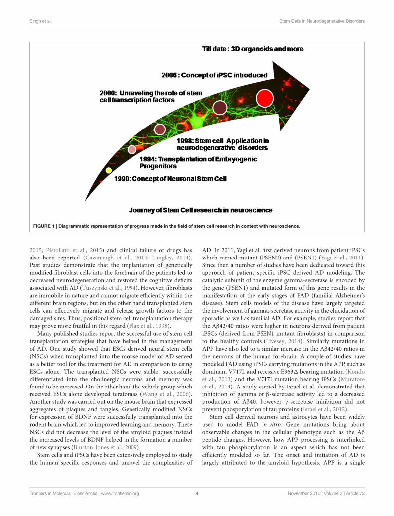
AKT deregulation, mitochondrial swelling, and reduction in theTH-positive neurons (Miller et al., 2013). These iPSCs harboringthe aging markers will thereby mimic the neurodegenerativedisorders more efficiently. In the upcoming sections we shalldiscuss the application of stem cells and iPSCs for the mostcommon and globally prevalent neurodegenerative disorders.Figure 1 shows a diagrammatic representation of progress madein the field of stem cell research in context with neuroscience.
ALZHEIMER’S DISEASE (AD)
Alzheimer’s disease (AD) is described as “Presenile dementia”by German psychiatrist Alois Alzheimer and is one of the mostprevalent neurodegenerative disorders of the world. It is a leadingcause of dementia in the aging population and has lately beendeclared the sixth major reason for death. Patients having ADare reflected with cognitive deficits, memory loss, and behavioralchanges and these changes are inherently associated withneurodegeneration (Blundell and Shah, 2015). Hippocampus,amygdala, neocortex, and basal forebrain regions of the brainare adversely affected leading to the severe impairment ofcognition and memory. Neurofibrillary tangles (NFT) and β-amyloid plaques are the pathological hallmarks of AD (Pengand Zeng, 2011). Hyperphosphorylation of tau proteins andamyloid peptide aggregates are responsible for the formationof NFT and β-amyloid plaques respectively. Alzheimer geneticsinvolves the mutated forms of presenilin 1 (PSEN1), presenilin 2(PSEN2), Amyloid Precursor Protein (APP), and apolipoproteinE. There are still no permanent treatments available for ADexcept acetylcholinesterase (AChE) inhibitors which provideonly temporary relief (Birks, 2006; Lindvall and Kokaia, 2006).Several drugs serve as potent acetylcholinesterase inhibitorssuch as tacrine, tacrine derivatives, donepizel, rivastigmine,galantamine, and the glutamate receptor agonist memantine(Romero et al., 2013; Cecilia Rodrigues Simoes et al., 2014; Ehretand Chamberlin, 2015). These FDA approved pharmacologicalinterventions provide only symptomatic relief for a limitedperiod and may also have side effects in the long run. Removalof the Amyloid β levels from the brain is considered an effectivetherapy for AD and physiologically, the enzyme neprilysin hasbeen reported to be involved in the clearance of the Amyloid β
plaques by degrading it (Iwata et al., 2001). Other proteinasessuch as cathepsin B (Mueller-Steiner et al., 2006) and plasmin(Melchor et al., 2003) too have a similar role and are used todecrease the levels of Aβ thus acting as potent therapeutic agentsfor AD. A number of past studies show the relevance of nervegrowth factor (NGF) in the prevention of neurodegeneration andamyloid toxicity (Tuszynski et al., 2005; Tuszynski, 2007) but asevere limitation with NGF is that it is unable to cross the bloodbrain barrier and therefore cannot be delivered peripherally.
Transgenic animal models of AD carrying the diseasemutations have given ample insight into the etiology andpathophysiology of the disease but have failed to entirelyrecapitulate the formation of NFT and β-amyloid plaquestogether. Human disease pathophysiology has not beencompletely depicted by mouse models so far (D’avanzo et al.,
Frontiers in Molecular Biosciences | www.frontiersin.org 3 November 2016 | Volume 3 | Article 72
Singh et al. Stem Cells in Neurodegenerative Disorders
FIGURE 1 | Diagrammatic representation of progress made in the field of stem cell research in context with neuroscience.
2015; Pistollato et al., 2015) and clinical failure of drugs hasalso been reported (Cavanaugh et al., 2014; Langley, 2014).Past studies demonstrate that the implantation of geneticallymodified fibroblast cells into the forebrain of the patients led todecreased neurodegeneration and restored the cognitive deficitsassociated with AD (Tuszynski et al., 1994). However, fibroblastsare immobile in nature and cannot migrate efficiently within thedifferent brain regions, but on the other hand transplanted stemcells can effectively migrate and release growth factors to thedamaged sites. Thus, positional stem cell transplantation therapymay prove more fruitful in this regard (Flax et al., 1998).
Many published studies report the successful use of stem celltransplantation strategies that have helped in the managementof AD. One study showed that ESCs derived neural stem cells(NSCs) when transplanted into the mouse model of AD servedas a better tool for the treatment for AD in comparison to usingESCs alone. The transplanted NSCs were stable, successfullydifferentiated into the cholinergic neurons and memory wasfound to be increased. On the other hand the vehicle group whichreceived ESCs alone developed teratomas (Wang et al., 2006).Another study was carried out on the mouse brain that expressedaggregates of plaques and tangles. Genetically modified NSCsfor expression of BDNF were successfully transplanted into therodent brain which led to improved learning and memory. TheseNSCs did not decrease the level of the amyloid plaques insteadthe increased levels of BDNF helped in the formation a numberof new synapses (Blurton-Jones et al., 2009).
Stem cells and iPSCs have been extensively employed to studythe human specific responses and unravel the complexities of
AD. In 2011, Yagi et al. first derived neurons from patient iPSCswhich carried mutant (PSEN2) and (PSEN1) (Yagi et al., 2011).Since then a number of studies have been dedicated toward thisapproach of patient specific iPSC derived AD modeling. Thecatalytic subunit of the enzyme gamma-secretase is encoded bythe gene (PSEN1) and mutated form of this gene results in themanifestation of the early stages of FAD (familial Alzheimer’sdisease). Stem cells models of the disease have largely targetedthe involvement of gamma-secretase activity in the elucidation ofsporadic as well as familial AD. For example, studies report thatthe Aβ42/40 ratios were higher in neurons derived from patientiPSCs (derived from PSEN1 mutant fibroblasts) in comparisonto the healthy controls (Livesey, 2014). Similarly mutations inAPP have also led to a similar increase in the Aβ42/40 ratios inthe neurons of the human forebrain. A couple of studies havemodeled FAD using iPSCs carrying mutations in the APP, such asdominant V717L and recessive E9631 bearing mutation (Kondoet al., 2013) and the V717I mutation bearing iPSCs (Muratoreet al., 2014). A study carried by Israel et al. demonstrated thatinhibition of gamma or β-secretase activity led to a decreasedproduction of Aβ40, however γ-secretase inhibition did notprevent phosporylation of tau proteins (Israel et al., 2012).
Stem cell derived neurons and astrocytes have been widelyused to model FAD in-vitro. Gene mutations bring aboutobservable changes in the cellular phenotype such as the Aβ
peptide changes. However, how APP processing is interlinkedwith tau phosphorylation is an aspect which has not beenefficiently modeled so far. The onset and initiation of AD islargely attributed to the amyloid hypothesis. APP is a single
Frontiers in Molecular Biosciences | www.frontiersin.org 4 November 2016 | Volume 3 | Article 72
Singh et al. Stem Cells in Neurodegenerative Disorders
pass transmembrane protein and its proteolytic cleavage formsshort Aβ peptides (Livesey, 2014). Mutation in the genes that areinvolved in the proteolysis of APP play a pivotal role in FAD.Aβ42 is the longer form of amino acid and its accumulationbrings about neurodegeneration and cell death (Sproul et al.,2014). Post translational and cellular localization changes in themicrotubule-associated tau protein play the second biggest rolein AD progression. Tau changes and amyloid plaques if modeledtogether via stem cells will provide the best models of AD (Mooreet al., 2015). Simple monogenic iPSCs’ derived neurons are notsufficient to model amyloidosis since they accumulate low levelsof toxic β-amyloid plaques. To overcome this hurdle stem celllines bearing multiple mutations have been generated, which alsoover express the mutated genes such as APP and PSEN1.
PARKINSON’S DISEASE (PD)
Parkinson’s disease ranks second after AD in being the mostcommon and widely prevalent neurodegenerative disorderinflicting almost one percent of the aging population globally.Dopaminergic neuron loss from the nigrostriatum and substantianigra pars compacta brain regions is the major characteristic ofthe disease (Marchetto et al., 2010). The other major hallmarkbeing the presence of lewy bodies (α-synuclein aggregates)(Spillantini et al., 1998) which further promotes neuronaldeath due to the altered firing pattern of the neurons (Janezicet al., 2013). The genetic involvement of ubiquitin carboxyterminal hydrolase L1, serine threonine kinase 1, parkin, DJ-1,α-synuclein, and leucine-rich repeat kinase 2 have been reportedin the case of genetically acquired familial PD (Dauer andPrzedborski, 2003). Environmental influence in conjunction withage, genetic polymorphisms and chemical exposure predisposean individual toward sporadic PD, however the complex etiologyis yet to be fully understood (Adami et al., 2014). Fitzmauriceet al. showed that variation in Aldehyde dehydrogenaseenhances the pesticide effect related to PD thereby proving thatenvironmental influences work in conjunction with genetics(Fitzmaurice et al., 2014).
Rigidity, resting tremor and bradykinesia are the majorsymptoms which make PD the most common movementdisorder of the world affecting individuals post 65 years of age(Fu et al., 2015). Mechanistic and pathophysiological studies havegiven us a deep understanding of the disease. PD is generallyassociated with a disrupted calcium homeostasis, inflammation,disrupted kinase pathways, generation of reactive oxygen species,and dysfunctional mitochondria (Schapira et al., 2014; Xiaoet al., 2016). Animal and cellular models have given us adeep understanding of the disease but data generated is notfully applicable to human subjects due to difference in diseasepathogenesis of animals and humans (Devine et al., 2011).
PD so far has been managed using monoamine oxidaseinhibitors, dopamine agonists, levodopa, and deep brainstimulation (Politis and Lindvall, 2012). The latter employsstimulation of the ventral intermediate nucleus, a part of thethalamus which can greatly reduce the symptoms of tremor.Other symptoms like rigidity and bradykinesia are also alleviated
after the stimulation of the subthalamic nucleus or internalsegment of globus pallldius. However, these treatments failto repair the damaged brain region and the oral drugs arenot effective beyond 5 years. Administration of L-DOPA (L-dihydroxy-phenyl alanine) can induce dyskinesis and fails to haltthe disease progression (Politis and Lindvall, 2012).
The earliest transplantation studies employed the use offetal ventral mesencephalic tissue of human origin (hfVM)which were engrafted in the striatum of the PD patients andlaid the basis for cell therapy for PD. The attempts weresuccessful and symptomatic relief was provided for almost 16years in the successful cases. However, the clinical trials producedanomalies and the success of the approach was further challengedby the presence of side effects such as GIDs (graft induceddyskinesis). The reason was the presence of the serotonergicneuroblasts in the hfVM that led to an imbalanced serotonin/DAtransporter ratios and false DA release (Politis and Lindvall,2012). Studies also indicated that the survival of the transplantedfetal mesenchymal cells was very low and ethical issues furtherblurred the scope of this therapy. Since PD is characterized bya regional loss of dopaminergic neurons, transplantation, andreplenishment therapy employing stem cell and iPSC deriveddopaminergic neurons (yielding a pure population) in the SNregion provides an excellent alternative.
Neurons with the DA phenotype have been developed fromESCs by using sonic hedgehog (Shh) and fibroblast growth factor-8 (FGF8) or the over expression of Nurr1 by using geneticallymodified NSCs (Kim et al., 2003). Co-culture of mouse bonemarrow stromal and monkey ESCs have successfully yieldeddopaminergic neurons in the past (Takagi et al., 2005). Studiesfurther demonstrate the successful intrastriatal transplantation offetal brain human NSCs in the MPTP lesioned monkeys whichbrought about improved behavioral changes (Redmond et al.,2007). A study reported that when NSCs were isolated fromthe patient brain, converted into dopaminergic neurons and re-implanted into the patient brain, the symptoms of trembling andrigidity were substantially reduced. The brain scans revealed anincrease in the dopamine production by almost 58% and evenwhen the levels did not increase further the symptoms did notrevert back, hinting at a possible restorative potential of stem cellderived dopaminergic neurons (Hassan et al., 2010).
Transition from pluripotent stem cells to iPSCs has shownpromising results and has opened new avenues for the modelingof PD. Dopaminergic neurons derived from patient specificiPSCs were successfully transplanted into a Parkinsonian ratstriatum and showed a considerable reduction in motorasymmetry (Hargus et al., 2010).The role of mitophagy has beenstrongly implicated in PD. The mitochondrion targeted kinasePINK1 accumulates on the outer membrane of mitochondria ondepolarization and further recruits Parkin which is instrumentalin initiating mitophagy (Van Laar et al., 2010; Cai et al., 2012).The data generated on animal models so far has generatedconflicts regarding the direct involvement of mitophagy in PD.However, PINK1 mutated dopaminergic neurons from humanpatient derived iPSCs have given us a clearer understanding of themitophagy theory, thus proving once again that stem cell derivedhuman diseasemodels are far superior and farmore edifying than
Frontiers in Molecular Biosciences | www.frontiersin.org 5 November 2016 | Volume 3 | Article 72
Singh et al. Stem Cells in Neurodegenerative Disorders
any animal model (Seibler et al., 2011). It is also implicated thatmitophagy may be a result of aging and may not have a directcorrelation with the disease; however the hypothesis is still undercontradiction. Progerin expression and long term culture havebeen employed to produce artificial aging of neurons in culture.These aged neurons have been treated as a model to study the lateonset of PD and to elucidate the disease phenotypes (Miller et al.,2013).
As mentioned earlier, apart from disease modeling, stemcells, and iPSCs hold great promise as the in-vitro screeningtools for therapeutic agents, drugs, and compounds. It has beenreported that rapamycin, GW5074 (LRRK2 kinase inhibitor) andcoenzyme Q10 diminish the cytotoxic effect of concanamycin Aalso known as valinomycin in patient derived iPSC neurons. Ithas been clearly seen that GW5074 does not reduce oxidativestress in the healthy neurons from control subjects whereaseffectively lowers oxidative stress in the patient derived neuronsbearing mutated PINK1 (Cooper et al., 2012). This differenceunderlines the importance of therapeutic compound screening indiseasemutation bearing cells as well as the healthy cells. Anotherexample where stem cells have been employed to study thePD physiology is the mutation correction of A53T α-synucleinvia genome editing which diminished the formation of lewybodies in iPSC derived dopaminergic neurons (Ryan et al., 2013).Mutation bearing iPSCs will thus serve as an excellent tool forscreening and assessing the biosafety of drugs and compoundsas well as identifying the underlying signaling cascades andnovel therapeutic targets. The generation and characterizationof iPSCs is cumbersome and the differentiated population ofdopaminergic neurons may contain traces of the undifferentiatedcells which may lead to teratoma formation. Thus, if patientderived fibroblasts are directly converted to dopaminergicneurons, the limitations with iPSCs can be overcome (Han et al.,2015). The successful differentiation of fibroblasts into the DAneurons are reported in literature and has paved way for potentialdisease modeling (Caiazzo et al., 2011; Kim et al., 2014). The stemcell technology can be used to identify the biochemical markersof the disease and can thus help in the diagnosis of early PD (Xiaoet al., 2016).
AMYOTROPHIC LATERAL SCLEROSIS(ALS)
A fatal neurodegenerative disease ALS is caused due to themotor neuron degeneration in the spinal cord, brain stem,and the primary motor cortex (Thomsen et al., 2014) whichresults in muscle wasting, paralysis and eventually death dueto respiratory failure (Hedges et al., 2016). First described byCharcot in 1874, ALS is also known as Lou Gehrig’s disease(Marchetto et al., 2010). ALS has been linked with FTD(frontotemporal lobar dementia) due to symptomatic, clinical,genetic, and pathological overlap. ALS and FTD when occurtogether further shorten the life span of a patient further. Thetwo diseases are often considered the two ends of the samedisease spectrum. Ling et al. gave evidence at the genetic levelby reporting that FTD-ALS and ALS patients carry similar
mutated genes (Lee and Huang, 2015). Many gene mutationsare responsible for causing familial ALS such as mutations inPFN1, FUS/TLS (fused in sarcoma/translocation in liposarcoma),TARDBP or TDP-43 (TAR-DNA-binding protein 43), UBQLN2,C9ORF72, SOD1 (superoxide dismutase 1), HNRNPA1, OPTN,and VCP (Adami et al., 2014). Recently a new gene namedTBK1 has been discovered which plays a crucial role ininflammation & autophagy which are inherently associatedwith ALS pathogenesis (Cirulli et al., 2015). Pathogenesisof sporadic ALS is attributed to glutamate excitotoxicity,protein mitochondrial dysfunction, aggregation, oxidative stress,deficiency of neurotrophic factors, glial cell dysfunction, andimpaired axonal transport, all of these together eventually leadto the accumulation of intracellular neurofilaments (Kiernanet al., 2011; Robberecht and Philips, 2013). Riluzole is theonly therapeutic drug commercially available which helps inthe disease management, but its effect does not last beyond 6months (Cetin et al., 2015). ALS has been widely studied usinganimal models; however reported failure of clinical trials hassomehow restricted the sole dependency on in-vivo research(Gordon and Meininger, 2011). Patient specific iPSC banks holdpromise for personalized medicine and are a good alternative forscreening the efficacy of a number of drugs and compounds forthe treatment of ALS (Giri and Bader, 2015).
Transplantation therapy employing stem cells can beeffectively used as a therapeutic measure to deal with thedevastating disease. Mesenchymal stem cells and hematopoieticstem cells have been efficiently employed as transplantsin the affected spinal cord and have favorably supportedALS management (Mazzini et al., 2012). However, studieswere conducted on a small group of patients and thusthorough research continues so as to be applicable for alarger pool of patients. Neural stem cells (NSCs) ESCs, glial-restricted progenitor cells (GRPs), and induced pluripotentstem cells (iPSCs) also offer a potential alternative fortransplantation approaches and can be used (Traub et al.,2011). It is hypothesized that when donor cells are engraftednear the damaged motor neurons, they not only have animmunomodulatory effect but secrete trophic factors whichimproves the overall therapeutic potential of the transplant. Suchtransplants can effectively delay the progression and even theinitiation of the disease (Teng et al., 2012).
The direct or peripheral injection of MSCs into the spinalcord of patients serves as a potent treatment for ALS and severalstudies have reported the therapeutic potential of MSCs (Maoet al., 2015). Several studies report the beneficial aspects oftransplantable MSCs, which are used to deliver the requiredneurotrophic factors aiding in the prevention of motor neuronloss, improve survival of experimental animals, and delay thedisease progression (Zhao et al., 2007; Vercelli et al., 2008;Uccelli et al., 2012; Krakora et al., 2013). Genetically manipulatedMSCs which secrete GDNF (glial-derived neurotrophic factor)have been reported to increase the life of ALS rats by rescuingmotor neuron loss (Suzuki et al., 2008). “NurOwn” developed byBrainStorm Cell Therapeutics are specialized MSCs which cansecrete neurotrophic factors, can successfully differentiate intoneuronal cells and can be used for ALS treatment. The cells are
Frontiers in Molecular Biosciences | www.frontiersin.org 6 November 2016 | Volume 3 | Article 72
Singh et al. Stem Cells in Neurodegenerative Disorders
under the clinical trial phase (Therapeutics, 2015). A number ofclinical trials have been conducted to assess the efficacy and safetyof MSCs and many are still in process.
Neural stem cells have a specific lineage commitment forthe cells of the CNS and find widespread applicability in theneurodegenerative disorders. When engrafted in the animalmodels of ALS, NSCs have been reported to exert a protectiveeffect on the adjacent motor neurons (Hefferan et al., 2012). Stemcells not only provide symptomatic relief but help in restorationof the brain damage via repair and neurogenesis which istriggered in the affected spinal cord. Successful transplantationof neural stem cells from aborted fetuses into the patient’s spinalcord has been reported (Xu et al., 2012). Results of the phaseI clinical trials have been documented and show that stem celltherapy is a reasonably safe and can be used to treat a largeenough pool of patients (Mazzini et al., 2015).
The familial cases of ALS can be modeled by ESCs harboringthe disease mutations but the sporadic cases require the use ofpatient specific iPSCs. Literature reports the use of iPSCs forALS modeling and hints at a possibility that motor neuronsderived from patient specific iPSCs can be employed for therecapitulation of disease phenotypes. ALS exhibits a complexphysiology and thus requires use of more than one cell typefor its modeling. It was shown that SOD1 bearing astrocytes(from ESCs) exerted toxicity on the adjacent motor neurons(Wada et al., 2012) whereas iPSCs derived astrocyte bearingthe TDP-43 mutation exerted no toxicity (Serio et al., 2013),hinting at the relevance of the co-culture of cells for modeling thecomplex disease. One drawback with in-vitro disease modeling ofALS is the short survival duration of motor neurons in culturewhich limits the study of phenotypic signs occurring in theaged diseased tissues. However, if iPSC derived motor neuronsare grafted in the animal model it will increase the survival ofthese cells. The grafted cells can be recovered lately to visualizethe disease phenotypes in the post mortem rodent tissues, thusoffering a possible solution (Coatti et al., 2015).
Studies report that iPSC derived glial rich population of neuralprogenitors can be successfully transplanted into the spinal cordof mice suffering from ALS. These transplanted cells show a goodsurvival, differentiation potential, and also enhanced the life spanof the treated animals (Kondo et al., 2014). Stem cell therapy hasbeen an area of debate for a long time. The beneficial aspectscannot be overlooked, but extensive clinical trials are in progressso as to generate an effective treatment and possible cure for ALSin the near future.
HUNTINGTON’S DISEASE (HD)
Huntington’s disease is a fatal genetic neurodegenerative disorderwith no cure so far. Genetically acquired in an autosomaldominant manner, the disease is caused by an increasedtrinucleotide CAG (encoding polyglutamine) repeat in the ITI5huntington gene. The CAG repeats are normally less than 36in a healthy individual however a repeat of more than 40will predispose an individual toward the disease. The striatum,cortex, substantia nigra pars compacta, substantia nigra pars
reticulata, globus pallidus are the major brain regions whichare subjected to severe degeneration (Carter and Chan, 2012).Enkephalin and gamma-aminobutyric acid containing mediumspiny neurons, glutaminergic, GABAergic, and parvalbuminergicstriatal projection neurons are severely affected. MutatedHuntington protein forms aggregates in the nuclei and cytoplasmof the brain tissues. Being a Cognitive, movement, psychiatricdisorder the disease brings about mitochondrial, synaptic, andaxonal transport dysfunction. The disease leads to a severetranscriptional dysregulation, proteolysis, and excitotoxicity(Peng and Zeng, 2011).
The disease, though incurable can be managed via genetherapy and drugs to alleviate symptoms. Drugs providetemporary symptomatic relief and largely target the motoraspects of HD. Tetrabenazine is dopamine depleting in natureand is used to reduce chorea, however severe side effects restrictsits widespread use as a drug of choice for HD (Frank, 2014).The pros and cons of the various animal models of HD havebeen reviewed by Pouladi et al. In 2013 (Pouladi et al., 2013).HD has been widely modeled via rodent models in-vivo, howeverhere too the success of the clinical trials has been limited. Theproblem lies not only with the animal model chosen but also withthe robustness of the preclinical studies (Menalled and Brunner,2014).
Gene therapy employs RNAi mediated silencing andreduction in the mutated HTT protein translation. However,gene therapy is effective only at the earliest stage of the diseasebut the symptoms are not visible until the later stages bywhich the major damage is already done. Replenishing thestratial neuron population by intracranial transplantation isan alternative cell therapy approach. Fetal striatal tissue graftshave been employed in the non human primate and rodent HDmodels as a means of transplantation therapy (Chen et al., 2014).These grafted tissues have a low survival rate and since theyare procured from aborted fetuses, ethical constraints are high.Limited availability of donors further restricts the scope of thispromising strategy. Neural progenitor cells (NPCs) derived fromstem cells and iPSCs are a good alternative in this regard.
Successful differentiation of human ESCs into the GABAergicand DARPP32 positive medium spiny neurons (MSNs) is wellreported. NPCs transplanted into the chemical lesion mousemodel of HD showed efficient connectivity with the host neuronsand also alleviated motor deficits (Ma et al., 2012). Anotherstudy by Carri et al. reports the successful differentiation ofESCs and iPSCs into MSNs carrying adenosine and dopaminereceptors (Carri et al., 2013). However, NPCs transplantationhas been more successful in chemical lesion HD mouse modelsas compared to the transgenic (R6/2) mouse model, the reasonspeculated being the short life span of the transgenic mouse(El-Akabawy et al., 2012). Along with disease modeling andregenerative transplantation therapy, stem cells and iPSCs canbe simply used as an in-vitro tool for screening the efficacy andbiosafety of drugs and small molecule compounds that help inrelieving the symptoms of the disease as well as bring about areduction in neurodegeneration (Carter and Chan, 2012). HDpatient specific cell lines have been generated which recapitulatethe disease specific phenotypes more closely (Consortium, 2012).
Frontiers in Molecular Biosciences | www.frontiersin.org 7 November 2016 | Volume 3 | Article 72
Singh et al. Stem Cells in Neurodegenerative Disorders
The gene therapy for HD involves targeting the mutantHTT allele (mHTT) transcript by RNA interference and ASOs(antisense oligodeoxynucleotides) specifically without disturbingthe normal HTT allele transcript (Carroll et al., 2011; Hu et al.,2012). The exact role HTT in normal conditions is still not clearlyunderstood but a study reports that HTT knockout mice do notsurvive and are lethal embryonically. Thus targeting only themutant allele is preferable. ASOs target complimentary mRNAvia RNase Hmediated degradation and are important in researchas they are capable of targeting the SNPs (single nucleotidepolymorphisms). These SNPs are responsible for the differencebetween normal and mutated HTT gene alleles (Carroll et al.,2011). Studies have been carried out in the non human primateand rodent models of HD and disease alleviation was observed(Kordasiewicz et al., 2012). This paves way for a possibilitythat patient derived iPSCs carrying the disease mutations canbe effectively corrected via ASOs and gene silencing and theNPCs thus derived can be successfully transplanted. Thus stemcell therapy in amalgamation with gene therapy can help in themanagement of the incurable disease in future (Chen et al., 2014).
THREE DIMENSIONAL (3D) STEM CELLBASED STUDIES: APPLICATION INNEURODEGENERATION ANDNEURODEGENERATIVE DISORDERS
In-vitro stem cell based studies are the best possible alternativeto animal models because cells of human origin are usedand thus data generated can be easily extrapolated to humansubjects. However, two dimensional in-vitro studies have manylimitations. The 2D culture comprise a homogenous populationof cells, complex intercellular interactions are lacking, the
cultures do not represent the complex microenvironment ofan organ like the brain and the intact organ physiology islacking. In this regard 3D cultures have played a pivotal rolein overcoming the hurdles of typical 2D studies and also findwidespread application in the field of neuroscience. Advanceshave been made to recapitulate the brain development andliterature supports successful formation of stem cell derived 3Dcortical and cerebral organoids (Lancaster et al., 2013; Sasai, 2013;Lancaster and Knoblich, 2014).
AD has been extensively modeled using stem cell and iPSC
derived 2D neuronal cell culture however, a major limitation
being the diffusion of Aβ aggregates, which are gradually, washed
off with successive media changes. Therefore, it is hypothesized
that 3D stem cell models of AD will provide a more compact
and comprehensive brain microenvironment where the local
niche will allow a sufficient Aβ aggregation (D’avanzo et al.,2015). This theory has been confirmed lately, where 3D matrigel
system has been used for the culture of ReN cells to model
familial AD. The 3D stem cell model of AD showed a significant
deposit of β-amyloid plaques in a 6 week differentiated culture.
The cells further showed elevated levels of phosphorylated tau
protein thereby confirming that 3D based stem cell models
are more efficient in recapitulating the disease pathophysiology
(Choi et al., 2014). A study by Zhang et al. reported the
successful recapitulation of in-vivo like responses in a 3D culture
model of AD. The study comprises neuroepithelial stem cell
derived neuronal culture in a PuraMatrix hydrogel comprising
self assembling peptide matrix and laminin. The study confirmed
that 3D microenvironment allowed Aβ sensing via p21-activated
kinase (Zhang et al., 2014). Genetically engineered hNPCsproduction and 3D cell culture protocols for AD modeling havebeen standardized and published (Kim et al., 2015).
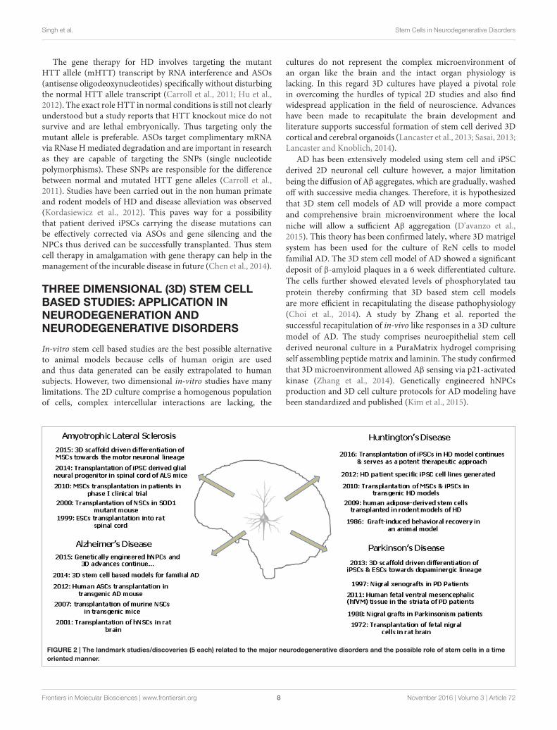
FIGURE 2 | The landmark studies/discoveries (5 each) related to the major neurodegenerative disorders and the possible role of stem cells in a time
oriented manner.
Frontiers in Molecular Biosciences | www.frontiersin.org 8 November 2016 | Volume 3 | Article 72
Singh et al. Stem Cells in Neurodegenerative Disorders
It is well reported that mouse iPSCs and ESCs show a betterdopaminergic differentiation potential in a three dimensionalpeptide derived nanofibre scaffold. The 3D culture provides abetter environment for the development of the DA neuronswhich showed appropriate action potential firing and expressedthe specific markers as well (Ni et al., 2013). Successfuldifferentiation of chorion derived MSCs into motor neurons hasbeen recently reported in 3D nanofibrous gelatin scaffold. Thesemotor neurons shall provide a possibility to model ALS in athree dimensional scenario (Faghihi et al., 2016). Figure 2 depictsthe landmark studies/discoveries (5 each) related to the majorneurodegenerative disorders and the possible role of stem cellsin a time oriented manner.
CONCLUDING REMARKS
The above discussion so far clearly sheds light on thewidespread applicability of stem cells and induced pluripotentstem cells in the field of neurodegeneration. Disease modeling,transplantation therapy, restoration of lost brain functionalitydue to injury and aging and regenerative therapeutics are someof the areas where stem cells have been abundantly used. Thearticle highlights the advances made especially in the past 5years as envisaging the entire applicability of stem cells inneurodegenerative medicine is beyond the scope of the presentdiscussion. Human stem cells and patient derived iPSCs havebeen instrumental in overcoming the major limitations of animalbased research providing a more profound understanding ofthe neurodegenerative disorders. Patient derived iPSCs are evenbetter models for understanding the disease pathophysiologyand mechanistics because they carry the patient’s genotype, bearthe disease mutations and also account for the environmentalinfluences. Stem cells have also been employed as simplisticin-vitro tools for screening of therapeutics and drugs. Threedimensional stem cell based studies and stem cell derivedorganoids have further contributed by providing a more in-vivolike microenvironment which is the closest possible mimic ofa live animal. With technological advancements and efficientimaging techniques have revolutionized the concept of 3Dstem cell based organoid research. Pharmacological interventionutilizing natural agents like curcumin has shown neuroprotectiveefficacy in clinical and experimental models of neurotoxicity andcan provide beneficial effects in the neurodegenerative disordersin future (Srivastava et al., 2014). However, the current prevalentpharmacological treatments provide symptomatic relief onlyfor a limited period of time and the drugs administered mayhave side effects. The advent of stem cell therapy has laid thefoundation keystone for a possible cure with minimized sideeffects. Personalized medical treatment using iPSCs is the currentface of modern medicine and constant efforts are being made toscale down the cost and increase the efficacy of the approach.Animal based clinical field trials cannot be completely surpassedand transplantation therapies will require validation. However, ifcells of human origin are employed for the preliminary diseasemodeling and therapeutic screening, a lot shall be saved in
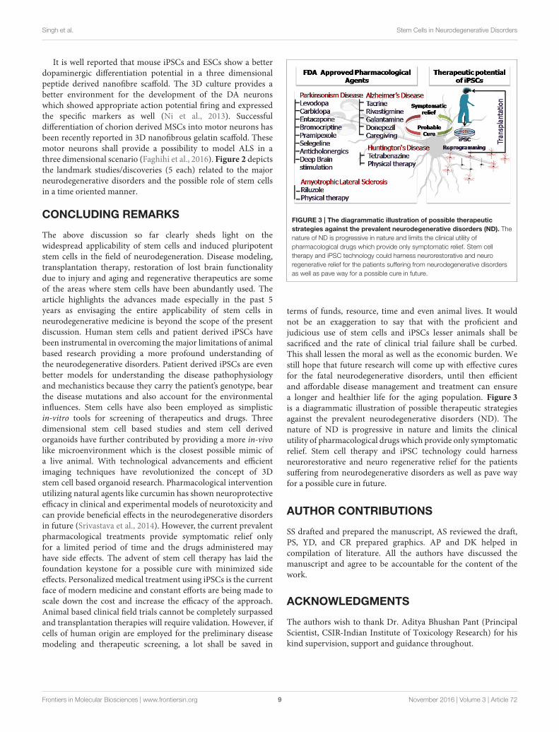
FIGURE 3 | The diagrammatic illustration of possible therapeutic
strategies against the prevalent neurodegenerative disorders (ND). The
nature of ND is progressive in nature and limits the clinical utility of
pharmacological drugs which provide only symptomatic relief. Stem cell
therapy and iPSC technology could harness neurorestorative and neuro
regenerative relief for the patients suffering from neurodegenerative disorders
as well as pave way for a possible cure in future.
terms of funds, resource, time and even animal lives. It wouldnot be an exaggeration to say that with the proficient andjudicious use of stem cells and iPSCs lesser animals shall besacrificed and the rate of clinical trial failure shall be curbed.This shall lessen the moral as well as the economic burden. Westill hope that future research will come up with effective curesfor the fatal neurodegenerative disorders, until then efficientand affordable disease management and treatment can ensurea longer and healthier life for the aging population. Figure 3is a diagrammatic illustration of possible therapeutic strategiesagainst the prevalent neurodegenerative disorders (ND). Thenature of ND is progressive in nature and limits the clinicalutility of pharmacological drugs which provide only symptomaticrelief. Stem cell therapy and iPSC technology could harnessneurorestorative and neuro regenerative relief for the patientssuffering from neurodegenerative disorders as well as pave wayfor a possible cure in future.
AUTHOR CONTRIBUTIONS
SS drafted and prepared the manuscript, AS reviewed the draft,PS, YD, and CR prepared graphics. AP and DK helped incompilation of literature. All the authors have discussed themanuscript and agree to be accountable for the content of thework.
ACKNOWLEDGMENTS
The authors wish to thank Dr. Aditya Bhushan Pant (PrincipalScientist, CSIR-Indian Institute of Toxicology Research) for hiskind supervision, support and guidance throughout.
Frontiers in Molecular Biosciences | www.frontiersin.org 9 November 2016 | Volume 3 | Article 72
Singh et al. Stem Cells in Neurodegenerative Disorders
REFERENCES
Aasen, T., Raya, A., Barrero, M. J., Garreta, E., Consiglio, A., Gonzalez, F., et al.
(2008). Efficient and rapid generation of induced pluripotent stem cells from
human keratinocytes. Nat. Biotechnol. 26, 1276–1284. doi: 10.1038/nbt.1503
Adami, R., Scesa, G., and Bottai, D. (2014). Stem cell transplantation in
neurological diseases: improving effectiveness in animal models. Front. Cell
Dev. Biol. 2:17. doi: 10.3389/fcell.2014.00017
Agrawal, M., and Biswas, A. (2015). Molecular diagnostics of neurodegenerative
disorders. Front. Mol. Biosci. 2:54. doi: 10.3389/fmolb.2015.00054
Aleynik, A., Gernavage, K. M., Mourad, Y. S. H., Sherman, L. S., Liu, K., Gubenko,
Y. A., et al. (2014). Stem cell delivery of therapies for brain disorders. Clin.
Transl. Med. 3, 3–24. doi: 10.1186/2001-1326-3-24
Avior, Y., Sagi, I., and Benvenisty, N. (2016). Pluripotent stem cells in disease
modelling and drug discovery. Nat. Rev. Mol. Cell Biol. 17, 170–182. doi:
10.1038/nrm.2015.27
Begley, C. G., and Ellis, L. M. (2012). Drug development: raise standards for
preclinical cancer research. Nature 483, 531–533. doi: 10.1038/483531a
Birks, J. S. (2006). Cholinesterase inhibitors for Alzheimer’s disease. Cochrane Libr.
6, 220–221. doi: 10.1002/14651858.cd005593
Blundell, R., and Shah, M. (2015). Neurodegenerative diseases and stem cell
transplantation. J. Stem Cell Res. Ther. 5:277. doi: 10.4172/2157-7633.1000277
Blurton-Jones, M., Kitazawa, M., Martinez-Coria, H., Castello, N. A., Müller, F.-
J., Loring, J. F., et al. (2009). Neural stem cells improve cognition via BDNF
in a transgenic model of Alzheimer disease. Proc. Natl. Acad. Sci. U.S.A. 106,
13594–13599. doi: 10.1073/pnas.0901402106
Cai, Q., Zakaria, H. M., Simone, A., and Sheng, Z.-H. (2012). Spatial parkin
translocation and degradation of damaged mitochondria via mitophagy
in live cortical neurons. Curr. Biol. 22, 545–552. doi: 10.1016/j.cub.2012.
02.005
Caiazzo, M., Dell’anno, M. T., Dvoretskova, E., Lazarevic, D., Taverna, S., Leo,
D., et al. (2011). Direct generation of functional dopaminergic neurons from
mouse and human fibroblasts. Nature 476, 224–227. doi: 10.1038/nature10284
Cananzi, M., and De Coppi, P. (2012). CD117+ amniotic fluid stem cells: state of
the art and future perspectives. Organogenesis 8, 77–88. doi: 10.4161/org.22426
Carri, A. D., Onorati, M., Lelos, M. J., Castiglioni, V., Faedo, A., Menon, R., et al.
(2013). Developmentally coordinated extrinsic signals drive human pluripotent
stem cell differentiation toward authentic DARPP-32+ medium-sized spiny
neurons. Development 140, 301–312. doi: 10.1242/dev.084608
Carroll, J. B., Warby, S. C., Southwell, A. L., Doty, C. N., Greenlee, S., Skotte,
N., et al. (2011). Potent and selective antisense oligonucleotides targeting
single-nucleotide polymorphisms in the Huntington disease gene/allele-
specific silencing of mutant huntingtin. Mol. Ther. 19, 2178–2185. doi:
10.1038/mt.2011.201
Carter, R. L., and Chan, A. W. (2012). Pluripotent stem cells models for
Huntington’s disease: prospects and challenges. J. Genet. Genomics 39, 253–259.
doi: 10.1016/j.jgg.2012.04.006
Cavanaugh, S. E., Pippin, J. J., and Barnard, N. D. (2014). Animal models of
Alzheimer disease: historical pitfalls and a path forward.Altex 31, 279–302. doi:
10.14573/altex.1310071
Cecilia Rodrigues Simoes, M., Pereira Dias Viegas, F., Soares Moreira, M.,
De Freitas Silva, M., Maximo Riquiel, M., Mattos Da Rosa, P., et al.
(2014). Donepezil: an important prototype to the design of new drug
candidates for Alzheimer’s disease. Mini Rev. Med. Chem. 14, 2–19. doi:
10.2174/1389557513666131119201353
Cetin, H., Rath, J., Füzi, J., Reichardt, B., Fülöp, G., Koppi, S., et al. (2015).
Epidemiology of amyotrophic lateral sclerosis and effect of riluzole on disease
course. Neuroepidemiology 44, 6–15. doi: 10.1159/000369813
Chen, Y., Carter, R. L., Cho, I. K., and Chan, A. W. (2014). Cell-based
therapies for Huntington’s disease. Drug Discov. Today 19, 980–984. doi:
10.1016/j.drudis.2014.02.012
Chiou, S.-H., Jiang, B.-H., Yu, Y.-L., Chou, S.-J., Tsai, P.-H., Chang, W.-C., et al.
(2013). Poly (ADP-ribose) polymerase 1 regulates nuclear reprogramming
and promotes iPSC generation without c-Myc. J. Exp. Med. 210, 85–98. doi:
10.1084/jem.20121044
Choi, S. H., Kim, Y. H., Hebisch, M., Sliwinski, C., Lee, S., D’avanzo, C., et al.
(2014). A three-dimensional human neural cell culture model of Alzheimer/’s
disease. Nature 515, 274–278. doi: 10.1038/nature13800
Cirulli, E. T., Lasseigne, B. N., Petrovski, S., Sapp, P. C., Dion, P. A., Leblond, C. S.,
et al. (2015). Exome sequencing in amyotrophic lateral sclerosis identifies risk
genes and pathways. Science 347, 1436–1441. doi: 10.1126/science.aaa3650
Coatti, G., Beccari, M., Olávio, T., Mitne-Neto, M., Okamoto, O., and Zatz, M.
(2015). Stem cells for amyotrophic lateral sclerosis modeling and therapy: myth
or fact? Cytometry A 87, 197–211. doi: 10.1002/cyto.a.22630
Consortium, H. I. (2012). Induced pluripotent stem cells from patients with
Huntington’s disease show CAG-repeat-expansion-associated phenotypes. Cell
Stem Cell 11, 264–278. doi: 10.1016/j.stem.2012.04.027
Cooper, O., Seo, H., Andrabi, S., Guardia-Laguarta, C., Graziotto, J., Sundberg, M.,
et al. (2012). Pharmacological rescue of mitochondrial deficits in iPSC-derived
neural cells from patients with familial Parkinson’s disease. Sci. Transl. Med. 4,
141ra190. doi: 10.1126/scitranslmed.3003985
Dauer, W., and Przedborski, S. (2003). Parkinson’s disease: mechanisms and
models. Neuron 39, 889–909. doi: 10.1016/S0896-6273(03)00568-3
D’avanzo, C., Aronson, J., Kim, Y. H., Choi, S. H., Tanzi, R. E., and Kim, D. Y.
(2015). Alzheimer’s in 3D culture: challenges and perspectives. Bioessays 37,
1139–1148. doi: 10.1002/bies.201500063
Devine, M. J., Ryten, M., Vodicka, P., Thomson, A. J., Burdon, T., Houlden, H.,
et al. (2011). Parkinson’s disease induced pluripotent stem cells with triplication
of the α-synuclein locus. Nat. Commun. 2, 440. doi: 10.1038/ncomms1453
Dimos, J. T., Rodolfa, K. T., Niakan, K. K., Weisenthal, L. M., Mitsumoto, H.,
Chung,W., et al. (2008). Induced pluripotent stem cells generated from patients
with ALS can be differentiated intomotor neurons. Science 321, 1218–1221. doi:
10.1126/science.1158799
Ehret, M. J., and Chamberlin, K. W. (2015). Current practices in the treatment of
alzheimer disease: where is the evidence after the phase iii trials? Clin. Ther. 37,
1604–1616. doi: 10.1016/j.clinthera.2015.05.510
El-Akabawy, G., Rattray, I., Johansson, S. M., Gale, R., Bates, G., and Modo, M.
(2012). Implantation of undifferentiated and pre-differentiated human neural
stem cells in the R6/2 transgenic mouse model of Huntington’s disease. BMC
Neurosci. 13:97. doi: 10.1186/1471-2202-13-97
Faghihi, F.,Mirzaei, E., Ai, J., Lotfi, A., Sayahpour, F. A., Barough, S. E., et al. (2016).
Differentiation potential of human chorion-derived mesenchymal stem cells
into motor neuron-like cells in two-and three-dimensional culture systems.
Mol. Neurobiol. 53, 1862–1872. doi: 10.1007/s12035-015-9129-y
Ferroni, L., Gardin, C., Tocco, I., Epis, R., Casadei, A., Vindigni, V., et al.
(2013). “Potential for neural differentiation of mesenchymal stem cells,” in
Mesenchymal Stem Cells-Basics and Clinical Application I eds B. Weyand, M.
Dominici, R. Hass, R. Jacobs, and C. Kasper (Berlin; Heidelberg: Springer),
89–115.
Fitzmaurice, A. G., Rhodes, S. L., Cockburn, M., Ritz, B., and Bronstein,
J. M. (2014). Aldehyde dehydrogenase variation enhances effect of
pesticides associated with Parkinson disease. Neurology 82, 419–426. doi:
10.1212/WNL.0000000000000083
Flax, J. D., Aurora, S., Yang, C., Simonin, C., Wills, A. M., Billinghurst, L. L.,
et al. (1998). Engraftable human neural stem cells respond to development cues,
replace neurons, and express foreign genes.Nat. Biotechnol. 16, 1033–1039. doi:
10.1038/3473
Frank, S. (2014). Treatment of Huntington’s disease. Neurotherapeutics 11,
153–160. doi: 10.1007/s13311-013-0244-z
Frattini, E., Ruggieri, M., Salani, S., Faravelli, I., Zanetta, C., Nizzardo, M., et al.
(2015). Pluripotent stem cell-based models of spinal muscular atrophy. Mol.
Cell. Neurosci. 64, 44–50. doi: 10.1016/j.mcn.2014.12.005
Freije, J. M., and López-Otín, C. (2012). Reprogramming aging and progeria. Curr.
Opin. Cell Biol. 24, 757–764. doi: 10.1016/j.ceb.2012.08.009
Fu, M.-H., Li, C.-L., Lin, H.-L., Chen, P.-C., Calkins, M. J., Chang, Y.-F., et al.
(2015). Stem cell transplantation therapy in Parkinson’s disease. Springerplus 4,
1–8. doi: 10.1186/s40064-015-1400-1
Giri, S., and Bader, A. (2015). A low-cost, high-quality new drug discovery process
using patient-derived induced pluripotent stem cells. Drug Discov. Today 20,
37–49. doi: 10.1016/j.drudis.2014.10.011
Glenn, J. D., and Whartenby, K. A. (2014). Mesenchymal stem cells: emerging
mechanisms of immunomodulation and therapy. World J. Stem Cells 6,
526–539. doi: 10.4252/wjsc.v6.i5.526
Gordon, P. H., and Meininger, V. (2011). How can we improve clinical
trials in amyotrophic lateral sclerosis? Nat. Rev. Neurol. 7, 650–654. doi:
10.1038/nrneurol.2011.147
Frontiers in Molecular Biosciences | www.frontiersin.org 10 November 2016 | Volume 3 | Article 72
Singh et al. Stem Cells in Neurodegenerative Disorders
Han, F., Baremberg, D., Gao, J., Duan, J., Lu, X., Zhang, N., et al. (2015).
Development of stem cell-based therapy for Parkinson’s disease. Transl.
Neurodegener. 4:16. doi: 10.1186/s40035-015-0039-8
Hargus, G., Cooper, O., Deleidi, M., Levy, A., Lee, K., Marlow, E., et al. (2010).
Differentiated Parkinson patient-derived induced pluripotent stem cells grow
in the adult rodent brain and reduce motor asymmetry in Parkinsonian rats.
Proc. Natl. Acad. Sci. U.S.A. 107, 15921–15926. doi: 10.1073/pnas.1010209107
Hassan, A. U., Hassan, G., and Rasool, Z. (2010). Role of stem cells in treatment of
neurological disorder. J. Health Sci. 3, 227–233.
Hedges, E. C., Mehler, V. J., and Nishimura, A. L. (2016). The use of stem cells
to model amyotrophic lateral sclerosis and frontotemporal dementia: from
basic research to regenerative medicine. Stem Cells Int. 2016:9279516. doi:
10.1155/2016/9279516
Hefferan, M. P., Galik, J., Kakinohana, O., Sekerkova, G., Santucci, C.,
Marsala, S., et al. (2012). Human neural stem cell replacement therapy for
amyotrophic lateral sclerosis by spinal transplantation. PLoS ONE 7:e42614.
doi: 10.1371/journal.pone.0042614
Hu, J., Liu, J., Yu, D., Chu, Y., and Corey, D. R. (2012). Mechanism of allele-
selective inhibition of huntingtin expression by duplex RNAs that target
CAG repeats: function through the RNAi pathway. Nucleic Acids Res. 40,
11270–11280. doi: 10.1093/nar/gks907
Israel, M. A., Yuan, S. H., Bardy, C., Reyna, S. M., Mu, Y., Herrera, C., et al. (2012).
Probing sporadic and familial Alzheimer/’s disease using induced pluripotent
stem cells. Nature 482, 216–220. doi: 10.1038/nature10821
Iwata, N., Tsubuki, S., Takaki, Y., Shirotani, K., Lu, B., Gerard, N. P., et al. (2001).
Metabolic regulation of brain Aβ by neprilysin. Science 292, 1550–1552. doi:
10.1126/science.1059946
Janezic, S., Threlfell, S., Dodson, P. D., Dowie, M. J., Taylor, T. N., Potgieter, D.,
et al. (2013). Deficits in dopaminergic transmission precede neuron loss and
dysfunction in a new Parkinsonmodel. Proc. Natl. Acad. Sci. 110, E4016–E4025.
doi: 10.1073/pnas.1309143110
Jiang, Y., Zhang, M.-J., and Hu, B.-Y. (2012). Specification of functional neurons
and glia from human pluripotent stem cells. Protein Cell 3, 818–825. doi:
10.1007/s13238-012-2086-6
Ju, R., Wen, Y., Gou, R., Wang, Y., and Xu, Q. (2014). The experimental therapy on
brain ischemia by improvement of local angiogenesis with tissue engineering in
the mouse. Cell Transplant. 23, S83–S95. doi: 10.3727/096368914X684998
Kiernan, M. C., Vucic, S., Cheah, B. C., Turner, M. R., Eisen, A., Hardiman,
O., et al. (2011). Amyotrophic lateral sclerosis. Lancet 377, 942–955. doi:
10.1016/S0140-6736(10)61156-7
Kim, H.-S., Kim, J., Jo, Y., Jeon, D., and Cho, Y. S. (2014). Direct lineage
reprogramming of mouse fibroblasts to functional midbrain dopaminergic
neuronal progenitors. Stem Cell Res. 12, 60–68. doi: 10.1016/j.scr.2013.09.007
Kim, T. E., Lee, H. S., Lee, Y. B., Hong, S. H., Lee, Y. S., Ichinose, H., et al. (2003).
Sonic hedgehog and FGF8 collaborate to induce dopaminergic phenotypes in
the Nurr1-overexpressing neural stem cell. Biochem. Biophys. Res. Commun.
305, 1040–1048. doi: 10.1016/S0006-291X(03)00879-9
Kim, Y. H., Choi, S. H., D’avanzo, C., Hebisch, M., Sliwinski, C., Bylykbashi, E.,
et al. (2015). A 3D human neural cell culture system for modeling Alzheimer’s
disease. Nat. Protoc. 10, 985–1006. doi: 10.1038/nprot.2015.065
Kondo, T., Asai, M., Tsukita, K., Kutoku, Y., Ohsawa, Y., Sunada, Y., et al. (2013).
Modeling Alzheimer’s disease with iPSCs reveals stress phenotypes associated
with intracellular Aβ and differential drug responsiveness. Cell Stem Cell 12,
487–496. doi: 10.1016/j.stem.2013.01.009
Kondo, T., Funayama, M., Tsukita, K., Hotta, A., Yasuda, A., Nori, S.,
et al. (2014). Focal transplantation of human iPSC-derived glial-rich neural
progenitors improves lifespan of ALS mice. Stem Cell Reports 3, 242–249. doi:
10.1016/j.stemcr.2014.05.017
Kordasiewicz, H. B., Stanek, L. M., Wancewicz, E. V., Mazur, C., Mcalonis, M.
M., Pytel, K. A., et al. (2012). Sustained therapeutic reversal of Huntington’s
disease by transient repression of huntingtin synthesis. Neuron 74, 1031–1044.
doi: 10.1016/j.neuron.2012.05.009
Krakora, D., Mulcrone, P., Meyer, M., Lewis, C., Bernau, K., Gowing, G., et al.
(2013). Synergistic effects of GDNF and VEGF on lifespan and disease
progression in a familial ALS rat model. Mol. Ther. 21, 1602–1610. doi:
10.1038/mt.2013.108
Kumar, V., Jahan, S., Singh, S., Khanna, V. K., and Pant, A. B. (2015). Progress
toward the development of in vitro model system for chemical-induced
developmental neurotoxicity: potential applicability of stem cells.Arch. Toxicol.
89, 265–267. doi: 10.1007/s00204-014-1442-0
Lancaster, M. A., and Knoblich, J. A. (2014). Generation of cerebral organoids
from human pluripotent stem cells. Nat. Protoc. 9, 2329–2340. doi:
10.1038/nprot.2014.158
Lancaster, M. A., Renner, M., Martin, C.-A., Wenzel, D., Bicknell, L. S., Hurles,
M. E., et al. (2013). Cerebral organoids model human brain development and
microcephaly. Nature 501, 373–379. doi: 10.1038/nature12517
Langley, G. R. (2014). Considering a new paradigm for Alzheimer’s disease
research. Drug Discov. Today 19, 1114–1124. doi: 10.1016/j.drudis.2014.03.013
Lapasset, L., Milhavet, O., Prieur, A., Besnard, E., Babled, A., Aït-Hamou,
N., et al. (2011). Rejuvenating senescent and centenarian human cells by
reprogramming through the pluripotent state. Genes Dev. 25, 2248–2253. doi:
10.1101/gad.173922.111
Lee, S., and Huang, E. J. (2015). Modeling ALS and FTD with iPSC-derived
neurons. Brain Res. doi: 10.1016/j.brainres.2015.10.003. [Epub ahead of print].
Lindvall, O., and Kokaia, Z. (2006). Stem cells for the treatment of neurological
disorders. Nature 441, 1094–1096. doi: 10.1038/nature04960
Liu, W., Deng, Y., Liu, Y., Gong, W., and Deng, W. (2013). Stem cell models for
drug discovery and toxicology studies. J. Biochem. Mol. Toxicol. 27, 17–27. doi:
10.1002/jbt.21470
Livesey, F. J. (2014). Human stem cell models of dementia. Hum. Mol. Genet. 23,
R35–R39. doi: 10.1093/hmg/ddu302
Lu, D., Chen, E. Y., Lee, P., Wang, Y.-C., Ching, W., Markey, C., et al. (2015).
Accelerated neuronal differentiation towardmotor neuron lineage from human
embryonic stem cell line (H9). Tissue Eng. Part C Methods. 21, 242–252. doi:
10.1089/ten.TEC.2013.0725
Lukovic, D., Moreno-Manzano, V., Lopez-Mocholi, E., Rodriguez-Jiménez, F. J.,
Jendelova, P., Sykova, E., et al. (2015). Complete rat spinal cord transection as
a faithful model of spinal cord injury for translational cell transplantation. Sci.
Reports 5:9640. doi: 10.1038/srep09640
Ma, L., Hu, B., Liu, Y., Vermilyea, S. C., Liu, H., Gao, L., et al. (2012).
Human embryonic stem cell-derived GABA neurons correct locomotion
deficits in quinolinic acid-lesioned mice. Cell Stem Cell 10, 455–464. doi:
10.1016/j.stem.2012.01.021
Mao, Z., Zhang, S., and Chen, H. (2015). Stem cell therapy for amyotrophic lateral
sclerosis. Cell Regen. 4:11. doi: 10.1186/s13619-015-0026-7
Marchetto, M. C., Brennand, K. J., Boyer, L. F., and Gage, F. H. (2011). Induced
pluripotent stem cells (iPSCs) and neurological disease modeling: progress and
promises. Hum. Mol. Genet. 20, R109–R115. doi: 10.1093/hmg/ddr336
Marchetto, M. C., Winner, B., and Gage, F. H. (2010). Pluripotent stem cells in
neurodegenerative and neurodevelopmental diseases. Hum. Mol. Genet. 19,
R71–R76. doi: 10.1093/hmg/ddq159
Mazzini, L., Gelati, M., Profico, D. C., Sgaravizzi, G., Pensi, M. P., Muzi, G., et al.
(2015). Human neural stem cell transplantation in ALS: initial results from a
phase I trial. J. Transl. Med. 13, 17. doi: 10.1186/s12967-014-0371-2
Mazzini, L., Mareschi, K., Ferrero, I., Miglioretti, M., Stecco, A., Servo, S.,
et al. (2012). Mesenchymal stromal cell transplantation in amyotrophic
lateral sclerosis: a long-term safety study. Cytotherapy 14, 56–60. doi:
10.3109/14653249.2011.613929
Melchor, J. P., Pawlak, R., and Strickland, S. (2003). The tissue plasminogen
activator-plasminogen proteolytic cascade accelerates amyloid-β (Aβ)
degradation and inhibits Aβ-induced neurodegeneration. J. Neurosci. 23,
8867–8871.
Menalled, L., and Brunner, D. (2014). Animal models of Huntington’s disease
for translation to the clinic: best practices. Mov. Disord. 29, 1375–1390. doi:
10.1002/mds.26006
Mendonça, L. S., Nóbrega, C., Hirai, H., Kaspar, B. K., and De Almeida, L.
P. (2015). Transplantation of cerebellar neural stem cells improves motor
coordination and neuropathology in Machado-Joseph disease mice. Brain 138,
320–335. doi: 10.1093/brain/awu352
Miller, J. D., Ganat, Y. M., Kishinevsky, S., Bowman, R. L., Liu, B., Tu, E.
Y., et al. (2013). Human iPSC-based modeling of late-onset disease via
progerin-induced aging. Cell Stem Cell 13, 691–705. doi: 10.1016/j.stem.2013.
11.006
Moore, S., Evans, L. D., Andersson, T., Portelius, E., Smith, J., Dias, T. B., et al.
(2015). APP metabolism regulates tau proteostasis in human cerebral cortex
neurons. Cell Rep. 11, 689–696. doi: 10.1016/j.celrep.2015.03.068
Frontiers in Molecular Biosciences | www.frontiersin.org 11 November 2016 | Volume 3 | Article 72
Singh et al. Stem Cells in Neurodegenerative Disorders
Mueller-Steiner, S., Zhou, Y., Arai, H., Roberson, E. D., Sun, B., Chen,
J., et al. (2006). Antiamyloidogenic and neuroprotective functions of
cathepsin B: implications for Alzheimer’s disease. Neuron 51, 703–714. doi:
10.1016/j.neuron.2006.07.027
Muratore, C. R., Rice, H. C., Srikanth, P., Callahan, D. G., Shin, T., Benjamin, L. N.,
et al. (2014). The familial Alzheimer’s disease APPV717I mutation alters APP
processing and Tau expression in iPSC-derived neurons. Hum. Mol. Genet. 23,
3523–3536. doi: 10.1093/hmg/ddu064
Nam, H., Lee, K.-H., Nam, D.-H., and Joo, K. M. (2015). Adult human neural stem
cell therapeutics: current developmental status and prospect. World J. Stem
Cells 7, 126–136. doi: 10.4252/wjsc.v7.i1.126
Ni, N., Hu, Y., Ren, H., Luo, C., Li, P., Wan, J.-B., et al. (2013). Self-assembling
peptide nanofiber scaffolds enhance dopaminergic differentiation of mouse
pluripotent stem cells in 3-dimensional culture. PLoS ONE 8:e84504. doi:
10.1371/journal.pone.0084504
Nicaise, C., Mitrecic, D., Falnikar, A., and Lepore, A. C. (2015). Transplantation of
stem cell-derived astrocytes for the treatment of amyotrophic lateral sclerosis
and spinal cord injury.World J. Stem Cells 7:380. doi: 10.4252/wjsc.v7.i2.380
Nikoletopoulou, V., and Tavernarakis, N. (2012). Embryonic and induced
pluripotent stem cell differentiation as a tool in neurobiology. Biotechnol. J. 7,
1156–1168. doi: 10.1002/biot.201200040
Okano, H., Nakamura, M., Yoshida, K., Okada, Y., Tsuji, O., Nori, S., et al. (2013).
Steps toward safe cell therapy using induced pluripotent stem cells. Circ. Res.
112, 523–533. doi: 10.1161/CIRCRESAHA.111.256149
Peng, J., and Zeng, X. (2011). The role of induced pluripotent stem cells in
regenerative medicine: neurodegenerative diseases. Stem Cell Res. Ther. 2, 32.
doi: 10.1186/scrt73
Pistollato, F., Cavanaugh, S. E., and Chandrasekera, P. C. (2015). A Human-Based
Integrated Framework forAlzheimer’s Disease Research. J. Alzheimer’s Dis. 47,
857–868. doi: 10.3233/JAD-150281
Politis, M., and Lindvall, O. (2012). Clinical application of stem cell therapy in
Parkinson’s disease. BMCMed. 10:1. doi: 10.1186/1741-7015-10-1
Pouladi, M. A., Morton, A. J., and Hayden, M. R. (2013). Choosing an animal
model for the study of Huntington’s disease. Nat. Rev. Neurosci. 14, 708–721.
doi: 10.1038/nrn3570
Prinz, F., Schlange, T., and Asadullah, K. (2011). Believe it or not: how much can
we rely on published data on potential drug targets? Nat. Rev. Drug Discov. 10,
712. doi: 10.1038/nrd3439-c1
Redmond, D. E. Jr., Bjugstad, K. B., Teng, Y. D., Ourednik, V., Ourednik,
J., Wakeman, D. R., et al. (2007). Behavioral improvement in a primate
Parkinson’s model is associated with multiple homeostatic effects of
human neural stem cells. Proce. Natl. Acad. Sci. 104, 12175–12180. doi:
10.1073/pnas.0704091104
Robberecht, W., and Philips, T. (2013). The changing scene of amyotrophic lateral
sclerosis. Nat. Rev. Neurosci. 14, 248–264. doi: 10.1038/nrn3430
Romero, A., Cacabelos, R., Oset-Gasque, M. J., Samadi, A., and Marco-
Contelles, J. (2013). Novel tacrine-related drugs as potential candidates for the
treatment of Alzheimer’s disease. Bioorg. Med. Chem. Lett. 23, 1916–1922. doi:
10.1016/j.bmcl.2013.02.017
Ryan, S. D., Dolatabadi, N., Chan, S. F., Zhang, X., Akhtar, M. W., Parker, J.,
et al. (2013). Isogenic human iPSC Parkinson’s model shows nitrosative stress-
induced dysfunction in MEF2-PGC1α transcription. Cell 155, 1351–1364. doi:
10.1016/j.cell.2013.11.009
Sareen, D., Ebert, A. D., Heins, B. M., Mcgivern, J. V., Ornelas, L., and Svendsen,
C. N. (2012). Inhibition of apoptosis blocks human motor neuron cell death
in a stem cell model of spinal muscular atrophy. PLoS ONE 7:e39113. doi:
10.1371/journal.pone.0039113
Sasai, Y. (2013). Next-generation regenerative medicine: organogenesis from stem
cells in 3D culture. Cell Stem Cell 12, 520–530. doi: 10.1016/j.stem.2013.
04.009
Schapira, A. H., Olanow, C. W., Greenamyre, J. T., and Bezard, E. (2014).
Slowing of neurodegeneration in Parkinson’s disease and Huntington’s disease:
future therapeutic perspectives. Lancet 384, 545–555. doi: 10.1016/S0140-
6736(14)61010-2
Seibler, P., Graziotto, J., Jeong, H., Simunovic, F., Klein, C., and Krainc, D.
(2011). Mitochondrial Parkin recruitment is impaired in neurons derived from
mutant PINK1 induced pluripotent stem cells. J. Neurosc. 31, 5970–5976. doi:
10.1523/JNEUROSCI.4441-10.2011
Serio, A., Bilican, B., Barmada, S. J., Ando, D. M., Zhao, C., Siller, R., et al. (2013).
Astrocyte pathology and the absence of non-cell autonomy in an induced
pluripotent stem cell model of TDP-43 proteinopathy. Proc. Natl. Acad. Sci.
U.S.A. 110, 4697–4702. doi: 10.1073/pnas.1300398110
Singh, S., Srivastava, A., Kumar, V., Pandey, A., Kumar, D., Rajpurohit, C., et al.
(2015). Stem cells in neurotoxicology/developmental neurotoxicology: current
scenario and future prospects. Mol. Neurobiol. 52, 1–12. doi: 10.1007/s12035-
015-9615-2
Spillantini,M. G., Crowther, R. A., Jakes, R., Hasegawa,M., andGoedert,M. (1998).
α-Synuclein in filamentous inclusions of Lewy bodies from Parkinson’s disease
and dementia with Lewy bodies. Proc. Natl. Acad. Sci. U.S.A. 95, 6469–6473.
doi: 10.1073/pnas.95.11.6469
Sproul, A. A., Jacob, S., Pre, D., Kim, S. H., Nestor, M. W., Navarro-Sobrino,
M., et al. (2014). Characterization and molecular profiling of PSEN1 familial
Alzheimer’s disease iPSC-derived neural progenitors. PLoS ONE 9:e84547. doi:
10.1371/journal.pone.0084547
Srivastava, P., Yadav, R. S., Chandravanshi, L. P., Shukla, R. K., Dhuriya, Y. K.,
Chauhan, L. K., et al. (2014). Unraveling the mechanism of neuroprotection
of curcumin in arsenic induced cholinergic dysfunctions in rats. Toxicol. Appl.
Pharmacol. 279, 428–440. doi: 10.1016/j.taap.2014.06.006
Sterneckert, J. L., Reinhardt, P., and Schöler, H. R. (2014). Investigating
human disease using stem cell models. Nat. Rev. Genet. 15, 625–639. doi:
10.1038/nrg3764
Strachan, T., Lindsay, S., and Wilson, D. I. (1997). Molecular Genetics of Early
Human Development. London: Bios Scientific Pub Limited.
Suzuki,M.,Mchugh, J., Tork, C., Shelley, B., Hayes, A., Bellantuono, I., et al. (2008).
Direct muscle delivery of GDNFwith humanmesenchymal stem cells improves
motor neuron survival and function in a rat model of familial ALS. Mol. Ther.
16, 2002–2010. doi: 10.1038/mt.2008.197
Takagi, Y., Takahashi, J., Saiki, H., Morizane, A., Hayashi, T., Kishi, Y., et al.
(2005). Dopaminergic neurons generated from monkey embryonic stem cells
function in a Parkinson primate model. J. Clin. Invest. 115, 102–109. doi:
10.1172/JCI21137
Takahashi, K., and Yamanaka, S. (2006). Induction of pluripotent stem cells from
mouse embryonic and adult fibroblast cultures by defined factors. Cell 126,
663–676. doi: 10.1016/j.cell.2006.07.024
Takahashi, K., and Yamanaka, S. (2013). Induced pluripotent stem cells inmedicine
and biology. Development 140, 2457–2461. doi: 10.1242/dev.092551
Tan, H.-K., Toh, C.-X. D., Ma, D., Yang, B., Liu, T. M., Lu, J., et al. (2014). Human
finger-prick induced pluripotent stem cells facilitate the development of stem
cell banking. Stem Cells Transl. Med. 3, 586–598. doi: 10.5966/sctm.2013-0195
Teng, Y. D., Benn, S. C., Kalkanis, S. N., Shefner, J. M., Onario, R. C., Cheng,
B., et al. (2012). Multimodal actions of neural stem cells in a mouse model of
ALS: a meta-analysis. Sci. Transl. Med. 4, 165ra164. doi: 10.1126/scitranslmed.
3004579
Therapeutics, B.-C. (2015). Phase 2, Randomized, Double Blind, Placebo
Controlled Multicenter Study of Autologous MSC-NTF Cells in Patients with
ALS (NurOwn). Boston; Worcester; Rochester. Available online at: https://
clinicaltrials.gov/ct2/show/record/NCT02017912
Thompson, L. H., and Björklund, A. (2015). Reconstruction of brain circuitry by
neural transplants generated from pluripotent stem cells. Neurobiol. Dis. 79,
28–40. doi: 10.1016/j.nbd.2015.04.003
Thomsen, G. M., Gowing, G., Svendsen, S., and Svendsen, C. N. (2014). The past,
present and future of stem cell clinical trials for ALS. Exp. Neurol. 262, 127–137.
doi: 10.1016/j.expneurol.2014.02.021
Traub, R., Mitsumoto, H., and Rowland, L. P. (2011). Research advances in
amyotrophic lateral sclerosis, 2009 to 2010. Curr. Neurol. Neurosci. Rep. 11,
67–77. doi: 10.1007/s11910-010-0160-0
Tuszynski, M. H. (2007). Nerve growth factor gene therapy in
Alzheimer disease. Alzheimer Dis. Assoc. Disord. 21, 179–189. doi:
10.1097/WAD.0b013e318068d6d2
Tuszynski, M. H., Peterson, D. A., Ray, J., Baird, A., Nakahara, Y., and Gages,
F. H. (1994). Fibroblasts genetically modified to produce nerve growth factor
induce robust neuritic ingrowth after grafting to the spinal cord. Exp. Neurol.
126, 1–14. doi: 10.1006/exnr.1994.1037
Tuszynski, M. H., Thal, L., Pay, M., Salmon, D. P., Bakay, R., Patel, P., et al. (2005).
A phase 1 clinical trial of nerve growth factor gene therapy for Alzheimer
disease. Nat. Med. 11, 551–555. doi: 10.1038/nm1239
Frontiers in Molecular Biosciences | www.frontiersin.org 12 November 2016 | Volume 3 | Article 72
Singh et al. Stem Cells in Neurodegenerative Disorders
Uccelli, A., Milanese, M., Cristina Principato, M., Morando, S., Bonifacino, T.,
Vergani, L., et al. (2012). Intravenous mesenchymal stem cells improve survival
and motor function in experimental amyotrophic lateral sclerosis. Mol. Med.
18, 794. doi: 10.2119/molmed.2011.00498
Van Laar, V. S., Arnold, B., Cassady, S. J., Chu, C. T., Burton, E. A., and Berman, S.
B. (2010). Bioenergetics of neurons inhibit the translocation response of Parkin
following rapid mitochondrial depolarization. Hum. Mol. Genet. 20, 927–940.
doi: 10.1093/hmg/ddq531
Vercelli, A., Mereuta, O. M., Garbossa, D., Muraca, G., Mareschi, K., Rustichelli,
D., et al. (2008). Human mesenchymal stem cell transplantation extends
survival, improves motor performance and decreases neuroinflammation in
mouse model of amyotrophic lateral sclerosis. Neurobiol. Dis. 31, 395–405. doi:
10.1016/j.nbd.2008.05.016
Wada, T., Goparaju, S. K., Tooi, N., Inoue, H., Takahashi, R., Nakatsuji, N., et al.
(2012). Amyotrophic lateral sclerosis model derived from human embryonic
stem cells overexpressing mutant superoxide dismutase 1. Stem Cells Transl.
Med. 1, 396–402. doi: 10.5966/sctm.2011-0061
Wang, Q., Matsumoto, Y., Shindo, T., Miyake, K., Shindo, A., Kawanishi,
M., et al. (2006). Neural stem cells transplantation in cortex in a mouse
model of Alzheimer’s disease. J. Med. Investig. 53, 61–69. doi: 10.2152/jmi.
53.61
Wang, Z.-B., Zhang, X., and Li, X.-J. (2013). Recapitulation of spinal motor
neuron-specific disease phenotypes in a human cell model of spinal muscular
atrophy. Cell Res. 23, 378–393. doi: 10.1038/cr.2012.166
Xiao, B., Ng, H. H., Takahashi, R., and Tan, E.-K. (2016). Induced pluripotent
stem cells in Parkinson’s disease: scientific and clinical challenges. J. Neurol.
Neurosurgery Psychiatry 87, 697–702. doi: 10.1136/jnnp-2015-312036
Xu, L., Mahairaki, V., and Koliatsos, V. E. (2012). Host induction by transplanted
neural stem cells in the spinal cord: further evidence for an adult spinal cord
neurogenic niche. Regen. Med. 7, 785–797. doi: 10.2217/rme.12.76
Yagi, T., Ito, D., Okada, Y., Akamatsu, W., Nihei, Y., Yoshizaki, T., et al. (2011).
Modeling familial Alzheimer’s disease with induced pluripotent stem cells.
Hum. Mol. Genet. 20, 4530–4539. doi: 10.1093/hmg/ddr394
Yamanaka, S. (2009). A fresh look at iPS cells. Cell 137, 13–17. doi:
10.1016/j.cell.2009.03.034
Zhang, D., Pekkanen-Mattila, M., Shahsavani, M., Falk, A., Teixeira, A. I., and
Herland, A. (2014). A 3D Alzheimer’s disease culture model and the induction
of P21-activated kinasemediated sensing in iPSC derived neurons. Biomaterials
35, 1420–1428. doi: 10.1016/j.biomaterials.2013.11.028
Zhao, C. P., Zhang, C., Zhou, S. N., Xie, Y.M.,Wang, Y. H., Huang, H., et al. (2007).
Human mesenchymal stromal cells ameliorate the phenotype of SOD1-G93A
ALS mice. Cytotherapy 9, 414–426. doi: 10.1080/14653240701376413
Conflict of Interest Statement: The authors declare that the research was
conducted in the absence of any commercial or financial relationships that could
be construed as a potential conflict of interest.
Copyright © 2016 Singh, Srivastava, Srivastava, Dhuriya, Pandey, Kumar and
Rajpurohit. This is an open-access article distributed under the terms of the Creative
Commons Attribution License (CC BY). The use, distribution or reproduction in
other forums is permitted, provided the original author(s) or licensor are credited
and that the original publication in this journal is cited, in accordance with accepted
academic practice. No use, distribution or reproduction is permitted which does not
comply with these terms.
Frontiers in Molecular Biosciences | www.frontiersin.org 13 November 2016 | Volume 3 | Article 72


![Recent advances in cardiac regeneration: Stem cell ...eprints.lums.ac.ir/475/1/cheraghi2017.pdf · to regulate stem cell function (Fig. 1) [1–4]. However, stem cell therapy has](https://img.pdfslide.us/doc/110x75/5f0426877e708231d40c8f91/recent-advances-in-cardiac-regeneration-stem-cell-to-regulate-stem-cell-function.jpg)