Embed Size (px)
Citation preview
20
Advances in Phytoremediation Research: A Case Study of Gynura pseudochina (L.) DC.
Woranan Nakbanpote, Natthawoot Panitlertumpai, Kannika Sukadeetad, Orapan Meesungneon and Wattchara Noisa-nguan
Mahasarakham University, Thailand
1. Introduction
Phytoremediation is the process through which contaminated land is ameliorated by growing plants that have the ability to remove the contaminating chemicals. The processes in phytoremediation include phytodegradation, phytostabilization, phytovolatilization and rhizofiltration. In addition, the association of plant and microorganism in the rhizosphere seems to enhance removal of the contaminants. Although relatively slow, phytoremediation is environmentally friendly, cheap, requires little equipment or labor, is easy to perform, and sites can be cleaned without removing the polluted soil; it is an in situ method. In addition, precious metals, such as gold, zinc and chromium, collected by the hyperaccumulator can be harvested and extracted as phytoextraction. However, the key factor for successful phytoremediation is identification of a plant that is tolerant and suitable for each area, one which can accumulate high concentrations of the required metal. In addition, the mechanism of heavy metal accumulated in the plant should be studied before the application. Advance research in phytoremediation has been focused on the analysis of the distribution and speciation of metals accumulated in the hyperaccumulative plant by using synchrotron radiation and the techniques of micro-X-ray fluorescence imaging (µ-XRF imaging) and X-ray adsorption fine edge structure (XAFS). Expression of metal-inducible proteins in plant such as glutathione, methallothionine and heat-shock proteins have been extracted by various suitable extraction buffers, separated and purified by the techniques of dialysis, chromatography and gel electrophoresis. Then, the amino acid sequences, the crystal structures of proteins, and the status of the metal bound in the proteins have been studied. In addition, the microorganisms in the rhizosphere of metal hyperaccumulative plants have been investigated by isolating the bacteria that are tolerant to heavy metals and contain plant growth promoting properties; such as nitrogen fixation, phosphate solubilization, indole-3-acetic acid (IAA) phytohormone and 1-aminocyclopropane-1-carboxylate (ACC) deaminase production, etc. Finally, the plant design for successful phytoremediation in any contaminated area should be concerned with ground water, running off, geology and the effect of growing the plant on biodiversity. Especially, harvesting management and byproduct utilization should be studied and investigated to convince local people and government of the usefulness of phytoremediation.
www.intechopen.com
Advanced Knowledge Application in Practice
354
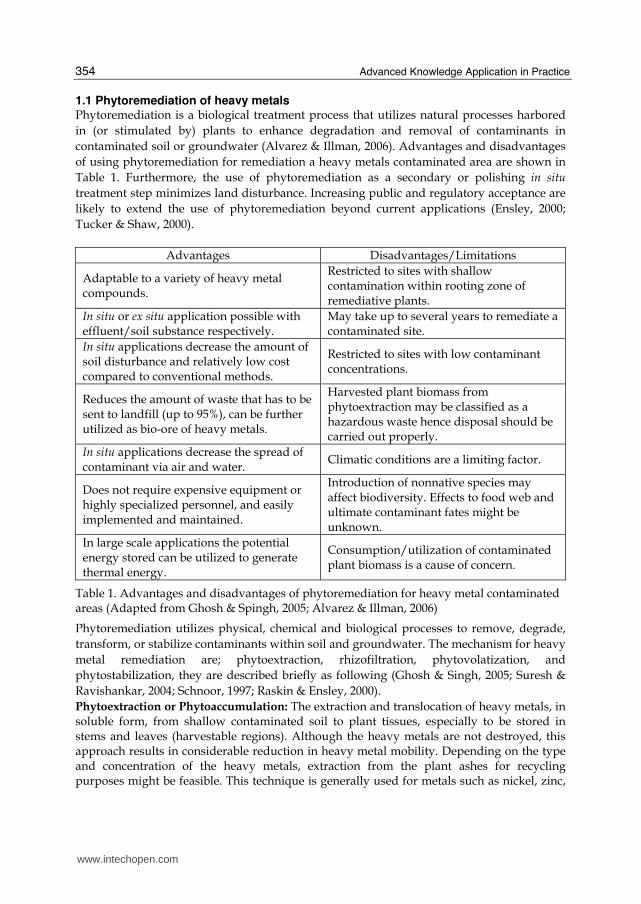
1.1 Phytoremediation of heavy metals Phytoremediation is a biological treatment process that utilizes natural processes harbored in (or stimulated by) plants to enhance degradation and removal of contaminants in contaminated soil or groundwater (Alvarez & Illman, 2006). Advantages and disadvantages of using phytoremediation for remediation a heavy metals contaminated area are shown in Table 1. Furthermore, the use of phytoremediation as a secondary or polishing in situ treatment step minimizes land disturbance. Increasing public and regulatory acceptance are likely to extend the use of phytoremediation beyond current applications (Ensley, 2000; Tucker & Shaw, 2000).
Advantages Disadvantages/Limitations
Adaptable to a variety of heavy metal compounds.
Restricted to sites with shallow contamination within rooting zone of remediative plants.
In situ or ex situ application possible with effluent/soil substance respectively.
May take up to several years to remediate a contaminated site.
In situ applications decrease the amount of soil disturbance and relatively low cost compared to conventional methods.
Restricted to sites with low contaminant concentrations.
Reduces the amount of waste that has to be sent to landfill (up to 95%), can be further utilized as bio-ore of heavy metals.
Harvested plant biomass from phytoextraction may be classified as a hazardous waste hence disposal should be carried out properly.
In situ applications decrease the spread of contaminant via air and water.
Climatic conditions are a limiting factor.
Does not require expensive equipment or highly specialized personnel, and easily implemented and maintained.
Introduction of nonnative species may affect biodiversity. Effects to food web and ultimate contaminant fates might be unknown.
In large scale applications the potential energy stored can be utilized to generate thermal energy.
Consumption/utilization of contaminated plant biomass is a cause of concern.
Table 1. Advantages and disadvantages of phytoremediation for heavy metal contaminated areas (Adapted from Ghosh & Spingh, 2005; Alvarez & Illman, 2006)
Phytoremediation utilizes physical, chemical and biological processes to remove, degrade, transform, or stabilize contaminants within soil and groundwater. The mechanism for heavy metal remediation are; phytoextraction, rhizofiltration, phytovolatization, and phytostabilization, they are described briefly as following (Ghosh & Singh, 2005; Suresh & Ravishankar, 2004; Schnoor, 1997; Raskin & Ensley, 2000). Phytoextraction or Phytoaccumulation: The extraction and translocation of heavy metals, in soluble form, from shallow contaminated soil to plant tissues, especially to be stored in stems and leaves (harvestable regions). Although the heavy metals are not destroyed, this approach results in considerable reduction in heavy metal mobility. Depending on the type and concentration of the heavy metals, extraction from the plant ashes for recycling purposes might be feasible. This technique is generally used for metals such as nickel, zinc,
www.intechopen.com
Advances in Phytoremediation Research: A Case Study of Gynura pseudochina (L.) DC.
355
copper, lead, chromium and cadmium. Plant productivity; accumulation in harvestable portion of plant > 3 tons dry matter/acre-yr; > 1,000 mg/kg metals lightly contaminated soil near to clean-up standard. Rhizofiltration: This mechanism refers to the use of aquatic plants in wetlands or hydroponic reactors. Generally, plants with large root systems are used. The submerged roots of such plants act as filters for the adsorption and absorption of a wide variety of contaminants. This mechanism is commonly used for treatment of industrial discharge, agricultural runoff, metals and radioactive contamination, with the plant densities of 200-1000 g m-2 and hydraulic detention time of several days. Phytovolatilization: The natural ability of a plant to volatilize a contaminant that has been taken up through its roots can be exploited as a natural air-stripping pump system. Volatile pollutants diffuse from the plant into the atmosphere through open stomata in leaves where gas exchange occurs. Phytostabilization: This application aims to prevent the dispersion of contaminated sediments and soil by using plants (mainly grasses) to minimize erosion by wind or rain action. Plants are used to reduce the bioavailability of environmental pollutants. Conditions for optimum likelihood of success are vigorously growing roots; hydrophobic or immobile chemicals.
1.2 Utilization of Phytoremediation by products In the case of phytoremediation of heavy metals, the utilization is based on hyperaccumulative plants and the phytoextraction mechanism. Where, hyperaccumulation is defined as concentration of metal in the harvestable above ground tissues of the plant, with levels in the range of 0.1-1% of the dry weight of the plant. Therefore, the phytomining and diet enrichment of trace elements in the edible parts of plants were mentioned for utilization of the byproducts (Suresh & Ravishankar, 2004; Raskin & Ensley, 2000) Phytomining is a green technology involving the use of hyperacumulative plants to grow and concentrate a metal. The basic principle of phytomining is combined with biomass generation and its commercial utilization as an energy source, so it can be turned into a profit making operation and the remaining ash can be used as bio-ore. One approach to the post-phytoremediation strategy would be to incinerate the plants in a incinerator, resulting in ash with high metal content, and there would be no emission to the air (Ghosh & Singh, 2005; Anderson et al., 1999). The aspects of diet enrichment by trace elements such as zinc, iron and silinium in the edible parts of plants were mentioned for application as supplements in feed or biofertilizer. More than 430 taxa to date have been reported to hyperaccumulate heavy metals, ranging from annual herbs to perennial shrubs and trees. However, the chemical forms of the heavy metals accumulated in these plants have to be clearly understood before application as a supplement can be achieved (Suresh & Ravishankar, 2004; Chantiratikul et al., 2008; Ensley et al., 2001).
1.3 Distribution and speciation of heavy metals in plants Plants absorb heavy metals from soil and they predominantly accumulate in the roots, then some portions are transported to other parts of the plant. Generally, the contents of heavy metals in the underground parts are higher than those found in the parts above the ground and follows a pattern of root>leaf>shoot (stem)>fruit and lateral root>main root, old leaf>young leaf (Cheng, 2003). Metals accumulated in plant tissues can cause toxic effects, especially when translocated to above ground tissues. The root epidermis served as a barrier
www.intechopen.com
Advanced Knowledge Application in Practice
356
to transport of any heavy metals to aboveground tissues. The endodermis casparian strip provided a barrier to the movement of the metals into the stele (the vascular bundles). Once in the leaves, however, metals were highest in the xylem, followed by the mesophyll and then hypodermal tissue. Concentrations of metals in the cell walls were also higher than in intracellular locations (Weis & Weis, 2004). The distribution and accumulation of heavy metals in plants is related to plant species, element species, chemical and bioavailability, and a number of environmental conditions such as redox, pH, cation exchange capacity, dissolved oxygen and temperature (Cheng, 2003; Weis & Weis, 2004). The tolerance of plants to heavy metals and the accumulation are depended on various physiological factors such as uptake and leakage of metal ions by roots, root cation exchange capacity (CEC), phytochelatin production, antioxidative stress, carbohydrate production and utilization (Suresh & Ravishankar, 2004). Generally, the transportation of heavy metals is related to the chemical status in plants. The transport activities of the ethanol extractive and water dissoluble metals were the highest, then the sodium chloride extractive metal, the acetic acid and the hydrochloric acid extractive metal being the lowest. In spite of the chemical extraction forms, heavy metals can combine with inorganic substances (e.g. sulphides), and some small-molecular organic substances such as glutathione (GSH), oxalic acid, histidine, citrate and metal-binding proteins in the plants (Cheng, 2003). In addition, metal ion interactions might be responsible for the regulation of metal uptake and translocation, for example, free proline acts as an antioxidant in Cd stressed cells, increasing phytochelator synthesis and sequestration of Cd, ultimately leading to hyperacculation (Suresh & Ravishankar, 2004; Chantiratikul et al., 2008).
1.4 Metal-inducible proteins Subsequent to metal uptake into the root symplasm, the movement to xylem involves 3 steps: metal sequestration inside root cells, symplastic transport into stele, and finally release into xylem mediated by membrane transport proteins. In addition, a mechanism of heavy metals detoxification and tolerance in plants involves the chelation of metals by organic acids, amino acids or peptides and metalloproteins (Suresh & Ravishankar, 2004; Callahan et al., 2006). Phytochelatin (PC) and metallothioneins (MT) play an important role in transport, translocation and detoxification of heavy metals (Memon et al., 2001; Cobbett & Goldsbrough, 2002). Phytochelatins are a group of proteins that are inducible in plants when plants are faced with heavy metal stress. They bind to free metal ions and carry them to vacuoles, where they are no longer toxic and if the metals are essential, for plant growth, like copper and zinc, they can be utilized by the plant itself. Phytochelatins consist of 3 amino acids, viz. cysteine, glycine and glutamic acid arranged in a (γ-GluCys) n-Gly confirmation. The synthesis of PCs starts with a response to a heavy metal by the plant by activation of an enzyme phytochelatin synthase, which acts upon a glutathione substrate to produce PCs. This action continues until the complexation of metals is completed. The action of PCs on heavy metals starts with the exposure of the plant to high levels of heavy metal concentrations (Suresh & Ravishankar, 2004; Cobbett & Goldsbrough, 2002). Metallothioneins (MT) resemble phytochelatins in many ways, structurally and functionally. They have high cysteine content, low molecular weight (6-7 KDa) and high metal content with coordination of metal ions in metallothionate clusters. Plant MTs are further classified into two types. The type 1 MTs have 12 cysteine residues arranged as 6Cys-Xaa-6Cys, with the Xaa consisting of approximately 40 amino acids. Type 2 MTs are
www.intechopen.com

Advances in Phytoremediation Research: A Case Study of Gynura pseudochina (L.) DC.
357
arranged either as 6Cys-6Cys or 6Cys-Xaa-Xaa-6Cys formation. Even though, the exact function of metallothioneins is not clear, two theories are hypothesized. One theory says that MTs create ion storage pools for free excess heavy metal ions, which are chelated until the plant can use them, if it is an essential metal for the plant. The second theory is that MTs are transport proteins responsible for moving excess heavy metals from sites where they have built up to toxic levels to areas of the plant where they are needed (Suresh & Ravishankar, 2004; Cobbett & Goldsbrough, 2002). The organic compounds use oxygen and sulfur groups in their structures to be bound to heavy metals; however, plants might be bound to different atoms, depending on the tissue (Cobbett & Goldsbrough, 2002; Hall, 2002). Salsola kali, a potential Cd-hyperaccumulator desert plant species, might synthesis phytochelatins in the stems, later coordinating the absorbed cadmium for transport and storage in leaves as a Cd-phytochelatin complex (Rosa et al., 2004). In order to understand the structure and function of these biological systems and the relationship of the metals with the proteins, a technique with high-resolution for protein separation and high sensitivity for metal analysis is required. Gel filtration, Sodium dodecyl sulfate-polyacrylamide gel electrophoresis (SDS-PAGE) and two dimension gel electrophoresis (2DE) are techniques commonly used to analyze proteins. The techniques have been applied to investigate protein detoxification of metals in both microorganisms and plants (Yoshida, et al., 2006; Sobkowiak & Deckert, 2006). In addition, X-ray absorption spectroscopy (XAS) is one of the premier tools for investigating the local structural environment of metal ions. X-ray absorption fine edge structure (XAFS) consists of two the complimentary techniques of X-ray absorption near edge structure (XANES) and extended X-ray absorption fine structure (EXAFS) (Gardea-Torresdey et al., 2005). XAFS has provided the nature of heavy metal complexes accumulated in metal tolerant and hyperaccumulating plant species, the invaluable information of metal-binding proteins, and the structure of metals-containing metalloproteins (Kelly et al., 2002; Verbi et al., 2005; Zhao et al., 2007).
1.5 Microorganism in rhizosphere Although metals are essential components of microbial cells; for example, sodium and potassium regulate gradients across the cell membrane, while copper, iron and manganese are required for activity of key metalloenzymes in photosynthesis and electron transport. Perhaps the most toxic metals are the non-essential metals such as cadmium, lead and mercury. As a consequence of metal toxicity, some microorganisms have developed various resistance mechanisms to prevent metal toxicity. The strategies are either to prevent entry of the metal into the cell or actively to pump the metal out of the cell. This can be accomplished by either sequestration, active transport or chemical transformation through metal oxidation or reduction. Sequestration involves metal complexation with microbial products such as extracellular polymeric substances (EPS) and metallothionine-like proteins. Following metal uptake, some cells sequester metals intracellularly utilizing low molecular weight, cysteine-rich proteins called metallothioneins. The complexed metal may then either be transported back out of the cell or stored as intracellular granules. Which mechanism predominates, intracellular or extracellular sequestration, is dependent on the organism involved. Active transport of metals out of the cell is one mechanism of microbial resistance to metal toxicity. Highly specific efflux systems can rapidly pump out toxic metal ions that have entered the cell. Such efflux pumps may derive their energy from membrane potential or from ATP, and in fact ATP-dependent efflux pumps have been identified that are specific for metals.
www.intechopen.com
Advanced Knowledge Application in Practice
358
Oxidation-reduction (redox) reactions constitute a third mechanism for microbial metal resistance. Microorganisms may either oxidize to mobilize or reduce to immobilize a metal and both reactions are used to prevent metal entry into the cell (Roane et al., 1996). Plant growth-promoting bacteria (PGPB) have the ability to promote a plant’s growth (increase biomass) and increase tolerance to toxic heavy metals by nitrogen fixation, phosphate solubilisation, sulfate oxidation and synthesis of phytohormones such as indole-3-acetic acids (IAA), cytokinins, gibberellins and aminocyclopropane-1-carboxylate (ACC) deaminase (Zhuang et al., 2007) and induced systemic resistance (ISR) mechanism in the plant. Other mechanisms are the release of antibiotics, extracellular enzymes, chemical and volatile compounds such as lumichrome that allow respiration in roots and lead to an increase in the size of plants (Mukerji, 2006). More than the plant promoting properties under stress conditions, PGPB could assist the phytoremediation process by increase the availability and mobility of heavy metals to plants through acidification, redox changes and releasing of chelating agents such as siderophores which are then transferred together with water and nutrient uptake into the plant (Jing, 2007; Khan, 2005; Zhuang et al., 2007). Some PGPB also have toxic reducing or are heavy metal resistant mechanical inducers for example extracellular polymeric substants (EPS) or polysaccharides that secreted outside the bacterial cells can modify the soil structure and induce the plants to respond to stress. This suggest that rhizosphere bacteria, especially plant-growth promoting bacteria (PGPB) that contain the properties of heavy metal resistance can increase the plants metal resistance and increase accumulation in the plants that leads to increased efficiency of phytoremediation (Jing et al., 2007; Siddiqui, 2006; Weis & Weis, 2004; Suresh & Ravishankar, 2004) Phytoremediation is still in the research and development phase, and could be most suitable for developing countries, such as Thailand. However, there are many technical barriers which need to be addressed. Our survey in a zinc mine, Tak province, Thailand, found Gynura pseudochina (L.) DC. (Wan-Maha-Kan), a tuber plant in the Genera Asteraceae. It can grow in areas with high zinc and cadmium contamination and highly accumulates the metals. In addition, it can survive all year, both dry and rainy season in Thailand. The plant has a potential to be used in the phytoremediation process. However, the mechanism of zinc and cadmium accumulation have to be made clear before application. This research aimed to study the distribution and chemical speciation of the metals by transmission electron microscopy (TEM), X-ray fluorescence (XRF) imaging and X-ray absorption fine structure (XAFS), using synchrotron light source. The proteins of G. pseudochina (L.) DC. involved in the accumulation and tolerance of zinc and cadmium were studied by extraction and separation with SDS-PAGE techniques. A plant growth promoting rhizobacteria (PGPR) from the rhizosphere of G. pseudochina (L.) DC. growing in the Zn/Cd contaminated soil was isolated and studied its capability to induce phosphate solubility, nitrogen fixation, IAA and ACC deaminase production. Finally, the phytoremediation byproduct was studied for their utilization as a zinc bio-ore and phytochemical extracts.
2. Materials and methods
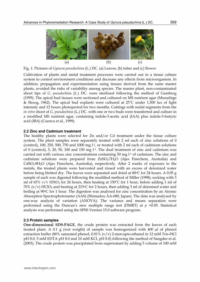
2.1 Plant samples Gynura pseudochina (L.) DC. (Fig.1) is a member of the Asteraceae Family. It is a dicotyledon, with elliptic, coarsely dentate leaves covered with multicellular hairs, and an upright unbranched stem. The tuber has several heads near the stem apex. The flower is a long scape with an orange corolla on a short peduncle and an inferior ovary.
www.intechopen.com
Advances in Phytoremediation Research: A Case Study of Gynura pseudochina (L.) DC.
359
(a) (b) (c)
Fig. 1. Pictures of Gynura pseudochina (L.) DC. (a) Leaves, (b) tuber and (c) flower
Cultivation of plants and metal treatment processes were carried out in a tissue culture system to control environment conditions and decrease any effects from microorganism. In addition, propagation and experimentation using tissues derived from the same master plants, avoided the risks of variability among species. The master plant, non-contaminated shoot tips of G. pseudochina (L.) DC. were sterilized following the method of Gambrog (1995). The apical bud tissues were sectioned and cultured on MS nutrient agar (Murashige & Skoog, 1962). The apical bud explants were cultured at 25˚C under 1,500 lux of light intensity and 12 hours photoperiod for two months. Cuttings with nodal segments from the in vitro shoot of G. pseudochina (L.) DC. with one or two buds were transferred and culture in a modified MS nutrient agar, containing indole-3-acetic acid (IAA) plus indole-3-butyric acid (IBA) (Cuenca et al., 1999).
2.2 Zinc and Cadmium treatment The healthy plants were selected for Zn and/or Cd treatment under the tissue culture system. The plant samples were separately treated with 2 ml each of zinc solutions of 0 (control), 100, 250, 500, 750 and 1000 mg l-1, or treated with 2 ml each of cadmium solutions of 0 (control), 5, 20, 50, 100 and 150 mg l-1. The dual treatment of zinc and cadmium was carried out with various zinc concentrations containing 50 mg l-1 of cadmium. The zinc and cadmium solutions were prepared from ZnSO4.7H2O (Ajax Finechem, Australia) and CdSO4.8H2O (Ajax Finechem, Australia), respectively. After 2 weeks of exposure to the metals, the treated plants were harvested and rinsed with an excess of deionized water before being blotted dry. The leaves were separated and dried at 80oC for 24 hours. A 0.05 g sample of each was digested following the modified method of Miller (1998); socking with 3 ml of 65% v/v HNO3 for 24 hours, then heating at 150˚C for 1 hour, before adding 1 ml of 70% (v/v) HClO4 and heating at 215oC for 2 hours, then adding 3 ml of deionised water and boiling at 90oC for 1 hour. The digestion was analysed for zinc concentration by an Atomic Absorption Spectrophotometer (AAS) (Shimadzu AA-680, Japan). The data was analyzed by one-way analysis of variation (ANOVA). The variance and means separation were performed using the Duncan’s new multiple range test (DMRT) at p <0.05. Statistical analysis was performed using the SPSS Version 15.0 software program.
2.3 Protein samples One-dimensional SDS-PAGE, the crude protein was extracted from the leaves of each treated plant. A 0.1 g (wet weight) of sample was homogenized with 400 µl of phenol extraction buffer (80% saturated phenol, 0.01% (v/v) 2-mercapto-ethanol in 12 mM Tris-HCl pH 8.0, 5 mM EDTA pH 8.0 and 10 mM KCl, pH 8.0) following the method of Sangdee et al. (2003). The crude protein was precipitated from supernatant by adding 5 volume of 100 mM
www.intechopen.com
Advanced Knowledge Application in Practice
360
ammonium acetate in 99.9% methanol then incubated at -20oC for 30 min, and centrifuged at 13,000 g for 20 minute. The protein pellet was washed with 99.96% acetone (AnalaR NORMAPUR, France) and resuspended in 50 µl of protein sample buffer (63 mM Tris-HCl pH 6.8, 2% (v/v) SDS, 5% (v/v) mercaptoethanol, 20% (v/v) glycine) and the protein concentration was determined by Bradford’s assay (Bradford, 1976). Each 40 µg protein sample was fractionated by polyacrylamide gel electrophoresis, resolving gel 12% and stacking gel 5%, for 2 hours at 20 mA per gel by electrophoresis (MiniVE GE Healthcare Bio-Sciences Corp., USA). The gels were stained with Coomassie Brilliant Blue R-250 staining buffer (Bollag et al., 1996). Molecular weight of protein band was measured based on the molecular weight of two broad range protein markers of 7.1 kDa to 209 kDa and 10 kDa to 250 kDa (prestained SDS-PAGE standard, broad range, Bio-Rad, USA). The expression of proteins in SDS-PAGE was analysed by Quantity One 1-D analysis software program (Bio-Rad, USA). Two-dimensional electrophoresis, a crude protein extract was extracted from the plant leaves (0.2 g) by grinding in a chilled mortar with liquid nitrogen to obtain a fine powder, then 200 µl of lysis buffer (7M urea, 2M Thiourea, 4% CHAPS, 2% IPG buffer pH 3-10, 40 mM dithiothreitol; (DTT)) was added directly to the mortar and continually ground for 30 sec. The extract was separated by centrifugation at 12000g, 4°C for 15 min. The supernatant was cleaned by 2-D Clean-up kit. Each 80 µg protein sample was isoelectric focused (IEF), fractionated on Immobiline DryStrip gel pH 3-7, 7 cm and IPG buffer for 4 hours by an isoelectric focusing electrophoresis (Ettan IPGphor II GE Healthcare, Sweden). The chemicals for 2-DE were ordered from GE Healthcare. After separating by first dimension of IEF, the strip was equilibrated in SDS equilibration buffer (50 mM Tris-HCl pH 8.8, 6 M urea, 30%(v/v) glycerol, 2%(w/v) SDS, 0.002%(w/v) bromophenol blue), before separated in the second dimension on SDS-PAGE with a 12% polyacrylamide gel for 2 hours at 20 mA per gel. The separated protein spots were visualized by silver staining (Bollag et al., 1996).
2.4 TEM The samples for imaging by transmission electron microscopy (TEM) were G. pseudochina (L.) DC. treated with 100 mg l-1 zinc solution, pH 5.5 ± 0.5 or watered with deionised water (control) for two months. Small sections of stem, leaf and tuber (1 mm length x 1 mm width) were fixed in 2.5-10% glutaraldehyde (v/v) in 0.1 M phosphate buffer at 4oC, pH 7.2 for 24 hours. Samples were washed 3 times for 30 minutes each in phosphate buffer before being postfixed in 1% OsO4 for 2 hours. The postfixed samples were washed with deionised water 3 times for 30 minutes each, then dehydrated by ethanol series (20, 40, 60, 80, and 100%) followed by infiltration with ethanol and Spurs’ resin in ratios of 3:1, 1:1 and 1:3 for 24 hours each. Embedding in Spurs’ resin and polymerization was carried out at 70°C for 80 hours. Thin slide sections (60-90 nm) were prepared on a grid for viewing by TEM (JEOL JEM 2010, Japan) at an accelerating voltage of 80.0 kV.
2.5 XAFS and XRF imaging Zinc treated plants were subjected to two-dimensional µ-XRF imaging and XAFS analysis. The plant samples were harvested and washed with an excess of running deionised water, before being separated into leaves, stem and tuber. A thin section of the freeze-dried sample was prepared in order to maintain its tissue structure and chemical state. The cross section of the tuber, stem and leaves were cut to a thickness of 200-300 µm using a vertical slicer
www.intechopen.com
Advances in Phytoremediation Research: A Case Study of Gynura pseudochina (L.) DC.
361
(HS-1 JASCO, Japan) and immediately put on dry ice. The sections were freeze-dried overnight using a lyophilizer (Labconco Lyph Lock 6 Freeze Dryer, USA). For bulk XAFS analysis, an amount of each freeze-dried plant part, tuber (medulla and periderm), stem and leaves, were ground and pressed into a pellet before being sealed in a mylar plastic bag (Mylar polyester film, No.100, Chemplex, USA). The whole plant treated with 1,000 mg l-1 of Zn solution was used to prepare a crude protein pellet for XAFS analysis. µ-XRF imaging was performed at beamline 4A, Photon Factory (PF), High Energy Accelerator Research Organization (KEK), and beamline 37XU, SPring-8, Japan. XAFS analysis was performed at beamline 12C, at PF, KEK. The XAFS analysis data was analyzed by Rigaku Rex2000 Version 2.3.2. The analytical chemicals used for reference materials were Zn(NO3)2, ZnS, ZnSO4.7H2O, ZnCl2 and ZnO. Zn-cellulose (prepared by absorption system) and Zn-cysthein (prepared by absorption system) were measured as reference materials.
2.6 Microbial isolation A soil sample was collected from the rhizosphere of G. pseudochina (L.) DC., growing in zinc and cadmium contaminated areas. The soil sample was suspended in sterile distilled water. Bacterial isolation was carried out by spread plate techniques on nutrient agar (NA) containing Zn and/or Cd of 5, 10, 15 and 20 mg l-1. The plates were incubated at 30oC for 3 days. Colonies with different characteristics from the Zn and/or Cd containing agar plate were picked and further isolated to single colonies on Nutrient agar containing Zn and/or Cd according to their resistant metal concentration. The isolates were given code names, for example, PDMZnCd2003 (PDM-Phadeand mining; ZnCd-agar containing Zn and Cd; 20-concentration of heavy metals in agar (mg l-1); 03-isolate number). All bacterial isolates were Gram stained and endospore stained to confirm each isolates was not the same and pre-grouped the isolates. The isolates containing the properties of Zn/Cd tolerance and plant growth promoting were identified by API 20E biochemical test (Koneman et al., 1997) and genetic characterization. Genetic characterization: Total genomic DNA of selected isolate was extracted by a modified phenol: chloroform procedure of Sambrook & Russel’s (2001). Two primers of fD1 (5’-AGAGTTTGATCCTGGCTCAG-3’) and rP2 (5’-ACGGCTACCTTGTTACGACTT-3’) (Weisburg et al., 1991) were used for 16S rDNA (ribosomal Deoxyribonucleic acid) amplification, and nif H-F (5’-AAAGGYGGWATCGGYAARTCCACCAC-3’) and nif H-R (5’-TTGTTSGCSGCRTACATSGCCATCAT-3’) primer pairs (Torok et al., 1981) were used for nif gene amplification. Each 50 µl of polymerase chain reaction (PCR) reaction contained: 100 ng of purified total DNA, 0.2 mM of each diethylnitrophenyl thiophosphate (dNTP), 5 unit of Tag DNA polymerase (Invitrogen, USA) in 5 µL of 10x Tag buffer, 1 mM MgCl2, 0.2 mM of each primer, and 35 µl sterile deionized water. Thermalcycling program for 16S rDNA amplification consisted of 1 cycle of 94oC for 5 min (denaturation), 57oC for 2 min (annealing for fD1 and rP2) and 72oC for 2 min (extension), and 29 cycles of 94oC for 2 min, 57oC for 30 sec and 72oC for 2 min, with a final elongation cycle of 72oC for 10 min (Wood et al., 1998). Amplification of nif gene was carried out under the thermalcycling program of 35 cycles of 94oC for 5 min, 54oC for 1 min (annealing for nif H-F and nif H-R) and 72oC for 1 min, with a final elongation cycle of 72oC for 10 min (Zehr et al., 1989). The PCR products obtained were purified with a HiYieldTM Gel/PCR DNA Fragments extraction kit (Real Biotech Corporation, Taiwan) and cloned into the pGEM-T-Easy vector (Promega, Madison, Wis.) according to the protocols of the manufacturers. The plasmids were transformed into
www.intechopen.com
Advanced Knowledge Application in Practice
362
competent E. coli JM109 by transformation and storage solution (TSS) method (Chung & Miller, 1993). Sequencing was performed on an 3730XL DNA sequencer, monitoring the whole experimental process through Laboratory Information Magement System (LIMS), Macrogen Inc, Korea. Sequence data of 16S rDNA (1500 bp) and nif gene (700 bp) were compared with sequences in the National Center for Biotechnology Information data bank using the BLAST program (Altschul et al., 1997).
2.7 Study the plant growth promoting (PGPB) properties All bacteria were screened for their plant growth promoting properties as measured by their IAA and ACC deaminase production, N2 fixation, and phosphate solubilisation. The isolates that exhibited high heavy metal tolerance and showed plant growth promoting properties were subjected to quantitative analysis. IAA production: Bacteria were cultured in Trypticase soya broth (TSB) containing 0.2% w/v tryptophan and were incubated in the dark at 30oC for 48 hours at 150 rpm by an incubated shaker (Innova 2100 Platform shaker, New Brunswick Scientific, USA). The cultures were then centrifuged at 6,000 rpm for 15 minutes at 4°C by a refrigerator centrifuge (MX-301, TOMY, USA). The supernatants were mixed with Salkowski’s reagent (ratio 2:1) and left in the dark for 20 min. The optical density was measured at an absorbance of 530 nm (Bric et al., 1991). The IAA concentration was determined by using a standard curve of authentic IAA (Sigma-Aldrich, St. Louis, MO, USA). N2 fixation: One loop of each bacterial culture (24 hours old) was spotted onto N-Free agar containing 0.0025 % w/v bromothymol blue and incubated for between 1-7 days. Isolates that showed growth within 7 days and intensified the color of bromotymol blue around the colony were considered to be N2-fixation bacteria. To measure quantity, bacteria were inoculated into N-free malate medium and incubated for 72 hours while being shaken. The cultures were then centrifuged at 6,000 rpm for 15 minutes at 4°C. Ammonia nitrogen (NH3-N), an inorganic dissolved form of nitrogen, in the supernatant was quantitatively analysed with Nessler’s reagent as described by Cappuccino & Sherman (1992). The amount of NH3-H was measured against a standard curve of ammonium choride (NH4Cl) (Ajax Finechem Pty Ltd, Australia). Phosphate solubilisation: One loop of each bacterial culture (24 hours old) was streaked onto NBRIP (National Botanical Research Institute’s phosphate growth) agar. After incubation for 7 days, the clear zone around each colony that indicated phosphate-solubilisation was observed. The amount of soluble phosphate was measured. Bacteria were inoculated in NBRIP medium consisting of 0.5% w/v Tricalcium phosphate and incubated aerobically on a rotary shaker for 72 hours. Bacterial cultures were centrifuged at 6,000 rpm for 15 minutes at 4°C. Soluble phosphate in the supernatant was measured by the modified ascorbic acid method of APHA, AWWA (Clesceri et al., 1998). Concentration of soluble phosphate was determined against a standard curve of potassium dihydrogen phosphste (KH2PO4) (Ajax Finechem Pty Ltd, Australia). ACC deaminase activity: Bacteria were grown in TSB until late log phase and harvested by centrifugation at 6000 rpm at 4˚C. Cell pellets were washed twice with DF-salt medium (Dworkin and Foster, 1958) and resuspended in DF-salt medium. The suspended cells were then added to a DF-salt medium containing 3 mM of ACC as the sole N- source and shaken at 200 rpm for 72 hours. ACC deaminase activity was measured by following the method of Penrose and Glick (Penrose & Glick, 2003).
www.intechopen.com
Advances in Phytoremediation Research: A Case Study of Gynura pseudochina (L.) DC.
363
2.8 Utilisation of phytoremediation by-product The zinc accumulative plant was dried and pyrolysed in a furnace at 550oC for 4 hours to obtain ash. The 98% (v/v) sulphuric acid (H2SO4), which is used in zinc mining, was used to extract zinc from the ash. In addition, the phytochemical extraction from leaves of G. pseudochina (L.) DC. and the antioxidant property were studied to support a sustainable development in the zinc contaminated area. The plant from a non-contaminated side was studied in the extraction and basis data of phytochemicals. The extracts were investigated in total phenolic content (TPC) (Liu et al., 2008), total flavonoid (TF) (Yoo et al., 2008), free radical scavenging activity (FRSA) (Cotelle et al., 1996), and high-performance liquid chromatography (HPLC) by modified of the conditions of Zuo et al. (2002). Then the extracts of plants grown in zinc contaminated soil were measured to determine the possibility of zinc contaminating the extracts.
3. Results and discussion
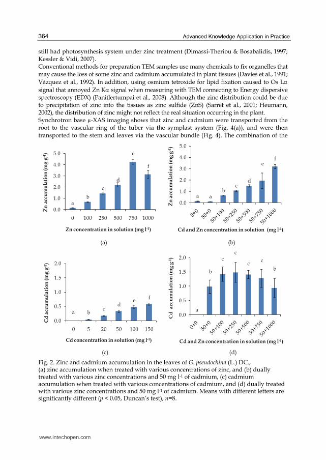
3.1 Zinc accumulation The concentration of zinc in G. pseudochina (L.) DC. leaves increased with increasing Zn levels in the treatment solution (0, 100, 250, 500 and 750 mg l-1). The accumulations of Zn in the leaves of treated plants were 0.2-4.2 mg Zn g-1 dry wt. (Fig. 2(a)). The plants dually treated with various zinc concentrations and 50 mg l-1 of cadmium resulted to lower Zn accumulation in a range of 0.1-3.1 mg Zn g-1 dry wt. (Fig. 2 (b)), compared with only Zn treatment. The amounts of Cd accumulated in treated plants’ leaves are shown in Fig.2(c), the Cd level significantly increased with the increasing concentration of Cd (p < 0.05, n=8). The accumulations of Cd in the leaves of treated plants were 0.1-0.6 mg Cd g-1 dry wt. The Cd concentration in the leaves when dually treated with various zinc concentrations and 50 mg l-1 of cadmium resulted in higher Cd accumulation in a range of 1.0-1.5 mg Cd g-1 dry wt. (Fig. 2(d)). The high accumulation of Zn and Cd in the leaves and the shoot, indicated the potential for application in phytoremediation (Reeves & Baker, 2000). The increase in zinc and cadmium accumulation when treated with higher concentrations of the metals was found in wheat plants, Triticum aestivum L.cv Klein Atalaya (Santa-Maria & Cogliatti, 1998), Linum usitatissimum (Chakravarty & Srivastava, 1997), gray mangrove Avicennia marina (Forsk.) Vierh (MacFarlane & Burchett, 2002), Sedum afredii (Li et al., 2006), Brassica juncea (Maruthi Sridhar et al., 2005), and Potentilla griffithii (Hu et al., 2009). The net Zn-uptake rate increased as the Zn-concentration in the growth solution increased, because the greater the external concentration of Zn, the higher were both Zn-influx and Zn-efflux (Santa-Maria & Cogliatti, 1998). In addition, the enhancement of Cd uptake under dually Zn and Cd treatment might be attributed to an altered and specialized transporter of metal ions in the plasma membrane system induced by the addition of Zn (Ebbs et al., 2009).
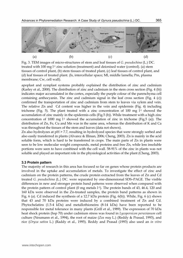
3.2 Distribution and speciation of Zn Ultra structure studied by transmission electron microscope (TEM) showed zinc treatment might be responsible for the rough cell walls observed in stem tissues (Fig. 3(b)), and the stoma of chloroplast in leaves containing starch grains and plastoglobule (Fig. 3(d)). The rough cell walls occurred in B. juncea and Armeria maritime, when they were treated with a high zinc concentration (Maruthi Sridhar et al., 2005; Heumann, 2002). However, the complete chloroplast, containing starch grains and plastoglobule, implied that the plants
www.intechopen.com
Advanced Knowledge Application in Practice
364
still had photosynthesis system under zinc treatment (Dimassi-Theriou & Bosabalidis, 1997; Kessler & Vidi, 2007). Conventional methods for preparation TEM samples use many chemicals to fix organelles that may cause the loss of some zinc and cadmium accumulated in plant tissues (Davies et al., 1991; Vázquez et al., 1992). In addition, using osmium tetroxide for lipid fixation caused to Os Lα signal that annoyed Zn Kα signal when measuring with TEM connecting to Energy dispersive spectroscopy (EDX) (Panitlertumpai et al., 2008). Although the zinc distribution could be due to precipitation of zinc into the tissues as zinc sulfide (ZnS) (Sarret et al., 2001; Heumann, 2002), the distribution of zinc might not reflect the real situation occurring in the plant. Synchrotron base µ-XAS imaging shows that zinc and cadmium were transported from the root to the vascular ring of the tuber via the symplast system (Fig. 4(a)), and were then transported to the stem and leaves via the vascular bundle (Fig. 4). The combination of the
(a) (b)
(c) (d)
a bc
de
f
0.0
0.5
1.0
1.5
2.0
0 5 20 50 100 150
Cd
acc
um
ula
tio
n (
mg
g-1
)
Cd concentration in solution (mg l-1)
ab
c
d
e
f
0.0
1.0
2.0
3.0
4.0
5.0
0 100 250 500 750 1000
Zn
acc
um
ula
tio
n (
mg
g-1
)
Zn concentration in solution (mg l-1)
a ab
cd
ef
0.0
1.0
2.0
3.0
4.0
5.0
Zn
acc
um
ula
tio
n (
mg
g-1
)
Cd and Zn concentration in solution (mg l-1)
a
b
cc
cc
b
0.0
0.5
1.0
1.5
2.0
Cd
acc
um
ula
tio
n (
mg
g-1
)
Cd and Zn concentration in solution (mg l-1)
Fig. 2. Zinc and cadmium accumulation in the leaves of G. pseudochina (L.) DC., (a) zinc accumulation when treated with various concentrations of zinc, and (b) dually treated with various zinc concentrations and 50 mg l-1 of cadmium, (c) cadmium accumulation when treated with various concentrations of cadmium, and (d) dually treated with various zinc concentrations and 50 mg l-1 of cadmium. Means with different letters are significantly different (p < 0.05, Duncan’s test), n=8.
www.intechopen.com
Advances in Phytoremediation Research: A Case Study of Gynura pseudochina (L.) DC.
365
(a) (b) (c) (d)
Fig. 3. TEM images of micro-structures of stem and leaf tissues of G. pseudochina (L.) DC. treated with 100 mg l-1 zinc solution (treatment) and deionised water (control). (a) stem tissues of control plant, (b) stem tissues of treated plant, (c) leaf tissues of control plant, and (d) leaf tissues of treated plant. (Is, intercellular space; Ml, middle lamella; Pm, plasma membrane; Cw, cell wall).
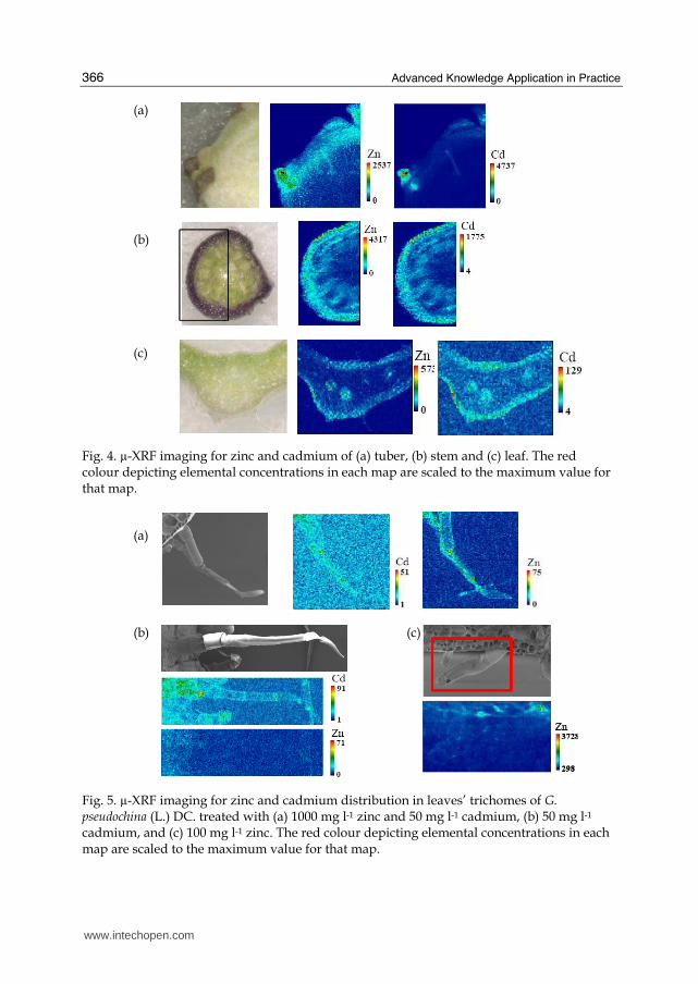
apoplast and symplast systems probably explained the distribution of zinc and cadmium (Karley et al., 2000). The distribution of zinc and cadmium in the stem cross section (Fig. 4 (b)) indicates major accumulated in the cortex, especially the purple colour of the parenchyma cell containing anthocyanin. The zinc and cadmium signal in the leaf cross section (Fig. 4 (c)) confirmed the transportation of zinc and cadmium from stem to leaves via xylem and vein. The relative Zn and Cd content was higher in the vein and epidermis (Fig. 4) including trichome (Fig. 5). The plant treated with a zinc concentration of 100 mg l-1 showed the accumulation of zinc mainly in the epidermis cells (Fig.5 (b)). While treatment with a high zinc concentration of 1000 mg l-1 showed the accumulation of zinc in trichome (Fig.5 (a)). The distribution of Zn, Fe, Cu and Mn was in the same area, whereas the distribution of K and Ca was throughout the tissues of the stem and leaves (data not shown). Zn also hydrolyzes at pH > 7.7, resulting in hydrolyzed species that were strongly sorbed and also easily transferred in plants (Alvarez & Illman, 2006; Cheng, 2003). Zn is mainly in the acid soluble form, which is hard to be transferred in crops. The main parts of Zn in plants were seen to be low molecular weight compounds, metal proteins and free Zn, while less insoluble portions were seen to have combined with the cell wall. 58-91% of the zinc in plants was not soluble and played an important role in the physiological activities of the plant (Cheng, 2003).
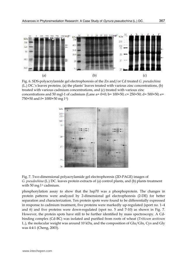
3.3 Protein pattern The majority of research in this area has focused so far on genes whose protein products are involved in the uptake and accumulation of metals. To investigate the effect of zinc and cadmium on the protein patterns, the crude protein extracted from the leaves of Zn and Cd treated G. pseudochina (L.) DC. were separated by one-dimensional SDS–PAGE. The visible differences in new and stronger protein band patterns were observed when compared with the protein pattern of control plant (0 mg metals l-1). The protein bands of 43, 46.4, 120 and 160 kDa were observed in the Zn-treated samples, the protein band patterns as shown in Fig. 6 (a). Cd induced the synthesis of a 12.7 kDa protein (Fig. 6(b)). While, Fig. 6 (c) shows that 43 and 70 kDa proteins were induced by a combined treatment of Zn and Cd. Phytochelatins (1.5-4 kDa) and metallothioneins (8-14 kDa) have been reported to be responsible for metal tolerance in many plants (Grill et al., 1989). The expression of 70 kDa heat shock protein (hsp 70) under cadmium stress was found in Lycopersicon peruvianum cell culture (Neumann et al., 1994), the root of maize (Zea may L.) (Reddy & Prasad, 1993), and rice (Oryza sativa L.) (Raddy et al., 1995). Reddy and Prasad (1993) also used an in vitro
www.intechopen.com
Advanced Knowledge Application in Practice
366
(a) (b) (c)
Fig. 4. µ-XRF imaging for zinc and cadmium of (a) tuber, (b) stem and (c) leaf. The red colour depicting elemental concentrations in each map are scaled to the maximum value for that map.
(a) (b) (c)
Fig. 5. µ-XRF imaging for zinc and cadmium distribution in leaves’ trichomes of G. pseudochina (L.) DC. treated with (a) 1000 mg l-1 zinc and 50 mg l-1 cadmium, (b) 50 mg l-1 cadmium, and (c) 100 mg l-1 zinc. The red colour depicting elemental concentrations in each map are scaled to the maximum value for that map.
www.intechopen.com
Advances in Phytoremediation Research: A Case Study of Gynura pseudochina (L.) DC.
367
(a) (b) (c)
Fig. 6. SDS-polyacrylamide gel electrophoresis of the Zn and/or Cd treated G. pseudochina (L.) DC.’s leaves proteins. (a) the plants’ leaves treated with various zinc concentrations, (b) treated with various cadmium concentrations, and (c) treated with various zinc concentrations and 50 mgl-1 of cadmium (Lane a= 0+0; b= 100+50; c= 250+50; d= 500+50; e= 750+50 and f= 1000+50 mg l-1)
Fig. 7. Two-dimensional polyacrylamide gel electrophoresis (2D-PAGE) images of G. pseudochina (L.) DC. leaves protein extracts of (a) control plants, and (b) plants treatment with 50 mg l-1 cadmium.
phosphorylation assay to show that the hsp70 was a phosphoprotein. The changes in protein patterns were analyzed by 2-dimensional gel electrophoresis (2-DE) for better separation and characterization. Ten protein spots were found to be differentially expressed in response to cadmium treatment, five proteins were markedly up-regulated (sport no. 1–4 and 6) and five proteins were down-regulated (spot no. 5 and 7-10) as shown in Fig. 7. However, the protein spots have still to be further identified by mass spectroscopy. A Cd-binding complex (Cd-BC) was isolated and purified from roots of wheat (Triticum aestivum L.), the molecular weight was around 10 kDa, and the composition of Glu/Gln, Cys and Gly was 4:4:1 (Cheng, 2003).
www.intechopen.com
Advanced Knowledge Application in Practice
368
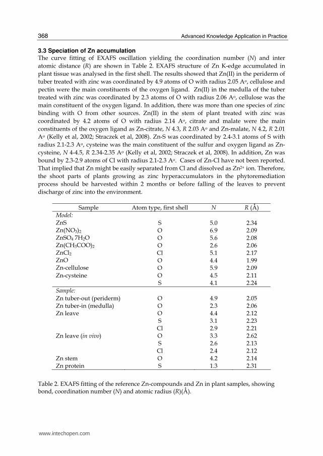
3.3 Speciation of Zn accumulation The curve fitting of EXAFS oscillation yielding the coordination number (N) and inter atomic distance (R) are shown in Table 2. EXAFS structure of Zn K-edge accumulated in plant tissue was analysed in the first shell. The results showed that Zn(II) in the periderm of tuber treated with zinc was coordinated by 4.9 atoms of O with radius 2.05 Ao, cellulose and pectin were the main constituents of the oxygen ligand. Zn(II) in the medulla of the tuber treated with zinc was coordinated by 2.3 atoms of O with radius 2.06 Ao, cellulose was the main constituent of the oxygen ligand. In addition, there was more than one species of zinc binding with O from other sources. Zn(II) in the stem of plant treated with zinc was coordinated by 4.2 atoms of O with radius 2.14 Ao, citrate and malate were the main constituents of the oxygen ligand as Zn-citrate, N 4.3, R 2.03 Ao and Zn-malate, N 4.2, R 2.01 Ao (Kelly et al, 2002; Straczek et al, 2008). Zn-S was coordinated by 2.4-3.1 atoms of S with radius 2.1-2.3 Ao, cysteine was the main constituent of the sulfur and oxygen ligand as Zn-cysteine, N 4-4.5, R 2.34-2.35 Ao (Kelly et al, 2002; Straczek et al, 2008). In addition, Zn was bound by 2.3-2.9 atoms of Cl with radius 2.1-2.3 Ao. Cases of Zn-Cl have not been reported. That implied that Zn might be easily separated from Cl and dissolved as Zn2+ ion. Therefore, the shoot parts of plants growing as zinc hyperaccumulators in the phytoremediation process should be harvested within 2 months or before falling of the leaves to prevent discharge of zinc into the environment.
Sample Atom type, first shell N R (Å) Model: ZnS Zn(NO3)2 ZnSO4 7H2O Zn(CH3COO)2 ZnCl2 ZnO Zn-cellulose
S O O O Cl O O
5.0 6.9 5.6 2.6 5.1 4.4 5.9
2.34 2.09 2.08 2.06 2.17 1.99 2.09
Zn-cysteine O S
4.5 4.1
2.11 2.24
Sample:
Zn tuber-out (periderm) O 4.9 2.05 Zn tuber-in (medulla) O 2.3 2.06 Zn leave O
S Cl
4.4 3.1 2.9
2.12 2.23 2.21
Zn leave (in vivo) O S Cl
3.3 2.6 2.4
2.62 2.13 2.12
Zn stem O 4.2 2.14 Zn protein S 1.3 2.31
Table 2. EXAFS fitting of the reference Zn-compounds and Zn in plant samples, showing bond, coordination number (N) and atomic radius (R)(Å).
www.intechopen.com
Advances in Phytoremediation Research: A Case Study of Gynura pseudochina (L.) DC.
369
XANES spectrum of zinc on protein indicated that zinc in protein was possibly ZnS (81% fitting) and ZnO (19% fitting). The curve fittings of EXAFS oscillation are shown in Table 2. The results obtained for the first Zn-S coordination shell were 1.3 (N), 2.31Ao(R). However, the first Zn-S coordination is limited by the number of free sulfur atoms on the protein and lower coordination numbers of ZnS might be due to our methodology which used a reducing agent such as mercaptoethanol (HOCH2CH2SH). It has the ability to cleave disulfide bonds so it might be decreasing the number of sulfur ligands on the protein of G.
pseudochina (L.) DC. Ideal geometrics of zinc forms four, five and six coordinate complexes by Tetrahedral, Trigonal Bipyramidal, Square Pyramidal and Octahedral, respectively (Patel et al., 2007). Zinc was bound into cysteine residue and histidine complex by coordination of S, O and N atoms (Bracey et al., 1994). In addition, Yu et al., (2008) reported that the formation of a stable Zn tetrahedral configuration with four sulfur ligands on a protein of KTI11. Therefore, the results indicated that Zn-S coordination in the first shell for proteins of G. pseudochina (L.) DC. by the amino acids containing sulfur groups might be involved in zinc binding proteins such as protein content a mainly of cysteine residue and histidine. Therefore, these results indicate that protein patterns of our experiment might be zinc binding proteins.
3.4 PGPB properties of Zn and Cd tolerance isolates The number of bacteria isolated from the rhizosphere of G. pseudochina (L.) DC). growing in zinc and cadmium contaminated soil in a zinc mining area, Phatat Phadaeng sub-distric, Mae sot, Tak province, Thailand, were 34 isolates (they were divided by NA agar plates containing Zn, Cd and Zn+Cd as 18, 10 and 6 isolates, respectively). 25 isolates were gram negative bacteria and 9 isolates were gram positive bacteria. The results indicated that 75% of the isolates were gram negative bacteria. That might be due to the cell wall structures of gram negative bacteria which are more complex and resulted in many functions to help survival in extreme environments when compared to gram positive bacteria (Willey et al., 2009; Ahmad et al., 2008). However, it depends on the kind of heavy metal, as Abou-shanab et al. (2007) found the genes that control Hg, Zn, Cr and Ni tolerance in both gram negative and positive bacteria. In nature, metal resistance-gene transformations can occur between bacteria and this mechanism is the main mechanism for bacterial resistance. Plant growth-promoting abilities of bacteria: From the 34 isolates, there were 24, 24 and 15 isolates of bacteria had the properties of IAA production, N2-fixation and phosphate solubilisation, respectively. The number of IAA producing and N2-fixing bacteria was more than the number of phosphate solubilising bacteria. In addition, 20 isolates contained the properties of N2-fixation and IAA production. These results suggested that IAA production and nitrogen fixation in free-living bacteria could be the major mechanisms needed to associate with the host plants. The large amount of IAA producing and N2-fixing bacteria could be related to their plant colonization due to the mutaulism relationship between plants and bacteria (Siddiqui., 2006). For instance IAA-producing bacteria obtained tryptophan that produced by plants as an IAA precursor (Spaepen et al., 2007). Four isolates capable of IAA production, nitrogen fixation and phosphate solubilisation were chosen for quantitative analysis of IAA production, nitrogen fixation, phosphate solubilisation and ACC deaminase activity. Table 3 shows that 4 isolates produced large
www.intechopen.com
Advanced Knowledge Application in Practice
370
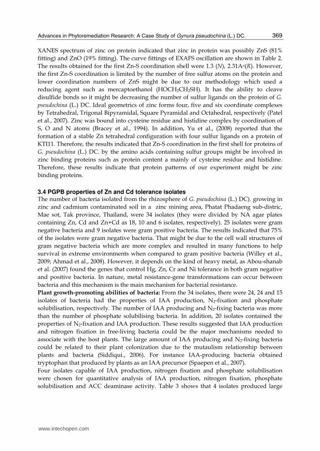
amounts of IAA. The large concentration of IAA produced by IAA producing bacterial isolates might support the growth of G. pseudochina (L.) DC. Although, ACC deaminase production was less than that detection by measuring the amount by the α-ketobutyrate production method. High IAA production implied their ability to product ACC deaminase. The results indicated that these 4 isolates have multiple properties to promote plant growth.
Isolates IAA (mg l-1)
N as NH3-N (mg l-1)
P as KH2PO4 (mg l-1)
ACC deaminase (unit ml-1)
PDMZn2008 275.3 16.2 13.9 N/D PDMCd0501 249.7 18.9 9.6 N/D PDMCd2007 311.3 12.5 7.5 N/D PDMZnCd2003 294.3 23.5 10.5 N/D
*N/D, not detectable
Table 3. IAA production, nitrogen fixation, phosphate solubilisation and ACC deaminase of the bacterial isolates
Identification of bacteria The isolate PDMZnCd2003 was chosen for identification because of its tolerance to high concentrations of Zn and Cd (20 mg l-1 of Zn and Cd) and its ability to produce IAA, fix nitrogen, and solubilise phosphate (Table 3). In addition, PDMZnCd2003 produced fluorescent pigments when cultured in Cd-containing medium. It is very interesting because it may be a siderophores with chelating agents for holding heavy metal ions. Moreover it could demonstrate other plant growth promoting properties that we did not test for in this study. The results of API 20E biochemical test identified that PDMZnCd2003 could possibly be Pseudomonas aeruginosa. The 16S rDNA and nif gene sequences of the isolate were compared with the sequence in Genbank according to the BLAST search tool. The organism identified from the matched sequences was P. aeruginosa. In addition, Pseudomonas sp. have been reported on heavy metals and to have chemical stress characteristics, while the bacterium has been suggested for improving phytoremediation process previously (Rajkumar &Freitas, 2008; Abou-shanab et al., 2008; Robinson et al., 2001). P. aeruginosa showed uranium accumulated by both passive diffusion and, in some instances, by a metabolism-dependent translocation process (Hughes & Poole, 1989; Strandberg et al., 1981), and the Pseudomonas putida GAM-1 was isolated as a Cd2+-resistant gram-negative bacterium (Horitsu et al., 1986).
3.5 Utilization of Zn phytoremediation by product Zinc was completely extracted from the ash of phytoremediation by-products using sulphuric acid. Therefore, the application of zinc phytoremediated by G. psuedochina (L.) DC. could be processed in the hydrometallurgy of zinc ore mining. For phytochemical extracts from G. pseudochina (L.) DC., an extraction in a column system was carried out by a series of solvents which were hexane, ethanol, 99.8% methanol and 50% methanol, in that order. Total phenolic content (TPC) in the series of leaf extracts by hexane, ethanol, 99.8% methanol and 50% methanol were 0.74 ± 0.18, 7.94 ± 0.18, 10.24 ± 0.33 and 25.78 mg GA g-1 dry wt., respectively. Total flavonoid (TF) in the series of leaf extracts by hexane, ethanol, 98.9% methanol and 50% methanol were 8.43 ± 7.15, 54.07, 89.07 and 138.3
www.intechopen.com
Advances in Phytoremediation Research: A Case Study of Gynura pseudochina (L.) DC.
371
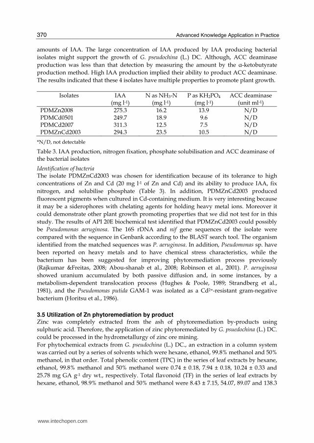
mg QE g-1 dry wt., respectively. The inhibitory concentration (IC50) of leaves extracted by ethanol, 99.8% methanol and 50% methanol were 0.0002, 0.0028 ± 0.0001 and 0.0040 mg GA g-1 ml reaction, respectively. The small IC50 values indicated that the extract contained high antioxidant compounds. The HPLC chromatogram (Fig.8) shows that the most common compounds in all extracts were caffeic acid, epicatechin, rutin, myricetin, quercetin and kaempferol. The contents of rutin and myricetin were the highest in leaf extracts by ethanol as 16,592.2 ± 5216.9 and 3228.1 ± 1362.5 µg g-1 dry wt., respectively. Whereas, the amounts of epicatechin and vanillin were highest, in leaves extracted by 50% methanol as 3592.4 ± 670.4 and 66.0 µg g-1 dry wt., respectively. Consequently, all results clearly showed that the amount of TPC, TF and the structure of phenolic compounds in the extracts related to their free radical scavenging activity. Moreover, the leaf extracts of plants grown in zinc contaminated soil contained 0.007 ± 0.001 mg in 5 ml of methanol, which was 4% of the zinc accumulated in the leaves. In which, the zinc contaminant was within the legal level for human consumption. However, the application of G. psuedochina (L.) DC., growing in the zinc mine in Tak province, Thailand, which has been reported to be contaminated with both zinc (100-8,036 mg kg-1) and cadmium (0.5-284 mg kg-1) (Simmons et al., 2005), should be more concerned with cadmium toxicity in the biomass and by product.
Fig. 8. HPLC chromatogram of (a) standards of polyphenol, and leaves extracts by
(b) methanol, (c) ethanol, (d) 50% methanol, and (e) hexane.
4. Conclusions
Phytoremediation is an environmental friendly method to remediate contaminated areas. However, the mechanism of the metal accumulated in the plant should be clearly studied before application. The bacterial rhizosphere, which can stand the toxicity of the metal and contain plant growth promoting properties, should be identified to promote plant growth. Moreover, the utilization of phytoremediation byproducts have to be determined to convince people to accept the plant for accumulating toxic metals. A case study of G.
pseudochina (L.) DC., growing in a Zn/Cd contaminated area of a zinc mine, showed that the
(a)
(b)
(c)
(d)
(e)
Gallic
Catechin Epicatechin
Caffeic
Vanillin
Rutin
Myricetin
Naringenin
Quercetin
Kaempferol
5 10 15 20 25 30 35 40 45 50
www.intechopen.com
Advanced Knowledge Application in Practice
372
plant has properties to be a zinc hyperaccumulator in Thailand. The mechanism of zinc accumulation involved the complexation of zinc with organocompounds, such as cellulose, anthocyanin and sulfur protein. The possible ZnCl2 complex in the plant indicated that the shoot parts of the zinc accumulative plant should be harvested before falling of leaves. The bacteria isolated from the plant’s rhizosphere that contained plant growth promoting properties and could grow in Zn and Cd was Pseudomonas aeruginosa PDMZnCd2003. The bacterium could be used to promote growth of G. pseudochina (L.) DC. during the phytoremediation process. Finally, for application, the polyphenolic compounds containing antioxidant properties could be extracted from the plant’s leaves, then unneeded biomass should be turned to ash in an incinerator before zinc extracted using sulphuric acid as in the mining process.
5. Acknowledgement
Nakbanpote, W. would like to thank Synchrotron Light Research Institute (Public Organization) (GRANT 1-2549/LS01 and GS-52-M07) and Thailand Research Fund (DBG5080016) for financial support, and be grateful for ASEA-uninet (one month research scholarship) and Prof. Dr. Klaus Stolze, Molecular Pharmacology and Toxicology Unit, Department of Biomedical Sciences, University of Veterinary Medicine, Vienna, for helping in HPLC analysis. The research facilities at beamline 4A and 12C were supported by Photon Factory (PF), High Energy Accelerator Research Organization (KEK), Tsukuba, Japan according to proposal number 2008G633, and we gratefully appreciate Prof. Dr. Izumi Nakai, Assoc. Prof. Dr. Akiko Hokura, Assist. Dr.Kriengkamol Tantrakarn, and Nakai’s laboratory, Tokyo University of Science, Japan, for helping research at PF and SPring-8. Natthawoot, P. gratefully thanks Junior Science Talent Project, National Science and Technology Development Agency, for grant JSTP-01-48-02R. Meesungneon, O. gratefully thanks Young Scientist and Technologist Programs, NSTDA (YSTP SP51-BT21), and National Synchrotron Research centre (GS-52-M07). Kannika, S. grateful thanks TRF Master Research Grants: TRF-MAG (MRG-WII515S097).
6. References
Abou-Shanab, R.A.I.; van Berkum, P. & Angle, J.S. (2007). Heavy metal resistance and genotypic analysis of metal resistance genes in gram-positive and gram-negative bacteria present in Ni-rich serpentine soil and in the rhizosphere of Alysum murale. Chemosphere, 68, 2, 360-367. ISSN: 0045-6535
Abou-Shanab, R. A.; Ghanem, K.; Ghanem, N. & Al-Kolaibe, A. (2008). The role of bacteria on heavy-metal extraction and uptake by plants growing on multi-metal-contaminated soils. World Journal of Microbiology & Biotechnology, 24, 2, 253-262. ISSN: 1573-0972
Altschul, S.F.; Madden, T.L.; Schaffer, A.A.; Zhang, J.; Zhang, Z.; Miller, W. & Lipman, D.J. (1997) Gapped BLAST and PSI-BLAST: a new generation of protein database search programs. Nucleic acids Research, 25, 17, 3389-3402. ISSN: 1362-4962
www.intechopen.com
Advances in Phytoremediation Research: A Case Study of Gynura pseudochina (L.) DC.
373
Alvarez, P.J.J. & Illman, W.A. (2006). Bioremediation and natural attenuation: process fundamentals and mathematical models, John Wiley & Sons, ISBN-13 978-0-471-65043-0, New Jersey.
Ahmad, F.; Ahmad, I. & Khan, M.S. (2008). Screening of free-living rhizospheric bacteria for their multiple plant growth promoting activities. Microbiological Research, 163, 2, 173-181. ISSN: 0944-5013
Anderson, C.W.N.; Brooks, R.R.; Chiarucci, A.; LaCoste, C.J.; Leblanc, M.; Robinson, B.H.; Simcock, R. & Stewart, R.B. (1999). Phytomining for nickel, thallium and gold, Journal of Geochemical Exploration, 67, 1-3, 407-415, ISSN: 0375-6742
Bollag, D.M.; Rozycki, M.D. & Edelstein, S.J. (1996). Protein method, Wiley-Liss, ISBN: 0-471-11837-0, New York.
Bradford, M.M. (1976). A rapid and sensitive method for the quantitation of microgram quantities of protein utilizing the principle of protein-dye binding, Analytical Biochemistry, 72, 1-2, 248-254. ISSN: 0003-2697
Bracey, M.H.; Christiansen, J.; Tovar, P.; Cramer, S.P. & Bartlett, S.G. (1994). Spinach carbonic anhydrase: Investigation of the zinc-binding ligands by site-directed mutagenesis, elemental analysis, and EXAFS. Biochemistry, 33, 44, 13126-13131. ISSN: 1520-4995
Bric, J.M.; Bostock, R.M. & Silversone, S.E. (1991). Rapid in situ assay for indole acetic acid production by bacteria immobilization on a nitrocellulose membrane. Applied and Environmental Microbiology, 57, 2, 535–538. ISSN: 1098-5336
Callahan, D.; Baker, A.J.M.; Kolev, S.D. & Wedd, A.G. (2006). Metal ion ligands in hyperaccumulating plants. Journal of Biological Inorganic Chemistry, 11, 1, 2–12. ISSN: 1432-1327
Cappuccino, J.G. & Sherman, N. (1999). Microbiology: A Laboratory Manual, 5th edition, Benjamin/cummings Science Publising, ISBN: 0-8053-7646-1, New York.
Chakravarty, B. & Srivastava, S. (1997). Effect of cadmium and zinc interaction on metal uptake and regeneration of tolerant plants in linseed. Agriculture, Ecosystems and Environment, 61, 1, 45-50. ISSN: 0167-8809
Chantiratikul, A.; Aengwanich, W.; Chinrasri, O. & Chantiratikul, P. (2008). Plasma selenium concentration and glutathione peroxidase activity in red blood cells of laying hens fed sodium selenite or zinc-L-selenomethionine. International Journal of Poultry Science, 7, 7, 692-695. ISSN: 16828356
Cheng, S. (2003). Review articles: Heavy metals in plants and phytoremediation. Environmental Science & Pollution Research, 10, 5, 335-340. ISSN: 1614-7499
Chung, C.T. & Miller, R.H. (1993). Preparation and storage of competent Escherichia coli cells. Methods in Enzymology, 218, 621-627. ISSN: 0076-6879
Clesceri, L.S.; Greenberg, A.E.; Eaton, A.D. (1998) Standard Methods for the Examination of Water and wastewater, 20th edition, American Public Health Association, American Water Works Association & Water Environment Federation, ISBN: 0-87553-235-7, Meryland.
Cobbett, C. & Goldsbrough, P. (2000). Mechanisms of metal resistance: Phytochelatins and metallothioneins, In: Phytoremediation of Toxic Metals: Using Plants to Clean up the Environment, Raskin, L. & Ensley, R.D. (Ed.), 33-49, John Wiley & Sons, ISBN 0-471-19254-6, New York.
www.intechopen.com
Advanced Knowledge Application in Practice
374
Cobbett, C. & Goldsbrough, P. (2002). Photochelatins and metallothioneins: Roles in heavy metal detoxification and homeostasis. Annual Reviews Plant Biology, 53, 1, 159-182. ISSN: 1543-5008.
Cotelle, N.; Bernier, J.L.; Catteau, J.P.; Pommery, J.; Wallet, J.C. & Gaydou, E.M. (1996) Antioxidant Property of hydroxyl- Flavones. Journal of Free Radical Biology & Medicine, 20, 1, 35-43. ISSN: 0891-5849
Cuenca, S., Amo-Marco, J.B. & Parra, R. (1999). Micropropagation from inflorescence stems of the Spanish endemic plant Centaurea paui Loscos ex Willk. (Compositae). Plant Cell Reports, 18, 7, 674-679. ISSN: 1432-203X
Davies, K.L.; Davies, M.S. & Francis, D. (1991) Zinc-induced vacuolation in root meristematic cells of Festuca rubra L. Plant, Cell and Environment, 14, 4, 399-406. ISSN: 1365-3040
Dimassi-Theriou, K. & Bosabalidis, A.M. (1997). Effects of light, magnesium and sucrose on leaf anatomy, photosynthesis, starch and total sugar accumulation, in kiwifruit cultured in vitro. Plant Cell, Tissue & Organ Culture, 47, 2, 127-134. ISSN: 1573-5044
Dworkin, M. & Foster, J.W. (1958) Experiments with some microorganisms which utilize ethane and hydrogen, Journal of Bacteriology, 75, 5, 592-603. ISSN: 1098-5530
Ebbs, S.D.; Zambrano, M.C.; Spiller, S.M. & Newville, M. (2009). Cadmium sorption, influx, and efflux at the mesophyll layer of leaves from ecotypes of the Zn/Cd hyperaccumulator Thlaspi caerulescens. The New Phytologist, 181, 3, 626–636. ISSN: 1469-8137
Ensley, B.D. (2000) Rationable for use of phytoremediation, In: Phytoremediation of toxic metals, Raskin, L. & Ensley, R.D. (Ed.), 3-11, John Wiley & Sons, ISBN 0-471-19254-6, New York.
Ensley, B.D.; Elless, M.; Blaylock, M.J. & Huang, J. (2001). Nutritionl supplements, US Patent 6,270,809B1.
Gamborg, O.L. & Phillips, G.C. (1995). Plant cell, tissue, and organ culture : fundamental methods, Springer, ISBN: 3540580689, New York.
Gardea-Torresdey, J.L.; Peralta-Videa, J.R.; Rosa, G. de la & Parsons, J.G. (2005). Review: Phytoremediation of heavy metals and study of the metal coordination by X-ray absorption spectroscopy. Coordination Chemistry Reviews, 249, 17-18, 1797–1810. ISSN: 0010-8545
Ghosh, M. & Singh, S.P. (2005). A review on phytoremediation of heavy metals and utilization of its by products. Applied and ecology and environmental research, 3, 1, 1-18. ISSN 1785 0037
Grill, E.; Loffler, S.; Winnacker, E.L. & Zenk, M.H. (1989). Phytochelatins, the heavy-metals binding peptindes of plants are synthesized from glutathione by a specific γ-glutamylcysteine dipeptidyl traspeptidase. Proceeding of the National Academy of Sciences of the United States of America. 86, 18, 6838-6842. ISSN: 1091-6490
Hall, J.L. (2002). Cellular mechanisms for heavy metal detoxification and tolerance. Journal experiment of Botany, 53, 366, 1-11. ISSN: 1460-2431
Heumann, H.G. (2002). Ultrastructural localization of zinc in zinc-tolerant Armeria maritime ssp. Helleri by autometallography. Journal of Plant Physiology, 159, 2, 191-203. ISSN: 0176-1617
www.intechopen.com
Advances in Phytoremediation Research: A Case Study of Gynura pseudochina (L.) DC.
375
Horitsu, H.; Yamamota, K.; Wachi, S.; Kawai, K. & Fukuchi, A. (1986). Plasmid-determined cadmium resistance in Pseudomonas putida GAM-1 isolated from soil. Journal of Bacteriology, 165, 1, 334-335. ISSN: 1098-5530
Hu, P.J.; Qiu, R.L.; Senthikumar, P.; Jiang, D.; Chen, Z.W.; Tang, Y.T. & Liu, F.J. (2009). Tolerance, accumulation and distribution of zinc and cadmium in hyperaccumulator Potentilla griffithii. Environmental and Experimental Botany, 66, 2, 317-325. ISSN: 0098-8472
Hughes, M.N. & Poole, R.K. (1989). Metals and Micro-organisms, Chapman & Hall, ISBN 0412244004, New York.
Jing, Y.; He, Z. & Yang, X. (2007). Role of soil rhizobacteria in phytoremediation of heavy metal contaminated soils. Journal of Zhejiang University: Science B, 8, 3, 192-207. ISSN: 1862-1783
Karley, A.J.; Leigh, R.A.& Sanders, D. (2000). Where do all the ions go? The cellular basis of differential ion accumulation in leaf cells. Trends in Plant Science, 5, 11, 465-470. ISSN: 1360-1385
Kelly, R.A.; Andrews, J.C. & DeWitt, J.G. (2002). An X-ray absorption spectroscopic investigation of the nature of the zinc complex accumulated in Datura innoxia plant tissue culture. Microchemical Journal, 71, 2-3, 231-245. ISSN: 0026-265X
Kessler, F. & Vidi, P.A. (2007). Plastoglobule lipid bodies: their functions in chloroplasts and their potential for applications. Advances in Biochemical Engineering/Biotechnol, 107, 153-172. ISSN: 0724-6145
Khan, A.G. (2005). Role of soil microbes in the rhizospheres of plants growing on trace metal contaminated soils in phytoremediation. Journal of Trace Elements in Medicine and Biology, 18, 4, 355-364. ISSN: 0946-672X
Koneman, E.W.; Allen, S.D.; Janda, W.M.; Schreckenberger, P.C. & Winn, W.C. (1997). Color Atlas and Textbook of Diagnostic Microbiology, 5th Edition, Lippincott-Raven Publishers, ISBN: 0-397-51529-4, Pennsylvania.
Li, T.Q.; Yand, X.E.; Yang, J.Y. & He, Z.L. (2006). Zn accumulation and subcellular distribution in the Zn hyperaccumulator Sedum alfredii Hance. Pedosphere, 16, 5, 616-623. ISSN: 1002-0160
Liu, H.; Qiu, N.; Ding, H. & Yao, R. (2008). Polyphenols contents and antioxidant capacity of 68 Chinese herbals suitable for medical or food uses. Food Research International. 41, 4, 363–370. ISSN: 0963-9969
MacFarlane, G.R. & Burchett, M.D. (2002). Toxicity, growth and accumulation relationships of copper, lead and zinc in the gray mangrove Avicennia marina (Forsk.) Vierh. Marine Environmental Research, 54, 1, 65-84. ISSN: 0141-1136
Miller, R.O. (1998). Nitric-Perchloric acid wet digestion in an open vessel, In: Handbook of Reference Methods for Plant Analysis, Kalra, Y.P. (Ed.), 57-62, CRC Press, ISBN: 1-57444-124-8, Florida.
Menon, A.R.; Aktoprakligül, D.; Zdemür, A. & Vertii, A. (2001). Heavy Metal Accumulation and Detoxification Mechanisms in Plants. Turkish Journal of Botany, 25, 3, 111-121. ISSN 1300-008X
Mukerji, K.; Manoharachary, C. & Singh, J. (2006). Microbial activity in the rhizosphere, Spinger, ISBN: 10 3-540-29182-2, Berlin.
Murashige, T. & Skoog, F. (1962). A revised medium for rapid growth and bioassays with tobacco tissue cultures. Physiologia Plantarum, 15, 3, 473-497. ISSN: 1399-3054
www.intechopen.com
Advanced Knowledge Application in Practice
376
Neumann, D.; Lichtenberger, O.; Gunther, D.; Tschersch, K. & Nover, L. (1994). Heat-shock proteins induce heavy metal tolerance in higher plants. Planta, 194, 3, 360-367. ISSN: 1432-2048
Panitlertumpai, N., Mongkhonsin, B., Nakbanpote, W. & Jitto, P. (2008). Zinc hyperaccumulation by Gynura pseudochina (L.) DC., Proceedings of Zinc processing 08, 25-26 Aug, Stamford Plaza Hotel, Brisbane, Australia.
Patel, K.; Kumar, A. & Durani, S. (2007). Analysis of the structural consensus of the zinc coordination centers of metalloprotein structures. Biochimica et Biophysica Acta - Proteins & Proteomics, 1774, 10, 1247-1253. ISSN: 1570-9639
Penrose, D.M. & Glick, B.R. (2003). Method for isolating and characterizing ACC deaminase-containing plant growth-promoting rhizobacteria. Physiologia Plantarum, 118, 1, 10-15. ISSN: 1399-3054
Rajkumar, M. & Freitas, H. (2008). Influence of metal resistant-plant growth-promoting bacteria on the growth of Ricinus communis in soil contaminated with heavy metals. Chemosphere, 71, 5, 834–842. ISSN: 0045-6535
Raskin, I. & Ensley, B.D. (2000). Phytoremediation of Toxic Metals: Using Plants to Clean up the Environment, John Wiley & Sons, ISBN 0-471-19254-6, New York.
Reddy, G. N. & Prasad, M. N.V. (1993). Tyrosine is not phosphorylated in cadmium induced hsp70 cognate in maize (Zea mays L.) seedlings: Role in chaperone function?. Biochemical Archives, 9, 25-32. ISSN: 0749-5331
Reddy, G. N. & Prasad, M. N.V. (1995). Cadmium induced protein phosphorylation changes in rice (Oryza sativa L.). Journal of Plant Physiology, 145, 1-2, 67-70. ISSN: 0176-1617
Reeves, R.D. & Baker, A.J.M. (2000). Metal-accumulating plants, In: Phyto-remediation of Toxic Metals: Using Plants to Clean up the Environment, Raskin, I. & Ensley, B.D. (Ed.), 193-230, John Wiley & Sons, ISBN: 0-471-19254-6, Toronto.
Roane, T.M.; Pepper, I.L. & Miller, R.M. (1996). Microbial remediation of metals, In: Bioremediation: Principles and applications, Crawford, R.L. & Crawford, D.L. (Ed.), 312-340, Cambridge University Press, ISBN 0-521-47041-2, Cambridge.
Robinson, B.; Russell, C.; Hedley, M. & Clothier, B. (2001). Cadmium adsorption by rhizobacteria: implications for New Zealand pastureland. Agriculture, Ecosystems and Environment, 87, 315-321. ISSN: 0167-8809
Rosa, G. de la; Peralta-Videa, J.R.; Montes, M.; Parsons, J.G.; Cano-Aguilera, I. & Gardea-Torresdey, J.L. (2004). Cadmium uptake and translocation in tumbleweed (Salsola kali), a potential Cd-hyperaccumulator desert plant species: ICP/OES and XAS studies. Chemosphere, 55, 9, 1159–1168. ISSN: 0045-6535
Sangdee, A.. Sirithorn, P. & Thummabenjapone, P. (2003). Virus and Viroid Causal Agent(s) of Bunchy Top and Severe Necrosis of Tomato, Khon Kaen Agricultural Journal, 31, 3, 161-170. ISSN 0125-2364
Santa-María, G.E. & Cogliatti, D.H. (1998) The regulation of zinc uptake in wheat plants. Plant Science, 137, 1, 1-12. ISSN: 0168-9452
Sarret, G.; Vangronsvele, J.; Manceau, A.; Musso, M.; Hean, J.D.; Menthonnex, J.J. & Hazemann, J.L. (2001). Accumulation froms of Zn and Pb in Phaseolus valgaris in the presence and absence of EDTA. Environmental Science Technology, 35, 13, 2854-2859. ISSN: 1520-5851
Schnoor, J.L. (1997). Phytoremediation, Technology Evaluation Report TE-98-01, GWRTAC, Pennsylvania.
www.intechopen.com
Advances in Phytoremediation Research: A Case Study of Gynura pseudochina (L.) DC.
377
Siddiqui, Z.A. (2006). PGPR: Biocontrol and Biofetilization, Springer, ISBN-10 1-4020-4002-4, Netherlands.
Simmons, R.W.; Pongsakul, P.; Saiyasitpanich, D. & Klinphoklap, S. (2005). Elevated levels of cadmium and zinc in paddy soils and elevated levels of cadmium in rice grain downstream of a zinc mineralized area in Thailand. Environmental Geochemistry & Health, 27, 5-6, 501–511. ISSN: 1573-2983
Sobkowiaka, R. & Deckertb, J. (2006). Proteins induced by cadmium in soybean cells. Journal of Plant Physiology, 163, 11, 1203–1206. ISSN: 0176-1617
Spaepen, S.. Vanderleyden, J. & Remans, R. (2007). Indole-3-acetic acid in microbial and microorganism-plant signaling. FEMS Microbiology Reviews, 31, 4, 425-448. ISSN: 1574-6976
Sridhar, B.B.M.; Diehl, S.V.; Han, F.X.; Monts, D.L. & Su, Y. (2005). Anatomical changes due to uptake and accumulation of Zn and Cd in Indian mustard (Brassica juncea). Environmental & Experimental Botany, 54, 2, 131–141. ISSN: 0098-8472
Strandberg, G.W.; Shumate, S.E. & Parrott, J.R.Jr. (1981). Microbial cells as biosorbents for heavy metals: accumulation of uranium by Saccharomyces cerevisiae and Pseudomonas aeruginosa. Applied and Environmental Microbiology, 41, 1, 237-245. ISSN: 1098-5336
Straczek, A.; Sarret, G.; Manceau, A.; Hinsinger, P.; Geoffroy, N. & Jaillard, B. (2008). Zinc distribution and speciation in roots of various genotypes of tobacco exposed to Zn. Environmental and Experimental Botany, 63, 1-3, 80-90. ISSN: 0098-8472
Suresh, B. & Ravishankar, G.A. (2004). Phytoremediation-A novel and promising approach for environmental clean-up. Critical reviews in biotechnology, 24, 2-3, 97-124. ISSN: 1549-7801
Tõrõk, I. & Kondorosim, Á. (1981). Nucleotide sequence of the R. meliloti nitrogenase reductase (nif H) gene. Nucleic Acids Research, 9, 21, 5711-5723. ISSN: 1362-4962
Tucker, R.K. & Shaw, J.A. (2000). Phytoremediation and public acceptance, In: Phytoremediation of Toxic Metals: Using Plants to Clean up the Environment, Raskin, L. & Ensley, R.D. (Ed.), 33-49, John Wiley & Sons, ISBN 0-471-19254-6, New York.
Verbi F.M.; Arruda, S.C.C.; Rodriguez, A.P.M.; Pe´rez, C.A. & Arruda, M.A.Z. (2005) Metal-binding proteins scanning and determination by combining gel electrophoresis, synchrotron radiation X-ray fluorescence and atomic spectrometry. Journal of Biochemical & Biophysical Methods, 62, 2, 97–109. ISSN: 0165-022X
Vázquez, M.D.; Barceló, J.; Poschenrieder, C.H.; Módico, J.; Hatton, P.; Baker, A.J.M. & Cope, G.H. (1992) Localization of zinc and cadmium in Thlaspi caerulescens (Brassicaceae), a metallophyte that can hyperaccumulate both metals. Journal of Plant Physiology, 140, 3, 350-355. ISSN: 0176-1617
Weis, J.S. & Weis, P. (2004). Metal uptake, transport and release by wetland plants: implications for phytoremediation and restoration. Environment International, 30, 5, 685-700. ISSN: 0160-4120
Weisburg, W.G.; Barns, S.M.; Pelletier, D.A. & Lane, D.J. (1991). 16s ribosomal DNA amplification for phylogenetic study. Journal of Bacteriology, 173, 2, 697–703. ISSN: 1098-5530
Willey, J.M.; Sherwood, L.M. & Woolverton, C.J. (2009). Prescott’s Principles of Microbiology, McGraw-Hill, ISBN 978-0-07-128367-0, New York.
Wood, J.; Scott, K.P.; Avgustin, G.; Newbold, C.J. & Flint, H.J. (1998). Estimation of the relative abundance of different Bacteroides and Prevotella ribotypes in gut samples
www.intechopen.com
Advanced Knowledge Application in Practice
378
by restriction enzyme profiling of PCR-amplified 16S rRNA gene sequences. Applied & Environmental Microbiology, 64, 10, 3683–3689. ISSN: 1098-5336
Yoo, K. M.; Lee, C. H.; Lee, H.; Moon, B. & Lee, C. Y. (2008). Relative antioxidant and cytoprotective activities of common herbs. Food Chemistry, 106, 3, 929–936. ISSN: 0308-8146
Yoshida, N.; Ishii, K.; Okuno, T. & Tanaka, K. (2006). Purification and Characterization of Cadmium-Binding Protein from Unicelluar Alga Chlorella sorokinian. Current Microbiology, 52, 6, 460–463. ISSN: 1432-0991
Yu, M.; Yang, F.; Chu, W.; Wang, Y.; Zhao, H.; Gao, B.; Zhao, W.; Gao, B.; Zhao, W.; Sun, J.; Wu, F.; Zhang, X.; Shi, Y. & Wu, Z. (2008). 3D local structure around Zn in Kti11p as a representative Zn-(Cys)4 motif as obtained by MXAN. Biochemical and Biophysical Research Communications, 374, 1, 28-32. ISSN: 1090-2104
Zehr, J.P. & McReynolds, L.A. (1989). Use of degenerate oligonucleotides for amplification of the nifH gene from the marine cyanobactertium Trichodesmium thiebautii. Applied & Environmental Microbiology, 55, 10, 2522-2526. ISSN: 1098-5336
Zhao, F.J.; Lombi, E.; Breedon, T. & McGrath, S.P. (2000). Zinc hyperaccumulation and cellular distribution in Arabidopsis halleri. Plant, Cell and Environment, 23, 5, 507–514. ISSN: 1365-3040
Zhuang, X.; Chen, J.; Shim, H. & Bai, Z. (2007). New advances in plant growth-promoting rhizobacteria for bioremediation. Environmental International, 33, 3, 406-413. ISSN: 0160-4120
Zuo, Y.; Chen, H. & Deng, Y. (2002). Simultaneous determination of catechins, caffeine and gallic acids in green, Oolong, black and pu-erh teas using HPLC with a photodiode array detector. Talanta, 57, 2, 307-316. ISSN: 0039-9140
www.intechopen.com
Advanced Knowledge Application in PracticeEdited by Igor Fuerstner
ISBN 978-953-307-141-1Hard cover, 378 pagesPublisher SciyoPublished online 02, November, 2010Published in print edition November, 2010
InTech EuropeUniversity Campus STeP Ri Slavka Krautzeka 83/A 51000 Rijeka, Croatia Phone: +385 (51) 770 447 Fax: +385 (51) 686 166www.intechopen.com
InTech ChinaUnit 405, Office Block, Hotel Equatorial Shanghai No.65, Yan An Road (West), Shanghai, 200040, China
Phone: +86-21-62489820 Fax: +86-21-62489821
The integration and interdependency of the world economy leads towards the creation of a global market thatoffers more opportunities, but is also more complex and competitive than ever before. Therefore widespreadresearch activity is necessary if one is to remain successful on the market. This book is the result of researchand development activities from a number of researchers worldwide, covering concrete fields of research.
How to referenceIn order to correctly reference this scholarly work, feel free to copy and paste the following:
Woranan Nakbanpote, Natthanwoot Paitlertumpai, Kannika Sukadeetad, Orapan Meesungeon and WattcharaNoisa-nguan (2010). Advances in Phytoremediation Research: A Case Study of Gynura Pseudochina (L.) DC.,Advanced Knowledge Application in Practice, Igor Fuerstner (Ed.), ISBN: 978-953-307-141-1, InTech,Available from: http://www.intechopen.com/books/advanced-knowledge-application-in-practice/advance-in-phytoremediation-research