Embed Size (px)
Citation preview

INSIDE THIS ISSUE
4PediatricSpinalImagingin3-D
6AAOSLeadershipAward
8ContinuingMedicalEducation
Vol.7,No.1,2013
Daniel J. Berry, M.D.Medical Editor and Chair, Mayo Clinic Department of Orthopedic Surgery
Advances in Articular Cartilage Defect Management
Articular (hyaline) cartilage restoration is a prized goal of orthopedic care because the clinical need is urgent and expanding. Articular cartilage enables the knee to tolerate shearing forces and absorb shock and loads up to 20 times the body's weight. As the population ages and people live longer—many of them active or overweight—articular cartilage increasingly bears prolonged and mounting skeletal stresses.
Easily Damaged, Hard to Renew "Articular cartilage damage is caused by both acute and repetitive trauma resulting in knee pain, effusion or mechanical symptoms," explains Michael J. Stuart, M.D., orthopedic surgeon at Mayo Clinic in Rochester, Minn. "Joint surface defects are ubiquitous, with numerous studies reporting a 60 percent prevalence in knees undergoing arthroscopy for pain."Adds his colleague, orthopedic surgeon Aaron J. Krych, M.D., "Full-thickness defects do not heal spontaneously and have limited ability to heal due to declining function of chondrocytes and the avascular nature of
cartilage. Untreated, these lesions can progress to more-serious degenerative joint conditions."
Articular cartilage has only a single type of cell for renewal — the chondrocyte — which becomes less active with age and injury. While long-term research goals focus on harnessing stem cell therapies for renewal, in the near term, Mayo Clinic orthopedists choose from multiple treatment strategies to manage cartilage injuries (Figures 1-5).
Three Mayo Clinic orthopedic innovations in cartilage-defect management are described below.
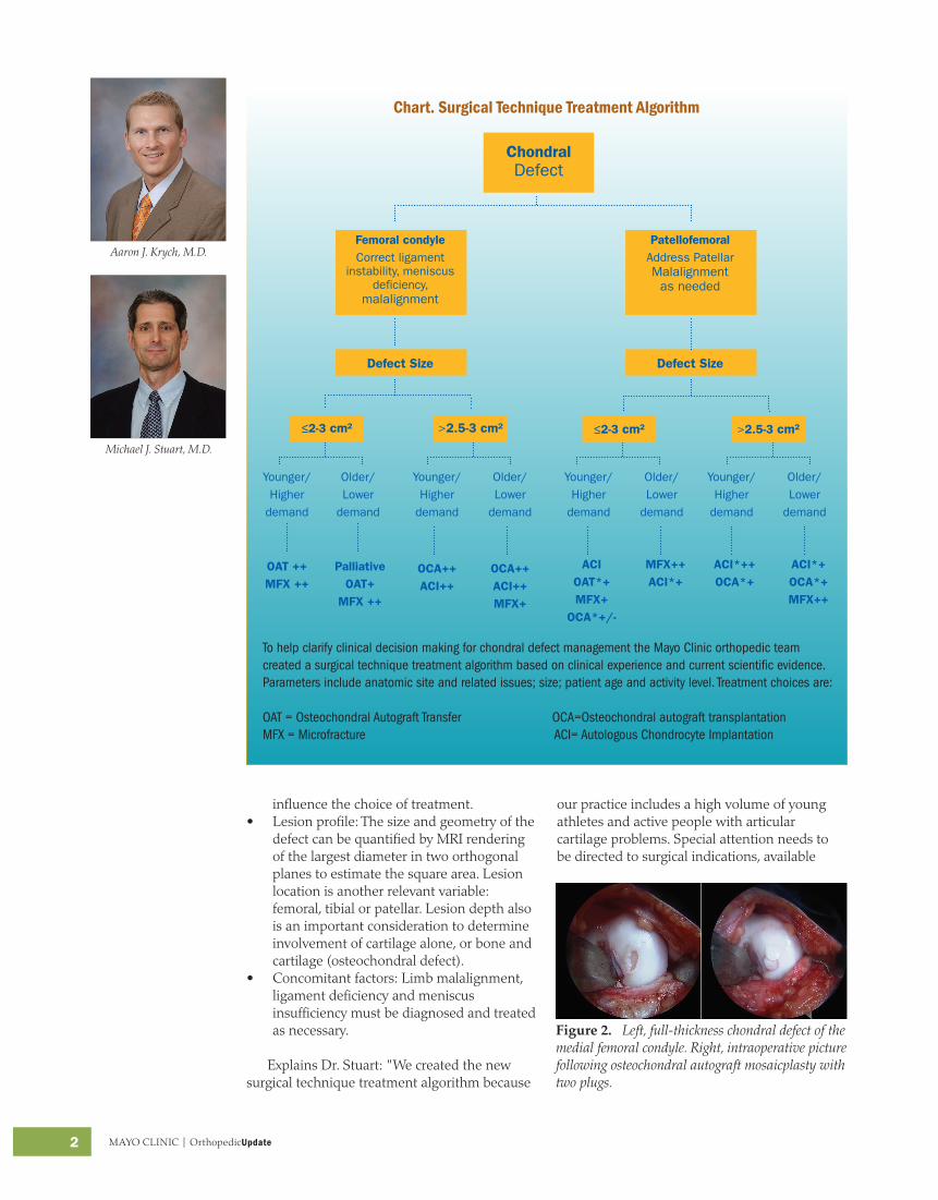
Surgical Technique Treatment Algorithm To help clarify clinical decision-making in this context of rapid change, the Mayo orthopedic team has created a surgical technique treatment algorithm (Chart). Based on clinical experience and current scientific evidence, the algorithm prioritizes the following factors in treatment decisions:
• Patient profile: Age, activity level, goals, expectations and demands of the individual
Figure 1. Left, cartilage defect following failed microfracture in the patella. Right, juvenile particulated chondral allograft transplant.
2 MAYO CLINIC | OrthopedicUpdate
Michael J. Stuart, M.D.
Aaron J. Krych, M.D.
Chart. Surgical Technique Treatment Algorithm
TohelpclarifyclinicaldecisionmakingforchondraldefectmanagementtheMayoClinicorthopedicteamcreatedasurgicaltechniquetreatmentalgorithmbasedonclinicalexperienceandcurrentscientificevidence.Parametersincludeanatomicsiteandrelatedissues;size;patientageandactivitylevel.Treatmentchoicesare:
OAT=OsteochondralAutograftTransferOCA=OsteochondralautografttransplantationMFX=MicrofractureACI=AutologousChondrocyteImplantation
Figure 2. Left, full-thickness chondral defect of the medial femoral condyle. Right, intraoperative picture following osteochondral autograft mosaicplasty with two plugs.
influence the choice of treatment.• Lesion profile: The size and geometry of the
defect can be quantified by MRI rendering of the largest diameter in two orthogonal planes to estimate the square area. Lesion location is another relevant variable: femoral, tibial or patellar. Lesion depth also is an important consideration to determine involvement of cartilage alone, or bone and cartilage (osteochondral defect).
• Concomitant factors: Limb malalignment, ligament deficiency and meniscus insufficiency must be diagnosed and treated as necessary.
Explains Dr. Stuart: "We created the new surgical technique treatment algorithm because
our practice includes a high volume of young athletes and active people with articular cartilage problems. Special attention needs to be directed to surgical indications, available
ChondralDefect
Femoral condyleCorrect ligament
instability, meniscus deficiency,
malalignment
Defect Size
Younger/Higher
demand
OAT ++MFX ++
PalliativeOAT+
MFX ++
≤2-3 cm²
Younger/Higher
demand
OCA++ACI++
OCA++ACI++MFX+
Older/Lower
demand
Older/Lower
demand
>2.5-3 cm²
Defect Size
PatellofemoralAddress Patellar Malalignment
as needed
Younger/Higher
demand
Older/Lower
demand
ACIOAT*+MFX+
OCA*+/-
MFX++ACI*+
Younger/Higher
demand
Older/Lower
demand
ACI*++OCA*+
ACI*+OCA*+MFX++
≤2-3 cm² >2.5-3 cm²

MAYO CLINIC | OrthopedicUpdate 3
Figure 3. Left, chondral defect of the mid weight-bearing condyle. Right, picture taken after placement of an osteochondral allograft transplant.
techniques, clinical outcome data and quality of postoperative care."
Randomized Trial Comparing NeoCart with Microfracture Microfracture is a marrow-stimulation technique that creates fibrocartilage at the site of the procedure. This type of cartilage is less durable, less resilient and less able to withstand shearing forces than is native articular cartilage. While this can have good results in smaller lesions, clinical studies reflect this lack of durability over a long-term follow-up. Autologous chondrocyte implantation (ACI) can result in more hyaline-like cartilage within the treated defect.
In ACI, healthy cartilage cells are harvested, cultured and then reimplanted into the defect under a patch at a second-stage surgery. Current drawbacks to this procedure are hypertrophy of the patch that can lead to further surgery and unreliable biological potential of the reimplanted cartilage cells.
NeoCart represents a novel approach to the treatment of cartilage defects, and Mayo Clinic has been chosen as a test center for the phase III Food and Drug Administration trial, randomizing patients to microfracture versus NeoCart. Similar to ACI, NeoCart is derived from the patient's own cartilage cells that are harvested arthroscopically. These cells are then embedded into a type 1 collagen matrix and incubated in a unique tissue processor that simulates the variation in pressure and low oxygen level of the knee joint. This environment ensures that the chondrocyte is maintained and stimulates the cells to produce the proteins that are crucial to the biomechanical function of cartilage.
Multicenter Study of Cartilage Repair and Osteoarthritis Evidence links repair of anterior cruciate ligament (ACL) injury with the subsequent development of osteoarthritis in the knee. To better understand this process, identify risk factors, and design interventions that could prevent or correct it, Mayo Clinic orthopedic specialists are developing a multicenter prospective study. It will document and analyze osteoarthritis in the knee after ACL injury utilizing biomarkers and advanced MRI imaging.
Figure 4. Left, articular cartilage defect of the medial femoral condyle. Right, arthroscopy picture following microfracture of this well-contained cartilage defect demonstrating flow of pluripotent cells into the defect.
Figure 5. Left, large pan-patellar cartilage defect. Right, ACI cells being injected under a collagen membrane.

4 MAYO CLINIC | OrthopedicUpdate
Anthony A. Stans, M.D.
A. Noelle Larson, M.D.
3-D Pediatric Spinal Imaging Expands Advanced Scoliosis and Spinal Deformity Practice
Conventional imaging of the pediatric spine has long been a challenge because of the well-documented cancer risk related to radiation exposures, which are cumulative over a lifetime.
Management of scoliosis in children typically entails repeated scans of the spine multiple times a year. This possibly exposes the youngest patients to as many as 20 imaging sessions throughout treatment. Clinicians and parents are therefore urgently looking for new imaging options.
Low-dose and 3-D Mayo Clinic orthopedics in Rochester, Minn., now uses the low-dose, three-dimensional (3-D), head-to-toe imaging option of the EOS technology for its pediatric spine patients. Mayo Clinic orthopedic specialists have always aimed
Advances at Mayo Clinic Department of Orthopedic Surgery in the management of spinal deformity are fueling the
growth and development of its collaborative, state-of-the-art spinal deformity center of excellence. Care for spinal
deformity is available to patients across the age spectrum, from infantile scoliosis to complex congenital cases and
revision surgery.
Highlights of this practice include:
1. Multidisciplinary evaluation, treatment and rehabilitation expertise across the entire spectrum of
spinal deformity
2. Full range of treatment modalities for the growing spine, including infantile casting (Mehta technique),
collaborative bracing program and support network, growing rod constructs, and Vertical Expandable
Prosthetic Titanium Rib (VEPTR)
3. Facilities and expertise to treat the most complex spinal deformities, including a Mayo-based team of experts
for intraoperative neurologic monitoring, admission for in-patient traction, intraoperative CT-guided navigation,
and a team-approach for the complex patient, including pediatric and adult orthopedic spine specialists
4. Clinical pathways for preoperative education and postoperative recovery plan
These developments open exciting opportunities to optimize and further individualize patient care. Notes Mayo
pediatric orthopedic specialist, A. Noelle Larson, M.D.: "The addition of new technology such as EOS and a center
for excellence in spine helps us provide the highest level of care for each patient, to better understand both the
3-D nature of the deformity and how it impacts other organ systems. Historic treatments for small children with
severe deformity included fusion surgery, which restricted pulmonary growth. We have a new appreciation of the
importance of preserving spinal growth while restoring coronal and sagittal plane alignment. A multidisciplinary
approach with collaboration with pulmonology and nutrition is essential to providing safe and effective treatment
for growing children with severe deformity."
Highlights of Mayo Clinic's Evolving Spinal Deformity Practice
for using the lowest possible radiation dose in pediatric radiographs.
In fact, Mayo previously used filters for scoliosis X-rays that significantly reduced the dose but required a high level of technical skill and frequent, repeated radiographs. With the addition of EOS, Mayo's pediatric patients may now have low-dose, 3-D head-to-toe imaging at a level that is one-third of most traditional X-ray radiation dose levels, and one-seventh of the dose delivered by Mayo's more technically demanding filtered X-ray system.
EOS reduces the amount of radiation a child receives because it is more efficient at capturing radiation. Mayo is among a small number of advanced orthopedic centers to offer EOS in the U.S. "The EOS technology provides accurate and reproducible 3-D modeling of spine deformity,

MAYO CLINIC | OrthopedicUpdate 5
Amy L. McIntosh, M.D.
Figure 1. Comparison of lateral views of scoliosis patient, (left) EOS vs. (right) standard x-ray. Note in EOS the full-length visual detail from cervical spine to pelvis, and EOS’ improved imaging of the abdominal cavity and lungs. In the standard x-ray film the typical limited view is present, marked by the “top-dark/bottom-light” distribution of radiation affected by air in the lungs and tissue mass of the abdomen.
Figure 2. Kyphosis, left to right, pre- and post-treatment. The enchanced visual detail of EOS is helpful in planning surgery and assessing outcomes.
Table 1. Patient characteristics and clinical indications most likely to benefit from EOS
Ideal Patient
Characteristics Clinical IndicationsThree-dimensional (3-D) imaging indicated
Spine deformity
Radiation exposure level concerns (all patients)
Lower-extremity deformity
Full-body scanning indicated
Kyphosis
Able to hold still for standing radiographs
Other deforming dorsopathies
Requires posteroanterior and lateral views
Lung volume issues
giving us a tool to achieve a more precise understanding of spinal deformity, which until now has been very difficult to obtain without significant radiation exposure for patients," explains Anthony A. Stans, M.D., a Mayo Clinic orthopedic surgeon who specializes in pediatric surgery. A major EOS strength is the enhanced detail of its visualizations (Figure 1).
This new level of visual detail improves understanding of spinal pathology, aids diagnosis and helps evaluate treatment strategies. With EOS data, physicians can conduct more accurate spinal deformity assessments pre-operatively, and then critically review results post-operatively to determine the effectiveness of a given intervention, compare outcomes and possibly revise approaches if data suggest it is warranted (Figure 2).
About EOSDeveloped in France, the EOS device has been used in Europe for approximately a decade. The Food and Drug Administration first approved EOS technology for imaging the spine of pediatric patients in 2010 and for use in the leg in early 2011. Using two perpendicular X-ray beams, the EOS system simultaneously captures two orthogonal X-ray images when the patient is standing in an upright, weight-bearing position. Standard two-dimensional
images are adequate for routine use, but when indicated, EOS computer software permits rendering of a 3-D representation from the two planar images. Prior to EOS, physicians could obtain 3-D images only by using computerized tomography, which carries a significant radiation burden.
Practice-changing TechnologyDr. Stans' Mayo colleague, orthopedic surgeon Amy L. McIntosh, M.D., believes this advance will help specialists think about spinal deformity differently and lead to changes in practice. Says Dr. McIntosh: "We will be able to compare pre- and postoperative images from EOS with pre- and postoperative images of the past and see a level of detail that was previously missing. As a result, the goals and methods of our practice in the future may change."
Mayo Clinic physicians are also beginning to use EOS for lower extremity imaging. "As with the spine, this application will likely improve our understanding and treatment of lower extremity deformity," she notes. While early results generate cautious enthusiasm for EOS, the Mayo team notes that more data and clinical experience are needed to fully assess its contributions.

6 MAYO CLINIC | OrthopedicUpdate
Michael J. Yaszemski Honored for Leadership in Orthopedics
Mary I. O'Connor, M.D.
Marco Rizzo, M.D.
Diane L. Dahm, M.D.
Mayo Clinic orthopedic researcher and surgeon Michael J. Yaszemski, M.D., Ph.D., received the 8th annual William W. Tipton Jr., M.D., Leadership Award at the annual meeting in March 2013 of the American Academy of Orthopaedic Surgeons (AAOS) in Chicago. Dr. Yaszemski is a member of Mayo Clinic Department of Orthopedic Surgery's spine surgery, where he served as division chair for 10 years. He also is a renowned researcher, directing the Department of Orthopedic Surgery's Tissue Engineering and Biomaterials Laboratory.
Dr. Yaszemski retired from the U.S. Air Force Reserve in February 2013 after 34 years of service, having attained the rank of brigadier general in 2008. General Yaszemski's last assignment prior to his retirement was as the reserve military adviser to the president of the Uniformed Services University of the Health Sciences in Bethesda, Md. He has been deployed on five occasions to Afghanistan, Iraq and other sites of conflict in southwest Asia over the past decade as adviser to the Air Force surgeon general on issues pertaining to wartime injuries, deployed medical personnel organization and critical care air transportation. He served as an orthopedic trauma surgeon and deputy commander of the Air Force Theater Hospital in Balad, Iraq in 2006.
About the Tipton Award Members of AAOS and the Orthopaedic Research and Education Foundation (OREF) established the award to honor the late orthopedic surgeon, William W. Tipton Jr., M.D., a former AAOS leader known for his untiring efforts to build coalitions and effect change. He died in 2005 of hereditary liver disease. The aim of the memorial is to recognize individuals who both contribute to and continue his exemplary leadership in orthopedics. The award carries a $5,000 stipend to further leadership in the field, which Dr. Yaszemski dedicated to OREF initiatives.
Collegial Praise Daniel J. Berry, M.D., chair of the Mayo Clinic Department of Orthopedic Surgery, praises Dr. Yaszemski's focus and ability to inspire teamwork. "Mike is a man who has strong beliefs about the value of hard work, as well as the value of achievement. He also is a man who is unfailingly kind and polite to everyone around him, and that helps him to get everybody working together." Always committed to furthering the field, Dr. Yaszemski encourages orthopedic specialists to continue their excellent work—and to raise
Mayo Clinic orthopedic researcher and surgeon Michael J. Yaszemski, M.D., Ph.D.
awareness of it so people understand the field's important contributions to public health.
AAOS president and chairman of Chicago's Rush Medical College Department of Orthopedic Surgery, Joshua J. Jacobs, M.D., praises Dr. Yaszemski's mentorship and strong sense of personal mission. "He is a role model who is dedicated to helping others. His selfless attitude is an inspiration to all of us in the orthopedic profession and the academy is proud to present him with this award."
Key Contributions At a GlanceAmongtheseminalcontributionsDr.Yaszemskihasmadetoorthopedicsthattheawardcommitteeconsideredinnaminghimthe2013TiptonLeadershipAwardrecipientarehisrolesas:
• Active contributor, AAOSaschairoftheOrthopedicDeviceForum,2006-2012
• Groundbreaking researcher, continuouslyfundedbytheNationalInstitutesofHealthsince1994andleadingthewayinresearchstudiesforbothboneregenerationviatissueengineeringtechnologiesandsarcomaresearch
• Minnesota representative,AAOSBoardofCouncilors2008-2014
• Member, AAOS CommitteeonOutsideInterests
• Mentor to more than 100 graduates,master'sanddoctoraldegreestudents,medicalstudents,residents,clinicalfellowsandpostdoctoralfellows,ofwhommorethan40percentarewomen
• First nonminority memberoftheJ.RobertGladdenOrthopaedicSociety
MAYO CLINIC | OrthopedicUpdate 7
Philosophy of Leadership Deeply grateful for the award, Dr. Yaszemski sees it as the product of a team effort growing out of his many collaborations across the continuum of care. He describes his philosophy of leadership this way: "I believe in people and (that) given the right situation, if you will, the right leadership, and a team to be a part of, most people are proud to excel and do a good job."
The ability to follow is a critical component of leadership, he believes. "Leadership, from my perspective, starts with followership. When we are leaders, we are leaders for some things. They are time limited. We do them for a while and then we go away from them. The rest of the time we have to be good followers, and it is that experience as a follower that really guides what one is to do when one is in a leadership position, from my perspective."
Background and EducationBorn in Nanticoke, Pa., Dr. Yaszemski received his bachelor's and master's degrees in chemical engineering from Lehigh University in Bethlehem, Pa., in 1977 and 1978. He received his M.D. from Georgetown University, Washington, D.C., in 1983 and his Ph.D. in chemical engineering from the Massachusetts Institute of Technology, Cambridge, Mass., in 1995. He performed his residency in orthopedic surgery at Wilford Hall Air Force Medical Center in San Antonio, Texas, and completed his fellowship in spine surgery with Augustus A. White, M.D., Ph.D., at Harvard Medical School. In 1979 Dr. Yaszemski joined the U.S. Air Force Ready Reserve, serving on active duty from 1983-1996.
Sources of Motivation A personal experience with a life-threatening condition deepened his commitment to work to save others' lives, notes David Wayne Polly Jr., M.D., the University of Minnesota orthopedic
John W. Sperling, M.D.
Robert T. Trousdale, M.D.
Joaquin Sanchez-Sotelo, M.D., Ph.D.
AttheMarchannualmeetingoftheAmericanAssociationofOrthopaedicSurgeons(AAOS)inChicago,MayoClinicDepartmentofOrthopedicSurgeryofferedanotheryearofunusuallystrongcontributionsintermsofleading,organizing,moderatingandservingasexpertpanelists,presentersandinstructionalcourselecturers.
Morethan50Mayoorthopedistsparticipated.TheseincludeMaryI.O'Connor,M.D.,moderatingafacultydevelopmentcourseforwomenorthopedists,aswellaspresentingonhipandkneetopics;MarcoRizzo,M.D.,chairingtheEducationalCommitteeonHandandWristaswellaspresentingandmoderatingonthosetopics;DianeL.Dahm,M.D.,chairingtheEducationalCommitteeonSportsMedicineandArthroscopyandpresentingandmoderatingonthosetopics;RobertT.Trousdale,M.D.,expertdebatepanelistontotalhipreplacementandpresenteronvarioushottopicsinhip;JoaquinSanchez-Sotelo,M.D.,Ph.D.,symposiummoderatorandalsopresentingonvariouselbowtopics;JohnW.Sperling,M.D.,chairingtheEducationalCommitteeonShoulderandElbow,aswellasservingasexpertpanelist,moderatoronvariousshoulderpanelsandinstructionalcourselectures.
Call to Action
To actively honor receiving the William W. Tipton, Jr. Leadership Award, Dr. Michael J. Yaszemski encourages orthopedic specialists to take two actions to support orthopedics.
First: Exceed. "Keep providing the excellent care that we provide to our patients. This is the cornerstone of what we do. Keep doing the research, the education of the next generation of orthopedists, and the service that makes us who we are and able to provide this outstanding care to patients. "
Second: Advocate. "Whenever you get an opportunity, tell people what orthopedic surgeons do, because people may not know all the things that we do day in and day out that improve their life and function."
surgeon who was among those who nominated Dr. Yaszemski for the award. "Mike was very ill at one point in time and nearly died. He was administered the last rites in the intensive care unit at Mayo for a bleed in his head and he recovered, and he's just about completely recovered … Mike is nearly unflappable now. And his story about this is, once you've had the last rites administered to you, any other problem you encounter doesn't seem so bad. And so he just has this wonderful sense of perspective."
Dr. Yaszemski draws motivation from the people he encounters and collaborates with, "be they family, friends, patients, colleagues, folks who are either wanting to help others or folks who themselves are in need," he explains. "And being part of a team that can offer some help to them from a medical perspective, I think, is the thing that makes me want to keep doing it."
Mayo Clinic Contributions to AAOS 2013
Mayo Clinic Orthopedic Update
Medical Editors: Daniel J. Berry, MDArlen D. Hanssen, MDMichael J. Stuart, MD
Orthopedic Update is written for physicians and should
be relied upon for medical education purposes only.
It does not provide a complete overview of the topics
covered and should not replace the independent
judgment of a physician about the appropriateness
or risks of a procedure for a given patient.
MC6247-0613
CME Opportunities
Minnesota Memorial Pediatric Orthopedic SymposiumOct. 11Rochester, MN
In this one-day course, faculty will focus on current approaches to some of the most challenging conditions seen in pediatric orthopedics. The course format includes lectures, case presentations, Q&A sessions and interactive discussion. It is designed to give participants a better understanding of new techniques in the diagnosis and treatment of pediatric orthopedic disorders, and the skills to apply them clinically.
For more information, call 800-323-2688 or email [email protected].
Mayo Clinic Symposium on Sports MedicineNov. 8-9 Rochester, MN
The 23rd annual Mayo Clinic Symposium on Sports Medicine is designed to provide physicians, physical therapists, athletic trainers and other sports medicine professionals with the latest diagnostic and treatment strategies for sports-related and musculoskeletal conditions. The program is multidisciplinary, featuring expert lecturers representing various sports medicine fields. Multiple educational formats will be used, including case presentations, as well as live demonstrations of physical examination, anatomy and arthroscopy.
To register, go to www.mayo.edu/cme. Direct questions to [email protected]
or 800-323-2688
Shoulder Arthroscopy, Arthroplasty and FracturesApril 25-26, 2014Rochester, MN
Thisisanadvancedcoursefororthopedicsurgeonstreatingdisordersoftheshoulder.Thecourseincludesdidacticsessionsandlaboratoryexperienceusingcadaverspecimens.Livevideodemonstrationsandpaneldiscussionareanintegralpartoftheoveralllearningexperience.Thiscourseteachestheprinciplesandtechniquesformanagementofrotatorcufftears,SLAPlesions,instabilityandarthritis.
Forinformation,gotowww.mayo.edu/cme/orthopedic-surgery-2014r295
To view all Mayo Clinic CME offerings visit www.mayo.edu/cme/
Contact UsReferralsandConsultations
Arizona866-629-6362
Florida800-634-1417
MinnesotaOrthopedicSurgery
507-538-4101AllOtherReferrals andConsultations
800-533-1564
www.mayoclinic.org/medicalprofs
2012