Embed Size (px)
Citation preview
Accepted Manuscript
Title: Advanced topical formulations (ATF)
Author: Jonathan Hadgraft Majella E. Lane
PII: S0378-5173(16)30455-0DOI: http://dx.doi.org/doi:10.1016/j.ijpharm.2016.05.065Reference: IJP 15804
To appear in: International Journal of Pharmaceutics
Received date: 23-4-2016Revised date: 28-5-2016Accepted date: 30-5-2016
Please cite this article as: Hadgraft, Jonathan, Lane, Majella E.,Advanced topical formulations (ATF).International Journal of Pharmaceuticshttp://dx.doi.org/10.1016/j.ijpharm.2016.05.065
This is a PDF file of an unedited manuscript that has been accepted for publication.As a service to our customers we are providing this early version of the manuscript.The manuscript will undergo copyediting, typesetting, and review of the resulting proofbefore it is published in its final form. Please note that during the production processerrors may be discovered which could affect the content, and all legal disclaimers thatapply to the journal pertain.
1
Advanced Topical Formulations (ATF)
Jonathan Hadgraft and Majella E Lane
Department of Pharmaceutics
UCL School of Pharmacy
29-39 Brunswick Square
London
WC1N 1AX
UK
*Corresponding author
Tel: +44 207 7535821
Fax: +44 870 1659275
Email: [email protected]
2
Graphical abstract
100 % b
0 % a
0 % b
100 % a
Binary Mixture
Saturated solubility
in pure solvent b
Saturated solubility
in pure solvent a
B
A
Solu
bili
ty
subsaturated
supersaturated
3
Abstract
Topical formulations aim to target the skin for a variety of cosmetic, protective or
therapeutic needs. Despite the use of creams and ointments over the millennia, the
bioavailability of actives from topical preparations remains quite low, often not exceeding 1-
2% of the applied dose. In this review we examine the reasons underlying the poor
performance of topical preparations. We also outline a rational approach, based on Fick’s
laws of diffusion, to develop advanced topical formulations. Methodologies which are
currently used in research and development are critically examined and the importance of
understanding the fate of the vehicle as well as the active is emphasised. Advanced topical
formulation development will also be facilitated by emerging and sophisticated analytical
techniques that are able to probe real time delivery of actives to the skin. A good
understanding of the underlying physical chemistry of both the formulation and the skin is
crucial in the development of optimised topical products.
Keywords: Advanced topical formulation, crystallisation, skin, supersaturation, penetration
4
Forenote
This is to acknowledge the contribution that Sandy Florence has made to the
pharmaceutical sciences, in particular, the role of physical chemistry in developing
optimised formulations.
5
1. Introduction - The Watershed
In the same way that our calendar runs from BC to AD, there was a marked
watershed in our understanding of topical drug deliver before steroids (BS) and after
steroids (AS). The major steroid in question was hydrocortisone which was developed some
60 years ago (Fourman et al., 1950). Delivery of agents to the skin has been documented by
the Egyptians (BC), the Romans (AD) and in mediaeval times when ‘flying ointment’ was
recorded (Hadgraft and Lane, 2016). It is interesting to note that, in the 13th – 14th centuries
that it was known how to extract hallucinogenic materials from plants (e.g. mandrake). The
extract had to be treated with lime to render the active more permeable through the skin
(Burton, 1972). We now know that the base form was liberated and it was then
compounded into a fat (i.e. a simple formulation was manufactured). Even though flying
ointment was known and it was recognised that nitroglycerin induced headaches in
armament workers by the late 1800s and also that nicotine could cause death as a result of
skin penetration, the skin was largely regarded as an impermeable barrier (Laws, 1910;
Faulkner, 1933; Rothman, 1943). Before steroids, very little was known about the role of the
formulation and how delivery through the skin could be markedly affected by simple
changes to the formulation.
2. Skin structure and route of penetration
For decades it has been appreciated that the stratum corneum, the outer layer of
the skin is the rate controlling membrane to permeation. It is a unique barrier that is only
some 15 micrometres thick (Hadgraft and Lane, 2011). It has evolved to prevent excessive
water loss from mammals. How does it achieve this? It is comprised of dead keratinised cells
that overlap, embedded in a lipid matrix, and the structure has been regarded as resembling
a brick wall (Michaels et al., 1975). This is interspersed by hair follicles and sweat glands
(Figure 1). There has been much debate concerning the route of penetration. Scheuplein
and co-workers published a series of papers in the mid sixties to mid seventies which
covered various aspects of percutaneous penetration. This included mechanisms and routes
of penetration (Scheuplein 1965,1966,1967), the effect of temperature (Blank et al., 1967)
and specific details on steroid permeation (Scheuplein et al., 1969). They also considered
effects of surfactants (Scheuplein and Ross, 1970) and penetration from solvent deposited
solids (Scheuplein and Ross, 1974).
6
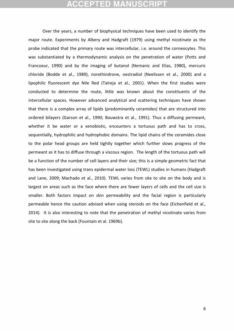
Over the years, a number of biophysical techniques have been used to identify the
major route. Experiments by Albery and Hadgraft (1979) using methyl nicotinate as the
probe indicated that the primary route was intercellular, i.e. around the corneocytes. This
was substantiated by a thermodynamic analysis on the penetration of water (Potts and
Francoeur, 1990) and by the imaging of butanol (Nemanic and Elias, 1980), mercuric
chloride (Bodde et al., 1989), norethindrone, oestradiol (Neelissen et al., 2000) and a
lipophilic fluorescent dye Nile Red (Talreja et al., 2001). When the first studies were
conducted to determine the route, little was known about the constituents of the
intercellular spaces. However advanced analytical and scattering techniques have shown
that there is a complex array of lipids (predominantly ceramides) that are structured into
ordered bilayers (Garson et al., 1990; Bouwstra et al., 1991). Thus a diffusing permeant,
whether it be water or a xenobiotic, encounters a tortuous path and has to cross,
sequentially, hydrophilic and hydrophobic domains. The lipid chains of the ceramides close
to the polar head groups are held tightly together which further slows progress of the
permeant as it has to diffuse through a viscous region. The length of the tortuous path will
be a function of the number of cell layers and their size; this is a simple geometric fact that
has been investigated using trans epidermal water loss (TEWL) studies in humans (Hadgraft
and Lane, 2009; Machado et al., 2010). TEWL varies from site to site on the body and is
largest on areas such as the face where there are fewer layers of cells and the cell size is
smaller. Both factors impact on skin permeability and the facial region is particularly
permeable hence the caution advised when using steroids on the face (Eichenfield et al.,
2014). It is also interesting to note that the penetration of methyl nicotinate varies from
site to site along the back (Fountain et al. 1969b).
7
Figure 1. Potential pathways through the stratum corneum. 1 - Transappendageal route through (A) hair follicles and (B) sweat glands; 2 - Transepidermal route across intact SC via (A) intercellular lipids and (B) via keratinised cells (or transcellular).
One of the other implications of the intercellular route is the fact that when a
formulation is spread over the skin surface only a small ‘active’ area for diffusion is
available. The bulk of the area, the large surface of the corneocytes will not be accessible for
a permeant. This is probably the major contributing factor to the low bioavailability (~1-3%)
of steroids when applied to the skin as solution in acetone (Feldmann and Maibach, 1965;
1966; 1969). It is probable that the steroids crystallise on and in the upper intercellular
channels of the skin and are therefore unavailable for continued diffusion; we have recently
presented evidence for this phenomenon (Hadgraft and Lane, 2016). This would also explain
the steroid reservoir mentioned above. It has been demonstrated that this is particularly
pronounced when DMSO was used as a solvent. DMSO is a particularly good solvent and an
excellent penetrant. It is does not stay on the skin or in the skin and when used as a vehicle
for a steroid the active will crystallise essentially leaving it stranded and unable to dissolve.
Intercellular
space
Lipid domain
Aqueous domain
Keratin
Minimal lipid
TranscellularIntercellular
1-A 1-B 2-A 2-B
Hair follicle
Sweat Gland
Corneocytes
1 – Transappendageal
route
2 – Transepidermal
route
SC
8
It will then be sloughed off the skin, which is a particularly dynamic membrane, the stratum
corneum having an average turnover time of 14 days.
3. Corticosteroids and nicotinic acid esters
Corticosteroids penetrate through the stratum corneum and constrict the blood
vessels at the epidermal – dermal junction. This manifests itself as a pallor, the degree of
which could be related to the potency of the steroid and the amount permeated (McKenzie
and Stoughton, 1962). On the other hand, the nicotinates cause vasodilatation of the blood
vessels and the onset of erythema could be related to the degree of penetration (Fountain
et al., 1969a). The advent of the more potent corticosteroids led to more investigations on
formulation effects. It was soon recognised that hydration of the skin, using occlusion,
improved the permeation of the steroids and that the degree of penetration was related to
the concentration applied (McKenzie and Stoughton, 1962). At the same time Cronin and
Stoughton (1962) used ethyl nicotinate to show that there was regional variation in skin
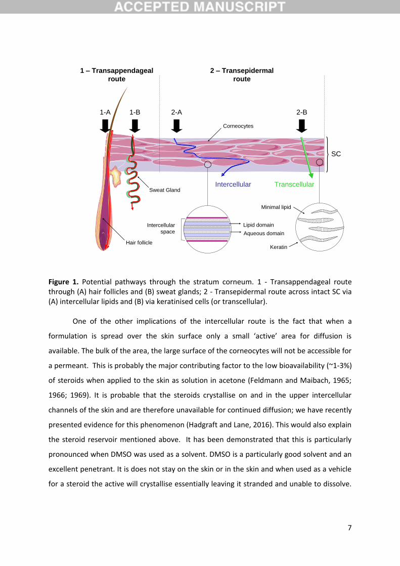
permeability. Barrett et al. (1964) used methyl nicotinate in various formulations to show
that there were considerable differences in absorption when equal concentrations of the
nicotinate were dissolved in aqueous cream, oily cream, white soft paraffin or macrogol
ointment (Table 1). These authors also showed permeation was directly related to
concentration.
Table 1: Penetration of methyl nicotinate from various topical vehicles. Adapted from Barrett et al. (1964) Concentration of methyl nicotinate
Time of onset of erythema (min)
Aqueous Cream Oily Cream White soft paraffin Macrogol
1 3.3 4.1 5.3 8.7
0.5 4.0 4.5 6.6 10.8
0.25 - - - 17.1
0.1 4.6 5.2 8.0 22/3
0.05 5.9 6.8 13.1 Not measured
0.01 7.4 10.5 19.7 Not measured
0.005 10.9 12.5 Not measured Not measured
0.001 Not measured Not measured Not measured Not measured
In the mid-60s there was an interest in enhancing skin penetration and one of the
most researched solvents was dimethyl sulphoxide (DMSO). Stoughton (1965) showed that
the penetration of fluocinolone acetonide was enhanced by DMSO and that a steroid
9
reservoir was induced. However, the reason for the formation of the reservoir was unclear.
Other agents were also examined as potential penetration enhancers such as dimethyl
formamide and dimethyl acetamide (Feldmann and Maibach, 1966). These vehicles also
influenced transepidermal water loss (TEWL) as reported by Baker (1968). Sarkany et al.
(1965) investigated formulation effects and conducted a clinical study on eczema and
psoriasis treatment. Unequivocal evidence showing a link between vasoconstrictor effect
and clinical efficacy could not be demonstrated.
4. Physicochemical determinants of topical delivery
When steroid treatment was coming to the fore in the late 50s, Higuchi (1960)
presented a paper showing the importance of Fick’s laws of diffusion in understanding
percutaneous absorption (Equation 1).
E Equation 1
Most simply this analysis shows the significance of the driving force for diffusion, D
across a membrane of thickness, h. At its most basic this is the concentration, c, of the
active but strictly it is the chemical potential, which is related to the concentration and the
activity coefficient. If an active is applied in a solvent (and the solvent does not interact with
the skin) the flux of the active across the skin will be related to the degree of saturation. It is
therefore possible to apply formulations which contain different concentrations of the drug
and their penetration rates will be the same. This was shown by Hadgraft et al. (1973) in
which methyl nicotinate, at different concentrations but at the same chemical potential was
applied in simple water – glycerol mixtures. (Figure 2) The time of onset of erythema was
the same thus demonstrating the importance of chemical potential. Similar results were
found for the steroids (Lippold and Schneeman, 1984). These workers showed that the
degree of blanching induced by a steroid was dose dependent up to the point at which the
steroid was at its saturated solubility in the formulation. Concentrations above the
solubility limit (i.e. suspensions) did not improve the vasoconstriction response; in simple
terms a 1% suspension will be as effective as a 5%.
10
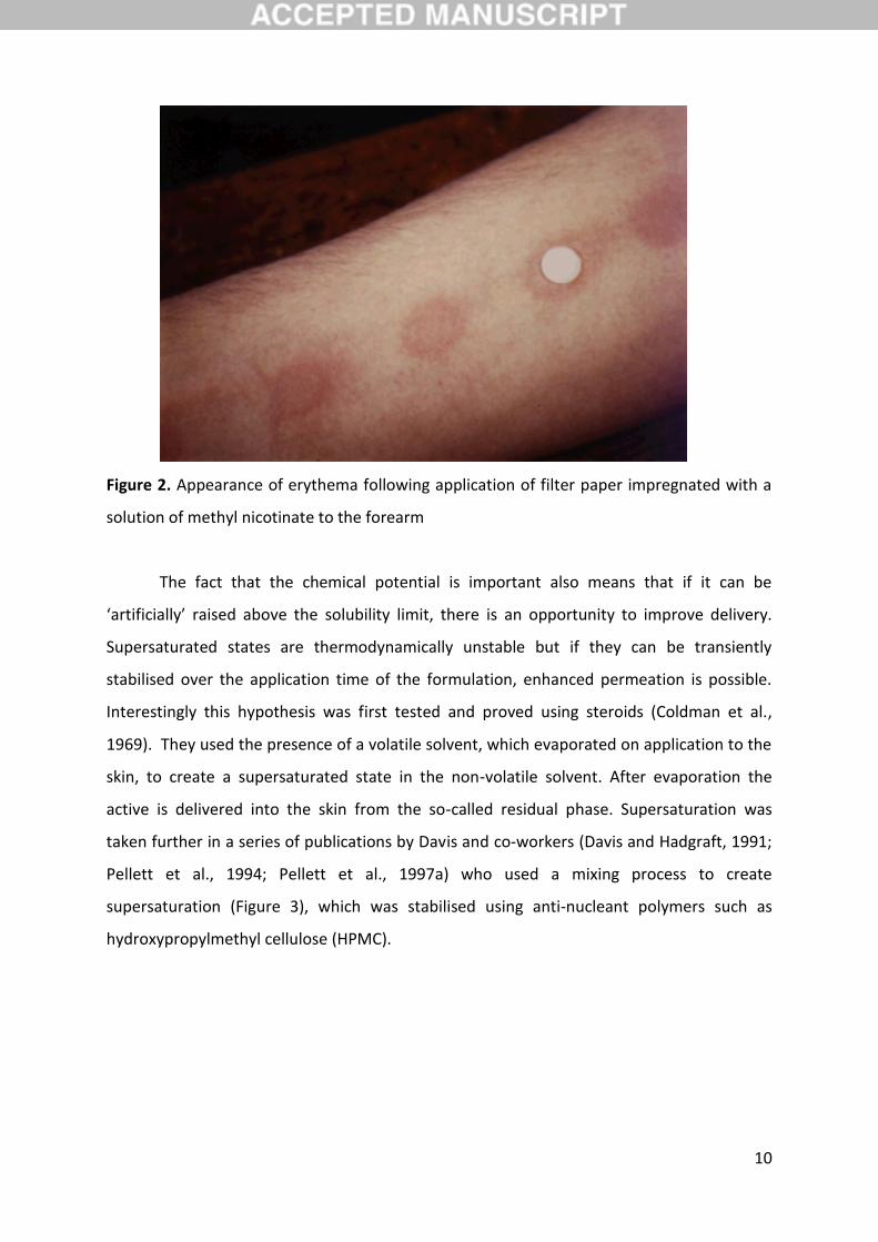
Figure 2. Appearance of erythema following application of filter paper impregnated with a
solution of methyl nicotinate to the forearm
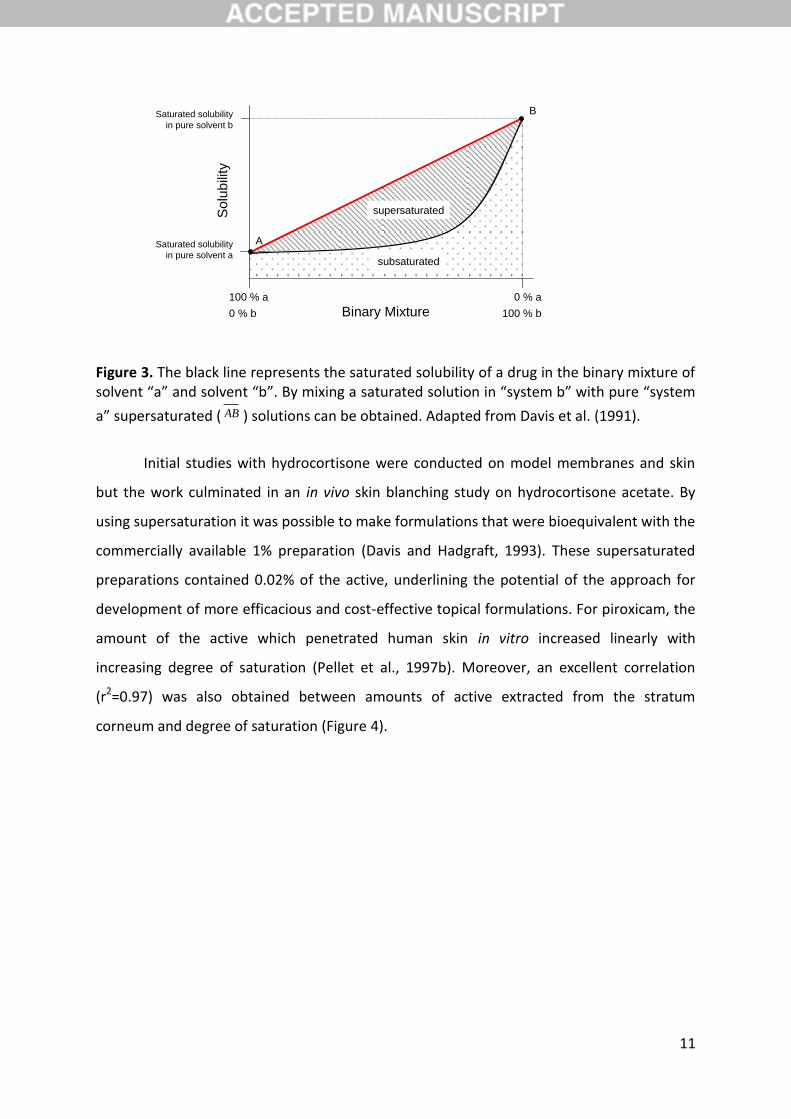
The fact that the chemical potential is important also means that if it can be
‘artificially’ raised above the solubility limit, there is an opportunity to improve delivery.
Supersaturated states are thermodynamically unstable but if they can be transiently
stabilised over the application time of the formulation, enhanced permeation is possible.
Interestingly this hypothesis was first tested and proved using steroids (Coldman et al.,
1969). They used the presence of a volatile solvent, which evaporated on application to the
skin, to create a supersaturated state in the non-volatile solvent. After evaporation the
active is delivered into the skin from the so-called residual phase. Supersaturation was
taken further in a series of publications by Davis and co-workers (Davis and Hadgraft, 1991;
Pellett et al., 1994; Pellett et al., 1997a) who used a mixing process to create
supersaturation (Figure 3), which was stabilised using anti-nucleant polymers such as
hydroxypropylmethyl cellulose (HPMC).
11
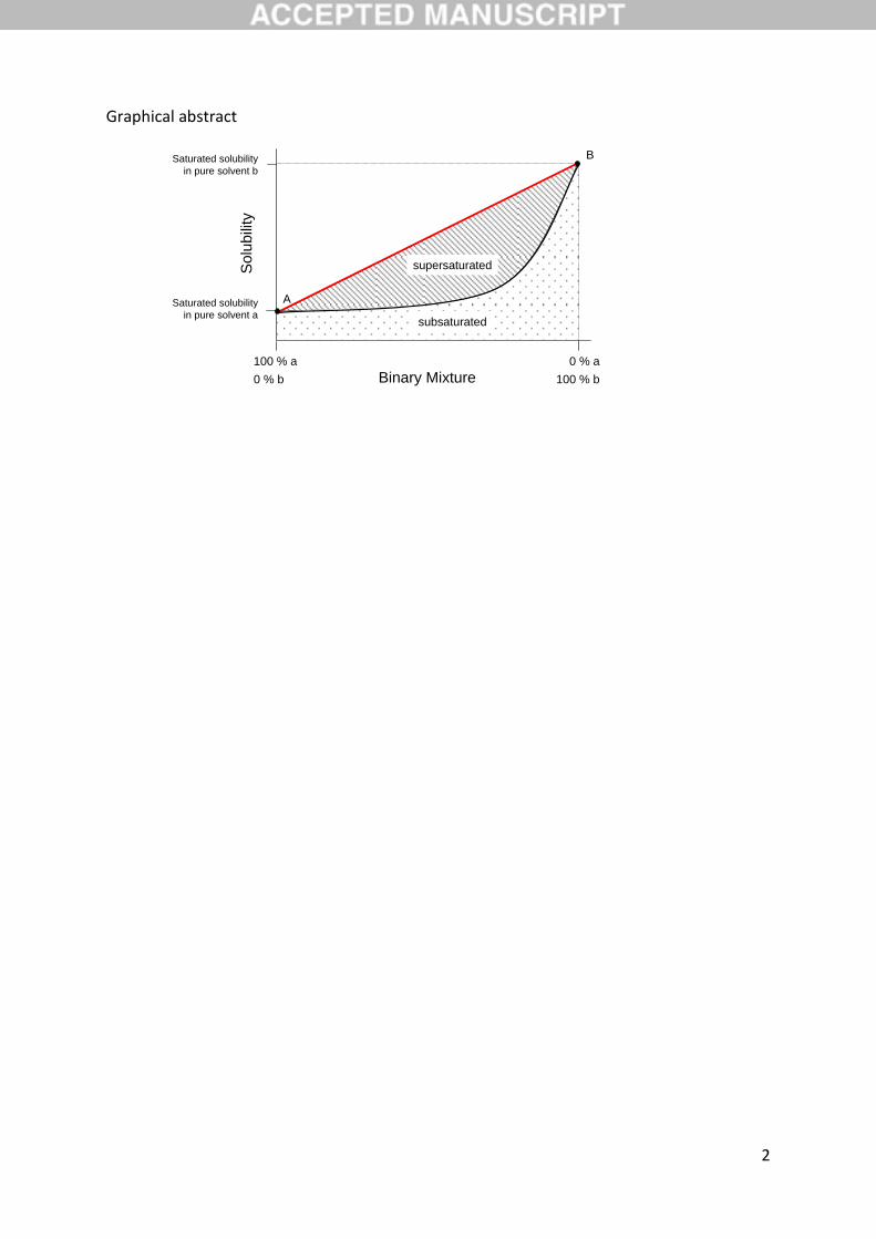
Figure 3. The black line represents the saturated solubility of a drug in the binary mixture of solvent “a” and solvent “b”. By mixing a saturated solution in “system b” with pure “system
a” supersaturated ( ) solutions can be obtained. Adapted from Davis et al. (1991).
Initial studies with hydrocortisone were conducted on model membranes and skin
but the work culminated in an in vivo skin blanching study on hydrocortisone acetate. By
using supersaturation it was possible to make formulations that were bioequivalent with the
commercially available 1% preparation (Davis and Hadgraft, 1993). These supersaturated
preparations contained 0.02% of the active, underlining the potential of the approach for
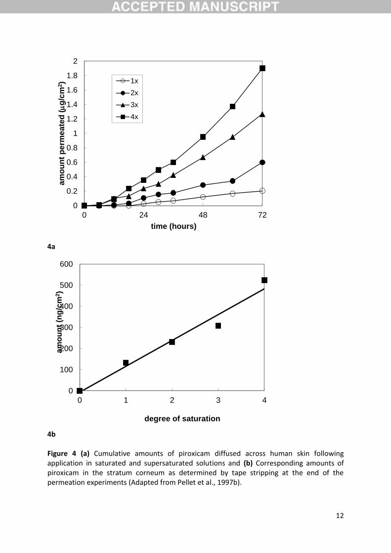
development of more efficacious and cost-effective topical formulations. For piroxicam, the
amount of the active which penetrated human skin in vitro increased linearly with
increasing degree of saturation (Pellet et al., 1997b). Moreover, an excellent correlation
(r2=0.97) was also obtained between amounts of active extracted from the stratum
corneum and degree of saturation (Figure 4).
100 % b
0 % a
0 % b
100 % a
Binary Mixture
Saturated solubility
in pure solvent b
Saturated solubility
in pure solvent a
B
A
Solu
bili
ty
subsaturated
supersaturated
AB
12
4a
4b Figure 4 (a) Cumulative amounts of piroxicam diffused across human skin following application in saturated and supersaturated solutions and (b) Corresponding amounts of piroxicam in the stratum corneum as determined by tape stripping at the end of the permeation experiments (Adapted from Pellet et al., 1997b).
0
0.2
0.4
0.6
0.8
1
1.2
1.4
1.6
1.8
2
0 24 48 72
am
ou
nt
pe
rme
ate
d (m
g/c
m2)
time (hours)
1x
2x
3x
4x
0
100
200
300
400
500
600
0 1 2 3 4
am
ou
nt
(ng
/cm
2)
degree of saturation
13
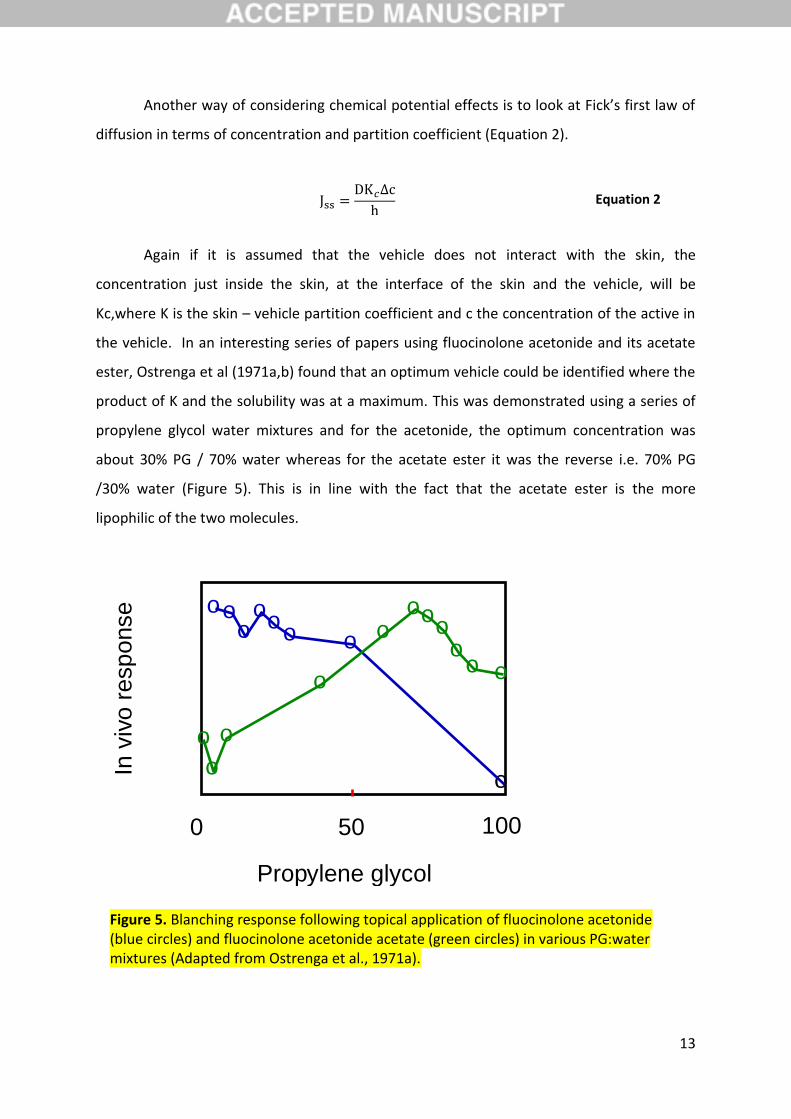
Another way of considering chemical potential effects is to look at Fick’s first law of
diffusion in terms of concentration and partition coefficient (Equation 2).
Equation 2
Again if it is assumed that the vehicle does not interact with the skin, the
concentration just inside the skin, at the interface of the skin and the vehicle, will be
Kc,where K is the skin – vehicle partition coefficient and c the concentration of the active in
the vehicle. In an interesting series of papers using fluocinolone acetonide and its acetate
ester, Ostrenga et al (1971a,b) found that an optimum vehicle could be identified where the
product of K and the solubility was at a maximum. This was demonstrated using a series of
propylene glycol water mixtures and for the acetonide, the optimum concentration was
about 30% PG / 70% water whereas for the acetate ester it was the reverse i.e. 70% PG
/30% water (Figure 5). This is in line with the fact that the acetate ester is the more
lipophilic of the two molecules.
Propylene glycol
0 50 100
o
o
o
o
o
o o o
o o o
o o
o
o o
o o
o
In v
ivo r
esp
onse
Figure 5. Blanching response following topical application of fluocinolone acetonide (blue circles) and fluocinolone acetonide acetate (green circles) in various PG:water mixtures (Adapted from Ostrenga et al., 1971a).
14
A further factor in Higuchi’s paper (1960), in which the role of Fick’s laws of diffusion
is discussed, is the diffusion coefficient, D. In general terms the smaller the active, and thus
the lower the molecular weight, the faster the active will diffuse through the skin. This
means that the three major determinants in controlling skin penetration are (i) solubility of
the active (ii) partition behaviour and (iii) diffusion coefficient. In general, those compounds
that permeate well have a log octanol – water partition coefficient (Log Po/w) of about 2,
good solubility in both oils and water, and are relatively small. These are the characteristics
of two of the drugs that are currently delivered via the transdermal route, nitroglycerin and
nicotine. All of the above statements have the caveat that the formulation components do
not interact with the skin, clearly some will and the implications of this can be profound.
Before discussing the interplay between excipients and the skin, it is important to identify
the rate controlling process in dermal penetration and how this is influenced by the
structure of the skin and the route of penetration.
5. Penetration enhancers
Since the barrier properties of the stratum corneum are formidable, it has been a
challenge to produce medicines which effectively treat skin disorders. Many actives do not
have ideal physicochemical properties and therefore the formulation scientist has significant
difficulties in optimising their delivery. These constraints led to extensive research into
compounds that enhance penetration. The first major compound was DMSO but this was
soon followed by many more. With the application of various biophysical techniques it
proved possible to describe some of the possible mechanisms of action. Considering Fick’s
first law of diffusion the two factors that can be modified are the partition behaviour and
the diffusion coefficient (Hadgraft, 2009; Lane, 2013). If a formulation component enters
the structured lipids of the stratum corneum it may alter their solubility characteristics and
will thereby alter the partition from the formulation into the skin. If this is favourable,
enhanced permeation will result. Alternatively, the excipient may intercalate into the
structured lipids and alter their fluidity. If it makes them more fluid diffusion through them
will be more rapid and, again, enhanced permeation could result.
It is very difficult to deconvolve the two effects of partition and diffusion on
membrane penetration of an active. However, using deuterated materials and Attenuated
Total Reflectance - Fourier Transform Infra Red (ATR-FTIR) spectroscopy it has been possible
15
to show that oleic acid forms pools in the structured lipids and that penetration of an active
is enhanced as a result of diffusion through these more fluid domains (Potts et al., 1991).
On the other hand, Azone also fluidises the structured lipids but intercalates into them in a
more homogenous way (Harrison et al., 1996b). The major structural difference is the cis
double bond in oleic acid which is probably responsible for the pool formation. Considering
the molecular structure of Azone, the seven membered ring interacts with the polar
headgroups of the skin lipids. This forces the alkyl chains apart and they therefore become
more fluid.
It has been suggested that small solvent molecules such as propylene glycol or
diethyleneglycol monoethylether (Transcutol), improve the solubility of penetrants in the
stratum corneum (Kasting et al., 1993; Watkinson et al., 2009; Osborne, 2011). This effect
has also been studied using ATR-FTIR (Harrison et al., 1996a). The fact that both D and K can
be influenced means that if excipients in a formulation can favourably alter both, then
synergistic effects are found. This had previously been reported for mixtures of Azone and
propylene glycol (Wotton et al., 1985). Supersaturation was described above and it is also
possible to obtain synergistic effects when supersaturation is combined with chemical
penetration enhancement (Davis et al., 2002).
6. Solvent effects
With the advent of in vivo confocal Raman spectroscopy it has been possible to
examine the effects of excipients on the distribution of actives within the stratum corneum.
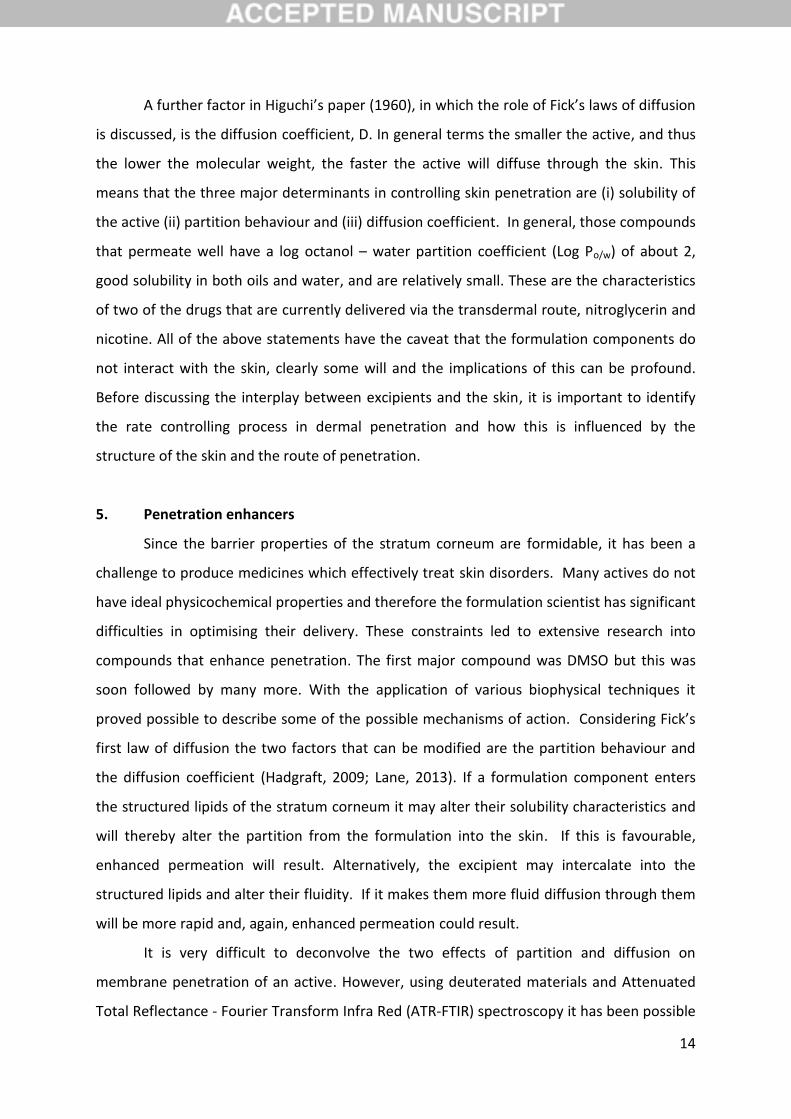
Mohammed et al. (2014) examined the penetration of nicotinamide from different
excipients, both singly, and in combinations (binary and ternary). One of the important
findings of the work was to show that the amount of active in the stratum corneum was
related to the uptake of the solvent in the stratum corneum (Figure 6). Very few studies
have been conducted on the uptake of emollients or their penetration across the skin.
Small molecules such as ethanol and propylene glycol have been examined and they
permeate quite quickly (Berner et al., 1989; Liu et al., 1991; Trottet et al., 2004). Larger,
more lipophilic emollients such as isopropyl myristate and isostearyl isostearate have not
been researched in any detail. Unpublished work from our laboratory has shown that the
uptake is small, and because of the high log P values of these materials they will not
permeate across the skin (Oliveira, 2009). They will have a high residence time in the skin.
16
Santos et al. (2010) studied the permeation of oxybutynin from finite doses in either
propylene glycol or octyl salicylate. They found that the propylene glycol permeated rapidly
and suggested that the active was left crystallised on and in the skin because of the
disappearance of the solvent. This contrasted with the formulations that contained octyl
salicylate where delivery of oxybutynin was better and more sustained. Because the octyl
salicylate had a longer residence time in the skin, it maintained the active in solution and
therefore available for delivery to the deeper layers.
Figure 6: Correlation between signal intensity of niacinamide and signal intensities of PG or
DMI which permeated into the SC in vivo with depth (0, 4, 8, 12, 16, 20 into the SC) for the
mid ventral forearm (n=8; Mean ± S.D.). Adapted from Mohammed et al. (2014).
7. Finite versus infinite doses in skin permeation studies
One of the important points in the studies conducted by Santos et al. (2010) was the
use of finite doses. In the clinic, a typical dose to the skin is 2 mg/cm2. Many studies, in
vitro, use infinite doses and often this does not give a true representation of how the
excipients will work in vivo. For example, a representative clinical dose of propylene glycol
will be subject to both evaporation, from the skin surface, and diffusion through the skin.
The levels of PG will therefore deplete very rapidly. At infinite doses there will be no
0
0.1
0.2
0.3
0.4
0.5
0.6
0.7
0.8
0 0.01 0.02 0.03 0.04 0.05 0.06 0.07
Solv
ent
sign
al in
ten
sity
(a.
u)
Niacinamide signal intenstiy (a.u)
17
depletion and the effects of PG in the skin may be significantly overestimated (Hadgraft and
Lane, 2016). .
There are not only problems associated with the use of finite doses, it should also be
noted that many experiments have been conducted on rodent skin, which also gives
misleading results. For a true representation of topical delivery to man, human skin should
be used with as close a clinical dose as possible. The closest one can get to this, in the
absence of human tissue, is pig tissue. A number of studies have reported the similarity
between pig and human skin tissue with reference to barrier properties (Dick and Scott,
1992; Singh et al., 2002; Barbero and Frasch, 2009; Luo et al., 2016).
8. Psychorheology
The cosmetics industry would not exist if it did not produce products that were
acceptable to consumers. It is extremely good at producing products that ‘feel good’ and
the public want to buy again and again. The pharmaceutical industry could learn from this
and perhaps patient compliance would be better if topical products were more cosmetically
acceptable. The subject has not been studied extensively but was recognised by Boyd
(1976). It is clear that medicines for treating mild eczema have to have much better ‘feel’
than, for example, a product to treat melanoma. However, in modern product design there
should be an element which considers the desirability of using the product on a regular and
repeated basis. There are many publications, in the pharmaceutical literature, which
consider the basic rheological properties of the product, but one should question their
relevance in relation to their ability to deliver the active and how the patient perceives
them.
9. Conclusions - Advanced Topical Formulations
Important principles underlying the basis for the design of advanced topical
formulations have been known for a number of years but all the elements are rarely
considered in an holistic approach. Firstly, the properties of the active should be taken into
account. Ideally it will have a log Po/w around 2, and will have reasonable solubility in both
oils and water, usually associated with a low melting point. If there are several candidates
that are structurally related then there will be a balance between the ideal physicochemical
properties and the potency.
18
The active needs to be delivered to the ‘active’ regions of the skin, i.e. the
intercellular spaces at as high a thermodynamic activity as possible. This means that it will
be close to saturation and therefore subject to the possibility of crystallisation. There needs
to be the possibility of lateral diffusion to the intercellular spaces from the formulation
deposited onto the corneocytes. It is possible that the enhanced penetration from some
liposomal formulations is a result of the formation of a lipid layer on the skin surface
through which lateral diffusion is facilitated. Rapid evaporation of a volatile solvent is likely
to result in supersaturation but this will need to be stabilised with the judicious choice of
anti-nucleant polymers.
It is important to recognise the evaporation of formulation components, such as
water, and the residual phase that remains is of most importance in the formulation design.
The residual phase should present the active at a high activity state but also deliver
appropriate enhancers into the skin, most particularly solvents that will remain in the
stratum corneum and maintain the active in solution. The solubility characteristics of the
residual phase both in terms of the active and the skin lipids need to be considered. In
order that the emollients also penetrate into the SC, it is important that they are also at a
high thermodynamic activity.
These simple rules may be difficult to achieve, in reality, but will result in more
effective topical delivery. Other factors that will also be built in to the final formulation are
clearly the feel of the product, as discussed above, and also its stability. The increased
sophistication of biophysical techniques, particularly those that can be used in vivo, will aid
the formulation process and give much more insight into the future developments of topical
formulation design.
19
References
Albery, W.J., Hadgraft, J. 1979. Percutaneous absorption: in vivo experiments. J. Pharm.
Pharmacol. 31(3):140-7.
Barrett, C.W., Hadgraft, J.W., Sarkany, I. 1964. The influence of vehicles on skin penetration.
J. Pharm. Pharmacol. 16(suppl)104T-107T.
Baker, H. 1968. The effects of dimethylsulfoxide dimethylformamide and
dimethylacetamide on the cutaneousbarrier to water in human skin. J. Invest. Dermatol.
50(4):283-8.
Barbero, A.M., Frasch, H.F. 2009. Pig and guinea pig skin as surrogates for human in vitro
penetration studies: a quantitative review. Toxicol. In Vitro. 23(1) 1-13.
Berner, B., Mazzenga, G.C., Otte, J.H., Steffens, R.J., Juang, R.H., Ebert, C.D. 1989. J. Pharm.
Sci. 78(5): 402-7.
Blank, I.H., Scheuplein, R.J., MacFarlane, D.J. 1967. Mechanism of percutaneous absorption.
III. The effect of temperature on the transport of non-electrolyes across the skin. J. Invest.
Dermatol. 49(6):582-589.
Bodde, H. E., Kruithof, M. A., Brussee, J., Koerten, H. K. 1989. Visualisation of normal and
enhanced HgCl2 transport through human skin in vitro. Int. J. Pharm. 53(1)13-24.
Bouwstra, J.A., Gooris, G.S., van der Spek, J.A., Bras, W. 1991. Structural investigations of
human stratum corneum by small-angle X-ray scattering. J Invest. Dermatol. ;97(6):1005-12.
Boyd, J.V. 1976. Psycho-rheology — the relevance of rheology to consumer acceptance. J.
Soc. Cosmet. Chem. 27(6):247-256
Burton, R,J. 1972. Witchcraft in the Middle Ages, Cornell University Press, New York.

20
Coldman, M.F., Poulsen, B.J., Higuchi, T. 1969. Enhancement of percutaneous absorption by
the use of volatile: nonvolatile systems as vehicles. J. Pharm. Sci. 58(9):1098-102.
Cronin, E., Stoughton, R.B. 1962. Percutaneous absorption. Regional variations and the
effect of hydration and epidermal stripping. Br. J. Dermatol. 74:265-72.
Davis, A.F., Hadgraft, J. 1991. Effect of supersaturation on membrane transport: 1.
Hydrocortisone acetate Int. J. Pharm. 76(1-2):1-8.
Davis, A.F., Hadgraft, J. 1993. Supersaturated solutions as topical drug delivery systems. In:
Pharmaceutical Skin Penetration Enhancement (Eds; K.A. Walters, J. Hadgraft). Marcel
Dekker, New York. Pp. 243-267.
Davis, A.F., Gyurik, R.J., Hadgraft, J., Pellett, M.A., Walters, K.A. 2002. Formulation strategies
for modulating skin penetration. In: Dermatological and Transdermal Formulations (Editor:
K.A. Walters). Marcel Dekker. Pp. 271-318.
Eichenfield L.F., Tom, W.L., Berger, T.G., Krol, A., Paller, A.S., Schwarzenberger, K., Bergman,
J.N., Chamlin, S.L., Cohen, D.E., Cooper, K.D., Cordoro, K.M., Davis, D.M., Feldman, S.R.,
Hanifin, J.M., Margolis, D.J., Silverman, R.A., Simpson, E.L., Williams, H.C., Elmets, C.A.,
Block, J., Harrod, C.G., Smith Begolka, W., Sidbury, R. 2014. Guidelines of care for the
management of atopic dermatitis: section 2. Management and treatment of atopic
dermatitis with topical therapies. J. Am. Acad. Dermatol. 71(1):116-32.
Faulkner, J.M. 1933. Nicotine poisoning by absorption through the skin. JAMA.
100(21):1664-1665.
Feldmann, R.J., Maibach, H.I. 1965. Penetration of 14C hydrocortisone through normal skin:
the effect of stripping and occlusion. Arch. Dermatol. 191:661-6.
21
Feldmann, R.J., Maibach, H.I. 1966. Percutaneous penetration of 14C hydrocortisone in
man. II. Effect of certain bases and pretreatments. Arch. Dermatol. 94(5):649-51.
Feldmann, R.J., Maibach, H.I. 1969. Percutaneous penetration of steroids in man. J. Invest.
Dermatol. 52(1):89-94
Fountain, R.B., Baker, B.S., Hadgraft, J.W., Sarkany, I. 1969a. The rate of absorption and
duration of action of four different solutions of methyl nicotinate. Br. J Dermatol. 81(3):202-
6.
Fountain, R.B., Baker, B.S., Hadgraft, J.W., Sarkany, I. 1969b. The rate of absorption and
duration of action of four different solutions of methyl nicotinate. Br. J. Dermatol.
81(3):202-6.
Fourman, P., Bartter, F.C., Albright, F., Dempsey, E., Carroll, E. 1950. Some effects of 17-
hydroxy-corticosterone (Compound F) in man. Rev. Canadin Biol. 9(1):72-72.
Garson, J.-C., Doucet, J., Leveque, J.-L., Tsoucaris, G. 1991. Oriented structure in human
stratum corneum revealed by X-ray diffraction. J. Invest. Dermatol. 96:43-49.
Hadgraft, J., Hadgraft, J.W., Sarkany, I. 1973. The effect of thermodynamic activity on the
percutaneous absorption of methyl nicotinate from water glycerol mixtures. J. Pharm.
Pharmacol. 25:Suppl:122P-123.
Hadgraft, J. 2004. Skin deep. Eur. J. Pharm. Biopharm. 58(2) 291-99.
Hadgraft, J., Lane, M.E. 2009. Transepidermal water loss and skin site: a hypothesis. Int. J.
Pharm. 373(1-2):1-3.
Hadgraft, J., Lane, M.E. 2011. Skin: the ultimate interface. Phys. Chem. Chem. Phys.
13(12):5215-22.

22
Hadgraft, J., Lane, M.E. 2016. Drug crystallization – implications for topical and transdermal
delivery. Expert Opin. Drug Deliv. DOI: 10.1517/17425247.2016.1140146
Harrison, J.E., Watkinson, A.C., Green, D.M., Hadgraft, J., Brain, K. 1996a. The relative effect
of Azone and Transcutol on permeant diffusivity and solubility in human stratum corneum.
Pharm Res. 13(4):542-6.
Harrison, J.E., Groundwater, P.W., Brain, K.R., Hadgraft, J. Azone induced fluidity in human
stratum corneum. A fourier transform spectroscopy investigation using the perdeuterated
analogue. 1996b. J. Control. Release. 41(3) 283-290.
Higuchi, T. 1960. Physical Chemical Analysis of Percutaneous Absorption Process from
Creams and Ointments. J. Soc. Cosmet. Chem. 11:85-97.
Kasting, G.B., Francis, W.R., Roberts, G.E. 1993. Skin penetration enhancement of
triprolidine base by propylene glycol. J. Pharm. Sci. 82(5):551-2.
Lane, M.E. 2013. Skin penetration enhancers. Int. J. Pharm. 15;447(1-2):12-21.
Laws, C.E. 1910. Nitroglycerin head. JAMA 54:793-793.
Lippold, B.C., Schneemann, H. 1984. The influence of vehicles on the local bioavailability of
betamethasone-17-benzoate from solution and suspension type ointments. Int J Pharm.
22(1):31-43.
Liu, P., Kurihara-Bergstrom, T., Good, W.R. 1991. Cotransport of estradiol and ethanol
through human skin in vitro: understanding the permeant/enhancer flux relationship.
Pharm. Res. 8:938-44.
Luo, L., Patel, A., Sinko, B., Bell, M., Wibawa, J., Hadgraft, J., Lane, M.E. 2016. A comparative
study of the in vitro permeation of ibuprofen in mammalian skin, the PAMPA model and
silicone membrane. Int. J. Pharm. 26;505(1-2):14-19.
23
Machado, M., Salgado, T.M., Hadgraft, J., Lane, M.E. 2010. The relationship between
transepidermal water loss and skin permeability. Int. J. Pharm. 384(1-2):73-7.
McKenzie, A.W., Stoughton, R.B. 1962. Method for comparing percutaneous absorption of
steroids. Arch. Dermatol. 86(5):608-10.
Michaels, A.S., Chandrasekaran, S.K., Shaw, J.E. 1975. Drug permeation through human skin:
Theory and in vitro experimental measurement. AIChE. 21(5):985-996.
Mohammed, D., Matts, P.J., Hadgraft, J., Lane, M.E. 2014. In vitro-in vivo correlation in skin
permeation. Pharm. Res. 31(2):394-400.
Neelissen, J.A., Arth, C., Wolff, M., Schrijvers, A.J., Junginger, H.E., Boddé, H.E. 2000.
Visualization of percutaneous 3H-estradiol and 3H-norethindrone acetate transport across
human epidermis as a function of time. Acta Derm. Venereol. Suppl (Stockh). 208:36-43.
Nemanic, M.K., Elias, P.M. 1980. In situ precipitation: a novel cytochemical technique for
visualization of permeability pathways in mammalian stratum corneum. J. Histochem.
Cytochem. 28(6):573-8.
Ostrenga, J., Steinmetz, C., Poulsen, B. 1971a. Significance of vehicle composition. I.
Relationship between topical vehicle composition, skin penetrability, and clinical efficacy.
J. Pharm. Sci. 60(8):1175-9.
Ostrenga, J., Steinmetz, C., Poulsen, B., Yett, S. 1971b. Significance of vehicle composition. II.
Prediction of optimal vehicle composition. J. Pharm. Sci. 60(8):1180-3.
Pellett, M.A., Davis, A.F., Hadgraft, J. 1994. Effect of supersaturation on membrane
transport:2. Piroxicam. Int. J. Pharm. 111(1):1-6.
24
Pellett, M.A., Castellano, S., Hadgraft, J., Davis, A.F. 1997a. The penetration of
supersaturated solutions of piroxicam across silicone membrane and human skin in vitro. J.
Control. Release. 46(3):205-214.
Pellett, M.A., Roberts, M.S., Hadgraft, J. 1997b. Supersaturated solutions evaluated with an
in vitro stratum corneum tape stripping techniques. Int. J. Pharm. 151(1):91-8.
Rothman, S. 1943. The principles of percutaneous absorption. J. Lab. Clin. Med. 28:1305-21.
Santos, P., Watkinson, A.C., Hadgraft, J., Lane, M.E. 2010. Oxybutynin permeation in skin:
the influence of drug and solvent activity. Int. J Pharm. 384(1-2):67-72.
Sarkany, I., Hadgraft, J.W., Caron, G.A., Barrett, C.W. 1965. The role of vehicles in the
percutaneous absorption of corticosteroids. An experimental and clinical study. Br. J.
Dermatol. 77(11):569-75.
Scheuplein, R.J. 1965. Mechanism of percutaneous absorption. I. Routes of penetration and
the influence of solubility. J. Invest. Dermatol. 45(5):334-346.
Scheuplein, R.J. 1966. Analysis of permeability data for the case of parallel diffusion
pathways. Biophys. J. 6(1):1-17.
Scheuplein, R.J. 1967. Mechanism of percutaneous absorption. II. Transient diffusion and
the relative importance of various routes of skin penetration. J. Invest. Dermatol. 18(1):79-
88.
Scheuplein, R.J., Blank, I.H., Brauner, G.J., MacFarlane, D.J. 1969. Percutaneous absorption
of steroids. J. Invest. Dermatol. 52(1):63-70.
Scheuplein, R.J., Ross, L. 1970. Effects of surfactants and solvents on the permeability of the
epidermis. J. Soc. Cosmet. Chem. 21:853-873.
25
Scheuplein, R.J., Ross, L.W. 1974. Mechanism of percutaneous absorption. V. Percutaneous
absorption of solvent deposited solids. J. Invest. Dermatol. 62(4):353-360.
Singh, S., Zhao, K., Singh, J. 2002. In vitro permeability and binding of hydrocarbons in pig
ear and human abdominal skin. Drug Chem. Toxicol. 25(1):83-92.
Stoughton, R.B. 1965. Dimethylsulfoxide (DMSO) induction of a steroid reservoir in human
skin. Arch. Dermatol. 91:657-60.
Talreja, P., Kleene, N.K., Pickens, W.L., Wang, T.F., Kasting, G.B. 2001. Visualization of the
lipid barrier and measurement of lipid pathlength in human stratum corneum. AAPS
PharmSci. 3(2):E13.
Trottet, L., Merly, C., Mirza, M., Hadgraft, J., Davis, A.F.2004. Effect of finite doses of
propylene glycol on enhancement of in vitro percutaneous permeation of loperamide
hydrochloride. Int J Pharm. 274(1-2):213-9.
Watkinson, R.M., Guy, R.H., Hadgraft, J., Lane, M.E. 2009. Optimisation of cosolvent
concentration for topical drug delivery - II: influence of propylene glycol on ibuprofen
permeation. Skin Pharmacol Physiol. 22(4):225-30.
Wotton, P.K., Møllgaard, B., Hadgraft, J. Hoelgaard, A. 1985. Vehicle effects on topical drug
delivery. III. Effect of Azone on the cutaneous penetration of metronidazole and propylene
glycol. Int. J. Pharm. 24(1), 19-26.