Embed Size (px)
Citation preview

Advanced OCT/SLO System

High-speed scan (53,000 A-scans / s)
High-resolution image of OCT & SLO
Extremely easy and fast operation with optimization
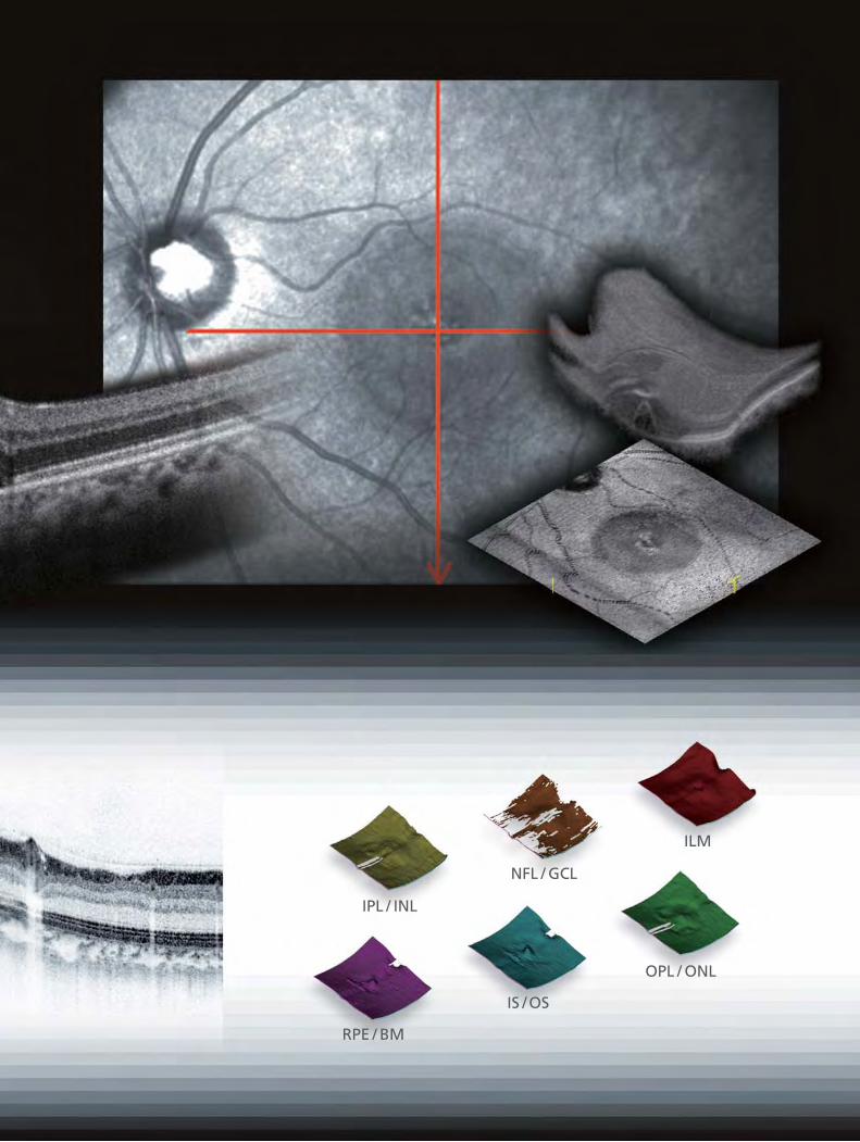
ILM
NFL / GCL
IPL / INL
OPL / ONL
IS / OS
RPE / BM
Max. 50 image averaging
53, 000
25, 000~27, 000
512
Conventional SD - OCT
Time Domain - OCTA-scans / s
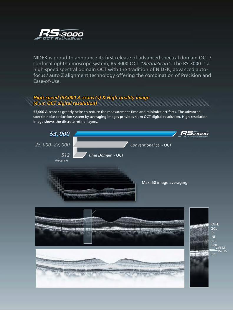
53,000 A-scans / s greatly helps to reduce the measurement time and minimize artifacts. The advanced speckle-noise-reduction system by averaging images provides 4 µm OCT digital resolution. High-resolution image shows the discrete retinal layers.
High-speed (53,000 A-scans / s) & High-quality image (4 m OCT digital resolution)
RNFLRNFLGCLGCLIPLIPLINLINLOPLOPLONLONL
RPERPE
RNFLGCLIPLINLOPLONL
RPE
ELMELMIS IS / / OSOSELMIS / OS
NIDEK is proud to announce its first release of advanced spectral domain OCT / confocal ophthalmoscope system, RS-3000 OCT "RetinaScan". The RS-3000 is a high-speed spectral domain OCT with the tradition of NIDEK, advanced auto-focus / auto Z alignment technology offering the combination of Precision and Ease-of-Use.
µ

1 1 2 2 3Step 1 2 3
The operation of RS-3000 is as easy as Auto-Refractometer. The focus of SLO fundus image and the alignment of OCT depth are adjusted automatically by pressing optimization button.
Fast and simple operation with optimization
33 ReleaseRelease
The focus of SLO fundus image and the alignment of OCT depth are adjusted automatically.
OptimizationOptimization22
Capturing both image of SLO and OCT by one shot.
11 Start ScanningStart Scanning
The position, length, angle of scanning line for the target are easily and flexibly changed on real-time confocal SLO image.
Real-time, high-contrast and wide view (40º x 30º) of confocal SLO imaging offers the accuracy for OCT scanning of the pathological target. OCT scanning position is precisely matched with SLO fundus image.
Accurate localization of pathology with real-time SLO image

Macula line Macula cross Macula multi
Macula map Disc circle Disc map
The RS-3000 provides 6 types of useful OCT scans to meet clinical requirements.
High contrast SLO fundus image and auto-tracking function achieve excellent reproducibility in follow-up examination. Auto-tracking function tracks eye movement and guides the OCT scanning to the previous examination position. Time frame monitoring results of examinations including NFL defect, Optic nerve head and macular thickness can be conducted easily.
Highly reproducible follow-up examination with auto-tracking function
Not match with baselineNot match with baselineNot match with baseline
Macula mapMacula mapMacula map
Match with baselineMatch with baselineMatch with baseline
Disc mapDisc mapDisc map
RelRelativelyatively match match with with baseaselinelineRelatively match with baseline
Disc circleDisc circleDisc circle

The morphological change on the surface of each layer is visually confirmed.
6 Layer Segmentation
Layer thickness map
High-speed and wide 3D imaging help to understand retinal condition quickly and comprehensively. Thickness map between each layer from ILM to RPE can be available.
High-speed (1.6 s) and wide (9 mm x 9 mm) 3D mapping
IS / OS OPL / ONL RPE / BM
ILM NFL / GCL IPL / INL
ILM - RPE/BM
ILM + NFL + GCL + IPL
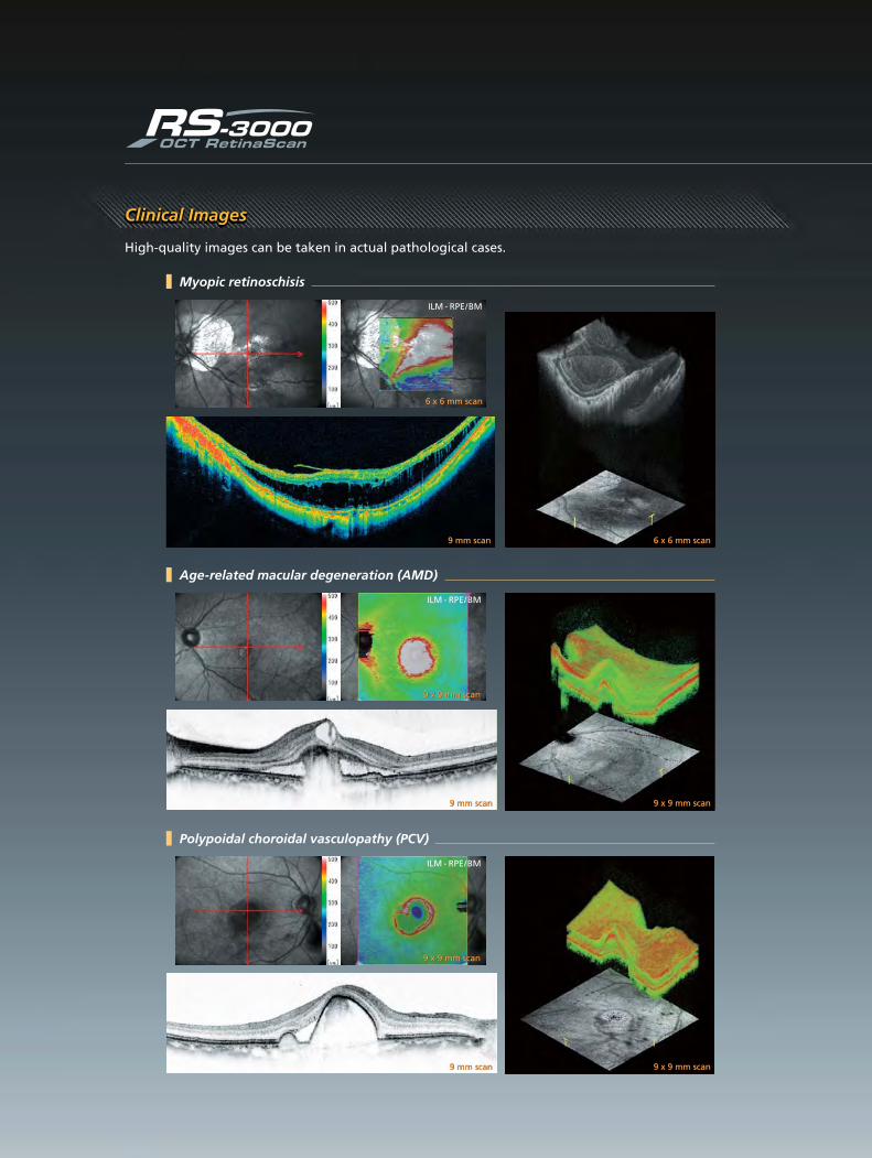
High-quality images can be taken in actual pathological cases.
Clinical Images
Myopic retinoschisis
6 x 6 mm scan6 x 6 mm scan
6 x 6 mm scan6 x 6 mm scan
ILM - RPE/BM
9 mm scan9 mm scan
9 mm scan9 mm scan 9 x 9 mm scan9 x 9 mm scan
Polypoidal choroidal vasculopathy (PCV)
9 mm scan9 mm scan 9 x 9 mm scan9 x 9 mm scan
Age-related macular degeneration (AMD)
ILM - RPE/BM
9 x 9 mm scan9 x 9 mm scan
ILM - RPE/BM
9 x 9 mm scan9 x 9 mm scan

9 mm scan9 mm scan
9 mm scan9 mm scan 9 x 9 mm scan9 x 9 mm scan
Pseudo macular hole
ILM - RPE/BM
9 x 9 mm scan9 x 9 mm scan
9 mm scan9 mm scan 9 x 9 mm scan9 x 9 mm scan
Epiretinal membrane (ERM)
ILM - RPE/BM
9 x 9 mm scan9 x 9 mm scan
Vogt-Koyanagi-Harada disease
Glaucoma
6 x 6 mm scan6 x 6 mm scan9 x 9 mm scan9 x 9 mm scan
ILM - RPE/BM ILM-IPL / INL ILM-IPL / INL
9 x 9 mm scan9 x 9 mm scan

Networking and review capability is a standard feature of the NAVIS-EX Image Filing Software. By having a review station on a LAN network, images from various instruments can be managed within unified software network on a single PC.
The NAVIS-EX Image Filing Software is included in any package of the RS-3000 OCT "RetinaScan" System. As well as filing, manipulating and analyzing the images from the RS-3000, NAVIS-EX is able to import the images from various external instruments.
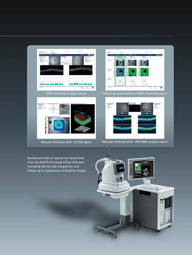
Numerous kinds of reports can be printed from the NAVIS-EX Image Filing Software, including side-by-side comparison and follow up in comparison to baseline image.
Macular thickness (ILM - RPE/BM) analysis report
Follow-up examination of RNFL thickness report
Macular thickness (ILM - IS /OS) report
RNFL thickness analysis report

HEAD OFFICE34-14 Maehama, HiroishiGamagori, Aichi, 443-0038, JapanTelephone : +81-533-67-6611Facsimile : +81-533-67-6610URL : http://www.nidek.co.jp
[Manufacturer ]
TOKYO OFFICE(International Div.)3F Sumitomo Fudosan Hongo Bldg., 3-22-5 Hongo, Bunkyo-ku, Tokyo, 113-0033, JapanTelephone : +81-3-5844-2641Facsimile : +81-3-5844-2642URL : http://www.nidek.com
NIDEK INC.47651 Westinghouse DriveFremont, CA 94539, U.S.A.Telephone : +1-510-226-5700 : +1-800-223-9044 (US only)Facsimile : +1-510-226-5750URL : http://usa.nidek.com
NIDEK TECHNOLOGIES SrlVia dell'Artigianato, 6 / A35020 Albignasego (Padova), ItalyTelephone : +39 049 8629200 / 8626399Facsimile : +39 049 8626824URL : http://www.nidektechnologies.it
NIDEK S.A.Europarc13, rue Auguste Perret94042 Creteil, FranceTelephone : +33-1-49 80 97 97Facsimile : +33-1-49 80 32 08URL : http://www.nidek.fr
CNIDEK 2009 Printed in Japan RS-3000 NRDNOK2
FDA 510(K) pending
Specifications and design are subject to change without notice.
RS-3000 Specifications Footprint (mm)OCT scanning
TechnologyOCT resolution
Scanning range
OCT light sourceScanning speedAcquisition time of 3D image Internal fixation lamp / WavelengthExternal fixation lampAuto alignmentMinimum pupil diameterFocus adjustment rangeWorking distanceScanning pattern
Software analysis
SLO imagingTechnologySLO light sourceField of viewFocusing method
PC networkingDisplayPower supply
Power consumptionMaximum power output (transformer)Dimensions / Weight
Motorized optical table (optional)Dimensions / Weight
Power supply
Power consumption
PC rack (optional)Dimensions / Weight
Spectral domain OCTOptical Z: 7 µm, XY: 20 µmDigital Z: 4 µm, XY: 3 µmZ: 2.1 mmXY: 3 to 9 mmSLD, 880 nm53,000 A-scans / s1.6 sCross shape (normal or large) / 635 nmRed / GreenZ directionø 2.5 mm-15 to +10 D (VD=12 mm)35.5 mm (from the objective lens to the pupil)Macula line (scan angle changeable by 15º)Macula crossMacula mapMacula multi (X - Y: 5 x 5)Disc circleDisc mapSegmentation of 6 retinal layersMacular thickness mapRNFL thickness mapOptic nerve analysisFollow-up examination of pathological progress
Confocal scanning laser ophthalmoscope785 nm40º x 30º (zoom: 20º x 15º)Auto focusAvailableTiltable 8.4-inch color LCDAC 100, 120, 230 V ±10%50 / 60 Hz300 VA1000 VA380 (W) x 524 (D) x 499 to 531 (H) mm / 34 kg14.96 (W) x 20.63 (D) x 19.65 to 20.91 (H)" / 75.0 lbs.
592 (W) x 472 (D) x 596 to 794 (H) mm / 27 kg23.31 (W) x 18.58 (D) x 23.46 to 31.26 (H)" / 59.5 lbs.AC 100 V50 / 60 Hz150 W
632 (W) x 452 (D) x 703 (H) mm / 34 kg24.88 (W) x 17.80 (D) x 27.68 (H)" / 75.0 lbs.
983632
452
592
472