Embed Size (px)
Citation preview
Advanced microscopy techniques to assess solid-state properties of
inhalation medicines
Jagdeep Shur and Robert Price
Pharmaceutical Surface Science Research Group Department of Pharmacy
and Pharmacology University of Bath Bath BA2 7AY UK
Corresponding author Robert Price
Tel +44 (0) 1225 383644
Fax+44 (0) 1225 386114
e-mail rpricebathacuk
1
1 Abstract
Efficient control and characterisation of the physico-chemical properties of
active pharmaceutical ingredients (APIs) and excipients for orally inhaled drug
products (OIDPs) are critical to successful product development Control and
reduction of risk requires the introduction of a material science based
approach to product development and the use of advanced analytical tools in
understanding how the solid-state properties of the input materials influence
structure and product functionality The key issues to be addressed at a
microscopic scale are understanding how the critical quality attributes of input
materials influence surface interfacial and particulate interactions within
OIDPs This review offers an in-depth discussion on the use of advanced
microscopy techniques in characterising of the solid-state properties of
particulate materials for OIDPs The review covers the fundamental principles
of the techniques instrumentation types data interpretation and specific
applications in relation to the product development of OIDPs
Keywords Raman chemical imaging dry powder inhalers metered dose
inhalers atomic force microscopy microscopy chemical imaging
tomography interferometry
2
2 Introduction
Material scientists have an ever-increasing array of analytical tools and
techniques available at their disposal to study the physical and chemical
properties of active pharmaceutical ingredients (APIs) and excipients These
tools are being widely applied in the pharmaceutical industry to support
product development of therapeutic medicines Whilst the solid oral dosage
tablet still forms the mainstay of drug delivery more complex medicines such
as those based on inhalation therapies are being developed These complex
medicines generally require the use of bespoke tools and techniques for
material characterisation This is particularly the case for particulate-based
medicaments such as orally inhaled drug products (OIDPs) where an
understanding of the role of the surface and interfacial properties of APIs and
excipients are key to successful product development
This understanding is critically important for the development of suspension
based pressurised metered dose inhaler (MDI) and dry powder inhaler (DPI)
formulations These portable delivery systems require the manufacture of a
product which are stable enough to withstand the manufacturing process and
provide a long shelf life while allowing the drug to be effectively and
reproducibly dispersed for delivery to the lung The development of
formulations which exhibit such properties presents considerable challenges
to formulators and manufacturers
3
The pressurised metered dose inhaler (MDI) is the most dominant and
recognised drug delivery vehicle for lung therapy [1] This dosage form
contains the active pharmaceutical ingredient (API) dissolved or suspended in
the propellant or a mixture of propellantssolvents (eg ethanol) [2] Owing to
the low solubility of many inhaled drug substances in the propellant
(HFA134a) most MDIs are formulated as suspensions Upon actuation of the
dose from a MDI the patient must co-ordinate their breathing to transport the
aerosol into the lungs [3]
In contrast passive dry powder inhalers (DPIs) are breath-actuated devices
and do not require propellants to aid the aerosolization of the APIs
Formulations for DPIs are typically prepared as homogenous adhesive
interactive mixtures comprising of micronized drug particles and a coarse
excipient carrier [4] The coarse carrier traditionally lactose monohydrate [5]
is employed within DPI formulations to improve flow properties and metering
of the highly cohesive API particles [6] The entrainment and subsequent
aerosolization of the formulation is achieved using the patientrsquos inspiratory
force which is required to elutriate the micronized API from the surface of the
carrier particle for delivery to the lower airways of the respiratory tract [7] DPI
formulations are also produced as agglomerated systems consisting of pure
micronised API or mixtures of API and excipients
In suspension MDI formulations the interfacial properties of the API particles
in suspension will be dominated by the van der Waals and electrostatic
double layer forces [8] For carrier based DPI formulations the interactive
4
forces between API and excipient are dominated by a composite of van der
Waals electrostatic and capillary forces [9] The preferential surface
interactions of the API particles with excipients and other components of the
container closure system of these drug product adds further complexity to
suspension MDI and DPI dosage forms These surface interactions may lead
to particle agglomeration segregation or adhesion of API to the inner walls of
a device which will contribute to inconsistent drug delivery and emitted
particle size distribution [10] [11] [12] Hence the United States
Pharmacopeia (USP) requires that the particle size distribution of the fine
particle mass is characterised [13] Additionally the guidance suggests that
appropriate characterisation of the particle size distribution of the API and any
excipients should be considered [14]
The use of appropriate tools for the characterisation and control of medicines
and their components is a vital part of the pharmaceutical development
process Validated analytical methods are used for the formal release of
excipients and medicines but additional tools may also be utilised to
understand various aspects of the medicine especially during early phase
product development This is particularly the case for analytical methods
based on microscopy
Excipients APIs and formulated drug products are routinely evaluated by an
lsquoappearancersquo test Whilst this is the lowest level of scrutiny for the evaluation
of a material the test is contained in excipient and product monographs and
provides useful information about a material concerning for example colour
5
and particulate appearance and also in detecting any contamination Prior to
the development of particle sizing methodologies such as inertial impactors
and laser light diffraction APIs excipients and formulations were routinely
monitored using optical microscopy Whilst this method of examination is
relatively crude subjective and does not provide relevant information
regarding the aerodynamic behaviour of drug particles current United States
of America (USA) Food and Drug Administration (FDA) chemistry
manufacturing and control (CMC) guidance believe this approach should be
retained for release and stability testing of OIDPs [14] The primary reason is
that visual microscopic analysis of formulations enables identification of
agglomerates within the formulation which are likely to affect formulation
aerodynamic particle size and therefore therapeutic efficacy [13] Hence the
use of basic optical microscopy is still regarded as a relevant and appropriate
methodology for probing MDI and DPI formulations to maintain control of drug
product performance and stability Whilst changes in MDI and DPI
aerodynamic particle size are related to the surface properties and interfacial
interactions of the API and other components of the drug product [10] there
remains only a limited understanding of the relationships between material
properties particle size and drug product performance Therefore any tool
including those based on microscopy should be utilised to investigate such
relationships
The term lsquomicroscopyrsquo was typically applied to methods that involve
visualising a material not possible with the naked eye The three key
microscopy techniques widely employed in the characterisation of OIDPs are
6
optical microscopy scanning electron microscopy (SEM) and scanning probe
microscopy (SPM) Optical and electron microscopy techniques utilise light
and electrons respectively to irradiate the sample of interest In the case of
optical microscopy this is achieved by wide-field irradiation of the sample of
interest with light [15] In contrast techniques such as confocal laser scanning
microscopy (CLSM) and scanning electron microscopy (SEM) utilise a fine
beam of the energy source to scan over the sample [16] [17] These
techniques have enabled in-depth investigation of the surface
physicochemical properties of pharmaceutical materials and dosage forms
Additionally methods based on scanning probe microscopy (SPM) which
involve the interaction of a scanning probe with the sample of interest have
enabled greater understanding of the effect of physical chemistry and
mechanical properties of surface and their role on interfacial adhesion and
cohesion in metered dose inhalers (MDIs) and dry powder inhalers (DPIs) [18]
[19] [20] [21] [22][23] The utilisation of these advanced microscopy
techniques in the development of OIDPs may enable the identification of
critical quality attributes of raw materials which affect the functionality of
these dosage forms
The use of advanced microscopic based techniques in studying microscopic
behaviour of particles and surfaces and their influence on the macroscopic
behaviour of OIDPs has been widely implemented during product
development and in manufacturing The ultimate aim in the use of these
techniques is to help reduce product failures and to limit the need of
conventional end-of-line testing in enabling intelligence based manufacturing
7
This is critical for the development of MDIs and DPIs where the impact of
input variability must be reduced through changes to process controls The
aim of this review is to provide an overview of these advanced microscopy
techniques with a particular emphasis on their application in investigating API
and excipients for the development of OIDPs
8
3 Imaging Techniques in OIDP Development
The use of microscopy in the development of inhaled products is a key
requirement in the USA FDA CMC guidance for the development of OIDPs
[14] The visualisation of raw materials and final formulations can be
performed from the macroscale down to the nanoscale depending on the
energy source deployed The application of microscopy to investigate surface
properties of raw materials and the structure of the processed formulation has
provided useful information during pharmaceutical development of inhaled
products A number of advanced techniques have also been employed These
techniques allow the analysis of individual particles and bulk particle
properties All of the techniques can be considered as destructive and range
from applying stress such as heat vacuum etc to the study of static
individual particles The following sections will describe some of these
techniques and their application in the development of OIDPs
31 Optical Microscopy
The use of traditional light microscopes requires the operator to visually
discriminate between particles on the basis of their lsquoappearancersquo [24]
However this approach is both operator dependent and labour intensive and
the results tend to be relatively subjective with low statistical significance and
are limited by the number of particles (sample mass) that can be evaluated
9
[25] However the use of upright and inverse microscopes with optical beam
paths for incident and transmitted light in combination with motorized sample
stages and image analysis have enabled automated investigations of large
numbers of samples and large sample areas [26]
311 Bright-field and cross-polarised microscopy
Many OIDP manufacturers have adopted the use of light microscopy to
characterize and control foreign particles in OIDP dosage forms These
particles are contaminants in the formulation and manufacturers are required
by USP 788 to ensure the highest levels of product purity [27] One of the
methods described in USP 788 is microscopic particle counting by light
obscuration to measure and analyse micron-range foreign particles in
pharmaceutical product manufacturing Light obscuration analysis is useful for
particle counting but not for characterization In light obscuration a sample
dose is suspended in a liquid and exposed to a laser Particles passing
through the laser will scatter or absorb the light leading to a change in voltage
in the detector The amount of voltage needed to return the detector to its
original voltage increases with increasing particle size Particles can thus be
counted in specific size ranges [28] This approach has been demonstrated by
Niemann et al which has shown this method meets International
Pharmaceutical Aerosol Consortium on Regulation and Science (IPAC-RS)
standards for the detection of foreign particles [29]
10
euro
Reliable and accurate measurement of the particle shape and size of an API
excipient and final formulation is critical in the development of an OIDP
Traditionally the naked eye and simple bright-field light microscopes have
been employed for the qualitative assessment of particle shape and
measurement of particle size In the development of different carrier particles
of lactose for DPIs Larhrib et al measured the elongation ratio of commercial
lactose and engineered crystals of lactose produced using the addition of
surfactants into the crystallization medium [30] They used an optical
microscope to calculate volume weighted median diameter of the lactose
crystals In addition the minimum Feret diameter (mF) and maximum Feret
diameter (MF) were also calculated from which the elongation ratio was
measured using equation 1
Elongation Ratio = MF (Eq 1) mF
These measurements were related to the flow properties content uniformity
and in vitro aerosolization performance of formulations containing lactose with
different elongation ratio It was found that increasing the elongation ratio of
the carrier or drug improved the deposition profiles of salbutamol sulphate
suggesting that the more elongated particles would be more aerodynamic and
favoured deep lung penetration [30]
This technique has also been utilised to characterise lactose crystals
produced from different ethanolbutanol co-solvent mixtures [31] In these
investigations a small amount of powder was dispersed on to a microscope
11
euro
slide from which the surface volume mean diameter Feret diameters
roundness and elongation ratios of a hundred particles were calculated using
image analysis software The elongation ratio was calculated in the same way
as Lahrib et al using Eq 1 however the roundness of the particles was
calculated using equation 2
Roundness = (perimeter)2
(Eq 2) 4πArea
Kaialy et al found that the crystallised lactose particles were less elongated
and more irregular in shape with rougher surfaces than commercial samples
These data where then related to the better content uniformity and
aerosolization performance of formulation blends produced using crystallised
lactose when compared to those produced using commercial grade lactose A
similar approach was utilised by Kaialy et al to investigate the effect of
crystallising mannitol from different binary mixtures of acetonewater on the in
vitro aerosolisation performance of carried based DPI formulations produced
with different mannitol crystals and salbutamol sulphate Their investigations
found that the aerosolisation performance of the formulations containing the
engineered mannitol had better aerosolization performance than that of the
commercial mannitol formulations [32] It was concluded that the improvement
in the DPI performance could be attributed to the presence of elongated
carrier particles with smooth surfaces since these are believed to have less
adhesive forces between carrier and the drug resulting in easier detachment
of the drug during the inhalation
12
In addition to simple bright-field microscopy the use of polarized light as an
illumination technique provides valuable insight into pharmaceutical material
properties [33] Polarized light microscopy is performed using a polarizing
element below the sample to produce plane polarized light and an analyser
that enables total distinction of the background which allows detection of any
birefringence Polarized light microscopy can distinguish between isotropic
and anisotropic materials Isotropic materials (eg gases liquids unstressed
glasses) demonstrate the same optical properties in all directions Anisotropic
materials in contrast have optical properties that vary with the orientation of
incident light with the crystallographic axes They exhibit a range of refractive
indices depending both on the propagation direction of light through the
substance and on the vibrational plane coordinates [34] Consequently
polarized light is very effective in investigating particle shape particle size and
in combination with image analysis the particle size distribution Moreover the
detection of birefringence enables investigation of crystal growth and
crystallinity of pharmaceutical materials [35]
Price and Young have utilised an environmentally controlled optical
microscope to further explore the effects of moisture on the metastable nature
of amorphous lactose [36] They were able to observe that at 75 RH not all
of the amorphous lactose particles underwent a re-crystallization event which
was suggested by a number of bulk analytical techniques such as isothermal
microcalorimetery Upon increasing the humidity to 94 RH they were able
to observe by optical microscopy that the increased partial water vapour
pressure was shown to induce primary nucleation and crystal growth of these
13
remaining particles They were also showed the formation of Newton ringrsquos
surrounding these crystals which was formed by a thin film covering the
crystals
The deliquescence behaviour of spray dried unfractionated heparin (UFH)
with and without leucine was also investigated using a similar environmentally
controlled optical microscope [37] In this study optical microscopy images
showed that at low humidity spray dried UFH was agglomerated whereas co
spray dried UFH with leucine appeared as a finely dispersed white powder
(Fig 1A and B) On exposure to 90 RH the spray-dried UFH material
began to deliquesce progressively over 15 min as shown by the growth of
transparent regions (Fig 1Arsquo) In contrast at the same time-point the sample
of co-spray-dried UFH with leucine remained as a finely dispersed powder
(Fig 1B and Brsquo)
312 Hot-stage microscopy
In combination with a hot-stage temperature-dependent phase
transformations can be observed by optical microscopy [38] Not only melting
points but also eventual solid-state transformations can be followed The hot
stage microscope is a polarizing microscope with a compartment that is
temperature controlled by a computer A material of interest can be mounted
on a microscope slide and placed into the chamber of the hot stage By
varying the temperature the melting point of the material of interest can be
determined [39]
14
Chan and Gonda successfully utilised hot-stage microscopy to demonstrate
phase-transformation of recombinant DNase (rhDNase)-lactose co spray-
dried materials [40] They were able to show that after exposure of the
material to high relative humidity the presence of birefringence in localised
areas of agglomerates was observed at 161 degC which suggested re-
crystallisation of the material This was further obviated by the disappearance
of birefringence at temperatures ranging between 200 and 210 degC which was
related to the melting of the lactose component of the co spray-dried material
In order to gain insight into the formation mechanisms of spray-dried mannitol
particles Littringer et al have utilised hot stage microscopy [41] In their
investigations they collected droplets of aqueous mannitol solutions on a
glass slide which were then placed on a hot stage preheated to 60 degC or 120
degC They showed that the re-crystallization process began at 60 degC at which
point the material formed small acicular crystals that grew in a radial manner
from several emerging nucleation centres Droplets placed on the hot stage at
120 degC shrunk quickly due to the fast evaporation of water but most of them
were observed to remain liquid and did not recrystallize indicating that the
nucleation rate is low at these conditions They also showed that a highly
supersaturated viscous solution recrystallization occurred instantly in the
presence of seed crystals or when the temperature of the hot stage was
lowered by 10 degC to 20 degC The hot stage microscopy experiments showed
that the differences in surface topography of the spray-dried particles at lab
scale are based on two different crystallization processes At low
15
temperatures fine needles crystallize from the supersaturated solution
resulting in smooth surfaces In contrast at high temperatures (around 120
degC) the mannitol solution dried quickly to a highly supersaturated viscous
liquid since the solvent evaporation rate is high and the nucleation of
crystalline mannitol is limited by lack of molecular mobility This metastable
supersaturated viscous liquid crystallizes to coarse crystals when sufficient
seeds are present resulting in particles or spheres with rough surfaces
313 Confocal laser scanning microscopy (CLSM)
Confocal microscopy is an imaging technique employed to obtain high-
resolution images and three-dimensional re-constructions This is achieved by
using a spatial pinhole to eliminate out-of-focus light in samples that are wider
than the focal plane [42] The key feature of confocal microscopy is its ability
to produce high-resolution images of thick specimens at various depths
Images are taken point-by-point and reconstructed with a computer rather
than projected through an eyepiece [43]
An image is formed using CLSM when a laser beam passes a light source
aperture which is then focused by an objective lens into a small focal volume
within a fluorescent specimen A mixture of emitted fluorescent light as well as
reflected laser light from the illuminated spot is then recollected by the
objective lens A beam splitter separates the light mixture by allowing only the
laser light to pass through into the detection apparatus After passing a
pinhole the fluorescent light is detected by a photodiode that transforms the
16
light into an electrical signal that is recorded by a computer The detector
aperture obstructs the light that is not coming from the focal point and most of
the returning light is blocked by the pinhole This results in sharper images
compared to conventional fluoresence microscopy techniques and permits
one to obtain images of various z axis planes (z-stacks) of the sample As the
laser scans over the plane of interest a whole image is obtained pixel by pixel
and line by line However the brightness of a resulting image pixel
corresponds to the relative intensity of detected fluorescent light As CLSM
depends on fluorescence a sample is usually treated with fluorescent dyes to
make samples visible
The interior structure of particles of spray dried bovine serum albumin (BSA)
has been previously investigated using CLSM In this study spray dried
powders were fixed to a glass coverslip and CLSM was used to detect voids
in the particle by continuous sectioning along the z-axis of the powder sample
[44] The CLSM images revealed that the spray dried samples were non-
porous In another study Maas et al successfully used CLSM to quantify the
surface roughness of spray dried mannitol particles [45] Operating a laser at
408 nm and with a lateral resolution of 120 nm they were able to detect
surface roughness down to 10 nm The development of this approach may
provide a simple quantitative technique to evaluate the surface roughness of
respirable particles
17
32 Microscopy based on the use high energy electron beams
321 Scanning electron microscopy
Scanning electron microscopy (SEM) is used to characterize particle
morphology and shape SEM achieves extremely high resolutions down to a
few nanometers and has a very flexible field of view making it a very powerful
tool [46] This form of microscopy also provides higher magnification and
depth of focus compared to optical microscopy and is suitable for particles of
sizes 01-1000 microm [47] SEM is routinely used to investigate particle
morphology and shape and structures of DPI formulations [48] [49] Whilst
these data remain largely qualitative they do provide important information
regarding the visual appearance of the dosage form that relates to product
functionality
As inhaled API particles are typically less than 5 microm SEM has the resolution
to extensively investigate the physical properties of these materials As such
SEM has been utilised to investigate the morphology of API particles
produced using different particle engineering strategies such as spray-drying
[50] spray-freeze drying [51] supercritical fluid engineering [52] solution
atomisation and crystallisation by sonication (SAX) [53] and controlled
precipitation [54] The morphology of API particles is known to affect the
performance of OIDPs [55] and therefore ability to investigate this property
by SEM is a critical in the development of inhaled products This is highlighted
in Fig2 which shows an array of different morphologies of budesonide
particles engineering via the SAX technology
18
In addition to characterising the API particles of in DPI dosage forms there is
an increasing use of SEM to investigate lactose carrier and final formulation
structure [56] Many studies have investigated the carrier particle shape and
roughness using SEM and the relationship between shaperoughness
formulation flowability and in vitro aerosolisation performance [57] [58] [59]
[60] [61] [62] Ferrari et al investigated the surface morphology of lactose
monohydrate following modification by a wet-smoothing process [63] Their
investigations utilised SEM to measure the rugosity of the lactose by fractal
descriptors The fractal descriptor of the roughness of the lactose materials
was calculated by means of gray level distribution analysis measured over the
lactose particle images which was performed with the IMAGE 14 program
(Wayne Rasband National Institutes of Health Bethesda MD) using the
algorithm called the box counting method In this process the SEM image
analysis is conducted in a fixed area selected on a flat base By scanning on
the selected area of the image the variability of gray level as a function of the
position is obtained In this way Ferrari et al were able to demonstrate that
the process of smoothing allowed the preparation of lactose particles with
different degrees of surface roughness for the control of flow and packing
properties and particle-particle interactions
In addition studies have also used SEM to investigate the role of mechanical
processing on the shape and morphology of carrier lactose materials [6465]
An example of an SEM micrograph of an adhesive DPI mixture of lactose
(SV003 DMV-Fonterra Netherlands) and budesonide is shown in Fig 3 The
19
image shows the surface of the lactose from which the surface topology of
the carrier can be distinguished Furthermore the images shows the presence
of particles of budesonide adhered to the surface of the lactose In this way
SEM can be used to investigate the effect of mechanical processing on the
size shape and morphology of carrier particles which may subsequently
affect fine particle delivery Similarly the shape and morphology of different
sugar carrier materials have been investigated using SEM and has been
related to powder flow properties and final drug product performance [66]
The use of as a ternary agents such as fine lactose particles in dry powder
inhalers (DPI) has been widely documented and is known to modify the
performance and stability of DPI drug products [67] SEM has been routinely
utilised to investigate the affect of these ternary agents on formulation
microstructure of carrier-based DPI formulations For example Adi et al
utilised SEM to investigate the effect of fine lactose particles on adhesive
mixtures of lactose with salmeterol xinafoate [68] Their examination of these
mixtures by SEM demonstrated agglomeration in mixtures containing the drug
and fine lactose particles They were also able to identify by SEM that the
drug adhered on the surface of the fine lactose formed agglomerates
approximately 17 and 30 microm in size Adi et al also showed that carried-based
formulations produced with 30 and 79 microm fine lactose particles
demonstrated different packing structures which was related to different
agglomeration behaviour and accounted for the difference in dispersion
behaviour
20
322 Advanced scanning electron microscopy techniques
An advance on SEM technology has been the development of the
environmental SEM (ESEM) The ESEM eliminates the need for many of the
sample preparation treatments related to conventional SEM In addition
samples are imaged in a partial pressure of gas and therefore are not
directly under high vacuum [69] High energy electron beams enter the
sample and generate secondary electrons as in a SEM Furthermore sample
do not need to be coated during ESEM investigations [70] This has enabled
the investigation of materials in their native state Recently Watling et al
have utilised ESEM to investigate the effect of different storage humidity
conditions on the properties of lactose [71] ESEM investigations were able to
identify that upon storage at high humidity the particle surface became much
smoother and that the fine particle lactose may form solid bridges which
results in the coarsening of the bulk powder
323 Focused ion beam-scanning electon microscopy (FIB-SEM)
The focused ion beam-scanning electron microscope (FIB-SEM) might be a
suitable microscope to study inhaled pharmaceutical samples This
microscope has been mostly used on non-biological samples [72] although
recently the FIB-SEM has also been used to study biological samples [73]
[74]
21
The FIB-SEM combines a scanning electron microscope with a focused ion
beam At relative low magnifications the SEM mode can be used to image a
large area of a sample In addition in scanning mode the system can navigate
to zoom in and out of the areas of interest The FIB can subsequently be used
to remove small volumes of material a process called sputtering or milling
ranging from tens of cubic nanometres up to thousands of cubic micrometres
at the areas of interest The sidewalls of the milled trenches reveal a cross-
section of the sample and can be visualized in the SEM mode This enables
the elucidation of 3D information of the architecture of the sample
Heng et al have utilised FIB-SEM to investigate the porosity of a number of
spray dried powders which consisted of bovine serum albumin (BSA)
mannitol and disodium cromoglycate [75] They conducted FIB milling of the
samples using a focused ion-beamscanning electron microscope dual beam
system (Quanta 200 3D FEI USA) As each material had different
mechanical properties the milling parameters of each material were altered
using parameters such as the accelerating voltage beam current and mill
depth Their investigations found that as the surface corrugation of spray dried
BSA particles increased their porosity decreased In addition FIB-SEM
studies suggested that spray-dried mannitol particles were porous whereas
FIB-SEM analysis of particles of spray dried disodium cromoglycate
suggested the material had a porosity rate of 0 ndash 10 This study highlighted
the novel used of FIB-SEM for investigating the internal structure of respirable
particles which may provide useful information regarding aerodynamic
properties of particles
22
324 Transmission electron microscopy
Transmission electron microscopy (TEM) is a related technology to SEM but
is rarely employed mainly due to the demanding sample preparation and
limited sample contrast However TEM provides the highest resolution of the
electron based microscopies Chew and Chan have demonstrated this in their
investigations of spray-dried mannitol [76] They utilized freeze-fracture to
examine the interior of individual particles of spray-dried mannitol A replica of
the fracture surface was made in carbon which was then viewed in the TEM
They were able to conclude that the spray-dried particles of mannitol were not
hollow which was contrary to the findings by the same group when they used
FIB-SEM to evaluate the porosity of spray dried mannitol [75] These findings
suggest that when examining particle porosity by electron microscopy it is
important to use complimentary techniques to enable through evaluation of
the material of interest
33 Microscopy based on the use of a scanning probe
331 Atomic Force Microscopy (AFM)
Scanning probe microscopy (SPM) is the name given to a range of
techniques which involves the formation of images and acquisition of surface
properties data from a range of physical optical and chemical interactions
between a sharp proximal probe and a surface one of which is atomic force
23
microscopy (AFM) [77] In 1986 the AFM was invented by Binnig et al which
allowed surfacing imaging of insulating materials at a nanoscale [78] The
AFM quickly become a routine tool for surface microscopy offering many
advantages such as minimal sample preparation and overcoming the need
for high vacuum conditions required for high-energy electron beam
microscopes [79]
In simple terms the AFM utilises a sharp pyramidal tip mounted on a spring-
like cantilever which is brought into close contact to the surface of interest
where the intermolecular forces acting between the tip and the surface cause
the cantilever to bend [80] Topographical images of the surface are obtained
by recording the cantilever deflection as detected by a laser beam which is
positioned at the free end of the cantilever as the sample is rastered back
and forth beneath the probe [23] The AFM can be operated in vacuum air or
in a liquid environment
Given the importance of interfacial forces on the blending dynamics and
aerosolisation behavior of DPI formulations and suspension properties of APIs
in MDIs it is not surprising that the colloidal probe technique has been widely
applied in the area of inhalation [81] [82] [83] [84] [85] In this approach a
single micronised particle is attached to the apex of a cantilever as shown in
Fig 4 In this way the particle is able to interact with the substrate and from
which the force of adhesion maybe calculated Force-distance curves can be
generated singly but in order to obtain a statistically relevant set of data in a
single operation force-volume mode can be employed In this mode the AFM
24
raster scans the substrate under the colloidal probe to produce a series of
force-distance curves each from a well-defined interval in the x and y
direction and a low-resolution topographical image These data can be
processed to calculate the force of adhesion from each individual force curve
which can be displayed as a force volume map showing variation in adhesion
over the surface (Fig 5) In this way the effect of environmental properties
such as humidity on the cohesion and adhesion properties of inhaled APIs
and lactose have been investigated [18] [86] Furthermore the adhesion of
lactose fines to pharmaceutical surfaces has also been investigated to
develop an understanding of the role of fine lactose on DPI formulation [87]
Investigations into the surface interfacial properties of inhaled APIs have been
carried out using a wet cell AFM system in which the medium utilised had
similar properties to HFA134a but was liquid at atmospheric pressure [88]
[89-91] In this way the surface interfacial properties of API materials were
investigated and related to their behaviour in MDI suspension systems [92]
The intrinsic roughness and irregular morphology of processed excipient and
APIs in OIDP formulations has severely limited the general use of colloid
probe microscopy [93] Most studies produced high variability in colloid probe
adhesion measurements between formulation components because it led to
significant variation in the contact area between the colloidal probe and
substrate to which adhesion is directly proportional [94]
25
A number of experimental approaches have been developed to overcome this
limitation One such technique employs a grid of extremely sharp spikes over
which the colloidal probe is scanned resulting in reconstructed image of the
interacting probe from which the morphology and the contact radius of the
particle can be calculated [95] It is then possible to normalise adhesion
measurements by the radius of contact and so calculate the work of adhesion
between the substrate and particle This approach has been utilised to
investigate adhesive properties of API materials processed by different
technologies and to calculate the surface energy of API and excipient
materials employed in MDI and DPI formulations [96] [21]
Another technique that overcomes the limitation of contact area and which
has been used to explain the influence of interfacial chemistry of APIs on
interfacial interactions within DPI systems is the cohesive-adhesive balance
(CAB) technique [9798] The CAB approach employs specially grown
molecularly smooth crystals as substrates to ensure that the contact area
between a given colloidal probe and various substrates is uniform and
constant
A number of colloidal probes of each material under investigation are
prepared and the interactive forces between each probe and a crystalline
substrate of each material under investigation measured These data are
used to produce a CAB graph by plotting the mean cohesive force for each
probe (the adhesive force between a probe and a substrate of the same
material) against the mean adhesive force between that probe and a
26
substrate of another material When data for a number of probes of the same
material interacting with the same substrate are plotted on the same axes a
straight line is formed allowing linear regression analysis of these data
Although the contact area of each probe may vary significantly the contact
area of an individual probe is the same for both the cohesive and adhesive
measurements and thus the ratio between cohesion and adhesion remains
consistent between different probes This ratio (known as the CAB ratio) can
be measured from the analysis of the gradient of the CAB graph
The CAB approach to colloid probe AFM has demonstrated that the cohesion
of budesonide is 119 times greater than its adhesion to lactose [99] [100] and
the adhesion of fluticasone propionate to lactose is 455 times greater than
the cohesion of fluticasone propionate [101] In this way the CAB procedure
is able to produce data that are independent of the contact area between the
colloidal probe and substrate and are therefore a quantitative analysis of the
influence of interfacial chemistry on API-lactose interactions The approach
has been shown to predict the behaviour and possibly the in vitro
performance of simple powder formulations (with and without force control
agents) of binary DPI systems [102] [66]
322 AFM Tapping-Modereg and Phase Imaging
One of the earliest AFM operating modes for imaging substrate surfaces was
in the contact mode In contact mode imaging the tip and sample are placed
in contact and the tip is rastered across the surface resulting in a
27
topographical image of the surface [103] One of the key disadvantages of the
contact mode is that the dragging motion of tip combined with adhesive and
lateral forces can cause substantial damage to both the tip and sample [79]
This may be problematic when imaging relatively soft pharmaceutical
materials To alleviate this problem non-contact or intermittent modes such
as TappingModereg have been developed In this mode rather than
encountering repulsive forces the cantilever is oscillated and changes in
phase or amplitude are measured whilst scanning Therefore attractive forces
between the tip and the surface are measured which are significantly smaller
than the force applied on samples in the contact mode operation [104] This
approach has enabled the measurement of surface rugosity of different APIs
and excipient materials such as lactose [61] erythritol [105] and trehalose
[106] The roughness of imaged areas are quantified using the mean (Ra) and
root mean squared roughness (Rq) of the variations in the height of the
asperities of the imaged topographical surface as calculated by the AFM
software using equations 3 and 4
(Eq 3)
(Eq 4)
28
where np is the number of points in the image and yi is the distance of the
asperities i from the central line In this way it has been possible to calculate
the roughness of lactose prepared by different surface modification
processes and the effect of materials with different roughness has been
related to the performance of DPI formulations [63] [107]
The TappingModereg operation also enables the measurement of a phase
signal that can give information about stiffnesselasticity viscoelasticity and
adhesion of the surface of materials This phase signal generates a Phase
image which is acquired simultaneously as the topography image Previous
investigations of pharmaceutical systems using phase imaging have
elucidated polymorphic variations in cimetidine crystals [108] and investigated
the internal chemical structure of starch granules [109] Phase imaging has
been used to characterise the crystalline disorder (amorphous content) on the
surface of micronized APIs [104] Young and Price utilised phase imaging to
investigate amorphous to crystalline transitions on the surface of lactose
materials [110] An example of AFM phase imaging is shown in Fig 6 which
shows a topographical and phase image of surface of an individual
micronized particle The image shows the presence of discrete regions which
resemble ldquopitscratersrdquo over the surface These well-defined regions produce
a significant phase shift response (gt100deg) upon interaction with the probing tip
as shown by Fig 6 which suggests significant variation in the surface
physico-mechanical properties of the material These regions are related to
regions of processed induce surface structural disorder caused by the
29
mechanical damage during micronization These highly energetic sites are
known to influence the surface interfacial properties of materials and their
stability of the particles in suspension MDIs and DPIs
323 White-Light Interferometry
White light interferometry has been used for many years as a reliable non-
contact optical profiling system for measuring step heights and surface
roughness [111] The main disadvantage compared to AFM measurements is
that the lateral resolution is limited to around 035microm [25] However white
light interferometry is routinely utilised in the semiconductor industry for
examining the surface roughness of semiconductor wafers [111]
The technique involves splitting an optical beam from the same source into
two separate beams [25] One of the beams is passed through or reflected
from the object to be measured whilst the other beam (the reference) follows
a known and constant optical path A light source provides a beam which is
passed through a filter and reflected down to an objective lens This combines
the light beams reflected from the sample surface and the reference surface
which creates an interference pattern of light and dark fringes (an
interferogram) which is magnified by the microscope optics and imaged by a
CCD camera As the objective lens is moved vertically between sample and
beam splitter a series of moving interference fringes which the camera will
detect The aim is to establish the point at which maximum constructive
interference occurs [111] Once this is achieved provided the vertical
30
movement of the lens can be accurately tracked it is possible to create a 3D
map of the sample surface by measuring the position of the lens required to
produce the brightest image at each point on the CCD array Each pixel of the
CCD array effectively acts as an individual interferometer and thus builds up a
very accurate map of the surface An example of white light interferometry for
investigating the surface roughness of lactose is shown in Fig 7 The surface
roughness of ML001 (DMV-Fonterra Netherlands) was investigated using a
Veeco WYKO NT1100 (Veeco Cambridge UK) The surface topology of
ML001 is shown in Fig 7 and indicates that material has high surface
rugosity The Ra and Rq surface roughness measurements were 132 and
220 microm respectively Surfaces with such high roughness are difficult to
image using AFM and therefore white light interferometry maybe suitable for
the measurement of the surface roughness of carrier lactose
Recently Adi et al have measured the surface roughness of the BSA and
lactose particles by white-light interferometry [112] Roughness values
determined by interferometry were in good agreement with AFM-derived
values Their data suggested that the roughness of BSA particles ranged from
18 ndash 110 nm In addition the roughness of commercial lactose was
determined as 300 nm but was smaller upon decantation of the lactose They
concluded that this approach was useful for rapid evaluation of surface
morphology and roughness of particles used in DPI formulations
324 Micro-thermal analysis using scanning thermal microscopy
31
There has been growing interest in the physical transformation at the surfaces
of pharmaceutical solids The scanning thermal microscope (SThM) has been
used to probe thermal properties of pharmaceutical materials at the sub-
microscopic scale The SThM uses nanofabricated thermal probes with a
resistive heater at the tip to achieve unprecedented high spatial and thermal
resolution and sensitivity with a unique signal detection system [113] The
SThM technique maps the thermal properties of the sample surface by
holding the probe temperature constant and measuring the power required to
maintain this temperature whilst the probe or sample is being rastered As the
probe encounters an area of the sample with high thermal conductivity more
heat is lost from the tip of the probe to the sample and thus more power is
required to maintain a constant temperature In this way the thermal
conductivity of a sample surface can be mapped
Harding et al have utilised SThM to discriminate between amorphous and
crystalline indomethacin material on a sub-micron scale [114] They were able
to achieve submicron lateral spatial resolution and sub-100 nm depth
penetration which enabled discrimination between amorphous and crystalline
material Whilst there are limited examples of this technique in characterising
inhaled APIs this approach may enable greater understanding of the
distribution of process induced surface disorder in micronized API
4 Chemical imaging
32
The combination of spectroscopy and microscopy has enhanced the ability to
characterise the properties of pharmaceutical materials Whist spectroscopic
investigations yield chemical and physical information microscopy provides
both lateral and spatial resolution [115] The development of these
combination systems enables spatial focusing and rastering of the exciting
radiation (eg Raman spectroscopy) or microscopy approaches that present
physicochemical information from secondary signals
Chemical imaging probes intrinsic properties of a molecule or atom often in a
non-invasive way Spatially resolved chemical information provides valuable
data which maybe represented in a colour-coded distribution map of a
system The utilisation of chemical imaging enables investigation of visual
appearance and chemicalphysical state of a material Typically chemical
imaging of MDI and DPI drug products has been used to identify ingredients
present within the formulation in addition to particle size morphology and
shape Furthermore content uniformity sample homogeneity and spatial
distribution of components of inhaled dosage forms have been investigated
using chemical imaging [116]
41 Energy-dispersive X-ray spectroscopy (EDX)
Energy-dispersive X-ray spectroscopy (EDX) is an analytical technique used
in combination with SEM for the elemental analysis of a sample The sampling
depth of EDX is approximately 10 nm and is performed using X-rays emitted
by the material in response to interacting with charged particles As each
33
element has a unique atomic structure they produce characteristic X-rays
which allow an elements atomic structure to identified uniquely from one
another [34] SEM requires coating of the sample surface with a thin
conducting layer of carbon platinum or gold Carbon is the coating material
used for EDX because it does not limit spectral resolution of EDX and does
not disturb the emission of the X-ray signals of the sample
Modern SEMs allow a spatial resolution down to a few nanometres For EDX
this resolution is difficult to achieve which is related to limited acquisition
times In addition an EDX map consists of a much lower number of pixels
than a typical SEM image As most API and excipients are organic materials
and therefore contain carbon oxygen and hydrogen atoms discrimination of
components by EDX is difficult because no intrinsic marker is present Hence
for substantial spatial discrimination of ingredients by EDX mapping the
presence of at least one component including a different element eg
sulphur phosphorous or chlorine is required If such an element is present in
the API or any of the excipients the distribution within the material can be
characterized
EDX has been utilised in the characterization of spray dried ipratropium
bromide particles [117] Corrigan et al used EDX to show that following spray
drying of ipratropium bromide from different ethanol solutions the material
remained as a bromide salt
34
42 Time-of-flight secondary ion mass spectroscopy (TOF-SIMS)
Time-of-flight secondary ion mass spectroscopy (TOF-SIMS) is another
method employed for chemical imaging It uses a focused pulsed primary ion
beam to produce secondary molecular ions from the surface monolayer of a
sample [118] The ejected secondary ions are collected and their mass is
determined by measuring the exact time at which they reach the detector It is
possible to measure the time-of-flight on a scale of nano-seconds which
produces high mass resolution down to 0001 atomic mass units (amu) at a
range of typically 0ndash10000 amu [119] The limit of detection can be as low as
part per billion although TOF-SIMS generally does not allow fully quantitative
analysis The ion beam can be scanned over a sample to produce maps at
sub-micrometre resolution TOF-SIMS is a highly surface sensitive method
being able to detect molecules within a depth of a few angstroms and is
useful to determine the elemental isotopic or molecular composition of the
surface [120] TOF-SIMS is ideal for probing flat surfaces however
microscopically rough surfaces maybe imaged The charging of materials due
to irradiating ions may also limit analysis of certain materials by TOF-SIMS
particularly those that are poorly conducting
Blister packaging material used in DPIs have been characterised by ToF-
SIMS [121] Bunker et al investigated two configurations of blisters that
consisted of either two strips of polymer coated metal foil and the other had a
series of pockets punched in a line The two sides are glued together to hold
the individual doses of powder in place in the pockets ToF-SIMS confirmed
35
that materials have different surface chemistry Furthermore ToF-SIMS was
able to show spatial mapping of the PVC and tin chloride when the two strips
were formed together
The ToF-SIMS has also been utilised in the mapping of lactose and lactose
processed with magnesium stearate [122] Zhou et al found that spatial
mapping of untreated lactose sample showed elements of carbon hydrogen
and oxygen with no presence of magnesium On processing lactose with
magnesium stearate using mechanofusion it was possible to detect
magnesium at the surface of the powder materials
43 Raman microscopy
Although automated image analysis offers significant practical advantage
relative to manual microscopy it shares the limitation of being unable to
discriminate between API and excipient particles that are visually identical
Adding an additional analytical probe for example the addition of a Raman
microprobe can increase the potential of automated imaging
Raman spectroscopy is based on the inelastic scattering of monochromatic
light when the frequency of photons changes upon interaction with a sample
within a given sample depth [123] The photons of the laser light are absorbed
by the sample and subsequently reemitted The frequency of the re-emitted
photons is shifted up or down in comparison with the original monochromatic
36
frequency and is known as the Raman effect [43] The Raman shift provides
information about vibrational and rotational energies of molecular bonds It
was realized that Raman spectroscopy was a convenient probe of the
vibrational energy levels within a molecule which easily provides molecular
fingerprints [124] Another unique advantage of Raman spectroscopy is it can
be used to selectively analyse components of a material by changing the
excitation wavelength In addition Raman spectroscopy does not require
invasive sample preparation and Raman spectra usually contain sharp bands
that are characteristic of the specific molecular bonds in the sample [15] The
intensity of the bands in a Raman spectrum is proportional to the
concentration of the corresponding molecules and thus can be used for
quantitative analysis of the surfaces of materials [125] The key to robust
Raman microscopy analysis of pharmaceutical materials is related to the spot
size of the laser and therefore the optical resolution which is diffraction
limited The optical resolution must be optimised for improved image quality
There are an increasing number of publications that have utilised Raman
microscopy to investigated inhaled pharmaceutical materials For example
confocal Raman microscopy has been used by Ward et al to identify and
map surface amorphous domains on particles of sorbitol They were able to
use Raman mapping to distinguish crystalline and amorphous regions The
distinction was possible due to the shift in the vibrational bands which were
altered by the molecules physical environment Confocal Raman microscopy
revealed the distribution of amorphous sorbitol material within the thermally
modified region This type of experiment was not possible with AFM due to
37
the large vertical height differences across the sample Using z-stacking they
were able to image the amorphous domain down to a depth of 20 nm This
profile qualitatively related to the heat transfer from the scanning thermal
probe tip which was used to generate the amorphous domain on the sorbitol
surface
Steele et al demonstrated the use of scanning Raman microscopy to map
aerosol particulate deposits produced from MDI [126] They aerosolized
commercially available combination asthma therapy MDI containing
salbutamol and beclometasone dipropionate into an Andersen cascade
impactor (ACI) and analyzed the deposition plated by conventional in vitro
quantitative analysis and scanning Raman microscopy Raman maps taken
from Andersen cascade impactor plate stages 3 and 5 (gt100 microm2 areas)
showed good correlation with chemical analysis of the respective stages
Another study has utilized Raman microscopy to investigate the co-deposition
of salmeterol and fluticasone propionate by a commercially available
combination MDI [127] This combination based therapy shows greater
efficacy compared with monotherapy treatments with the individual
components due to synergistic interactions of the two classes of compounds
at the receptor molecular and cellular level [128] In order to investigate the
co-deposition profile of the two APIs the MDI was aerosolized into an ACI
and the APIs deposited on stage 4 were investigated by Raman microscopy
In this study Theophilus et al used the Jaccard coefficient to measure the
co-association of the two drugs upon deposition in the ACI which was
38
computed from the statistically threshold Raman images Furthermore the
statistical validity of the co-deposition of the two drugs was determined using
the bootstrapping technique In this way it was found significant co-
association of salmeterol and fluticasone propionate leading to increased co-
deposition A similar finding was also found by Rogueda et al who found
using Raman microscopy that the fluticasone and salmeterol agglomerated
more extensively than budesonide and formoterol upon aerosolization into an
ACI In this study AFM measurements also confirmed greater chemical
affinity between fluticasone and salmeterol in comparison to budesonide and
formoterol [92] It was thought this occurrence provided greater opportunity for
synergistic interaction between the two drugs in the airways upon
aerosolization
Raman chemical imaging has much potential for the investigation of DPI
formulations Since DPI dosage forms are complex the ability to chemically
identify components of the formulation may enable greater investigation into
the structure of the formulation An example of bright-field reflectance and
Raman chemical image of fluticasone propionate and lactose collected on
stage 3 of a next generation impactor (NGI) following aerosolization of an
Advair DPI (50050) is shown in Fig 8 These data were collected using a
ChemImage Falcon II Raman imaging system (Pittsburgh USA) In addition
the particle size of each component on stage 3 was determined These data
showed that lactose and fluticasone were delivered independent of each
other In addition the volume-weighted median diameter of the API was larger
than that of the lactose
39
Recently Sasic and Harding investigated combination DPI formulations using
global illumination Raman chemical imaging [129] In this study two APIs
were mixed with carrier lactose particles using a Diosna high shear mixer
Raman chemical imaging enabled imaging of APIs adhered to the surface of
large particles of lactose or as agglomerates on the lactose surface
Furthermore mechanical and light dispersion method for dispersing particles
for imaging was investigated The method of dispersion was found to have a
profound effect on the API deposits because the mechanical dispersion leads
to complete separation of lactose and API particles previously adhered to its
surface These results suggested a significant potential of this imaging
technique for fast and reliable visualization of DPI formulations
Raman chemical imaging and scanning electron microscopy (RamanSEM)
have been used in a preliminary study to determine the size morphology
elemental and molecular composition and molecular structure of fine
particulate matter in several test samples and one ambient air sample Raman
chemical imaging and SEM respectively provide a way to spatially
characterize a sample based on its molecular and elemental makeup When
combined Raman chemical imaging and SEM provide detailed spatial
elemental and molecular information for particulate matter as small as
250nm Initial studies demonstrate the potential of RamanSEM for molecular
and elemental determination of organic and inorganic fine particulate matter
[130] This has been accomplished by analyzing samples with fine particulate
40
matter using each method independently Since both techniques are
nondestructive particles of interest can be relocated between instruments
Scanning electron microscopy (SEM) is a useful tool to examine drug
formulations If low vacuum scanning electron microscopy is used then un-
coated sections can be imaged using backscattered electrons ndash these yield
atomic number contrast which is useful for distinguishing between phases
that might appear similar optically In combination with Raman spectroscopy
can be used to analyse their chemistry Renishawrsquos SEM is a standard
tungsten-filament model (JEOL JSM-6060LV) capable of low vacuum
operation and fitted with a dual-channel (VISNIR) SEM-SCA
This system has recently been utilised by Shur and Price in the investigation
of the distribution of budesonide and formoterol within carrier based DPI
formulations [130] Their investigations suggested the presence of separate
budesonide only and formoterol only agglomerates on the lactose surface
while there is little or no interaction of BUD to FFD and vice versa This
system shows great promise in the investigation of the formulation
microstructure of carrier based DPI formulations
5 Application of Tomography Image Analysis
It is of significant interest to determine the structural features of OIDP
formulations One drawback of some of the techniques is that their invasive
nature can destroy the sample and prevent any further testing Another is the
41
techniques limited penetration and resolution Thus it is probably fair to say
that the ideal experimental approach for the three-dimensional structural
imaging of pharmaceutical dosage forms has not yet been realized
X-ray microtomography is a relatively new approach to imaging the internal
structure of solid dosage forms This technique has been widely used for the
in vivo imaging of plants insects animals and humans X-ray
microtomography is a non-destructive technique that has high penetration
ability and provides a reasonable level of resolution (~5ndash20 microm) [131]
The X-ray microtomography utilises X-rays that are directed from a high-
power source toward a sample and a detector on the opposite side of the
sample measures the intensity of the transmitted X-rays A two-dimensional
shadow image is produced by accurately rastering the X-ray beam across
the sample The sample then is carefully moved (usually rotated) relative to
the X-ray beam and the process is repeated to produce additional two-
dimensional images from various view points [132] Using a sophisticated
Fourier transform algorithm the two-dimensional images then are combined
to generate a complete three-dimensional map of the sample In very simple
terms X-ray microtomography can be thought of as creating a three-
dimensional map of the relative atomic density of the sample under
evaluation
X-ray microtomography is being utilised to investigate pharmaceutical
materials however there are limited examples in the literature with regards to
42
their application for characterizing OIDPs This technique has however been
utilised by valve manufactures to assess the crimping of valves to MDI cans
and in assessing the leakage potential with certain valve and can
combinations [133] A study by Miller and Dey used X-ray microtomography to
make non-destructive density measurements in compacted lactose powder
samples [134] In a recent study X-ray microtomography has been utilised in
the development of inhaler hardware during development [135] The capability
of this technique to form 3D constructions of the sample interest may afford
the opportunity to investigate the structure of DPI formulations in greater
detail
6 Future Directions
As the role of surface and interfacial properties of materials are critically
important to the processing structure and functionality of OIDPs the
identification and measurement of their critical quality attributes has become
the key area for OIDP development The need to implement a Quality by
Design (QbD) approach during product development and manufacturing of
OIDPs will manoeuvre the chemistry manufacturing and controls (CMC) of
excipients and APIs towards a greater understanding of their impact on
product quality Identifying these parameters requires greater implementation
of physical and chemical analyses at the microscopic level With the
continuing development of scanning microscopes and their coupling with
spectroscopic techniques greater understanding of these critical quality
43
attributes may enhance our control handling and processing of particulate
matter for the development of OIDPs
44
6 References
[1] HD Smyth Propellant-driven metered-dose inhalers for pulmonary drug delivery Expert Opin Drug Deliv 2 (2005) pp 53ndash74
[2] SP Newman Principles of metered-dose inhaler design Respir Care 50 (2005) pp 1177ndash1190
[3] KJ McDonald GP Martin Transition to CFC-free metered dose inhalers--into the new millennium Int J Pharm 201 (2000) pp 89ndash 107
[4] MJ Telko AJ Hickey Dry powder inhaler formulation Respir Care 50 (2005) pp 1209ndash1227
[5] S Edge S Mueller R Price J Shur Factors affecting defining the quality and functionality of excipients used in the manufacture of dry powder inhaler products Drug Dev Ind Pharm 34 (2008) pp 966ndash 973
[6] H-K Chan Dry powder aerosol drug delivery - Opportunities for colloid and surface scientists Colloid Surface A 284 (2006) pp 50ndash 55
[7] T Srichana G Martin C Marriott On the relationship between drug and carrier deposition from dry powder inhalers in vitro Int J Pharm 167 (1998) pp13ndash23
[8] P Rogueda Novel hydrofluoroalkane suspension formulations for respiratory drug delivery Expert Opin Drug Deliv 2 (2005) pp 625ndash 638
[9] S Newman Evolution of dry powder inhaler design formulation and performance Respir Med 96 (2002) pp 293ndash304
[10] AJ Hickey HM Mansour MJ Telko Z Xu HDC Smyth T Mulder R McLean J Langridge D Papadopoulos Physical characterization of component particles included in dry powder inhalers I Strategy review and static characteristics J Pharm Sci 96 (2007) pp 1282ndash1301
[11] V Lehto T Lankinen Moisture transfer into medicament chambers equipped with a double-barrier-desiccant system Int J Pharm 275 (2004) pp155ndash164
[12] WK Ng JW Kwek RBH Tan Anomalous particle size shift during post-milling storage Pharm Res 25 (2008) pp 1175ndash1185
45
[13] United States Pharmacopoeia (31st Rev ed) Aerosol lt601gt (2008) Rockville MD The United States Pharmacopoeial Convention Inc Retrieved September 8 2008 from wwwuspnfcom
[14] Food and Drug Administration Guidance for Industry Metered dose inhaler (MDI) and dry powder inhaler (DPI) drug products Chemistry manufacturing and controls (1998) Retrieved September 8 2008 from wwwfdagov
[15] P Cooke Chemical microscopy Anal Chem 72 (2000) pp169Rndash 188R
[16] S Surman J Walker D Goddard L Morton C Keevil W Weaver et al Comparison of microscope techniques for the examination of biofilms J Microbiol Meth 25 (1996) pp 57ndash70
[17] B Ruozi D Belletti A Tombesi G Tosi L Bondioli F Forni MA Vandelli AFM ESEM TEM and CLSM in liposomal characterization a comparative study Int J Nanomed 6 (2011) pp 557ndash563
[18] PM Young R Price MJ Tobyn M Buttrum F Dey Effect of humidity on aerosolization of micronized drugs Drug Dev Ind Pharm 29 (2003) pp 959ndash966
[19] N Islam P Stewart I Larson P Hartley Lactose surface modification by decantation are drug-fine lactose ratios the key to better dispersion of salmeterol xinafoate from lactose-interactive mixtures Pharm Res 21 (2004) pp 492ndash499
[20] M Bunker M Davies C Roberts Towards screening of inhalation formulations measuring interactions with atomic force microscopy Expert Opin Drug Deliv 2 (2005) pp 613ndash624
[21] M Davies A Brindley X Chen M Marlow S Doughty I Shrubb CJ Roberts Characterization of drug particle surface energetics and Youngs modulus by atomic force microscopy and inverse gas chromatography Pharm Res 22 (2005) pp 1158ndash1166
[22] D Traini P Rogueda P Young R Price Surface Energy and Interparticle Force Correlation in Model pMDI Formulations Pharm Res 22 (2005) pp 816ndash825
[23] X Liao T Wiedmann Characterization of pharmaceutical solids by scanning probe microscopy J Pharm Sci 93 (2004) pp 2250ndash 2258
[24] D Shotton Electronic Light-Microscopy - Present Capabilities and Future-Prospects Histochem Cell Biol 104 (1995) pp 97ndash137
46
[25] AS Elkady Scanning transmitted and reflected light microscopy A novel microscopy for visualizing biomaterials at interfaces Micron 38 (2007) pp 848ndash853
[26] J Mitchell S Newman H-K Chan In vitro and in vivo aspects of cascade impactor tests and inhaler performance a review AAPS Pharm Sci Tech 8 (2007) pp E1-E12
[27] United States Pharmacopoeia (31st Rev ed) Particulate matter in injections lt788gt (2008) Rockville MD The United States Pharmacopoeial Convention Inc Retrieved September 8 2008 from wwwuspnfcom
[28] P OShaughnessy M Barsotti J Fay S Tighe Evaluating particle counters J Am Water Works Ass 89 (1997) pp 60ndash70
[29] M Niemann M Fusser L Scaffidi A critical comparison Particle counting with light obscuration and automated Raman microscopy in RN Dalby PR Byron J Peart JD Suman and SJ Farr Respiratory Drug Delivery X Boca Raton USA 2006 pp 529ndash532
[30] H Larhrib G Martin C Marriott D Prime The influence of carrier and drug morphology on drug delivery from dry powder formulations Int J Pharm 257 (2003) pp 283ndash296
[31] W Kaialy GP Martin MD Ticehurst P Royall MA Mohammad J Murphy et al Characterisation and deposition studies of recrystallised lactose from binary mixtures of ethanolbutanol for improved drug delivery from dry powder inhalers AAPS J 13 (2011) pp 30ndash43
[32] W Kaialy MN Momin MD Ticehurst J Murphy A Nokhodchi Engineered mannitol as an alternative carrier to enhance deep lung penetration of salbutamol sulphate from dry powder inhaler Colloids Surf B 79 (2010) pp 345ndash356
[33] R Oldenbourg Polarized light field microscopy an analytical method using a microlens array to simultaneously capture both conoscopic and orthoscopic views of birefringent objects J Microsc (Oxf) 231 (2008) pp 419ndash432
[34] K Knowles F Freeman Microscopy and microanalysis of crystalline glazes J Microsc (Oxf) 215 (2004) pp 257ndash270
[35] Y Omura R Okamoto M Konno M Shiro Problems in polarized light microscopy observation of birefringence of calcium pyrophosphate dihydrate crystals Micron 41 (2010) pp 974ndash982
[36] R Price PM Young Visualization of the crystallization of lactose
47
from the amorphous state J Pharm Sci 93 (2003) pp 155ndash164
[37] J Shur TG Nevell RJ Ewen R Price A Smith E Barbu JH Conway MP Carroll JK Shute JR Smith Cospray-dried unfractionated heparin with L-leucine as a dry powder inhaler mucolytic for cystic fibrosis therapy J Pharm Sci 97 (2008) pp 4857ndash4868
[38] SAE Boyer JM Haudin Crystallization of polymers at constant and high cooling rates A new hot-stage microscopy set-up Polym Test 29 (2010) pp 445ndash452
[39] I Vitez A Newman M Davidovich C Kiesnowski The evolution of hot-stage microscopy to aid solid-state characterizations of pharmaceutical solids Thermochim Acta 327 (1998) pp 187ndash196
[40] H Chan I Gonda Solid state characterization of spray-dried powders of recombinant human deoxyribonuclease (RhDNase) J Pharm Sci 87 (1998) pp 647ndash654
[41] EM Littringer A Mescher SG Maas P Walzel NA Urbanetz Influence of droplet size on the crystallization behaviour of aqueous D-mannitol solutions during spray drying in 24th European Conference on Liquid Atomization and Spray Systems Estoril Portugal 2011 pp 1ndash8
[42] X Sun L Tolbert J Hildebrand Using Laser-Scanning Confocal Microscopy as a Guide for Electron-Microscopic Study - a Simple Method for Correlation of Light and Electron-Microscopy J Histochem Cytochem 43 (1995) pp 329ndash335
[43] C Maggiano T Dupras M Schultz J Biggerstaff Confocal laser scanning microscopy a flexible tool for simultaneous polarization and three-dimensional fluorescence imaging of archaeological compact bone J Archaeol Sci 36 (2009) pp 2392ndash2401
[44] NY Chew HK Chan Use of solid corrugated particles to enhance powder aerosol performance Pharm Res 18 (2001) pp 1570ndash 1577
[45] SG Maas G Schaldach EM Littringer A Mescher UJ Griesser DE Braun et al The impact of spray drying outlet temperature on the particle morphology of mannitol Powder Technology 213 (2011) pp 27ndash35
[46] G Casuccio S Schlaegle T Lersch G Huffman Y Chen N Shah Measurement of fine particulate matter using electron microscopy techniques Fuel Process Technol 85 (2004) pp 763ndash 779
48
[47] C Srinivasan TJ Mullen JN Hohman ME Anderson AA Dameron AM Andrews EC Dickey MW Horn PS Weiss Scanning electron microscopy of nanoscale chemical patterns ACS Nano 1 (2007) pp 191ndash201
[48] V Berard E Lesniewska C Andres D Pertuy C Laroche Y Pourcelot Dry powder inhaler influence of humidity on topology and adhesion studied by AFM Int J Pharm 232 (2002) pp 213ndash224
[49] M Murtomaa V Mellin P Harjunen T Lankinen E Laine V Lehto Effect of particle morphology on the triboelectrification in dry powder inhalers Int J Pharm 282 (2004) pp 107ndash114
[50] J Shur TG Nevell JK Shute JR Smith The spray drying of unfractionated heparin optimization of the operating parameters Drug Dev Ind Pharm 34 (2008) pp 559ndash568
[51] Y Maa P Nguyen T Sweeney S Shire C Hsu Protein inhalation powders Spray drying vs spray freeze drying Pharm Res 16 (1999) pp 249ndash254
[52] B Shekunov J Feeley A Chow H Tong P York Aerosolisation behaviour of micronised and supercritically-processed powders J Aerosol Sci 34 (2003) pp 553ndash568
[53] J Kaerger R Price Processing of spherical crystalline particles via a novel solution atomization and crystallization by sonication (SAXS) technique Pharm Res 21 (2004) pp 372ndash381
[54] D Murnane C Marriott GP Martin In situ and Ex situ analysis of salmeterol xinafoate microcrystal formation from poly(ethylene glycol) 400 - Water cosolvent mixtures Cryst Growth Des 8 (2008) pp1855ndash1862
[55] H Adi D Traini H-K Chan PM Young The influence of drug morphology on aerosolisation efficiency of dry powder inhaler formulations J Pharm Sci 97 (2008) pp 2780ndash2788
[56] C Pitchayajittipong R Price J Shur JS Kaerger S Edge Characterisation and functionality of inhalation anhydrous lactose Int J Pharm 390 (2010) pp 134ndash141
[57] Y Kawashima T Serigano T Hino H Yamamoto H Takeuchi Effect of surface morphology of carrier lactose on dry powder inhalation property of pranlukast hydrate Int J Pharm 172 (1998) pp 179ndash188
[58] X Zeng G Martin C Marriott J Pritchard Lactose as a carrier in dry powder formulations The influence of surface characteristics on drug delivery J Pharm Sci 90 (2001) pp1424ndash1434
49
[59] P Heng L Chan L Lim Quantification of the surface morphologies of lactose carriers and their effect on the in vitro deposition of salbutamol sulphate Chem Pharm Bull 48 (2000) pp 393ndash398
[60] Z Xu HM Mansour T Mulder R McLean J Langridge AJ Hickey Dry powder aerosols generated by standardized entrainment tubes from drug blends with lactose monohydrate 2 Ipratropium bromide monohydrate and fluticasone propionate J Pharm Sci 99 (2010) pp 3415ndash3429
[61] PM Young P Kwok H Adi H-K Chan D Traini Lactose Composite Carriers for Respiratory Delivery Pharm Res 26 (2008) pp 802ndash810
[62] MJ Donovan HDC Smyth Influence of size and surface roughness of large lactose carrier particles in dry powder inhaler formulations Int J Pharm 402 (2010) pp 1ndash9
[63] F Ferrari D Cocconi R Bettini F Giordano P Santi M Tobyn R Price PM Young C Caramella P Colombo The surface roughness of lactose particles can be modulated by wet-smoothing using a high-shear mixer AAPS Pharm Sci Tech 5 (2004) pp 1ndash6
[64] PM Young H-K Chan H Chiou S Edge THS Tee D Traini The influence of mechanical processing of dry powder inhaler carriers on drug aerosolization performance J Pharm Sci 96 (2007) pp 1331ndash1341
[65] J Shur H Harris MD Jones JS Kaerger R Price The role of fines in the modification of the fluidization and dispersion mechanism within dry powder inhaler formulations Pharm Res 25 (2008) pp 1631ndash1640
[66] MD Jones H Harris JC Hooton J Shur GS King CA Mathoulin K Nichol TL Smith ML Dawson AR Ferrie R Price An investigation into the relationship between carrier-based dry powder inhalation performance and formulation cohesive-adhesive force balances Eur J Pharm Biopharm 69 (2008) pp 496ndash507
[67] MD Jones R Price The Influence of Fine Excipient Particles on the Performance of Carrier-Based Dry Powder Inhalation Formulations Pharm Res 23 (2006) pp 1665ndash1674
[68] H Adi I Larson H Chiou P Young D Traini P Stewart Agglomerate Strength and Dispersion of Salmeterol Xinafoate from Powder Mixtures for Inhalation Pharm Res 23 (2006) pp 2556ndash 2565
[69] SE Kirk JN Skepper AM Donald Application of environmental
50
scanning electron microscopy to determine biological surface structure J Microsc (Oxf) 233 (2009) pp 205ndash224
[70] S Wight C Zeissler Environmental Scanning Electron-Microscope Imaging Examples Related to Particle Analysis Microsc Res Techniq 25 (1993) pp 393ndash397
[71] CP Watling JA Elliott C Scruton RE Cameron Surface modification of lactose inhalation blends by moisture Int J Pharm 391 (2010) pp 29ndash37
[72] G Huyang J Canning BC Gibson T Khoury TJ Sum C Neto MJ Crossely Focused ion beam processing and engineering of devices in self-assembled supramolecular structures Nanotechnology 20 (2009) pp 1-6
[73] A Friedmann A Hoess A Cismak A Heilmann Investigation of cell-substrate interactions by focused ion beam preparation and scanning electron microscopy Acta Biomater 7 (2011) pp 2499ndash 2507
[74] PK Wallace B Arey WF Mahaffee Subsurface examination of a foliar biofilm using scanning electron- and focused-ion-beam microscopy Micron 42 (2011) 579ndash585
[75] D Heng P Tang JM Cairney H-K Chan DJ Cutler R Salama et al Focused-ion-beam milling A novel approach to probing the interior of particles used for inhalation aerosols Pharm Res 24 (2007) pp 1608ndash1617
[76] N Chew H Chan Influence of particle size air flow and inhaler device on the dispersion of mannitol powders as aerosols Pharm Res 16 (1999) pp 1098ndash1103
[77] HG Hansma L Pietrasanta Atomic force microscopy and other scanning probe microscopies Curr Opin Chem Biol 2 (1998) pp 579ndash584
[78] G Binnig C Quate C Gerber Atomic force microscope Phys Rev Lett 56 (1986) pp 930ndash933
[79] HG Hansma LI Pietrasanta ID Auerbach C Sorenson R Golan PA Holden Probing biopolymers with the atomic force microscope a review J Biomater Sci Polym Ed 11 (2000) pp 675ndash683
[80] F Giessibl Advances in atomic force microscopy Rev Mod Phys 75 (2003) pp 949ndash983
[81] M Louey P Mulvaney PJ Stewart Characterisation of adhesional
51
properties of lactose carriers using atomic force microscopy Journal of Pharmaceutical and Biomedical Analysis 25 (2001) pp 559ndash567
[82] R Price PM Young S Edge JN Staniforth The influence of relative humidity on particulate interactions in carrier-based dry powder inhaler formulations Int J Pharm 246 (2002) pp 47ndash59
[83] CJ Roberts What can we learn from atomic force microscopy adhesion measurements with single drug particles Eur J Pharm Sci 24 (2005) pp153ndash157
[84] N Islam P Stewart I Larson P Hartley Surface roughness contribution to the adhesion force distribution of salmeterol xinafoate on lactose carriers by atomic force microscopy J Pharm Sci 94 (2005) pp1500ndash1511
[85] SC Strathmann MA Murphy BA Goeckner PW Carter J-BD Green Forces between insulin microspheres and polymers surfaces for a dry powder inhaler Int J Pharm 372 (2009) pp147ndash153
[86] PM Young R Price The influence of humidity on the aerosolisation of micronised and SEDS produced salbutamol sulphate Eur J Pharm Sci 22 (2004) pp 235ndash240
[87] MD Louey PJ Stewart Particle interactions involved in aerosol dispersion of ternary interactive mixtures Pharm Res 19 (2002) pp 1524ndash1531
[88] PM Young R Price D Lewis S Edge D Traini Under pressure predicting pressurized metered dose inhaler interactions using the atomic force microscope J Colloid Interf Sci 262 (2003) pp 298ndash 302
[89] D Traini P Rogueda P Young R Price Surface energy and interparticle force correlation in model pMDI formulations Pharm Res 22 (2005) pp 816ndash825
[90] D Traini PM Young P Rogueda R Price In Vitro investigation of drug particulates interactions and aerosol performance of pressurised metered dose Inhalers Pharm Res 24 (2006) pp 125ndash 135
[91] PM Young H Adi T Patel K Law P Rogueda D Traini The influence of micronised particulates on the aerosolisation properties of pressurised metered dose inhalers J Aerosol Sci 40 (2009) pp 324ndash337
[92] PGA Rogueda R Price T Smith PM Young D Traini Particle synergy and aerosol performance in non-aqueous liquid of two combinations metered dose inhalation formulations An AFM and
52
Raman investigation J Colloid Interf Sci 361 (2011) pp 649ndash655
[93] M Esayanur S Yeruva Y Rabinovich B Moudgil Interaction force measurements using atomic force microscopy for characterization and control of adhesion dispersion and lubrication in particulate systems J Adhes Sci Technol 19 (2005) pp 611ndash626
[94] YTA Turner CJ Roberts MC Davies Scanning probe microscopy in the field of drug delivery Adv Drug Deliv Rev 59 (2007) pp 1453ndash1473
[95] JC Hooton CS German S Allen MC Davies CJ Roberts SJB Tendler PM Williams Characterization of particle-interactions by atomic force microscopy effect of contact area Pharm Res 20 (2003) pp 508ndash514
[96] JC Hooton CS German S Allen MC Davies CJ Roberts SJB Tendler PM Williams An Atomic Force Microscopy Study of the Effect of Nanoscale Contact Geometry and Surface Chemistry on the Adhesion of Pharmaceutical Particles Pharm Res 21 (2004) pp 953ndash961
[97] P Begat D Morton J Staniforth R Price The cohesive-adhesive balances in dry powder inhaler formulations I Direct quantification by atomic force microscopy Pharm Res 21 (2004) pp 1591ndash1597
[98] P Begat D Morton J Staniforth R Price The cohesive-adhesive balances in dry powder inhaler formulations II Influence on fine particle delivery characteristics Pharm Res 21 (2004) pp1826ndash 1833
[99] JC Hooton MD Jones R Price Predicting the behavior of novel sugar carriers for dry powder inhaler formulations via the use of a cohesive-adhesive force balance approach J Pharm Sci 95 (2006) pp 1288ndash1297
[100] JC Hooton MD Jones H Harris J Shur R Price The influence of crystal habit on the prediction of dry powder inhalation formulation performance using the cohesive-adhesive force balance approach Drug Dev Ind Pharm 34 (2008) pp 974ndash983
[101] MD Jones JC Hooton ML Dawson AR Ferrie R Price An Investigation into the dispersion mechanisms of ternary dry powder inhaler formulations by the quantification of interparticulate forces Pharm Res 25 (2007) pp 337ndash348
[102] J Shur JS Kaerger R Price Effect of surface amorphous content of active pharmaceutical ingredients on the performance of dry powder inhaler formulations in RN Dalby PR Byron J Peart and JD Suman Respiratory Drug Delivery 2007 Paris France 2007
53
pp 341ndash344
[103] S Morita S Fujisawa E Kishi M Ohta H Ueyama Y Sugawara Contact and non-contact mode imaging by atomic force microscopy Thin Solid Films 273 (1996) pp 138ndash142
[104] P Begat P Young S Edge J Kaerger R Price The effect of mechanical processing on surface stability of pharmaceutical powders Visualization by atomic force microscopy J Pharm Sci 92 (2003) pp 611ndash620
[105] D Traini P Young M Jones S Edge R Price Comparative study of erythritol and lactose monohydrate as carriers for inhalation Atomic force microscopy and in vitro correlation Eur J Pharm Sci 27 (2006) pp 243ndash251
[106] M Jones J Hooton M Dawson A Ferrie R Price Dehydration of trehalose dihydrate at low relative humidity and ambient temperature Int J Pharm 313 (2006) pp 87ndash98
[107] PM Young D Cocconi P Colombo R Bettini R Price DF Steele MJ Tobyn Characterization of a surface modified dry powder inhalation carrier prepared by ldquoparticle smoothingrdquo J Pharm Pharmacol 54 (2002) pp 1339ndash1344
[108] A Danesh X Chen M Davies C Roberts G Sanders S Tendler et al Polymorphic discrimination using atomic force microscopy Distinguishing between two polymorphs of the drug cimetidine Langmuir 16 (2000) pp 866ndash870
[109] A Baker M Miles W Helbert Internal structure of the starch granule revealed by AFM Carbohydr Res 330 (2001) pp 249ndash256
[110] R Price P Young On the physical transformations of processed pharmaceutical solids Micron 36 (2005) pp 519ndash524
[111] B Bowe V Toal White light interferometric surface profiler Opt Eng 37 (1998) pp 1796ndash1799
[112] S Adi H Adi H-K Chan PM Young D Traini R Yang A Yu Scanning white-light interferometry as a novel technique to quantify the surface roughness of micron-sized particles for inhalation Langmuir 24 (2008) pp 11307ndash11312
[113] L Harding J Wood M Reading DQM Craig Two- and three-dimensional imaging of multicomponent systems using scanning thermal microscopy and localized thermomechanical analysis Anal Chem 79 (2007) pp 129ndash139
[114] L Harding WP King X Dai DQM Craig M Reading Nanoscale
54
characterisation and imaging of partially amorphous materials using local thermomechanical analysis and heated tip AFM Pharm Res 24 (2007) pp 2048ndash2054
[115] U Schmidt W Ibach J Mueller K Weishaupt O Hollricher Raman spectral imaging - A nondestructive high resolution analysis technique for local stress measurements in silicon Vib Spectrosc 42 (2006) pp 93ndash97
[116] HM Mansour AJ Hickey Raman characterization and chemical imaging of biocolloidal self-assemblies drug delivery systems and pulmonary inhalation aerosols a review AAPS Pharm Sci Tech 8 (2007) pp E1-E16
[117] DO Corrigan OI Corrigan AM Healy Physicochemical and in vitro deposition properties of salbutamol sulphateipratropium bromide and salbutamol sulphateexcipient spray dried mixtures for use in dry powder inhalers Int J Pharm 322 (2006) pp 22ndash30
[118] S Rabbani AM Barber JS Fletcher NP Lockyer JC Vickerman TOF-SIMS with argon gas cluster ion beams a comparison with C60+ Anal Chem 83 (2011) pp 3793ndash3800
[119] R Ogaki IS Gilmore MR Alexander FM Green MC Davies JLS Lee Surface mass spectrometry of two component drug-polymer systems novel chromatographic separation method using gentle-secondary ion mass spectrometry (G-SIMS) Anal Chem 83 (2011) pp 3627ndash3631
[120] A-C Almstrand M Josefson A Bredberg J Lausmaa P Sjoumlvall P Larsson A-C Olin TOF-SIMS analysis of exhaled particles from patients with asthma and healthy controls Eur Respir J (2011) In Press
[121] MJ Bunker MC Davies X Chen MB James CJ Roberts Single particle friction on blister packaging materials used in dry powder inhalers Eur J Pharm Sci 29 (2006) pp 405ndash413
[122] QT Zhou L Qu T Gengenbach JA Denman I Larson PJ Stewart DAV Morton Investigation of the extent of surface coating via mechanofusion with varying additive levels and the influences on bulk powder flow properties Int J Pharm 413 (2011) pp 36ndash43
[123] A Kudelski Raman spectroscopy of surfaces Surf Sci 603 (2009) pp 1328ndash1334
[124] C Schoenherr T Haefele K Paulus G Francese Confocal Raman microscopy to probe content uniformity of a lipid based powder for inhalation A quality by design approach Eur J Pharm Sci 38 (2009) pp 47ndash54
55
[125] RM Connatser SM Prokes OJ Glembocki RL Schuler CW Gardner SA Lewis LA Lewis Toward surface-enhanced Raman imaging of latent fingerprints J Forensic Sci 55 (2010) pp 1462ndash 1470
[126] DF Steele PM Young R Price T Smith S Edge D Lewis The potential use of raman mapping to investigate in vitro deposition of combination pressurized metered-dose inhalers AAPS Pharm Sci Tech 6 (2004) pp 1-4
[127] A Theophilus A Moore D Prime S Rossomanno B Whitcher H Chrystyn Co-deposition of salmeterol and fluticasone propionate by a combination inhaler Int J Pharm 313 (2006) pp 14ndash22
[128] PJ Barnes Scientific rationale for inhaled combination therapy with long-acting 2-agonists and corticosteroids European Respiratory Journal 19 (2002) pp 182ndash191
[129] S Šašić L Harding Global illumination Raman chemical imaging of a combination of two drug molecules in a dry powder inhaler formulation Anal Methods 2 (2010) pp1528-1535
[130] J Shur R Price Investigating the Structure-Function Relationship of Combination Dry Powder Inhaler Formulations in RN Dalby PR Byron J Peart JD Suman and PM Young Respiratory Drug Delivery 2011 Berlin Germany 2011 pp 479ndash482
[131] X Fu M Dutt AC Bentham BC Hancock RE Cameron JA Elliott Investigation of particle packing in model pharmaceutical powders using X-ray microtomography and discrete element method Powder Tech 167 (2006) pp 134ndash140
[132] AM Migueacutelez-Moraacuten C-Y Wu H Dong JPK Seville Characterisation of density distributions in roller-compacted ribbons using micro-indentation and X-ray micro-computed tomography Eur J Pharm Biopharm 72 (2009) pp 173ndash182
[133] G Williams Computational X-Ray Tomography Techniques as Development Tools for Nasal and Pulmonary Device Optimization in RN Dalby PR Byron J Peart JD Suman and PM Young Respiratory Drug Delivery 2010 Orlando USA 2010 pp 895ndash898
[134] N Miller F Dey Non-destructive density measurements of compacted lactose using X-ray microtomography in Drug Delivery to the Lungs 16 Edinburgh UK 2005 pp18ndash22
[135] A Stuart D Clarke The Role of X-Ray Computed Tomography as an Investigation Tool in Hardware Design in RN Dalby PR Byron J Peart JD Suman and PM Young Respiratory Drug
56
Delivery 2009 Lisbon Portugal 2009 pp 213ndash216
57
Figures
Figure 1 Light microscope images (30X magnification) of spray-dried UFH
(1A) and co spray-dried UFH with leucine (1 ww) (1B) at 0 RH Images
of spray-dried UFH (1Arsquo) and co spray-dried UFH with leucine (1 ww) (1Brsquo)
and after 15 min exposure to 90 RH
58
Figure 2 Scanning electron micrographs of engineered particles of
budesonide produced by SAX using (A) ethanol (B) acetone and (C)
dichloromethane as the carrier solvent
59
Figure 3 Scanning electron micrograph of a carrier based DPI formulation
containing lactose monohydrate (SV003) and micronized budesonide
60
Figure 4 Colloid probe consisting of micronized salbutamol sulphate adhered
to the apex of a V-shaped cantilever
61
Figure 5 Representative force-volume images of ML001 (left) and SV003
(right) The area of each image is 20 microm x 20 microm and each pixel represents a
force-distance measurements
62
Figure 6 Topography (left) and Phase-image (right) of a particle of micronized
salbutamol sulphate generated by TappingMode AFM
63
Figure 7 Surface of lactose particles of ML001 (milled lactose) using
profilometry (A) by white light interferometry (B) height image of the lactose
particle
64
Figure 8 Bright-field reflectance (left) and Raman chemical image (right) of
images of particles of fluticasone and lactose collected on stage 3 of the NGI
following aerosolization of the Advair 50050 DPI
65
euro
slide from which the surface volume mean diameter Feret diameters
roundness and elongation ratios of a hundred particles were calculated using
image analysis software The elongation ratio was calculated in the same way
as Lahrib et al using Eq 1 however the roundness of the particles was
calculated using equation 2
Roundness = (perimeter)2
(Eq 2) 4πArea
Kaialy et al found that the crystallised lactose particles were less elongated
and more irregular in shape with rougher surfaces than commercial samples
These data where then related to the better content uniformity and
aerosolization performance of formulation blends produced using crystallised
lactose when compared to those produced using commercial grade lactose A
similar approach was utilised by Kaialy et al to investigate the effect of
crystallising mannitol from different binary mixtures of acetonewater on the in
vitro aerosolisation performance of carried based DPI formulations produced
with different mannitol crystals and salbutamol sulphate Their investigations
found that the aerosolisation performance of the formulations containing the
engineered mannitol had better aerosolization performance than that of the
commercial mannitol formulations [32] It was concluded that the improvement
in the DPI performance could be attributed to the presence of elongated
carrier particles with smooth surfaces since these are believed to have less
adhesive forces between carrier and the drug resulting in easier detachment
of the drug during the inhalation
12
In addition to simple bright-field microscopy the use of polarized light as an
illumination technique provides valuable insight into pharmaceutical material
properties [33] Polarized light microscopy is performed using a polarizing
element below the sample to produce plane polarized light and an analyser
that enables total distinction of the background which allows detection of any
birefringence Polarized light microscopy can distinguish between isotropic
and anisotropic materials Isotropic materials (eg gases liquids unstressed
glasses) demonstrate the same optical properties in all directions Anisotropic
materials in contrast have optical properties that vary with the orientation of
incident light with the crystallographic axes They exhibit a range of refractive
indices depending both on the propagation direction of light through the
substance and on the vibrational plane coordinates [34] Consequently
polarized light is very effective in investigating particle shape particle size and
in combination with image analysis the particle size distribution Moreover the
detection of birefringence enables investigation of crystal growth and
crystallinity of pharmaceutical materials [35]
Price and Young have utilised an environmentally controlled optical
microscope to further explore the effects of moisture on the metastable nature
of amorphous lactose [36] They were able to observe that at 75 RH not all
of the amorphous lactose particles underwent a re-crystallization event which
was suggested by a number of bulk analytical techniques such as isothermal
microcalorimetery Upon increasing the humidity to 94 RH they were able
to observe by optical microscopy that the increased partial water vapour
pressure was shown to induce primary nucleation and crystal growth of these
13
remaining particles They were also showed the formation of Newton ringrsquos
surrounding these crystals which was formed by a thin film covering the
crystals
The deliquescence behaviour of spray dried unfractionated heparin (UFH)
with and without leucine was also investigated using a similar environmentally
controlled optical microscope [37] In this study optical microscopy images
showed that at low humidity spray dried UFH was agglomerated whereas co
spray dried UFH with leucine appeared as a finely dispersed white powder
(Fig 1A and B) On exposure to 90 RH the spray-dried UFH material
began to deliquesce progressively over 15 min as shown by the growth of
transparent regions (Fig 1Arsquo) In contrast at the same time-point the sample
of co-spray-dried UFH with leucine remained as a finely dispersed powder
(Fig 1B and Brsquo)
312 Hot-stage microscopy
In combination with a hot-stage temperature-dependent phase
transformations can be observed by optical microscopy [38] Not only melting
points but also eventual solid-state transformations can be followed The hot
stage microscope is a polarizing microscope with a compartment that is
temperature controlled by a computer A material of interest can be mounted
on a microscope slide and placed into the chamber of the hot stage By
varying the temperature the melting point of the material of interest can be
determined [39]
14
Chan and Gonda successfully utilised hot-stage microscopy to demonstrate
phase-transformation of recombinant DNase (rhDNase)-lactose co spray-
dried materials [40] They were able to show that after exposure of the
material to high relative humidity the presence of birefringence in localised
areas of agglomerates was observed at 161 degC which suggested re-
crystallisation of the material This was further obviated by the disappearance
of birefringence at temperatures ranging between 200 and 210 degC which was
related to the melting of the lactose component of the co spray-dried material
In order to gain insight into the formation mechanisms of spray-dried mannitol
particles Littringer et al have utilised hot stage microscopy [41] In their
investigations they collected droplets of aqueous mannitol solutions on a
glass slide which were then placed on a hot stage preheated to 60 degC or 120
degC They showed that the re-crystallization process began at 60 degC at which
point the material formed small acicular crystals that grew in a radial manner
from several emerging nucleation centres Droplets placed on the hot stage at
120 degC shrunk quickly due to the fast evaporation of water but most of them
were observed to remain liquid and did not recrystallize indicating that the
nucleation rate is low at these conditions They also showed that a highly
supersaturated viscous solution recrystallization occurred instantly in the
presence of seed crystals or when the temperature of the hot stage was
lowered by 10 degC to 20 degC The hot stage microscopy experiments showed
that the differences in surface topography of the spray-dried particles at lab
scale are based on two different crystallization processes At low
15
temperatures fine needles crystallize from the supersaturated solution
resulting in smooth surfaces In contrast at high temperatures (around 120
degC) the mannitol solution dried quickly to a highly supersaturated viscous
liquid since the solvent evaporation rate is high and the nucleation of
crystalline mannitol is limited by lack of molecular mobility This metastable
supersaturated viscous liquid crystallizes to coarse crystals when sufficient
seeds are present resulting in particles or spheres with rough surfaces
313 Confocal laser scanning microscopy (CLSM)
Confocal microscopy is an imaging technique employed to obtain high-
resolution images and three-dimensional re-constructions This is achieved by
using a spatial pinhole to eliminate out-of-focus light in samples that are wider
than the focal plane [42] The key feature of confocal microscopy is its ability
to produce high-resolution images of thick specimens at various depths
Images are taken point-by-point and reconstructed with a computer rather
than projected through an eyepiece [43]
An image is formed using CLSM when a laser beam passes a light source
aperture which is then focused by an objective lens into a small focal volume
within a fluorescent specimen A mixture of emitted fluorescent light as well as
reflected laser light from the illuminated spot is then recollected by the
objective lens A beam splitter separates the light mixture by allowing only the
laser light to pass through into the detection apparatus After passing a
pinhole the fluorescent light is detected by a photodiode that transforms the
16
light into an electrical signal that is recorded by a computer The detector
aperture obstructs the light that is not coming from the focal point and most of
the returning light is blocked by the pinhole This results in sharper images
compared to conventional fluoresence microscopy techniques and permits
one to obtain images of various z axis planes (z-stacks) of the sample As the
laser scans over the plane of interest a whole image is obtained pixel by pixel
and line by line However the brightness of a resulting image pixel
corresponds to the relative intensity of detected fluorescent light As CLSM
depends on fluorescence a sample is usually treated with fluorescent dyes to
make samples visible
The interior structure of particles of spray dried bovine serum albumin (BSA)
has been previously investigated using CLSM In this study spray dried
powders were fixed to a glass coverslip and CLSM was used to detect voids
in the particle by continuous sectioning along the z-axis of the powder sample
[44] The CLSM images revealed that the spray dried samples were non-
porous In another study Maas et al successfully used CLSM to quantify the
surface roughness of spray dried mannitol particles [45] Operating a laser at
408 nm and with a lateral resolution of 120 nm they were able to detect
surface roughness down to 10 nm The development of this approach may
provide a simple quantitative technique to evaluate the surface roughness of
respirable particles
17
32 Microscopy based on the use high energy electron beams
321 Scanning electron microscopy
Scanning electron microscopy (SEM) is used to characterize particle
morphology and shape SEM achieves extremely high resolutions down to a
few nanometers and has a very flexible field of view making it a very powerful
tool [46] This form of microscopy also provides higher magnification and
depth of focus compared to optical microscopy and is suitable for particles of
sizes 01-1000 microm [47] SEM is routinely used to investigate particle
morphology and shape and structures of DPI formulations [48] [49] Whilst
these data remain largely qualitative they do provide important information
regarding the visual appearance of the dosage form that relates to product
functionality
As inhaled API particles are typically less than 5 microm SEM has the resolution
to extensively investigate the physical properties of these materials As such
SEM has been utilised to investigate the morphology of API particles
produced using different particle engineering strategies such as spray-drying
[50] spray-freeze drying [51] supercritical fluid engineering [52] solution
atomisation and crystallisation by sonication (SAX) [53] and controlled
precipitation [54] The morphology of API particles is known to affect the
performance of OIDPs [55] and therefore ability to investigate this property
by SEM is a critical in the development of inhaled products This is highlighted
in Fig2 which shows an array of different morphologies of budesonide
particles engineering via the SAX technology
18
In addition to characterising the API particles of in DPI dosage forms there is
an increasing use of SEM to investigate lactose carrier and final formulation
structure [56] Many studies have investigated the carrier particle shape and
roughness using SEM and the relationship between shaperoughness
formulation flowability and in vitro aerosolisation performance [57] [58] [59]
[60] [61] [62] Ferrari et al investigated the surface morphology of lactose
monohydrate following modification by a wet-smoothing process [63] Their
investigations utilised SEM to measure the rugosity of the lactose by fractal
descriptors The fractal descriptor of the roughness of the lactose materials
was calculated by means of gray level distribution analysis measured over the
lactose particle images which was performed with the IMAGE 14 program
(Wayne Rasband National Institutes of Health Bethesda MD) using the
algorithm called the box counting method In this process the SEM image
analysis is conducted in a fixed area selected on a flat base By scanning on
the selected area of the image the variability of gray level as a function of the
position is obtained In this way Ferrari et al were able to demonstrate that
the process of smoothing allowed the preparation of lactose particles with
different degrees of surface roughness for the control of flow and packing
properties and particle-particle interactions
In addition studies have also used SEM to investigate the role of mechanical
processing on the shape and morphology of carrier lactose materials [6465]
An example of an SEM micrograph of an adhesive DPI mixture of lactose
(SV003 DMV-Fonterra Netherlands) and budesonide is shown in Fig 3 The
19
image shows the surface of the lactose from which the surface topology of
the carrier can be distinguished Furthermore the images shows the presence
of particles of budesonide adhered to the surface of the lactose In this way
SEM can be used to investigate the effect of mechanical processing on the
size shape and morphology of carrier particles which may subsequently
affect fine particle delivery Similarly the shape and morphology of different
sugar carrier materials have been investigated using SEM and has been
related to powder flow properties and final drug product performance [66]
The use of as a ternary agents such as fine lactose particles in dry powder
inhalers (DPI) has been widely documented and is known to modify the
performance and stability of DPI drug products [67] SEM has been routinely
utilised to investigate the affect of these ternary agents on formulation
microstructure of carrier-based DPI formulations For example Adi et al
utilised SEM to investigate the effect of fine lactose particles on adhesive
mixtures of lactose with salmeterol xinafoate [68] Their examination of these
mixtures by SEM demonstrated agglomeration in mixtures containing the drug
and fine lactose particles They were also able to identify by SEM that the
drug adhered on the surface of the fine lactose formed agglomerates
approximately 17 and 30 microm in size Adi et al also showed that carried-based
formulations produced with 30 and 79 microm fine lactose particles
demonstrated different packing structures which was related to different
agglomeration behaviour and accounted for the difference in dispersion
behaviour
20
322 Advanced scanning electron microscopy techniques
An advance on SEM technology has been the development of the
environmental SEM (ESEM) The ESEM eliminates the need for many of the
sample preparation treatments related to conventional SEM In addition
samples are imaged in a partial pressure of gas and therefore are not
directly under high vacuum [69] High energy electron beams enter the
sample and generate secondary electrons as in a SEM Furthermore sample
do not need to be coated during ESEM investigations [70] This has enabled
the investigation of materials in their native state Recently Watling et al
have utilised ESEM to investigate the effect of different storage humidity
conditions on the properties of lactose [71] ESEM investigations were able to
identify that upon storage at high humidity the particle surface became much
smoother and that the fine particle lactose may form solid bridges which
results in the coarsening of the bulk powder
323 Focused ion beam-scanning electon microscopy (FIB-SEM)
The focused ion beam-scanning electron microscope (FIB-SEM) might be a
suitable microscope to study inhaled pharmaceutical samples This
microscope has been mostly used on non-biological samples [72] although
recently the FIB-SEM has also been used to study biological samples [73]
[74]
21
The FIB-SEM combines a scanning electron microscope with a focused ion
beam At relative low magnifications the SEM mode can be used to image a
large area of a sample In addition in scanning mode the system can navigate
to zoom in and out of the areas of interest The FIB can subsequently be used
to remove small volumes of material a process called sputtering or milling
ranging from tens of cubic nanometres up to thousands of cubic micrometres
at the areas of interest The sidewalls of the milled trenches reveal a cross-
section of the sample and can be visualized in the SEM mode This enables
the elucidation of 3D information of the architecture of the sample
Heng et al have utilised FIB-SEM to investigate the porosity of a number of
spray dried powders which consisted of bovine serum albumin (BSA)
mannitol and disodium cromoglycate [75] They conducted FIB milling of the
samples using a focused ion-beamscanning electron microscope dual beam
system (Quanta 200 3D FEI USA) As each material had different
mechanical properties the milling parameters of each material were altered
using parameters such as the accelerating voltage beam current and mill
depth Their investigations found that as the surface corrugation of spray dried
BSA particles increased their porosity decreased In addition FIB-SEM
studies suggested that spray-dried mannitol particles were porous whereas
FIB-SEM analysis of particles of spray dried disodium cromoglycate
suggested the material had a porosity rate of 0 ndash 10 This study highlighted
the novel used of FIB-SEM for investigating the internal structure of respirable
particles which may provide useful information regarding aerodynamic
properties of particles
22
324 Transmission electron microscopy
Transmission electron microscopy (TEM) is a related technology to SEM but
is rarely employed mainly due to the demanding sample preparation and
limited sample contrast However TEM provides the highest resolution of the
electron based microscopies Chew and Chan have demonstrated this in their
investigations of spray-dried mannitol [76] They utilized freeze-fracture to
examine the interior of individual particles of spray-dried mannitol A replica of
the fracture surface was made in carbon which was then viewed in the TEM
They were able to conclude that the spray-dried particles of mannitol were not
hollow which was contrary to the findings by the same group when they used
FIB-SEM to evaluate the porosity of spray dried mannitol [75] These findings
suggest that when examining particle porosity by electron microscopy it is
important to use complimentary techniques to enable through evaluation of
the material of interest
33 Microscopy based on the use of a scanning probe
331 Atomic Force Microscopy (AFM)
Scanning probe microscopy (SPM) is the name given to a range of
techniques which involves the formation of images and acquisition of surface
properties data from a range of physical optical and chemical interactions
between a sharp proximal probe and a surface one of which is atomic force
23
microscopy (AFM) [77] In 1986 the AFM was invented by Binnig et al which
allowed surfacing imaging of insulating materials at a nanoscale [78] The
AFM quickly become a routine tool for surface microscopy offering many
advantages such as minimal sample preparation and overcoming the need
for high vacuum conditions required for high-energy electron beam
microscopes [79]
In simple terms the AFM utilises a sharp pyramidal tip mounted on a spring-
like cantilever which is brought into close contact to the surface of interest
where the intermolecular forces acting between the tip and the surface cause
the cantilever to bend [80] Topographical images of the surface are obtained
by recording the cantilever deflection as detected by a laser beam which is
positioned at the free end of the cantilever as the sample is rastered back
and forth beneath the probe [23] The AFM can be operated in vacuum air or
in a liquid environment
Given the importance of interfacial forces on the blending dynamics and
aerosolisation behavior of DPI formulations and suspension properties of APIs
in MDIs it is not surprising that the colloidal probe technique has been widely
applied in the area of inhalation [81] [82] [83] [84] [85] In this approach a
single micronised particle is attached to the apex of a cantilever as shown in
Fig 4 In this way the particle is able to interact with the substrate and from
which the force of adhesion maybe calculated Force-distance curves can be
generated singly but in order to obtain a statistically relevant set of data in a
single operation force-volume mode can be employed In this mode the AFM
24
raster scans the substrate under the colloidal probe to produce a series of
force-distance curves each from a well-defined interval in the x and y
direction and a low-resolution topographical image These data can be
processed to calculate the force of adhesion from each individual force curve
which can be displayed as a force volume map showing variation in adhesion
over the surface (Fig 5) In this way the effect of environmental properties
such as humidity on the cohesion and adhesion properties of inhaled APIs
and lactose have been investigated [18] [86] Furthermore the adhesion of
lactose fines to pharmaceutical surfaces has also been investigated to
develop an understanding of the role of fine lactose on DPI formulation [87]
Investigations into the surface interfacial properties of inhaled APIs have been
carried out using a wet cell AFM system in which the medium utilised had
similar properties to HFA134a but was liquid at atmospheric pressure [88]
[89-91] In this way the surface interfacial properties of API materials were
investigated and related to their behaviour in MDI suspension systems [92]
The intrinsic roughness and irregular morphology of processed excipient and
APIs in OIDP formulations has severely limited the general use of colloid
probe microscopy [93] Most studies produced high variability in colloid probe
adhesion measurements between formulation components because it led to
significant variation in the contact area between the colloidal probe and
substrate to which adhesion is directly proportional [94]
25
A number of experimental approaches have been developed to overcome this
limitation One such technique employs a grid of extremely sharp spikes over
which the colloidal probe is scanned resulting in reconstructed image of the
interacting probe from which the morphology and the contact radius of the
particle can be calculated [95] It is then possible to normalise adhesion
measurements by the radius of contact and so calculate the work of adhesion
between the substrate and particle This approach has been utilised to
investigate adhesive properties of API materials processed by different
technologies and to calculate the surface energy of API and excipient
materials employed in MDI and DPI formulations [96] [21]
Another technique that overcomes the limitation of contact area and which
has been used to explain the influence of interfacial chemistry of APIs on
interfacial interactions within DPI systems is the cohesive-adhesive balance
(CAB) technique [9798] The CAB approach employs specially grown
molecularly smooth crystals as substrates to ensure that the contact area
between a given colloidal probe and various substrates is uniform and
constant
A number of colloidal probes of each material under investigation are
prepared and the interactive forces between each probe and a crystalline
substrate of each material under investigation measured These data are
used to produce a CAB graph by plotting the mean cohesive force for each
probe (the adhesive force between a probe and a substrate of the same
material) against the mean adhesive force between that probe and a
26
substrate of another material When data for a number of probes of the same
material interacting with the same substrate are plotted on the same axes a
straight line is formed allowing linear regression analysis of these data
Although the contact area of each probe may vary significantly the contact
area of an individual probe is the same for both the cohesive and adhesive
measurements and thus the ratio between cohesion and adhesion remains
consistent between different probes This ratio (known as the CAB ratio) can
be measured from the analysis of the gradient of the CAB graph
The CAB approach to colloid probe AFM has demonstrated that the cohesion
of budesonide is 119 times greater than its adhesion to lactose [99] [100] and
the adhesion of fluticasone propionate to lactose is 455 times greater than
the cohesion of fluticasone propionate [101] In this way the CAB procedure
is able to produce data that are independent of the contact area between the
colloidal probe and substrate and are therefore a quantitative analysis of the
influence of interfacial chemistry on API-lactose interactions The approach
has been shown to predict the behaviour and possibly the in vitro
performance of simple powder formulations (with and without force control
agents) of binary DPI systems [102] [66]
322 AFM Tapping-Modereg and Phase Imaging
One of the earliest AFM operating modes for imaging substrate surfaces was
in the contact mode In contact mode imaging the tip and sample are placed
in contact and the tip is rastered across the surface resulting in a
27
topographical image of the surface [103] One of the key disadvantages of the
contact mode is that the dragging motion of tip combined with adhesive and
lateral forces can cause substantial damage to both the tip and sample [79]
This may be problematic when imaging relatively soft pharmaceutical
materials To alleviate this problem non-contact or intermittent modes such
as TappingModereg have been developed In this mode rather than
encountering repulsive forces the cantilever is oscillated and changes in
phase or amplitude are measured whilst scanning Therefore attractive forces
between the tip and the surface are measured which are significantly smaller
than the force applied on samples in the contact mode operation [104] This
approach has enabled the measurement of surface rugosity of different APIs
and excipient materials such as lactose [61] erythritol [105] and trehalose
[106] The roughness of imaged areas are quantified using the mean (Ra) and
root mean squared roughness (Rq) of the variations in the height of the
asperities of the imaged topographical surface as calculated by the AFM
software using equations 3 and 4
(Eq 3)
(Eq 4)
28
where np is the number of points in the image and yi is the distance of the
asperities i from the central line In this way it has been possible to calculate
the roughness of lactose prepared by different surface modification
processes and the effect of materials with different roughness has been
related to the performance of DPI formulations [63] [107]
The TappingModereg operation also enables the measurement of a phase
signal that can give information about stiffnesselasticity viscoelasticity and
adhesion of the surface of materials This phase signal generates a Phase
image which is acquired simultaneously as the topography image Previous
investigations of pharmaceutical systems using phase imaging have
elucidated polymorphic variations in cimetidine crystals [108] and investigated
the internal chemical structure of starch granules [109] Phase imaging has
been used to characterise the crystalline disorder (amorphous content) on the
surface of micronized APIs [104] Young and Price utilised phase imaging to
investigate amorphous to crystalline transitions on the surface of lactose
materials [110] An example of AFM phase imaging is shown in Fig 6 which
shows a topographical and phase image of surface of an individual
micronized particle The image shows the presence of discrete regions which
resemble ldquopitscratersrdquo over the surface These well-defined regions produce
a significant phase shift response (gt100deg) upon interaction with the probing tip
as shown by Fig 6 which suggests significant variation in the surface
physico-mechanical properties of the material These regions are related to
regions of processed induce surface structural disorder caused by the
29
mechanical damage during micronization These highly energetic sites are
known to influence the surface interfacial properties of materials and their
stability of the particles in suspension MDIs and DPIs
323 White-Light Interferometry
White light interferometry has been used for many years as a reliable non-
contact optical profiling system for measuring step heights and surface
roughness [111] The main disadvantage compared to AFM measurements is
that the lateral resolution is limited to around 035microm [25] However white
light interferometry is routinely utilised in the semiconductor industry for
examining the surface roughness of semiconductor wafers [111]
The technique involves splitting an optical beam from the same source into
two separate beams [25] One of the beams is passed through or reflected
from the object to be measured whilst the other beam (the reference) follows
a known and constant optical path A light source provides a beam which is
passed through a filter and reflected down to an objective lens This combines
the light beams reflected from the sample surface and the reference surface
which creates an interference pattern of light and dark fringes (an
interferogram) which is magnified by the microscope optics and imaged by a
CCD camera As the objective lens is moved vertically between sample and
beam splitter a series of moving interference fringes which the camera will
detect The aim is to establish the point at which maximum constructive
interference occurs [111] Once this is achieved provided the vertical
30
movement of the lens can be accurately tracked it is possible to create a 3D
map of the sample surface by measuring the position of the lens required to
produce the brightest image at each point on the CCD array Each pixel of the
CCD array effectively acts as an individual interferometer and thus builds up a
very accurate map of the surface An example of white light interferometry for
investigating the surface roughness of lactose is shown in Fig 7 The surface
roughness of ML001 (DMV-Fonterra Netherlands) was investigated using a
Veeco WYKO NT1100 (Veeco Cambridge UK) The surface topology of
ML001 is shown in Fig 7 and indicates that material has high surface
rugosity The Ra and Rq surface roughness measurements were 132 and
220 microm respectively Surfaces with such high roughness are difficult to
image using AFM and therefore white light interferometry maybe suitable for
the measurement of the surface roughness of carrier lactose
Recently Adi et al have measured the surface roughness of the BSA and
lactose particles by white-light interferometry [112] Roughness values
determined by interferometry were in good agreement with AFM-derived
values Their data suggested that the roughness of BSA particles ranged from
18 ndash 110 nm In addition the roughness of commercial lactose was
determined as 300 nm but was smaller upon decantation of the lactose They
concluded that this approach was useful for rapid evaluation of surface
morphology and roughness of particles used in DPI formulations
324 Micro-thermal analysis using scanning thermal microscopy
31
There has been growing interest in the physical transformation at the surfaces
of pharmaceutical solids The scanning thermal microscope (SThM) has been
used to probe thermal properties of pharmaceutical materials at the sub-
microscopic scale The SThM uses nanofabricated thermal probes with a
resistive heater at the tip to achieve unprecedented high spatial and thermal
resolution and sensitivity with a unique signal detection system [113] The
SThM technique maps the thermal properties of the sample surface by
holding the probe temperature constant and measuring the power required to
maintain this temperature whilst the probe or sample is being rastered As the
probe encounters an area of the sample with high thermal conductivity more
heat is lost from the tip of the probe to the sample and thus more power is
required to maintain a constant temperature In this way the thermal
conductivity of a sample surface can be mapped
Harding et al have utilised SThM to discriminate between amorphous and
crystalline indomethacin material on a sub-micron scale [114] They were able
to achieve submicron lateral spatial resolution and sub-100 nm depth
penetration which enabled discrimination between amorphous and crystalline
material Whilst there are limited examples of this technique in characterising
inhaled APIs this approach may enable greater understanding of the
distribution of process induced surface disorder in micronized API
4 Chemical imaging
32
The combination of spectroscopy and microscopy has enhanced the ability to
characterise the properties of pharmaceutical materials Whist spectroscopic
investigations yield chemical and physical information microscopy provides
both lateral and spatial resolution [115] The development of these
combination systems enables spatial focusing and rastering of the exciting
radiation (eg Raman spectroscopy) or microscopy approaches that present
physicochemical information from secondary signals
Chemical imaging probes intrinsic properties of a molecule or atom often in a
non-invasive way Spatially resolved chemical information provides valuable
data which maybe represented in a colour-coded distribution map of a
system The utilisation of chemical imaging enables investigation of visual
appearance and chemicalphysical state of a material Typically chemical
imaging of MDI and DPI drug products has been used to identify ingredients
present within the formulation in addition to particle size morphology and
shape Furthermore content uniformity sample homogeneity and spatial
distribution of components of inhaled dosage forms have been investigated
using chemical imaging [116]
41 Energy-dispersive X-ray spectroscopy (EDX)
Energy-dispersive X-ray spectroscopy (EDX) is an analytical technique used
in combination with SEM for the elemental analysis of a sample The sampling
depth of EDX is approximately 10 nm and is performed using X-rays emitted
by the material in response to interacting with charged particles As each
33
element has a unique atomic structure they produce characteristic X-rays
which allow an elements atomic structure to identified uniquely from one
another [34] SEM requires coating of the sample surface with a thin
conducting layer of carbon platinum or gold Carbon is the coating material
used for EDX because it does not limit spectral resolution of EDX and does
not disturb the emission of the X-ray signals of the sample
Modern SEMs allow a spatial resolution down to a few nanometres For EDX
this resolution is difficult to achieve which is related to limited acquisition
times In addition an EDX map consists of a much lower number of pixels
than a typical SEM image As most API and excipients are organic materials
and therefore contain carbon oxygen and hydrogen atoms discrimination of
components by EDX is difficult because no intrinsic marker is present Hence
for substantial spatial discrimination of ingredients by EDX mapping the
presence of at least one component including a different element eg
sulphur phosphorous or chlorine is required If such an element is present in
the API or any of the excipients the distribution within the material can be
characterized
EDX has been utilised in the characterization of spray dried ipratropium
bromide particles [117] Corrigan et al used EDX to show that following spray
drying of ipratropium bromide from different ethanol solutions the material
remained as a bromide salt
34
42 Time-of-flight secondary ion mass spectroscopy (TOF-SIMS)
Time-of-flight secondary ion mass spectroscopy (TOF-SIMS) is another
method employed for chemical imaging It uses a focused pulsed primary ion
beam to produce secondary molecular ions from the surface monolayer of a
sample [118] The ejected secondary ions are collected and their mass is
determined by measuring the exact time at which they reach the detector It is
possible to measure the time-of-flight on a scale of nano-seconds which
produces high mass resolution down to 0001 atomic mass units (amu) at a
range of typically 0ndash10000 amu [119] The limit of detection can be as low as
part per billion although TOF-SIMS generally does not allow fully quantitative
analysis The ion beam can be scanned over a sample to produce maps at
sub-micrometre resolution TOF-SIMS is a highly surface sensitive method
being able to detect molecules within a depth of a few angstroms and is
useful to determine the elemental isotopic or molecular composition of the
surface [120] TOF-SIMS is ideal for probing flat surfaces however
microscopically rough surfaces maybe imaged The charging of materials due
to irradiating ions may also limit analysis of certain materials by TOF-SIMS
particularly those that are poorly conducting
Blister packaging material used in DPIs have been characterised by ToF-
SIMS [121] Bunker et al investigated two configurations of blisters that
consisted of either two strips of polymer coated metal foil and the other had a
series of pockets punched in a line The two sides are glued together to hold
the individual doses of powder in place in the pockets ToF-SIMS confirmed
35
that materials have different surface chemistry Furthermore ToF-SIMS was
able to show spatial mapping of the PVC and tin chloride when the two strips
were formed together
The ToF-SIMS has also been utilised in the mapping of lactose and lactose
processed with magnesium stearate [122] Zhou et al found that spatial
mapping of untreated lactose sample showed elements of carbon hydrogen
and oxygen with no presence of magnesium On processing lactose with
magnesium stearate using mechanofusion it was possible to detect
magnesium at the surface of the powder materials
43 Raman microscopy
Although automated image analysis offers significant practical advantage
relative to manual microscopy it shares the limitation of being unable to
discriminate between API and excipient particles that are visually identical
Adding an additional analytical probe for example the addition of a Raman
microprobe can increase the potential of automated imaging
Raman spectroscopy is based on the inelastic scattering of monochromatic
light when the frequency of photons changes upon interaction with a sample
within a given sample depth [123] The photons of the laser light are absorbed
by the sample and subsequently reemitted The frequency of the re-emitted
photons is shifted up or down in comparison with the original monochromatic
36
frequency and is known as the Raman effect [43] The Raman shift provides
information about vibrational and rotational energies of molecular bonds It
was realized that Raman spectroscopy was a convenient probe of the
vibrational energy levels within a molecule which easily provides molecular
fingerprints [124] Another unique advantage of Raman spectroscopy is it can
be used to selectively analyse components of a material by changing the
excitation wavelength In addition Raman spectroscopy does not require
invasive sample preparation and Raman spectra usually contain sharp bands
that are characteristic of the specific molecular bonds in the sample [15] The
intensity of the bands in a Raman spectrum is proportional to the
concentration of the corresponding molecules and thus can be used for
quantitative analysis of the surfaces of materials [125] The key to robust
Raman microscopy analysis of pharmaceutical materials is related to the spot
size of the laser and therefore the optical resolution which is diffraction
limited The optical resolution must be optimised for improved image quality
There are an increasing number of publications that have utilised Raman
microscopy to investigated inhaled pharmaceutical materials For example
confocal Raman microscopy has been used by Ward et al to identify and
map surface amorphous domains on particles of sorbitol They were able to
use Raman mapping to distinguish crystalline and amorphous regions The
distinction was possible due to the shift in the vibrational bands which were
altered by the molecules physical environment Confocal Raman microscopy
revealed the distribution of amorphous sorbitol material within the thermally
modified region This type of experiment was not possible with AFM due to
37
the large vertical height differences across the sample Using z-stacking they
were able to image the amorphous domain down to a depth of 20 nm This
profile qualitatively related to the heat transfer from the scanning thermal
probe tip which was used to generate the amorphous domain on the sorbitol
surface
Steele et al demonstrated the use of scanning Raman microscopy to map
aerosol particulate deposits produced from MDI [126] They aerosolized
commercially available combination asthma therapy MDI containing
salbutamol and beclometasone dipropionate into an Andersen cascade
impactor (ACI) and analyzed the deposition plated by conventional in vitro
quantitative analysis and scanning Raman microscopy Raman maps taken
from Andersen cascade impactor plate stages 3 and 5 (gt100 microm2 areas)
showed good correlation with chemical analysis of the respective stages
Another study has utilized Raman microscopy to investigate the co-deposition
of salmeterol and fluticasone propionate by a commercially available
combination MDI [127] This combination based therapy shows greater
efficacy compared with monotherapy treatments with the individual
components due to synergistic interactions of the two classes of compounds
at the receptor molecular and cellular level [128] In order to investigate the
co-deposition profile of the two APIs the MDI was aerosolized into an ACI
and the APIs deposited on stage 4 were investigated by Raman microscopy
In this study Theophilus et al used the Jaccard coefficient to measure the
co-association of the two drugs upon deposition in the ACI which was
38
computed from the statistically threshold Raman images Furthermore the
statistical validity of the co-deposition of the two drugs was determined using
the bootstrapping technique In this way it was found significant co-
association of salmeterol and fluticasone propionate leading to increased co-
deposition A similar finding was also found by Rogueda et al who found
using Raman microscopy that the fluticasone and salmeterol agglomerated
more extensively than budesonide and formoterol upon aerosolization into an
ACI In this study AFM measurements also confirmed greater chemical
affinity between fluticasone and salmeterol in comparison to budesonide and
formoterol [92] It was thought this occurrence provided greater opportunity for
synergistic interaction between the two drugs in the airways upon
aerosolization
Raman chemical imaging has much potential for the investigation of DPI
formulations Since DPI dosage forms are complex the ability to chemically
identify components of the formulation may enable greater investigation into
the structure of the formulation An example of bright-field reflectance and
Raman chemical image of fluticasone propionate and lactose collected on
stage 3 of a next generation impactor (NGI) following aerosolization of an
Advair DPI (50050) is shown in Fig 8 These data were collected using a
ChemImage Falcon II Raman imaging system (Pittsburgh USA) In addition
the particle size of each component on stage 3 was determined These data
showed that lactose and fluticasone were delivered independent of each
other In addition the volume-weighted median diameter of the API was larger
than that of the lactose
39
Recently Sasic and Harding investigated combination DPI formulations using
global illumination Raman chemical imaging [129] In this study two APIs
were mixed with carrier lactose particles using a Diosna high shear mixer
Raman chemical imaging enabled imaging of APIs adhered to the surface of
large particles of lactose or as agglomerates on the lactose surface
Furthermore mechanical and light dispersion method for dispersing particles
for imaging was investigated The method of dispersion was found to have a
profound effect on the API deposits because the mechanical dispersion leads
to complete separation of lactose and API particles previously adhered to its
surface These results suggested a significant potential of this imaging
technique for fast and reliable visualization of DPI formulations
Raman chemical imaging and scanning electron microscopy (RamanSEM)
have been used in a preliminary study to determine the size morphology
elemental and molecular composition and molecular structure of fine
particulate matter in several test samples and one ambient air sample Raman
chemical imaging and SEM respectively provide a way to spatially
characterize a sample based on its molecular and elemental makeup When
combined Raman chemical imaging and SEM provide detailed spatial
elemental and molecular information for particulate matter as small as
250nm Initial studies demonstrate the potential of RamanSEM for molecular
and elemental determination of organic and inorganic fine particulate matter
[130] This has been accomplished by analyzing samples with fine particulate
40
matter using each method independently Since both techniques are
nondestructive particles of interest can be relocated between instruments
Scanning electron microscopy (SEM) is a useful tool to examine drug
formulations If low vacuum scanning electron microscopy is used then un-
coated sections can be imaged using backscattered electrons ndash these yield
atomic number contrast which is useful for distinguishing between phases
that might appear similar optically In combination with Raman spectroscopy
can be used to analyse their chemistry Renishawrsquos SEM is a standard
tungsten-filament model (JEOL JSM-6060LV) capable of low vacuum
operation and fitted with a dual-channel (VISNIR) SEM-SCA
This system has recently been utilised by Shur and Price in the investigation
of the distribution of budesonide and formoterol within carrier based DPI
formulations [130] Their investigations suggested the presence of separate
budesonide only and formoterol only agglomerates on the lactose surface
while there is little or no interaction of BUD to FFD and vice versa This
system shows great promise in the investigation of the formulation
microstructure of carrier based DPI formulations
5 Application of Tomography Image Analysis
It is of significant interest to determine the structural features of OIDP
formulations One drawback of some of the techniques is that their invasive
nature can destroy the sample and prevent any further testing Another is the
41
techniques limited penetration and resolution Thus it is probably fair to say
that the ideal experimental approach for the three-dimensional structural
imaging of pharmaceutical dosage forms has not yet been realized
X-ray microtomography is a relatively new approach to imaging the internal
structure of solid dosage forms This technique has been widely used for the
in vivo imaging of plants insects animals and humans X-ray
microtomography is a non-destructive technique that has high penetration
ability and provides a reasonable level of resolution (~5ndash20 microm) [131]
The X-ray microtomography utilises X-rays that are directed from a high-
power source toward a sample and a detector on the opposite side of the
sample measures the intensity of the transmitted X-rays A two-dimensional
shadow image is produced by accurately rastering the X-ray beam across
the sample The sample then is carefully moved (usually rotated) relative to
the X-ray beam and the process is repeated to produce additional two-
dimensional images from various view points [132] Using a sophisticated
Fourier transform algorithm the two-dimensional images then are combined
to generate a complete three-dimensional map of the sample In very simple
terms X-ray microtomography can be thought of as creating a three-
dimensional map of the relative atomic density of the sample under
evaluation
X-ray microtomography is being utilised to investigate pharmaceutical
materials however there are limited examples in the literature with regards to
42
their application for characterizing OIDPs This technique has however been
utilised by valve manufactures to assess the crimping of valves to MDI cans
and in assessing the leakage potential with certain valve and can
combinations [133] A study by Miller and Dey used X-ray microtomography to
make non-destructive density measurements in compacted lactose powder
samples [134] In a recent study X-ray microtomography has been utilised in
the development of inhaler hardware during development [135] The capability
of this technique to form 3D constructions of the sample interest may afford
the opportunity to investigate the structure of DPI formulations in greater
detail
6 Future Directions
As the role of surface and interfacial properties of materials are critically
important to the processing structure and functionality of OIDPs the
identification and measurement of their critical quality attributes has become
the key area for OIDP development The need to implement a Quality by
Design (QbD) approach during product development and manufacturing of
OIDPs will manoeuvre the chemistry manufacturing and controls (CMC) of
excipients and APIs towards a greater understanding of their impact on
product quality Identifying these parameters requires greater implementation
of physical and chemical analyses at the microscopic level With the
continuing development of scanning microscopes and their coupling with
spectroscopic techniques greater understanding of these critical quality
43
attributes may enhance our control handling and processing of particulate
matter for the development of OIDPs
44
6 References
[1] HD Smyth Propellant-driven metered-dose inhalers for pulmonary drug delivery Expert Opin Drug Deliv 2 (2005) pp 53ndash74
[2] SP Newman Principles of metered-dose inhaler design Respir Care 50 (2005) pp 1177ndash1190
[3] KJ McDonald GP Martin Transition to CFC-free metered dose inhalers--into the new millennium Int J Pharm 201 (2000) pp 89ndash 107
[4] MJ Telko AJ Hickey Dry powder inhaler formulation Respir Care 50 (2005) pp 1209ndash1227
[5] S Edge S Mueller R Price J Shur Factors affecting defining the quality and functionality of excipients used in the manufacture of dry powder inhaler products Drug Dev Ind Pharm 34 (2008) pp 966ndash 973
[6] H-K Chan Dry powder aerosol drug delivery - Opportunities for colloid and surface scientists Colloid Surface A 284 (2006) pp 50ndash 55
[7] T Srichana G Martin C Marriott On the relationship between drug and carrier deposition from dry powder inhalers in vitro Int J Pharm 167 (1998) pp13ndash23
[8] P Rogueda Novel hydrofluoroalkane suspension formulations for respiratory drug delivery Expert Opin Drug Deliv 2 (2005) pp 625ndash 638
[9] S Newman Evolution of dry powder inhaler design formulation and performance Respir Med 96 (2002) pp 293ndash304
[10] AJ Hickey HM Mansour MJ Telko Z Xu HDC Smyth T Mulder R McLean J Langridge D Papadopoulos Physical characterization of component particles included in dry powder inhalers I Strategy review and static characteristics J Pharm Sci 96 (2007) pp 1282ndash1301
[11] V Lehto T Lankinen Moisture transfer into medicament chambers equipped with a double-barrier-desiccant system Int J Pharm 275 (2004) pp155ndash164
[12] WK Ng JW Kwek RBH Tan Anomalous particle size shift during post-milling storage Pharm Res 25 (2008) pp 1175ndash1185
45
[13] United States Pharmacopoeia (31st Rev ed) Aerosol lt601gt (2008) Rockville MD The United States Pharmacopoeial Convention Inc Retrieved September 8 2008 from wwwuspnfcom
[14] Food and Drug Administration Guidance for Industry Metered dose inhaler (MDI) and dry powder inhaler (DPI) drug products Chemistry manufacturing and controls (1998) Retrieved September 8 2008 from wwwfdagov
[15] P Cooke Chemical microscopy Anal Chem 72 (2000) pp169Rndash 188R
[16] S Surman J Walker D Goddard L Morton C Keevil W Weaver et al Comparison of microscope techniques for the examination of biofilms J Microbiol Meth 25 (1996) pp 57ndash70
[17] B Ruozi D Belletti A Tombesi G Tosi L Bondioli F Forni MA Vandelli AFM ESEM TEM and CLSM in liposomal characterization a comparative study Int J Nanomed 6 (2011) pp 557ndash563
[18] PM Young R Price MJ Tobyn M Buttrum F Dey Effect of humidity on aerosolization of micronized drugs Drug Dev Ind Pharm 29 (2003) pp 959ndash966
[19] N Islam P Stewart I Larson P Hartley Lactose surface modification by decantation are drug-fine lactose ratios the key to better dispersion of salmeterol xinafoate from lactose-interactive mixtures Pharm Res 21 (2004) pp 492ndash499
[20] M Bunker M Davies C Roberts Towards screening of inhalation formulations measuring interactions with atomic force microscopy Expert Opin Drug Deliv 2 (2005) pp 613ndash624
[21] M Davies A Brindley X Chen M Marlow S Doughty I Shrubb CJ Roberts Characterization of drug particle surface energetics and Youngs modulus by atomic force microscopy and inverse gas chromatography Pharm Res 22 (2005) pp 1158ndash1166
[22] D Traini P Rogueda P Young R Price Surface Energy and Interparticle Force Correlation in Model pMDI Formulations Pharm Res 22 (2005) pp 816ndash825
[23] X Liao T Wiedmann Characterization of pharmaceutical solids by scanning probe microscopy J Pharm Sci 93 (2004) pp 2250ndash 2258
[24] D Shotton Electronic Light-Microscopy - Present Capabilities and Future-Prospects Histochem Cell Biol 104 (1995) pp 97ndash137
46
[25] AS Elkady Scanning transmitted and reflected light microscopy A novel microscopy for visualizing biomaterials at interfaces Micron 38 (2007) pp 848ndash853
[26] J Mitchell S Newman H-K Chan In vitro and in vivo aspects of cascade impactor tests and inhaler performance a review AAPS Pharm Sci Tech 8 (2007) pp E1-E12
[27] United States Pharmacopoeia (31st Rev ed) Particulate matter in injections lt788gt (2008) Rockville MD The United States Pharmacopoeial Convention Inc Retrieved September 8 2008 from wwwuspnfcom
[28] P OShaughnessy M Barsotti J Fay S Tighe Evaluating particle counters J Am Water Works Ass 89 (1997) pp 60ndash70
[29] M Niemann M Fusser L Scaffidi A critical comparison Particle counting with light obscuration and automated Raman microscopy in RN Dalby PR Byron J Peart JD Suman and SJ Farr Respiratory Drug Delivery X Boca Raton USA 2006 pp 529ndash532
[30] H Larhrib G Martin C Marriott D Prime The influence of carrier and drug morphology on drug delivery from dry powder formulations Int J Pharm 257 (2003) pp 283ndash296
[31] W Kaialy GP Martin MD Ticehurst P Royall MA Mohammad J Murphy et al Characterisation and deposition studies of recrystallised lactose from binary mixtures of ethanolbutanol for improved drug delivery from dry powder inhalers AAPS J 13 (2011) pp 30ndash43
[32] W Kaialy MN Momin MD Ticehurst J Murphy A Nokhodchi Engineered mannitol as an alternative carrier to enhance deep lung penetration of salbutamol sulphate from dry powder inhaler Colloids Surf B 79 (2010) pp 345ndash356
[33] R Oldenbourg Polarized light field microscopy an analytical method using a microlens array to simultaneously capture both conoscopic and orthoscopic views of birefringent objects J Microsc (Oxf) 231 (2008) pp 419ndash432
[34] K Knowles F Freeman Microscopy and microanalysis of crystalline glazes J Microsc (Oxf) 215 (2004) pp 257ndash270
[35] Y Omura R Okamoto M Konno M Shiro Problems in polarized light microscopy observation of birefringence of calcium pyrophosphate dihydrate crystals Micron 41 (2010) pp 974ndash982
[36] R Price PM Young Visualization of the crystallization of lactose
47
from the amorphous state J Pharm Sci 93 (2003) pp 155ndash164
[37] J Shur TG Nevell RJ Ewen R Price A Smith E Barbu JH Conway MP Carroll JK Shute JR Smith Cospray-dried unfractionated heparin with L-leucine as a dry powder inhaler mucolytic for cystic fibrosis therapy J Pharm Sci 97 (2008) pp 4857ndash4868
[38] SAE Boyer JM Haudin Crystallization of polymers at constant and high cooling rates A new hot-stage microscopy set-up Polym Test 29 (2010) pp 445ndash452
[39] I Vitez A Newman M Davidovich C Kiesnowski The evolution of hot-stage microscopy to aid solid-state characterizations of pharmaceutical solids Thermochim Acta 327 (1998) pp 187ndash196
[40] H Chan I Gonda Solid state characterization of spray-dried powders of recombinant human deoxyribonuclease (RhDNase) J Pharm Sci 87 (1998) pp 647ndash654
[41] EM Littringer A Mescher SG Maas P Walzel NA Urbanetz Influence of droplet size on the crystallization behaviour of aqueous D-mannitol solutions during spray drying in 24th European Conference on Liquid Atomization and Spray Systems Estoril Portugal 2011 pp 1ndash8
[42] X Sun L Tolbert J Hildebrand Using Laser-Scanning Confocal Microscopy as a Guide for Electron-Microscopic Study - a Simple Method for Correlation of Light and Electron-Microscopy J Histochem Cytochem 43 (1995) pp 329ndash335
[43] C Maggiano T Dupras M Schultz J Biggerstaff Confocal laser scanning microscopy a flexible tool for simultaneous polarization and three-dimensional fluorescence imaging of archaeological compact bone J Archaeol Sci 36 (2009) pp 2392ndash2401
[44] NY Chew HK Chan Use of solid corrugated particles to enhance powder aerosol performance Pharm Res 18 (2001) pp 1570ndash 1577
[45] SG Maas G Schaldach EM Littringer A Mescher UJ Griesser DE Braun et al The impact of spray drying outlet temperature on the particle morphology of mannitol Powder Technology 213 (2011) pp 27ndash35
[46] G Casuccio S Schlaegle T Lersch G Huffman Y Chen N Shah Measurement of fine particulate matter using electron microscopy techniques Fuel Process Technol 85 (2004) pp 763ndash 779
48
[47] C Srinivasan TJ Mullen JN Hohman ME Anderson AA Dameron AM Andrews EC Dickey MW Horn PS Weiss Scanning electron microscopy of nanoscale chemical patterns ACS Nano 1 (2007) pp 191ndash201
[48] V Berard E Lesniewska C Andres D Pertuy C Laroche Y Pourcelot Dry powder inhaler influence of humidity on topology and adhesion studied by AFM Int J Pharm 232 (2002) pp 213ndash224
[49] M Murtomaa V Mellin P Harjunen T Lankinen E Laine V Lehto Effect of particle morphology on the triboelectrification in dry powder inhalers Int J Pharm 282 (2004) pp 107ndash114
[50] J Shur TG Nevell JK Shute JR Smith The spray drying of unfractionated heparin optimization of the operating parameters Drug Dev Ind Pharm 34 (2008) pp 559ndash568
[51] Y Maa P Nguyen T Sweeney S Shire C Hsu Protein inhalation powders Spray drying vs spray freeze drying Pharm Res 16 (1999) pp 249ndash254
[52] B Shekunov J Feeley A Chow H Tong P York Aerosolisation behaviour of micronised and supercritically-processed powders J Aerosol Sci 34 (2003) pp 553ndash568
[53] J Kaerger R Price Processing of spherical crystalline particles via a novel solution atomization and crystallization by sonication (SAXS) technique Pharm Res 21 (2004) pp 372ndash381
[54] D Murnane C Marriott GP Martin In situ and Ex situ analysis of salmeterol xinafoate microcrystal formation from poly(ethylene glycol) 400 - Water cosolvent mixtures Cryst Growth Des 8 (2008) pp1855ndash1862
[55] H Adi D Traini H-K Chan PM Young The influence of drug morphology on aerosolisation efficiency of dry powder inhaler formulations J Pharm Sci 97 (2008) pp 2780ndash2788
[56] C Pitchayajittipong R Price J Shur JS Kaerger S Edge Characterisation and functionality of inhalation anhydrous lactose Int J Pharm 390 (2010) pp 134ndash141
[57] Y Kawashima T Serigano T Hino H Yamamoto H Takeuchi Effect of surface morphology of carrier lactose on dry powder inhalation property of pranlukast hydrate Int J Pharm 172 (1998) pp 179ndash188
[58] X Zeng G Martin C Marriott J Pritchard Lactose as a carrier in dry powder formulations The influence of surface characteristics on drug delivery J Pharm Sci 90 (2001) pp1424ndash1434
49
[59] P Heng L Chan L Lim Quantification of the surface morphologies of lactose carriers and their effect on the in vitro deposition of salbutamol sulphate Chem Pharm Bull 48 (2000) pp 393ndash398
[60] Z Xu HM Mansour T Mulder R McLean J Langridge AJ Hickey Dry powder aerosols generated by standardized entrainment tubes from drug blends with lactose monohydrate 2 Ipratropium bromide monohydrate and fluticasone propionate J Pharm Sci 99 (2010) pp 3415ndash3429
[61] PM Young P Kwok H Adi H-K Chan D Traini Lactose Composite Carriers for Respiratory Delivery Pharm Res 26 (2008) pp 802ndash810
[62] MJ Donovan HDC Smyth Influence of size and surface roughness of large lactose carrier particles in dry powder inhaler formulations Int J Pharm 402 (2010) pp 1ndash9
[63] F Ferrari D Cocconi R Bettini F Giordano P Santi M Tobyn R Price PM Young C Caramella P Colombo The surface roughness of lactose particles can be modulated by wet-smoothing using a high-shear mixer AAPS Pharm Sci Tech 5 (2004) pp 1ndash6
[64] PM Young H-K Chan H Chiou S Edge THS Tee D Traini The influence of mechanical processing of dry powder inhaler carriers on drug aerosolization performance J Pharm Sci 96 (2007) pp 1331ndash1341
[65] J Shur H Harris MD Jones JS Kaerger R Price The role of fines in the modification of the fluidization and dispersion mechanism within dry powder inhaler formulations Pharm Res 25 (2008) pp 1631ndash1640
[66] MD Jones H Harris JC Hooton J Shur GS King CA Mathoulin K Nichol TL Smith ML Dawson AR Ferrie R Price An investigation into the relationship between carrier-based dry powder inhalation performance and formulation cohesive-adhesive force balances Eur J Pharm Biopharm 69 (2008) pp 496ndash507
[67] MD Jones R Price The Influence of Fine Excipient Particles on the Performance of Carrier-Based Dry Powder Inhalation Formulations Pharm Res 23 (2006) pp 1665ndash1674
[68] H Adi I Larson H Chiou P Young D Traini P Stewart Agglomerate Strength and Dispersion of Salmeterol Xinafoate from Powder Mixtures for Inhalation Pharm Res 23 (2006) pp 2556ndash 2565
[69] SE Kirk JN Skepper AM Donald Application of environmental
50
scanning electron microscopy to determine biological surface structure J Microsc (Oxf) 233 (2009) pp 205ndash224
[70] S Wight C Zeissler Environmental Scanning Electron-Microscope Imaging Examples Related to Particle Analysis Microsc Res Techniq 25 (1993) pp 393ndash397
[71] CP Watling JA Elliott C Scruton RE Cameron Surface modification of lactose inhalation blends by moisture Int J Pharm 391 (2010) pp 29ndash37
[72] G Huyang J Canning BC Gibson T Khoury TJ Sum C Neto MJ Crossely Focused ion beam processing and engineering of devices in self-assembled supramolecular structures Nanotechnology 20 (2009) pp 1-6
[73] A Friedmann A Hoess A Cismak A Heilmann Investigation of cell-substrate interactions by focused ion beam preparation and scanning electron microscopy Acta Biomater 7 (2011) pp 2499ndash 2507
[74] PK Wallace B Arey WF Mahaffee Subsurface examination of a foliar biofilm using scanning electron- and focused-ion-beam microscopy Micron 42 (2011) 579ndash585
[75] D Heng P Tang JM Cairney H-K Chan DJ Cutler R Salama et al Focused-ion-beam milling A novel approach to probing the interior of particles used for inhalation aerosols Pharm Res 24 (2007) pp 1608ndash1617
[76] N Chew H Chan Influence of particle size air flow and inhaler device on the dispersion of mannitol powders as aerosols Pharm Res 16 (1999) pp 1098ndash1103
[77] HG Hansma L Pietrasanta Atomic force microscopy and other scanning probe microscopies Curr Opin Chem Biol 2 (1998) pp 579ndash584
[78] G Binnig C Quate C Gerber Atomic force microscope Phys Rev Lett 56 (1986) pp 930ndash933
[79] HG Hansma LI Pietrasanta ID Auerbach C Sorenson R Golan PA Holden Probing biopolymers with the atomic force microscope a review J Biomater Sci Polym Ed 11 (2000) pp 675ndash683
[80] F Giessibl Advances in atomic force microscopy Rev Mod Phys 75 (2003) pp 949ndash983
[81] M Louey P Mulvaney PJ Stewart Characterisation of adhesional
51
properties of lactose carriers using atomic force microscopy Journal of Pharmaceutical and Biomedical Analysis 25 (2001) pp 559ndash567
[82] R Price PM Young S Edge JN Staniforth The influence of relative humidity on particulate interactions in carrier-based dry powder inhaler formulations Int J Pharm 246 (2002) pp 47ndash59
[83] CJ Roberts What can we learn from atomic force microscopy adhesion measurements with single drug particles Eur J Pharm Sci 24 (2005) pp153ndash157
[84] N Islam P Stewart I Larson P Hartley Surface roughness contribution to the adhesion force distribution of salmeterol xinafoate on lactose carriers by atomic force microscopy J Pharm Sci 94 (2005) pp1500ndash1511
[85] SC Strathmann MA Murphy BA Goeckner PW Carter J-BD Green Forces between insulin microspheres and polymers surfaces for a dry powder inhaler Int J Pharm 372 (2009) pp147ndash153
[86] PM Young R Price The influence of humidity on the aerosolisation of micronised and SEDS produced salbutamol sulphate Eur J Pharm Sci 22 (2004) pp 235ndash240
[87] MD Louey PJ Stewart Particle interactions involved in aerosol dispersion of ternary interactive mixtures Pharm Res 19 (2002) pp 1524ndash1531
[88] PM Young R Price D Lewis S Edge D Traini Under pressure predicting pressurized metered dose inhaler interactions using the atomic force microscope J Colloid Interf Sci 262 (2003) pp 298ndash 302
[89] D Traini P Rogueda P Young R Price Surface energy and interparticle force correlation in model pMDI formulations Pharm Res 22 (2005) pp 816ndash825
[90] D Traini PM Young P Rogueda R Price In Vitro investigation of drug particulates interactions and aerosol performance of pressurised metered dose Inhalers Pharm Res 24 (2006) pp 125ndash 135
[91] PM Young H Adi T Patel K Law P Rogueda D Traini The influence of micronised particulates on the aerosolisation properties of pressurised metered dose inhalers J Aerosol Sci 40 (2009) pp 324ndash337
[92] PGA Rogueda R Price T Smith PM Young D Traini Particle synergy and aerosol performance in non-aqueous liquid of two combinations metered dose inhalation formulations An AFM and
52
Raman investigation J Colloid Interf Sci 361 (2011) pp 649ndash655
[93] M Esayanur S Yeruva Y Rabinovich B Moudgil Interaction force measurements using atomic force microscopy for characterization and control of adhesion dispersion and lubrication in particulate systems J Adhes Sci Technol 19 (2005) pp 611ndash626
[94] YTA Turner CJ Roberts MC Davies Scanning probe microscopy in the field of drug delivery Adv Drug Deliv Rev 59 (2007) pp 1453ndash1473
[95] JC Hooton CS German S Allen MC Davies CJ Roberts SJB Tendler PM Williams Characterization of particle-interactions by atomic force microscopy effect of contact area Pharm Res 20 (2003) pp 508ndash514
[96] JC Hooton CS German S Allen MC Davies CJ Roberts SJB Tendler PM Williams An Atomic Force Microscopy Study of the Effect of Nanoscale Contact Geometry and Surface Chemistry on the Adhesion of Pharmaceutical Particles Pharm Res 21 (2004) pp 953ndash961
[97] P Begat D Morton J Staniforth R Price The cohesive-adhesive balances in dry powder inhaler formulations I Direct quantification by atomic force microscopy Pharm Res 21 (2004) pp 1591ndash1597
[98] P Begat D Morton J Staniforth R Price The cohesive-adhesive balances in dry powder inhaler formulations II Influence on fine particle delivery characteristics Pharm Res 21 (2004) pp1826ndash 1833
[99] JC Hooton MD Jones R Price Predicting the behavior of novel sugar carriers for dry powder inhaler formulations via the use of a cohesive-adhesive force balance approach J Pharm Sci 95 (2006) pp 1288ndash1297
[100] JC Hooton MD Jones H Harris J Shur R Price The influence of crystal habit on the prediction of dry powder inhalation formulation performance using the cohesive-adhesive force balance approach Drug Dev Ind Pharm 34 (2008) pp 974ndash983
[101] MD Jones JC Hooton ML Dawson AR Ferrie R Price An Investigation into the dispersion mechanisms of ternary dry powder inhaler formulations by the quantification of interparticulate forces Pharm Res 25 (2007) pp 337ndash348
[102] J Shur JS Kaerger R Price Effect of surface amorphous content of active pharmaceutical ingredients on the performance of dry powder inhaler formulations in RN Dalby PR Byron J Peart and JD Suman Respiratory Drug Delivery 2007 Paris France 2007
53
pp 341ndash344
[103] S Morita S Fujisawa E Kishi M Ohta H Ueyama Y Sugawara Contact and non-contact mode imaging by atomic force microscopy Thin Solid Films 273 (1996) pp 138ndash142
[104] P Begat P Young S Edge J Kaerger R Price The effect of mechanical processing on surface stability of pharmaceutical powders Visualization by atomic force microscopy J Pharm Sci 92 (2003) pp 611ndash620
[105] D Traini P Young M Jones S Edge R Price Comparative study of erythritol and lactose monohydrate as carriers for inhalation Atomic force microscopy and in vitro correlation Eur J Pharm Sci 27 (2006) pp 243ndash251
[106] M Jones J Hooton M Dawson A Ferrie R Price Dehydration of trehalose dihydrate at low relative humidity and ambient temperature Int J Pharm 313 (2006) pp 87ndash98
[107] PM Young D Cocconi P Colombo R Bettini R Price DF Steele MJ Tobyn Characterization of a surface modified dry powder inhalation carrier prepared by ldquoparticle smoothingrdquo J Pharm Pharmacol 54 (2002) pp 1339ndash1344
[108] A Danesh X Chen M Davies C Roberts G Sanders S Tendler et al Polymorphic discrimination using atomic force microscopy Distinguishing between two polymorphs of the drug cimetidine Langmuir 16 (2000) pp 866ndash870
[109] A Baker M Miles W Helbert Internal structure of the starch granule revealed by AFM Carbohydr Res 330 (2001) pp 249ndash256
[110] R Price P Young On the physical transformations of processed pharmaceutical solids Micron 36 (2005) pp 519ndash524
[111] B Bowe V Toal White light interferometric surface profiler Opt Eng 37 (1998) pp 1796ndash1799
[112] S Adi H Adi H-K Chan PM Young D Traini R Yang A Yu Scanning white-light interferometry as a novel technique to quantify the surface roughness of micron-sized particles for inhalation Langmuir 24 (2008) pp 11307ndash11312
[113] L Harding J Wood M Reading DQM Craig Two- and three-dimensional imaging of multicomponent systems using scanning thermal microscopy and localized thermomechanical analysis Anal Chem 79 (2007) pp 129ndash139
[114] L Harding WP King X Dai DQM Craig M Reading Nanoscale
54
characterisation and imaging of partially amorphous materials using local thermomechanical analysis and heated tip AFM Pharm Res 24 (2007) pp 2048ndash2054
[115] U Schmidt W Ibach J Mueller K Weishaupt O Hollricher Raman spectral imaging - A nondestructive high resolution analysis technique for local stress measurements in silicon Vib Spectrosc 42 (2006) pp 93ndash97
[116] HM Mansour AJ Hickey Raman characterization and chemical imaging of biocolloidal self-assemblies drug delivery systems and pulmonary inhalation aerosols a review AAPS Pharm Sci Tech 8 (2007) pp E1-E16
[117] DO Corrigan OI Corrigan AM Healy Physicochemical and in vitro deposition properties of salbutamol sulphateipratropium bromide and salbutamol sulphateexcipient spray dried mixtures for use in dry powder inhalers Int J Pharm 322 (2006) pp 22ndash30
[118] S Rabbani AM Barber JS Fletcher NP Lockyer JC Vickerman TOF-SIMS with argon gas cluster ion beams a comparison with C60+ Anal Chem 83 (2011) pp 3793ndash3800
[119] R Ogaki IS Gilmore MR Alexander FM Green MC Davies JLS Lee Surface mass spectrometry of two component drug-polymer systems novel chromatographic separation method using gentle-secondary ion mass spectrometry (G-SIMS) Anal Chem 83 (2011) pp 3627ndash3631
[120] A-C Almstrand M Josefson A Bredberg J Lausmaa P Sjoumlvall P Larsson A-C Olin TOF-SIMS analysis of exhaled particles from patients with asthma and healthy controls Eur Respir J (2011) In Press
[121] MJ Bunker MC Davies X Chen MB James CJ Roberts Single particle friction on blister packaging materials used in dry powder inhalers Eur J Pharm Sci 29 (2006) pp 405ndash413
[122] QT Zhou L Qu T Gengenbach JA Denman I Larson PJ Stewart DAV Morton Investigation of the extent of surface coating via mechanofusion with varying additive levels and the influences on bulk powder flow properties Int J Pharm 413 (2011) pp 36ndash43
[123] A Kudelski Raman spectroscopy of surfaces Surf Sci 603 (2009) pp 1328ndash1334
[124] C Schoenherr T Haefele K Paulus G Francese Confocal Raman microscopy to probe content uniformity of a lipid based powder for inhalation A quality by design approach Eur J Pharm Sci 38 (2009) pp 47ndash54
55
[125] RM Connatser SM Prokes OJ Glembocki RL Schuler CW Gardner SA Lewis LA Lewis Toward surface-enhanced Raman imaging of latent fingerprints J Forensic Sci 55 (2010) pp 1462ndash 1470
[126] DF Steele PM Young R Price T Smith S Edge D Lewis The potential use of raman mapping to investigate in vitro deposition of combination pressurized metered-dose inhalers AAPS Pharm Sci Tech 6 (2004) pp 1-4
[127] A Theophilus A Moore D Prime S Rossomanno B Whitcher H Chrystyn Co-deposition of salmeterol and fluticasone propionate by a combination inhaler Int J Pharm 313 (2006) pp 14ndash22
[128] PJ Barnes Scientific rationale for inhaled combination therapy with long-acting 2-agonists and corticosteroids European Respiratory Journal 19 (2002) pp 182ndash191
[129] S Šašić L Harding Global illumination Raman chemical imaging of a combination of two drug molecules in a dry powder inhaler formulation Anal Methods 2 (2010) pp1528-1535
[130] J Shur R Price Investigating the Structure-Function Relationship of Combination Dry Powder Inhaler Formulations in RN Dalby PR Byron J Peart JD Suman and PM Young Respiratory Drug Delivery 2011 Berlin Germany 2011 pp 479ndash482
[131] X Fu M Dutt AC Bentham BC Hancock RE Cameron JA Elliott Investigation of particle packing in model pharmaceutical powders using X-ray microtomography and discrete element method Powder Tech 167 (2006) pp 134ndash140
[132] AM Migueacutelez-Moraacuten C-Y Wu H Dong JPK Seville Characterisation of density distributions in roller-compacted ribbons using micro-indentation and X-ray micro-computed tomography Eur J Pharm Biopharm 72 (2009) pp 173ndash182
[133] G Williams Computational X-Ray Tomography Techniques as Development Tools for Nasal and Pulmonary Device Optimization in RN Dalby PR Byron J Peart JD Suman and PM Young Respiratory Drug Delivery 2010 Orlando USA 2010 pp 895ndash898
[134] N Miller F Dey Non-destructive density measurements of compacted lactose using X-ray microtomography in Drug Delivery to the Lungs 16 Edinburgh UK 2005 pp18ndash22
[135] A Stuart D Clarke The Role of X-Ray Computed Tomography as an Investigation Tool in Hardware Design in RN Dalby PR Byron J Peart JD Suman and PM Young Respiratory Drug
56
Delivery 2009 Lisbon Portugal 2009 pp 213ndash216
57
Figures
Figure 1 Light microscope images (30X magnification) of spray-dried UFH
(1A) and co spray-dried UFH with leucine (1 ww) (1B) at 0 RH Images
of spray-dried UFH (1Arsquo) and co spray-dried UFH with leucine (1 ww) (1Brsquo)
and after 15 min exposure to 90 RH
58
Figure 2 Scanning electron micrographs of engineered particles of
budesonide produced by SAX using (A) ethanol (B) acetone and (C)
dichloromethane as the carrier solvent
59
Figure 3 Scanning electron micrograph of a carrier based DPI formulation
containing lactose monohydrate (SV003) and micronized budesonide
60
Figure 4 Colloid probe consisting of micronized salbutamol sulphate adhered
to the apex of a V-shaped cantilever
61
Figure 5 Representative force-volume images of ML001 (left) and SV003
(right) The area of each image is 20 microm x 20 microm and each pixel represents a
force-distance measurements
62
Figure 6 Topography (left) and Phase-image (right) of a particle of micronized
salbutamol sulphate generated by TappingMode AFM
63
Figure 7 Surface of lactose particles of ML001 (milled lactose) using
profilometry (A) by white light interferometry (B) height image of the lactose
particle
64
Figure 8 Bright-field reflectance (left) and Raman chemical image (right) of
images of particles of fluticasone and lactose collected on stage 3 of the NGI
following aerosolization of the Advair 50050 DPI
65
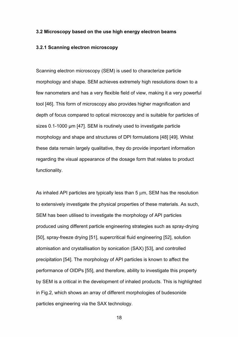
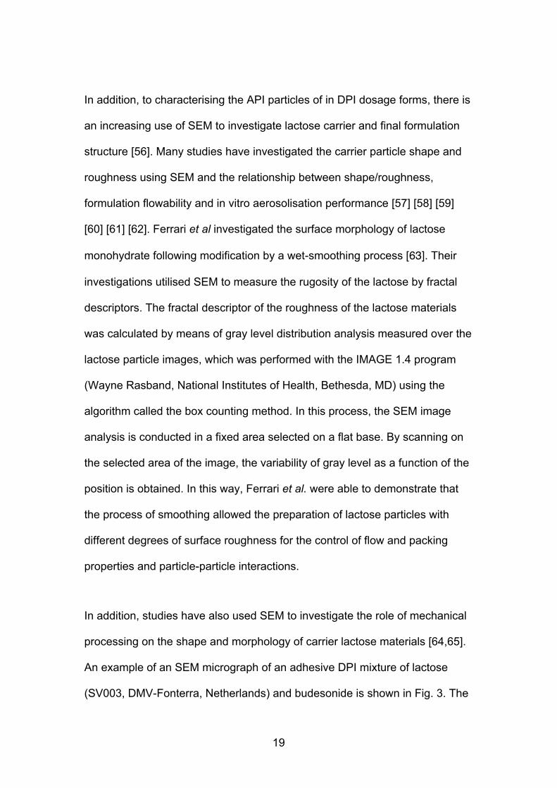
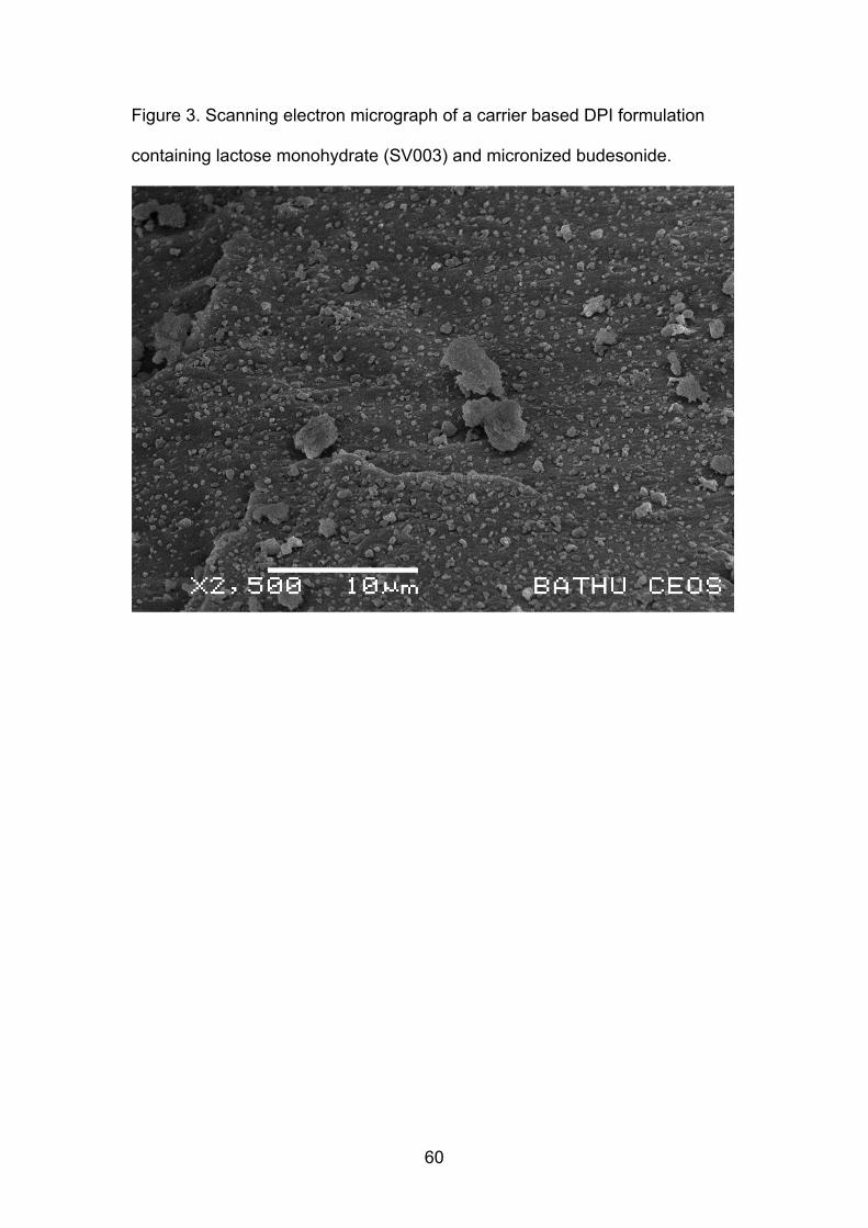
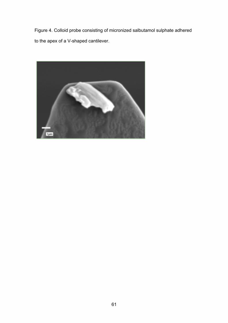
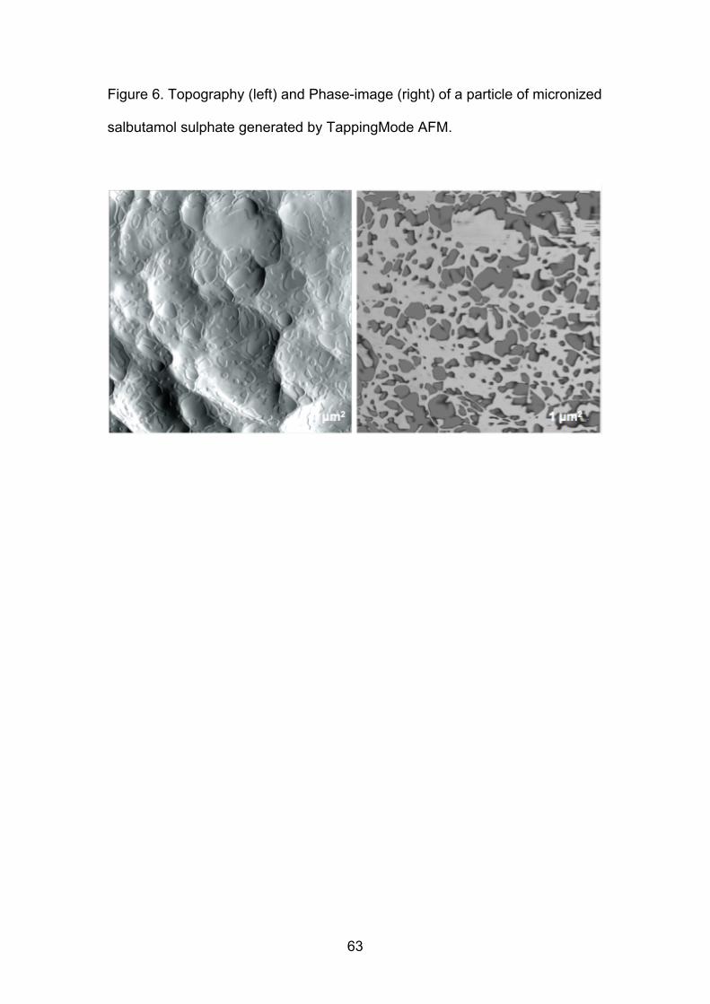
Figure 8 Bright-field reflectance (left) and Raman chemical image (right) of
images of particles of fluticasone and lactose collected on stage 3 of the NGI
following aerosolization of the Advair 50050 DPI
65