Embed Size (px)
Citation preview
For OSUWMC USE ONLY. To license, please contact the OSU Technology Commercialization Office at https://tco.osu.edu.
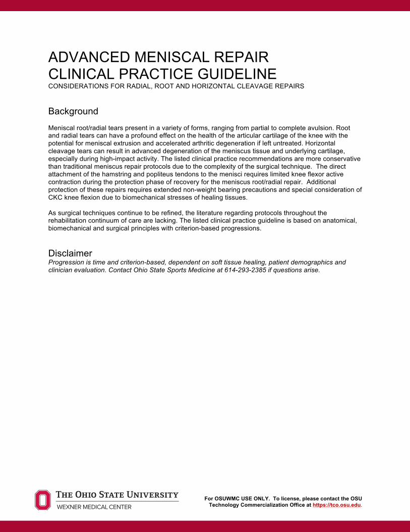
ADVANCED MENISCAL REPAIR CLINICAL PRACTICE GUIDELINE CONSIDERATIONS FOR RADIAL, ROOT AND HORIZONTAL CLEAVAGE REPAIRS Background Meniscal root/radial tears present in a variety of forms, ranging from partial to complete avulsion. Root and radial tears can have a profound effect on the health of the articular cartilage of the knee with the potential for meniscal extrusion and accelerated arthritic degeneration if left untreated. Horizontal cleavage tears can result in advanced degeneration of the meniscus tissue and underlying cartilage, especially during high-impact activity. The listed clinical practice recommendations are more conservative than traditional meniscus repair protocols due to the complexity of the surgical technique. The direct attachment of the hamstring and popliteus tendons to the menisci requires limited knee flexor active contraction during the protection phase of recovery for the meniscus root/radial repair. Additional protection of these repairs requires extended non-weight bearing precautions and special consideration of CKC knee flexion due to biomechanical stresses of healing tissues. As surgical techniques continue to be refined, the literature regarding protocols throughout the rehabilitation continuum of care are lacking. The listed clinical practice guideline is based on anatomical, biomechanical and surgical principles with criterion-based progressions. Disclaimer Progression is time and criterion-based, dependent on soft tissue healing, patient demographics and clinician evaluation. Contact Ohio State Sports Medicine at 614-293-2385 if questions arise.
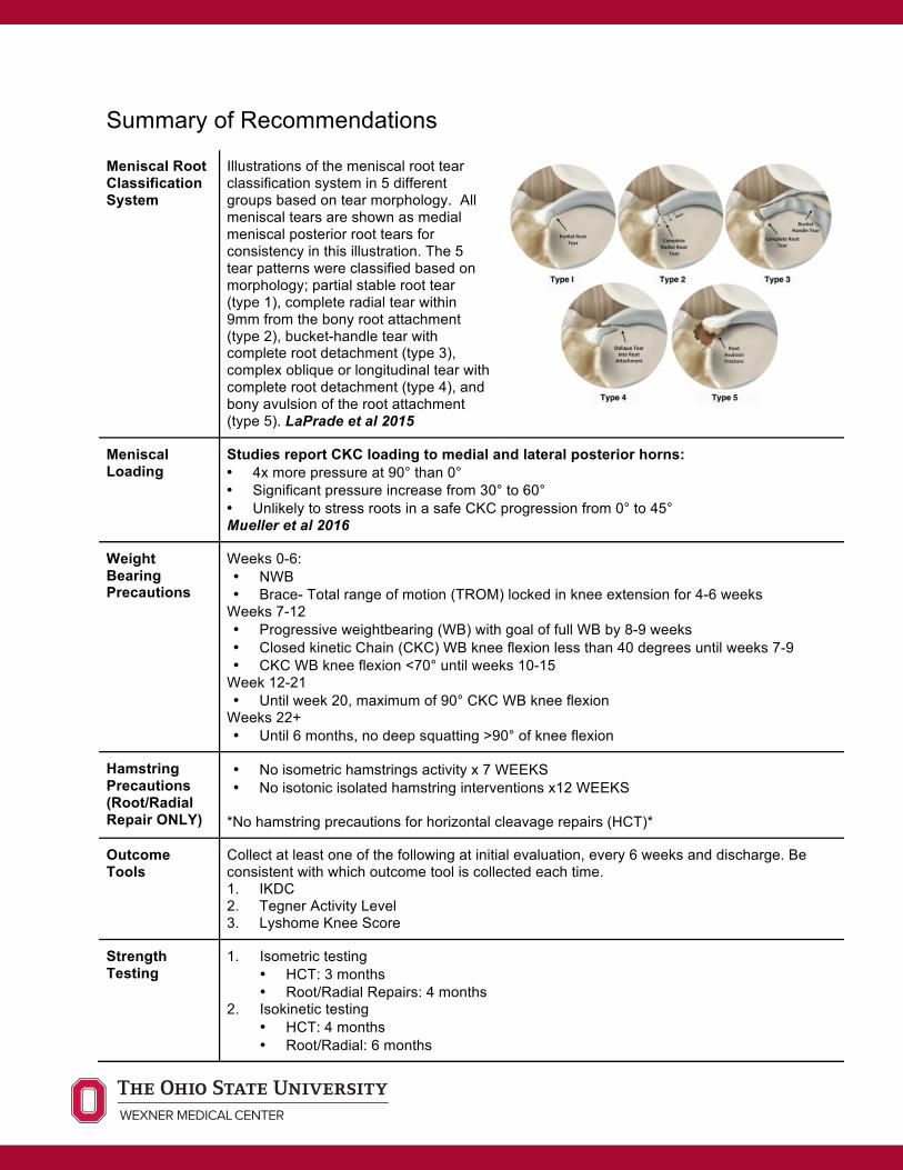
Summary of Recommendations Meniscal Root Classification System
Illustrations of the meniscal root tear classification system in 5 different groups based on tear morphology. All meniscal tears are shown as medial meniscal posterior root tears for consistency in this illustration. The 5 tear patterns were classified based on morphology; partial stable root tear (type 1), complete radial tear within 9mm from the bony root attachment (type 2), bucket-handle tear with complete root detachment (type 3), complex oblique or longitudinal tear with complete root detachment (type 4), and bony avulsion of the root attachment (type 5). LaPrade et al 2015
Meniscal Loading
Studies report CKC loading to medial and lateral posterior horns: • 4x more pressure at 90° than 0° • Significant pressure increase from 30° to 60° • Unlikely to stress roots in a safe CKC progression from 0° to 45° Mueller et al 2016
Weight Bearing Precautions
Weeks 0-6: • NWB • Brace- Total range of motion (TROM) locked in knee extension for 4-6 weeks
Weeks 7-12 • Progressive weightbearing (WB) with goal of full WB by 8-9 weeks • Closed kinetic Chain (CKC) WB knee flexion less than 40 degrees until weeks 7-9 • CKC WB knee flexion <70° until weeks 10-15
Week 12-21 • Until week 20, maximum of 90° CKC WB knee flexion
Weeks 22+ • Until 6 months, no deep squatting >90° of knee flexion
Hamstring Precautions (Root/Radial Repair ONLY)
• No isometric hamstrings activity x 7 WEEKS • No isotonic isolated hamstring interventions x12 WEEKS
*No hamstring precautions for horizontal cleavage repairs (HCT)*
Outcome Tools
Collect at least one of the following at initial evaluation, every 6 weeks and discharge. Be consistent with which outcome tool is collected each time. 1. IKDC 2. Tegner Activity Level 3. Lyshome Knee Score
Strength Testing
1. Isometric testing • HCT: 3 months • Root/Radial Repairs: 4 months
2. Isokinetic testing • HCT: 4 months • Root/Radial: 6 months
*Isokinetic testing only warranted if patient’s goal is to return to sport* • Many of these patients will be older with a goal of return to low-impact activity (ie- IADLS,
recreational walking, biking)
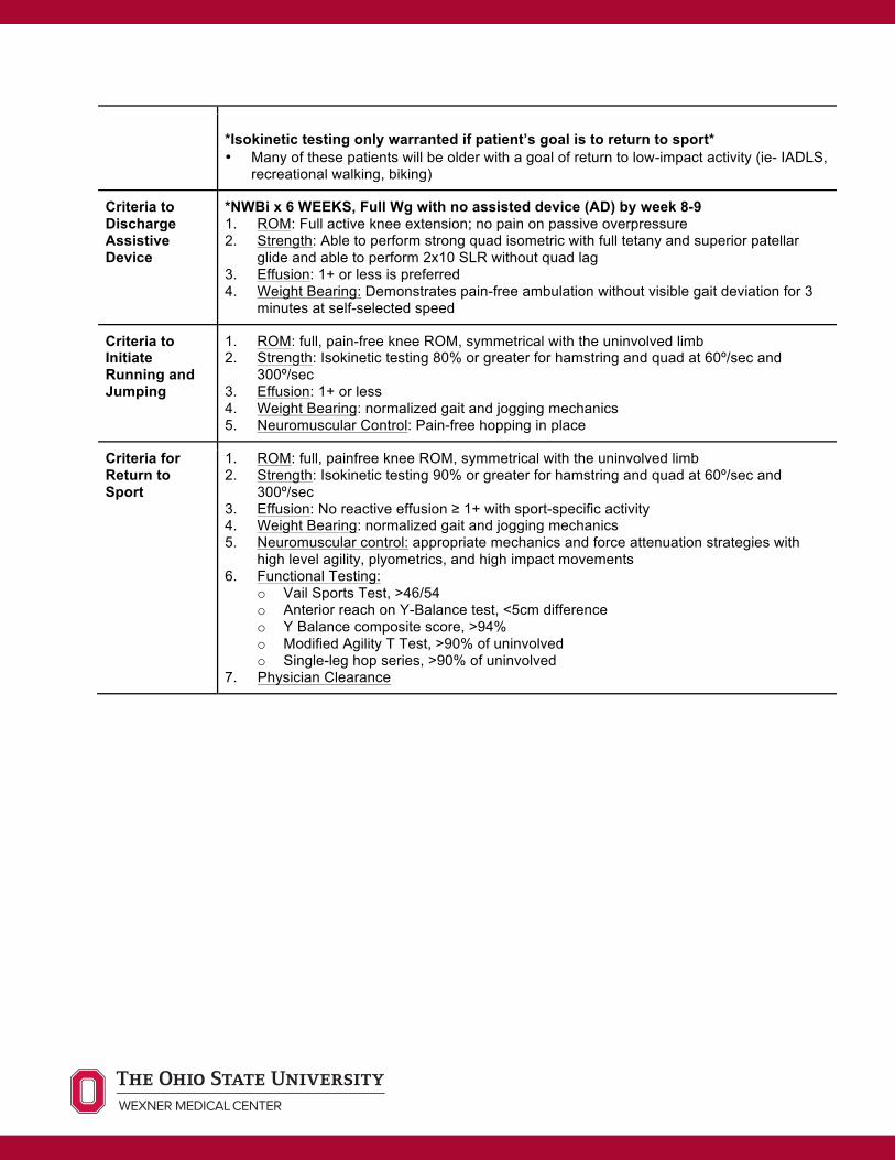
Criteria to Discharge Assistive Device
*NWBi x 6 WEEKS, Full Wg with no assisted device (AD) by week 8-9 1. ROM: Full active knee extension; no pain on passive overpressure 2. Strength: Able to perform strong quad isometric with full tetany and superior patellar
glide and able to perform 2x10 SLR without quad lag 3. Effusion: 1+ or less is preferred 4. Weight Bearing: Demonstrates pain-free ambulation without visible gait deviation for 3
minutes at self-selected speed
Criteria to Initiate Running and Jumping
1. ROM: full, pain-free knee ROM, symmetrical with the uninvolved limb 2. Strength: Isokinetic testing 80% or greater for hamstring and quad at 60º/sec and
300º/sec 3. Effusion: 1+ or less 4. Weight Bearing: normalized gait and jogging mechanics 5. Neuromuscular Control: Pain-free hopping in place
Criteria for Return to Sport
1. ROM: full, painfree knee ROM, symmetrical with the uninvolved limb 2. Strength: Isokinetic testing 90% or greater for hamstring and quad at 60º/sec and
300º/sec 3. Effusion: No reactive effusion ≥ 1+ with sport-specific activity 4. Weight Bearing: normalized gait and jogging mechanics 5. Neuromuscular control: appropriate mechanics and force attenuation strategies with
high level agility, plyometrics, and high impact movements 6. Functional Testing:
o Vail Sports Test, >46/54 o Anterior reach on Y-Balance test, <5cm difference o Y Balance composite score, >94% o Modified Agility T Test, >90% of uninvolved o Single-leg hop series, >90% of uninvolved
7. Physician Clearance
Phase 1 (Weeks 0-6): Protection, ROM, Muscle Activation
Goals 1. Protect surgical repair 2. Resolve joint effusion to 1+ or less 3. Restore full ROM
Precautions 1. NWB 2. PROM: 0-90 degrees for 2 weeks 3. Progress ROM as tolerated thereafter 4. No isolated hamstrings activation for radial/root repairs
Pain and Effusion
≥ 1+ (using Modified Stroke Test)
ROM Extension: Emphasis on achieving full knee extension immediately following surgery. If full extension is not achieved by 4 weeks, contact surgeon regarding ROM concerns. Flexion: • Root/Radial Only: Limited to PROM due to hamstring/popliteus attachment to
meniscus • Limited 0-90 degrees for first 2 weeks • After 2 weeks, gentle full PROM is allowed
Therapeutic Exercise
1. Emphasis on quad activation without gluteal co-contraction 2. Restore patellar mobility 3. Symmetrical ROM 4. Decrease effusion
Suggested Interventions
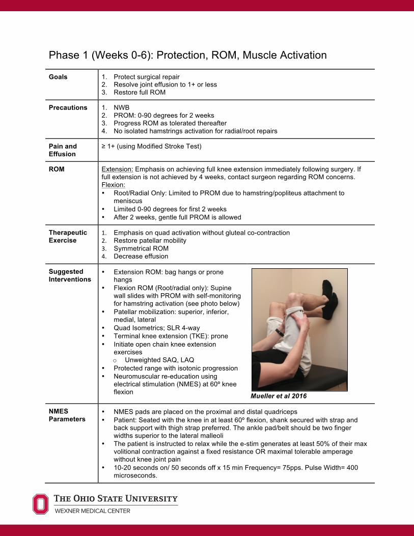
• Extension ROM: bag hangs or prone hangs
• Flexion ROM (Root/radial only): Supine wall slides with PROM with self-monitoring for hamstring activation (see photo below)
• Patellar mobilization: superior, inferior, medial, lateral
• Quad Isometrics; SLR 4-way • Terminal knee extension (TKE): prone • Initiate open chain knee extension
exercises o Unweighted SAQ, LAQ
• Protected range with isotonic progression • Neuromuscular re-education using
electrical stimulation (NMES) at 60º knee flexion
Mueller et al 2016
NMES Parameters
• NMES pads are placed on the proximal and distal quadriceps • Patient: Seated with the knee in at least 60º flexion, shank secured with strap and
back support with thigh strap preferred. The ankle pad/belt should be two finger widths superior to the lateral malleoli
• The patient is instructed to relax while the e-stim generates at least 50% of their max volitional contraction against a fixed resistance OR maximal tolerable amperage without knee joint pain
• 10-20 seconds on/ 50 seconds off x 15 min Frequency= 75pps. Pulse Width= 400 microseconds.
Criteria to Discharge Assistive Device
1. ROM: Full active knee extension; no pain on passive overpressure 2. Strength: Able to perform strong quad isometric with full tetany and superior patellar
glide and able to perform 2x10 SLR without quad lag 3. Effusion: 1+ or less is preferred (2+ acceptable if all other criteria are met) 4. Weight Bearing: Demonstrates pain-free ambulation without visible gait deviation
Criteria to Progress to Phase 2
1. ROM: Symmetrical to uninvolved limb 2. Strength: Quadriceps set with normal superior patellar translation, SLR x 10 seconds
without extensor lag 3. Effusion: 1+ or less with Modified stroke test
Phase 2 (7- 9 weeks): Weight Bearing Tolerance
Precautions 1. Gradual progression of WB to full WB with no AD by weeks 8-9 2. Knee flexion <40 degrees with CKC activity 3. CKC activity limited to WBstatus
Pain and Effusion
Cryotherapy/compression as needed for reactive effusion.
ROM • Monitor and progress knee ROM, patellar mobility, and LE flexibility • Begin more aggressive techniques to achieve/maintain full knee extension (i.e.
weighted bag hang) as needed • Continue bike for ROM and warmup • If full AROM knee extension is not achieved by 4 weeks, contact surgeon
regarding ROM concerns.
Suggested Interventions and timelines
• Multi-angle knee isometrics from 60-90⁰ for patients unable to tolerate high-intensity NMES
• Progress WB quadriceps and hamstring exercises with emphasis on proper LE mechanics
o Root/Radial Repairs: no isometric HS activity until 8 weeks, no isolated hamstring isotonics until 12 weeks
• Progress gluteal and lumbopelvic strength and stability • Progress single leg balance • NMES (see parameters in week 0-6)
Criteria to Progress to Phase 3
1. Achieve full WBing 2. Normalize gait pattern on flat ground 3. Maintain trace to zero joint effusion 4. Tolerate 25 minutes or standing and walking activity
Phase 3 (weeks 10-15): Endurance
Precautions Knee flexion <70 degrees with CKC activity
Pain and Effusion
Effusion may increase with increased activity, ≤1+ and/or non-reactive effusion for progression of endurance activities
ROM Full, symmetrical to contralateral limb, and pain free with overpressure
Therapeutic Exercise
Performance of the quadriceps, hamstrings and trunk dynamic stability with low load, high repetitions
Suggested Interventions
Therapeutic Exercise/Neuromuscular Re-education • Double-leg squats (<70 degrees) • Stationary lunges progressing to walking lunges • Step down- starting at 2” step and progressing to 6” Cardiovascular Conditioning: permitted week 12 • Stationary bike with resistance • Treadmill walking • Freestyle swimming (no fins until week 16)
Criteria to d/c NMES
<20% quadriceps deficit on isometric testing OR- If a Biodex machine in not available:
1. 10 SLR without quad lag 2. Normal gait
Criteria to Progress to Phase 3
90 second hold in single leg squat position at 45 degrees of knee flexion
Phase 4 (weeks 16-21): Strength
Precautions Until week 20, maximum of 90 degrees of knee flexion with CKC activity
Pain and Effusion
Effusion may increase with increased activity, ≤1+ and/or non-reactive effusion
ROM Full, symmetrical to contralateral limb, and pain-free with overpressure
Therapeutic Exercise
Performance of the quadriceps, hamstrings and trunk dynamic stability with high resistance, low repetitions
Suggested Interventions
Therapeutic Exercise/Neuromuscular Re-education • Single-leg squats • Single-leg deadlifts • Single-leg sit to stand • Multi-directional lunges
Criteria to Progress to Phase 5
• Quadriceps index >80% (isokinetic testing) • Anterior reach on Y balance test, <8-cm difference compared to uninvolved side
Phase 5 (weeks 22-Return to Sport (RTS)): Power, Running and Return to Sport
Precautions • No deep squatting for 6 months • Expected RTS by 9 months
Pain and Effusion Effusion may increase with increased activity, ≤1+ and/or non-reactive effusion for progression of plyometrics
ROM Full, symmetrical to contralateral limb, and pain-free with overpressure
Therapeutic Exercise
Performance of the quadriceps, hamstrings and trunk dynamic stability with sports-specific activity
Suggested Interventions
Therapeutic Exercise/Neuromuscular Re-education • Double and single leg jump training • Ladder drill agility • Lateral hops with and without resistance • Progressive cutting activities
Criteria to Progress to RTS
1. Pass Vail Sport Test, >46/54 2. Anterior reach on Y balance test, <5 cm difference 3. Y balance test composite scores, >94 4. Quadriceps index >90% (isokinetic) 5. Modified Agility T Test >90% of contralateral limb 6. Single-leg hop series >90% LSI
• SL hop for distance • Triple hop • Cross over hop • Timed 6m hop
Authors: Caroline Lewis, PT, DPT, SCS and Josh Pintar, PT, DPT, SCS References Arno S, Bell CP, Uquillas C, Borukhov I, Walker PS. “Tibiofemoral Contact Mechanics Following a Horizontal Cleavage Lesion in the Posterior Horn of the Medial Meniscus.” J Orthop Res. 2015; 33(4): 584-590.
Becker R, Wirz D, Wolf C, Göpfert B, Nebelung W, Friederich N. “Measurement of meniscofemoral contact pressure after repair of bucket-handle tears with biodegradable implants.” Arch Orthop Trauma Surg. 2005;125:254-260. http://dx.doi. org/10.1007/s00402-004-0739-5.
Bhatia S, LaPrade CM, Ellman MB, LaPrade RF. “Meniscal Root Tears: Significance, Diagnosis and Treatment.” Am J Sports Med. 2014; 42(12): 3016-3030.
Chung KS, Ha JK, Ra HJ, Kim JG. “A Menta-Analysis of Clinical and Radiographic Outcomes of Posterior Horn Medial Meniscus Root Repairs.” Knee Surg sports Traumatol Arthrosc. 2016; 24: 1455-1468.
Frizziero A, Ferrari R, Giannotti E, Ferroni C, Poli P, Masiero S. “The meniscus tear: state of the art of rehabilitation protocols related to surgical procedures.” Muscles, Ligaments and Tendons Journal. 2012;2(4):295-301.
Kurzweil PR, Lynch NM, Coleman S, Kearney B. “Repair of Horizontal Meniscus Tears: A Systematic Review.” Arthroscopy. 2014; 30(11): 1513-1519.
LaPrade CM, et al. “Meniscal Root Tears: A Classification System Based on Tear Morphology.” Am J Sports Med. 2015; 43(2): 363-369.
LaParade CM, et al. “Biomechanical Consequences of a Nonanatomic Posterior Medial Meniscus Root Repair.” Am J Sports Med. 2015; 43(4): 912-920.
Lavender CD, Hanzlik SR, Caldwell PE, Pearson SE. “Transosseous Medial Meniscal Root Repair Using a Modified Mason-Allen Suture Configuration.” Arthroscopy Techniques. 2015; 4(6): e781-e784.
Mueller BT, Moulton SG, O'Brien L, LaPrade RF. “Rehabilitation Following Meniscal Root Repair: A Clinical Commentary.” J Orthop Sports Phys Ther. 2016; Feb;46(2):104-13.
Chimera NJ, Warren M. Use of clinical movement screening tests to predict injury in sport. World Journal of Orthopedics. 2016; 7(4):202-217
Myer GD, Schmitt LC, Brent JL, et al. Utilization of Modified NFL Combine Testing to Identify Functional Deficits in Athletes Following ACL Reconstruction. The Journal of orthopedic and sports physical therapy. 2011:41(6):377-387
Garrison JC, Shanley E, Thigpen C, et al. The reliability of the Vail Sport Test as a measure of physical performance following anterior cruciate ligament recontruction. Int J Sports Phys Ther. 2012; 7(1):20-30.
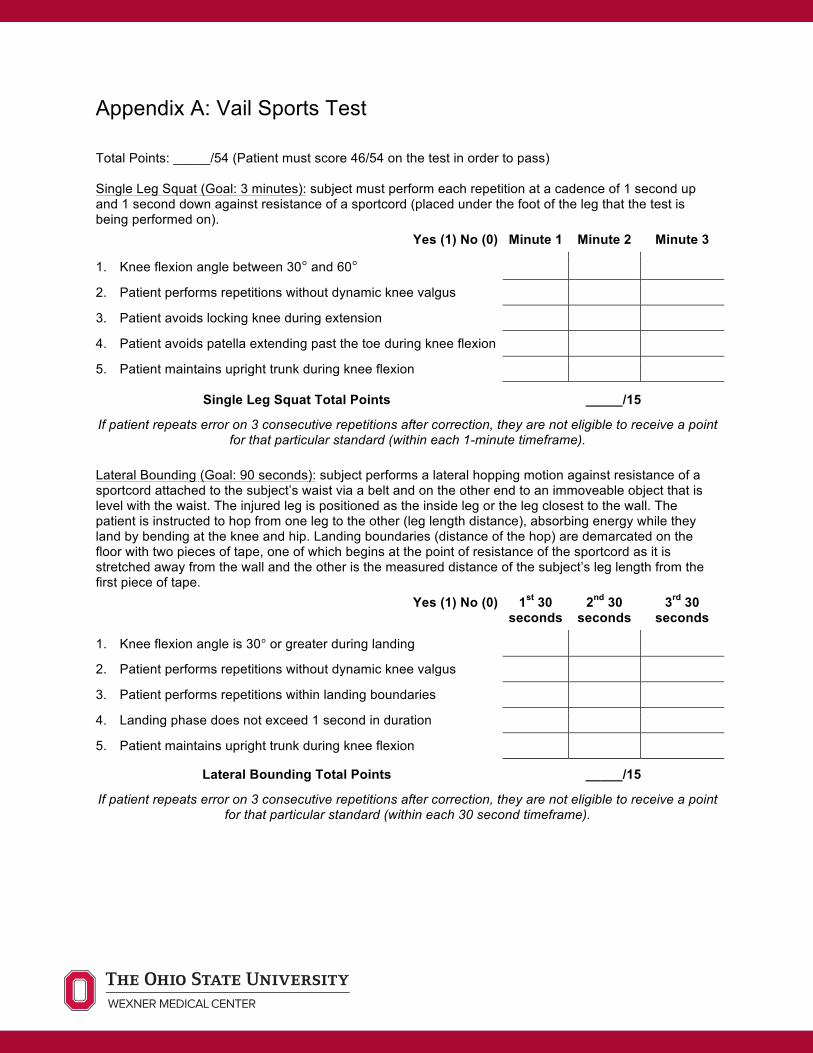
Appendix A: Vail Sports Test Total Points: _____/54 (Patient must score 46/54 on the test in order to pass) Single Leg Squat (Goal: 3 minutes): subject must perform each repetition at a cadence of 1 second up and 1 second down against resistance of a sportcord (placed under the foot of the leg that the test is being performed on).
Yes (1) No (0) Minute 1 Minute 2 Minute 3
1. Knee flexion angle between 30° and 60°
2. Patient performs repetitions without dynamic knee valgus
3. Patient avoids locking knee during extension
4. Patient avoids patella extending past the toe during knee flexion
5. Patient maintains upright trunk during knee flexion
Single Leg Squat Total Points _____/15
If patient repeats error on 3 consecutive repetitions after correction, they are not eligible to receive a point for that particular standard (within each 1-minute timeframe).
Lateral Bounding (Goal: 90 seconds): subject performs a lateral hopping motion against resistance of a sportcord attached to the subject’s waist via a belt and on the other end to an immoveable object that is level with the waist. The injured leg is positioned as the inside leg or the leg closest to the wall. The patient is instructed to hop from one leg to the other (leg length distance), absorbing energy while they land by bending at the knee and hip. Landing boundaries (distance of the hop) are demarcated on the floor with two pieces of tape, one of which begins at the point of resistance of the sportcord as it is stretched away from the wall and the other is the measured distance of the subject’s leg length from the first piece of tape.
Yes (1) No (0) 1st 30 seconds
2nd 30 seconds
3rd 30 seconds
1. Knee flexion angle is 30° or greater during landing
2. Patient performs repetitions without dynamic knee valgus
3. Patient performs repetitions within landing boundaries
4. Landing phase does not exceed 1 second in duration
5. Patient maintains upright trunk during knee flexion
Lateral Bounding Total Points _____/15
If patient repeats error on 3 consecutive repetitions after correction, they are not eligible to receive a point for that particular standard (within each 30 second timeframe).
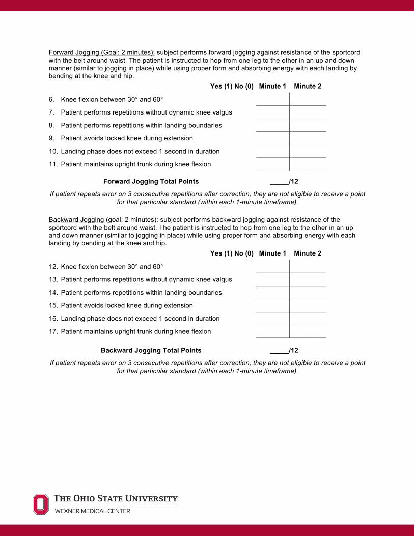
Forward Jogging (Goal: 2 minutes): subject performs forward jogging against resistance of the sportcord with the belt around waist. The patient is instructed to hop from one leg to the other in an up and down manner (similar to jogging in place) while using proper form and absorbing energy with each landing by bending at the knee and hip.
Yes (1) No (0) Minute 1 Minute 2
6. Knee flexion between 30° and 60°
7. Patient performs repetitions without dynamic knee valgus
8. Patient performs repetitions within landing boundaries
9. Patient avoids locked knee during extension
10. Landing phase does not exceed 1 second in duration
11. Patient maintains upright trunk during knee flexion
Forward Jogging Total Points _____/12
If patient repeats error on 3 consecutive repetitions after correction, they are not eligible to receive a point for that particular standard (within each 1-minute timeframe).
Backward Jogging (goal: 2 minutes): subject performs backward jogging against resistance of the sportcord with the belt around waist. The patient is instructed to hop from one leg to the other in an up and down manner (similar to jogging in place) while using proper form and absorbing energy with each landing by bending at the knee and hip.
Yes (1) No (0) Minute 1 Minute 2
12. Knee flexion between 30° and 60°
13. Patient performs repetitions without dynamic knee valgus
14. Patient performs repetitions within landing boundaries
15. Patient avoids locked knee during extension
16. Landing phase does not exceed 1 second in duration
17. Patient maintains upright trunk during knee flexion
Backward Jogging Total Points _____/12
If patient repeats error on 3 consecutive repetitions after correction, they are not eligible to receive a point for that particular standard (within each 1-minute timeframe).
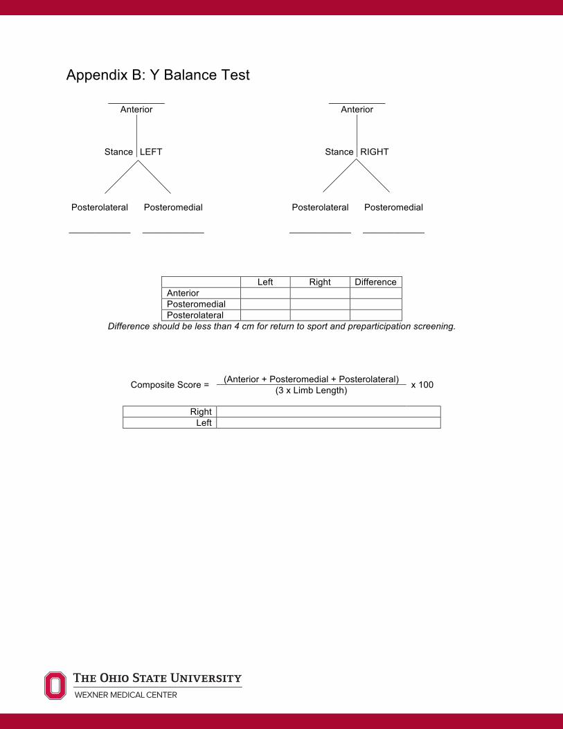
Appendix B: Y Balance Test
___________ Anterior
___________ Anterior
Stance LEFT Stance RIGHT
Posterolateral
____________
Posteromedial
____________
Posterolateral
____________
Posteromedial
____________
Left Right Difference Anterior Posteromedial Posterolateral
Difference should be less than 4 cm for return to sport and preparticipation screening.
Composite Score =
(Anterior + Posteromedial + Posterolateral) x 100 (3 x Limb Length)
Right
Left
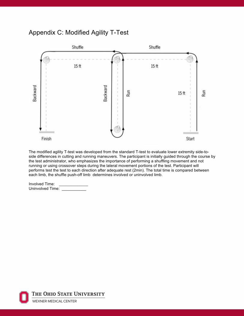
Appendix C: Modified Agility T-Test
The modified agility T-test was developed from the standard T-test to evaluate lower extremity side-to-side differences in cutting and running maneuvers. The participant is initially guided through the course by the test administrator, who emphasizes the importance of performing a shuffling movement and not running or using crossover steps during the lateral movement portions of the test. Participant will performs test the test to each direction after adequate rest (2min). The total time is compared between each limb, the shuffle push-off limb determines involved or uninvolved limb. Involved Time: _____________ Uninvolved Time: ___________
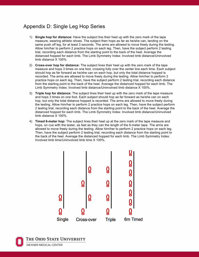
Appendix D: Single Leg Hop Series
1) Single hop for distance: Have the subject line their heel up with the zero mark of the tape measure, wearing athletic shoes. The subject then hops as far as he/she can, landing on the same push off leg, for at least 3 seconds. The arms are allowed to move freely during the testing. Allow him/her to perform 2 practice hops on each leg. Then, have the subject perform 2 testing trial, recording each distance from the starting point to the back of the heel. Average the distanced hopped for each limb. The Limb Symmetry Index: Involved limb distance/Uninvolved limb distance X 100%.
2) Cross-over hop for distance: The subject lines their heel up with the zero mark of the tape measure and hops 3 times on one foot, crossing fully over the center line each time. Each subject should hop as far forward as he/she can on each hop, but only the total distance hopped is recorded. The arms are allowed to move freely during the testing. Allow him/her to perform 2 practice hops on each leg. Then, have the subject perform 2 testing trial, recording each distance from the starting point to the back of the heel. Average the distanced hopped for each limb. The Limb Symmetry Index: Involved limb distance/Uninvolved limb distance X 100%.
3) Triple hop for distance: The subject lines their heel up with the zero mark of the tape measure and hops 3 times on one foot. Each subject should hop as far forward as he/she can on each hop, but only the total distance hopped is recorded. The arms are allowed to move freely during the testing. Allow him/her to perform 2 practice hops on each leg. Then, have the subject perform 2 testing trial, recording each distance from the starting point to the back of the heel. Average the distanced hopped for each limb. The Limb Symmetry Index: Involved limb distance/Uninvolved limb distance X 100%.
4) Timed 6-meter hop: The subject lines their heel up at the zero mark of the tape measure and hops, on cue with the tester, as fast as they can the length of the 6-meter tape. The arms are allowed to move freely during the testing. Allow him/her to perform 2 practice hops on each leg. Then, have the subject perform 2 testing trial, recording each distance from the starting point to the back of the heel. Average the distanced hopped for each limb. The Limb Symmetry Index: Involved limb time/Uninvolved limb time X 100%.