Embed Size (px)
Citation preview
ADVANCED IMAGING IN PORTOSYSTEMIC SHUNT DIAGNOSIS Nathan C. Nelson, DVM, MS, DACVR
Department of Small Animal Clinical Sciences, Michigan State University, East Lansing, MI
Key Points: • Various advanced imaging modalities may be used to diagnose extrahepatic
portosystemic shunts (PSS), including ultrasound, transsplenic scintigraphy, computed tomography (CT) and magnetic resonance imaging (MRI), each with unique advantages and disadvantages.
• CT angiography provides the best detail of overall shunt anatomy and is also helpful in rechecking patients with persistent clinical signs or biochemical abnormalities.
Ultrasound: Ultrasound examinations are readily available and may be performed without anesthesia,
with a sensitivity and specificity of 92% and 98% for shunt diagnosis, respectively, for skilled users.1 Examinations may be time-consuming, and typically require patient sedation and fasting. Use of color Doppler techniques allows characterization of blood flow direction and may alert to coexisting pathologies such as portal hypertension or acquired portostystemic shunts. Even if the shunting vessel itself is not identified, a combination of microhepatia, renomegaly, and cystic calculi has high positive predictive value (100%) for the presence of a congenital PSS.1 Transsplenic scintigraphy:
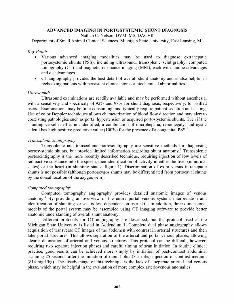
Transsplenic and transcolonic portoscintigraphy are sensitive methods for diagnosing portosystemic shunts, but provide limited information regarding shunt anatomy.2 Transsplenic portoscintigraphy is the more recently described technique, requiring injection of low levels of radioactive substance into the spleen, then identification of activity in either the liver (in normal states) or the heart (in shunting states; figure 1). Discrimination of extra versus intrahepatic shunts is not possible (although portoazygos shunts may be differentiated from portocaval shunts by the dorsal location of the azygos vein). Computed tomography:
Computed tomography angiography provides detailed anatomic images of venous anatomy.3 By providing an overview of the entire portal venous system, interpretation and identification of shunting vessels is less dependent on user skill. In addition, three-dimensional models of the portal system may be assembled using CT imaging software to provide better anatomic understanding of overall shunt anatomy.
Different protocols for CT angiography are described, but the protocol used at the Michigan State University is listed in Addendum 1. Complete dual phase angiography allows acquisition of transverse CT images of the abdomen with contrast in arterial structures and then later portal structures. This allows separation of the arterial and portal venous phases, allowing clearer delineation of arterial and venous structures. This protocol can be difficult, however, requiring two separate injection phases and careful timing of scan initiation. In routine clinical practice, good results can be achieved more simply by initiation of post-contrast abdominal scanning 25 seconds after the initiation of rapid bolus (3-5 ml/s) injection of contrast medium (814 mg I/kg). The disadvantage of this technique is the lack of a separate arterial and venous phase, which may be helpful in the evaluation of more complex arteriovenous anomalies.
302
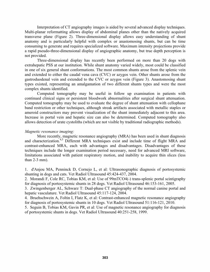
Interpretation of CT angiography images is aided by several advanced display techniques. Multi-planar reformatting allows display of abdominal planes other than the natively acquired transverse plane (Figure 2). Three-dimensional display allows easy understanding of shunt anatomy and is particularly helpful with complex or anastomosing shunts, but can be time consuming to generate and requires specialized software. Maximum intensity projections provide a rapid pseudo-three-dimensional display of angiographic anatomy, but true depth perception is not provided.
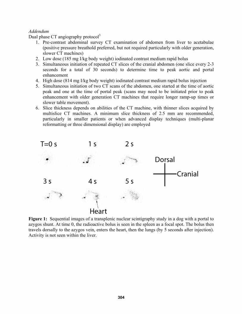
Three-dimensional display has recently been performed on more than 20 dogs with extrahepatic PSS at our institution. While shunt anatomy varied widely, most could be classified in one of six general shunt conformations. The most common shunts arose from the splenic vein and extended to either the caudal vena cava (CVC) or azygos vein. Other shunts arose from the gastroduodenal vein and extended to the CVC or azygos vein (Figure 3). Anastomosing shunt types existed, representing an amalgamation of two different shunts types and were the most complex shunts identified.
Computed tomography may be useful in follow up examination in patients with continued clinical signs or persistent bloodwork abnormalities after surgical shunt attenuation. Computed tomography may be used to evaluate the degree of shunt attenuation with cellophane band restriction or other techniques, although streak artifacts associated with metallic staples or ameroid constructiors may prevent visualization of the shunt immediately adjacent to this area. Increase in portal vein and hepatic size can also be determined. Computed tomography also allows detection of urate cystoliths (which are not visible by traditional radiographic methods). Magnetic resonance imaging:
More recently, magnetic resonance angiography (MRA) has been used in shunt diagnosis and characterization.4,5 Different MRA techniques exist and include time of flight MRA and contrast-enhanced MRA, each with advantages and disadvantages. Disadvantages of these techniques include the longer examination period necessary, need for advanced MRI software, limitations associated with patient respiratory motion, and inability to acquire thin slices (less than 2-3 mm). 1. d'Anjou MA, Penninck D, Cornejo L, et al: Ultrasonographic diagnosis of portosystemic shunting in dogs and cats. Vet Radiol Ultrasound 45:424-437, 2004. 2. Morandi F, Cole RC, Tobias KM, et al: Use of 99mTCO4(-) trans-splenic portal scintigraphy for diagnosis of portosystemic shunts in 28 dogs. Vet Radiol Ultrasound 46:153-161, 2005. 3. Zwingenberger AL, Schwarz T: Dual-phase CT angiography of the normal canine portal and hepatic vasculature. Vet Radiol Ultrasound 45:117-124, 2004. 4. Bruehschwein A, Foltin I, Flatz K, et al: Contrast-enhanced magnetic resonance angiography for diagnosis of portosystemic shunts in 10 dogs. Vet Radiol Ultrasound 51:116-121, 2010. 5. Seguin B, Tobias KM, Gavin PR, et al: Use of magnetic resonance angiography for diagnosis of portosystemic shunts in dogs. Vet Radiol Ultrasound 40:251-258, 1999.
303
Addendum Dual phase CT angiography protocol3
1. Pre-contrast abdominal survey CT examination of abdomen from liver to acetabulae (positive pressure breathold preferred, but not required particularly with older generation, slower CT machines)
2. Low dose (185 mg I/kg body weight) iodinated contrast medium rapid bolus 3. Simultaneous initiation of repeated CT slices of the cranial abdomen (one slice every 2-3
seconds for a total of 30 seconds) to determine time to peak aortic and portal enhancement
4. High dose (814 mg I/kg body weight) iodinated contrast medium rapid bolus injection 5. Simultaneous initiation of two CT scans of the abdomen, one started at the time of aortic
peak and one at the time of portal peak (scans may need to be initiated prior to peak enhancement with older generation CT machines that require longer ramp-up times or slower table movement).
6. Slice thickness depends on abilities of the CT machine, with thinner slices acquired by multislice CT machines. A minimum slice thickness of 2.5 mm are recommended, particularly in smaller patients or when advanced display techniques (multi-planar reformatting or three dimensional display) are employed
Figure 1: Sequential images of a transplenic nuclear scintigraphy study in a dog with a portal to azygos shunt. At time 0, the radioactive bolus is seen in the spleen as a focal spot. The bolus then travels dorsally to the azygos vein, enters the heart, then the lungs (by 5 seconds after injection). Activity is not seen within the liver.
304
Figure 2: Transverse (left) and dorsal plane (right) reformatted images of a dog with a portal to caval shunt. The shunt (SH) enters the caudal vena (CVC) near the level of the diaphragm, and its relation to the portal vein is evident on dorsal plane images. Note that the tortuous nature of the shunt is not well represented in the dorsal plane provided, as it enters the dorsal plane multiple times.
Figure 3: Three dimensional image of an extrahepatic PSS as viewed from the ventral aspect, created using a CT angiogram. The shunt extends from the gastroduodenal vein to the caudal vena cava (PV=portal vein, CVC=caudal vena cava, GD=gastroduodenal vein, SH=shunt).
305