Embed Size (px)
Citation preview

Cailan L. Cheng M.D.Ying Tang M.D.Zhenda D. Zheng M.M.Xun Liu M.D.Zengchun C Ye M.D.Cheng Wang M.D.Tanqi Q Lou M.D.
Department of Nephrology, �e �ird A�li-ated Hospital of SunYat-Sen University, Guangzhou, Guangdong, China
Advanced glycation end-products activate the renin-angiotensin system through the RAGE/PI3-K signaling pathway in podocytes
AbstractPurpose: �e purpose of this study was to investigate the e�ects of advanced glycation end-products (AGEs) on the components of the renin-angiotensin system (RAS) in podo-cytes and to understand the mechanism of these e�ects.
Methods: Immortalized mouse podocytes were exposed to various concentrations of AGEs for di�erent time intervals. �e expression levels of angiotensinogen (AGT), angio-tensin II type 1 and 2 receptors (AT1R and AT2R) and renin were examined by real-time PCR and western blot; the receptor for AGEs (RAGE) and both Akt and phosphorylated Akt were examined by western blot; levels of angiotensin II (Ang II) were assayed by ELISA, and the activity of angiotensin-converting enzyme (ACE) was evaluated by meas-uring the production of hippuric acid in vitro.
Results: Treatment with AGEs resulted in signi�cant increases in the expression of AGT (62%, P=0.002) and AT1R (59%, P=0.01). Moreover, Ang II levels increased signi�cantly in both cell lysates (70%, P=0.018) and conditioned media (65%, P=0.01). ACE activity was also signi�cantly higher in cell lysates (68% , P= 0.035) and conditioned media (65%, P=0.023). �ere were no changes in renin or AT2R expression (P>0.05). AGEs did in-crease the expression of RAGE by 50% (P=0.012) and the phosphorylation of Akt by 100% (P=0.001). When podocytes were pretreated with anti-RAGE antibody (50 μg/ml) or the phosphoinositide 3-kinase (PI3-K) inhibitor, LY294002 (10 μM), the AGEs-induced increases in AGT and AT1R expression were reduced. Likewise, Ang II levels and ACE activity decreased signi�cantly.
Conclusion: AGEs activate the RAS in podocytes through the RAGE-PI3-K/Akt-dependent pathway and lead to an increase in podocyte apoptosis.
ORIGINAL RESEARCH
© 2012 CIM Clin Invest Med • Vol 35, no 5, October 2012 E282
Correspondence to:Lou Tan QiDepartment of Nephrology, �e �ird A�liated Hospital of SunYat-Sen University, Guangzhou, Guangdong, ChinaTel:13609760315E-mail: [email protected]
Manuscript submitted 23rd February, 2012. Manuscript accepted 7th August, 2012.
Clin Invest Med 2012; 35 (5): E282-E293.
Diabetic nephropathy (DN) is a serious complication of diabe-tes and, ultimately, it progresses to glomerular sclerosis and end-stage renal disease (ESRD). Current evidence indicates that AGEs increase at an accelerated rate in diabetes and are associated with the development of severe diabetic complica-tions1. Since the glomerular capillary wall functions as an e�-cient and selective barrier, podocytes are critically involved in maintaining the glomerular �ltration barrier; damage to podo-cytes can lead to proteinuria and initiate glomerulosclerosis, which can result in the progressive loss of kidney function[2].
Accumulating evidence supports the idea that the intrare-nal RAS plays an important role in progressive kidney disease; suppression of the RAS with either ACE inhibitors or AT1R antagonists reduces proteinuria and retards the progression of DN[3,4]. Singh et al. found an increased Ang II and renin con-tent in glomerular extracts obtained from streptozotocin-induced diabetic rats[5]. Local RAS is activated in mesangial cells[6] and proximal tubular cells[7] by high glucose and in podocytes by mechanical stress[8]. However, the e�ects of AGEs on the RAS in podocytes are still uncertain and a clear understanding of the mechanism of how AGEs activate the RAS in podocytes is lacking.
Phosphoinositide 3-kinase (PI3-K) and its downstream mediator, Akt, play a central role in a diverse range of cellular responses including cell growth, survival, proteolysis and ma-lignant transformation[9.10]. While recent studies have shown that the activation of PI3-K may play a part in diabetic complications[11-13], its role in diabetic nephropathy, espe-cially in the context of activated RAS, is relatively unstudied.
�e purpose of this investigation was to characterize the e�ects of AGEs on the components of the RAS. How AGEs activated the RAS was examined with anti-RAGE antibody and PI3-K inhibitor to identify the underlying pathway in-volved.
Methods
Podocytes culture
A conditionally-immortalized mouse podocyte cell line was generously provided by Dr. Peter Mundel (Harvard Medical School, Boston, MA, USA). Cells were harvested as previously described[14]. Cells were cultured in a non-permissive condi-tion in 10% fetal bull serum with 10 U/ml INF-γ in RPMI 1640 media at 33°C. When podocytes were 60-80% con�uent, they were transferred to 37°C and incubated for 7-10 days to allow di�erentiation. Passages 8–14 were used for all experi-ments. Before treatment, di�erentiated podocytes were cul-
tured in media containing 1% fetal bull serum for 24 hours. LY294002 was purchased from Sigma-Aldrich.
MTT assay for cell proliferation
Podocyte viability was assessed using the MTT (3-[4,5] di-methylthiazol- 2,5-diphenyltetrazolium bromide) assay (Promega, Madison, WI, USA) according to the manufac-turer's instructions. �is assay is a nonradioactive cell prolifera-tion assay that identi�es living cells and is based on the cellular conversion of a tetrazolium salt into a formazan product; a chromophore that can be quanti�ed by spectrophotometry. Brie�y, podocytes were plated in a 96-well plate and were al-lowed to adhere overnight; then, an appropriate concentration of AGEs was added and incubated with the podocytes for 24 hours. A labeling dye included in the kit was added 4 hours prior to the end point. �e stop solution was then added to each well to stop the reaction and solubilize the cells and the absorbance was read at 490 nm using a SPECTRAmax (Mo-lecular Devices, Sunnyvale, CA, USA).
Lactate dehydrogenase (LDH) assay
Con�uent podocytes were cultured under control and experi-mental conditions. Supernatants were collected, centrifuged for 10 min at 2000 x g and were assessed for LDH release using a LDH cytotoxicity kit according to the manufacturer’s in-structions (Nanjing Jiancheng Bioengineering Institute, Nan-jing City, China). �e cytotoxicity of the control and experi-mental samples was expressed as the percentage of LDH release compared with the total intracellular LDH content. �e latter was determined by lysis of representative cell monolayers using 2% Triton X-100 (v/v).
Angiotensin-converting enzyme (ACE) activity
ACE activity in the media and cell lysates was determined as previously described[15] using a commercial kit (Navy General Hospital, Beijing, China). Hippuryl-histidyl-leucine is hydro-lyzed to hippuric acid and histidyl-leucine in the presence of exogenous ACE. Hippuric acid was extracted with ethyl ace-tate. �e extracted liquor was evaporated to dryness at 130°C for 15 min and was then dissolved in 1 M NaCl. �e mixture was monitored by UV spectrophotometry at 228 nm. One unit of ACE activity was de�ned as the amount of enzyme required to release 1 nmol of hippuric acid per minute per milliliter of sample.
Cheng et al. AGEs and the renin-angiotensin system in podocytes
© 2012 CIM Clin Invest Med • Vol 35, no 5, October 2012 E283

Measurement of Ang II
Ang II levels were determined in cell lysates and media. Podo-cytes were harvested in 1% FBS medium for 24 hours and then cultured in control and experimental conditions. �e cells were washed with ice-cold PBS, scraped in extraction bu�er and homogenized. Cell lysates and media were centrifuged at 12,000 x g for 10 minutes at 4°C and supernatants were col-lected. Ang II levels in the cell lysates and the media were de-termined using a commercial enzyme-linked immunosorbent assay (ELISA) kit (RayBiotech, Norcross, GA, USA) according to the manufacturer’s instructions.
Real-time polymerase chain reaction (PCR)
Total RNA was extracted from podocytes using Trizol reagent (Invitrogen, Carlsbad CA, USA) according to the manufac-turer’s instructions. Total RNA (500 ng) from each group was reverse transcribed using the reverse transcriptase (RT) pro-vided in a SYBRPremix Ex Taq kit (Perfect Real Time,TaKaRa, Otsu, Shiga, Japan). Reactions were carried out at 37 °C for 15 min and then 85 °C for 5 s. �e primers used in the experi-ments are listed in Table 1. PCR was performed by using an ABI Prism 7000 sequence detection system (AppliedBiosys-tems, Foster City, CA, USA) and a SYBRPremix Ex Taq Per-fect Real Time kit,
Western blot
Podocytes were washed twice with cold PBS and scraped with lysis bu�er containing 20 mM Tris-HCl (pH 7.5), 150 mM NaCl, 1 nM Na2EDTA, 1 mM EGTA, 1% Triton, 2.5 mM sodium pyrophosphate, 1 mM β-glycerophosphate, 1 mM
Na3VO4, 1 μg/ml leupeptin (Cell Signaling Technology, Bev-erly, MA, USA) and protease and phosphatase inhibitor cock-tail tablets (Roche Diagnostics, Mannheim, Germany). Pro-tein concentrations were determined with the Bradford reac-tion. A total of 50 μg of boiled extracts was loaded on 8% so-dium dodecylsulfate polyacrylamide gel electrophoresis (SDS-PAGE) gels and transferred to polyvinylidene �uoride mem-branes (Bio-Rad Laboratories, Hercules, CA, USA). �e membranes were blocked in 5% fat-free milk before incubation with antibody. �e following primary antibodies were used: rabbit anti-AGT (1:2000, Epitomics, Birlingame, CA, USA), rabbit anti-AT1R, rabbit anti-AT2R and rabbit anti-renin (1:200, Santa Cruz Biotechnology, Santa Cruz, CA, USA). A total of 20 μg of boiled extracts was loaded on 8% SDS-PAGE gels and incubated with rabbit anti-RAGE (1:2000, Sigma-Aldrich, St. Louis, MO, USA ), rabbit anti-phospho-Akt and mouse anti-Akt (1:2000, Cell Signal Technology, Danvers, MA, USA). Blots were then incubated with horseradish peroxidase-conjugated secondary antibodies (Santa Cruz Bio-technology Inc); bands were detected using the ECL chemilu-minescence system (Millipore, Billerica, MA, USA).
Measuring of apoptosis
Apoptosis of podocytes was measured at 24 hours by �ow cy-tometry (Annexin-FITC apoptosis diction Kit, BD Biosci-ences Pharmingen, city, country). Cells were washed twice with cold PBS, were scraped into 1×binding bu�er to make a cell suspension at a concentration of 1×10⁶ cells/ml and then 100 μL of the cell suspension were transferred to a 5 ml tube. With 5 μL of Annexin V-FITC and 5 μL of propidium iodide, the cells were gently mixed and incubated for 15 min at room tem-
Cheng et al. AGEs and the renin-angiotensin system in podocytes
© 2012 CIM Clin Invest Med • Vol 35, no 5, October 2012 E284
TABLE 1. Sequences o of primers
Se equence (5’→3’)
AGTSense TGACCCAGTTCTTGCCACTGAG
AGTAntisense ACACCGAGATGCTGTTGTCCAC
reninSense CTCCTGGCAGATCACGATGAAG
reninAntisense GGAGCTCGTAGGAGCCGAGATA
AT1RSense GGTGGCTGAAGCCAGTACCA
AT1RAntisense TGAGTTGGTCTCAGACACTGTTCAA
AT2RSense GTGCATGCGGGAGCTGAGTA
AT2RAntisense ATTGGTGCCAGTTGCGTTGA
AGT: angiotensinogen n; AT1R: angiotensin II type 1 receptor; AT AT2R: angiotensin II type 2 receptor

perature in the dark and then 400μL 1×binding bu�er were added to each tube. Samples were analyzed by �ow cytometry (BD FACSCalibur, San Jose, CA, USA) within 1 h, with exci-tation at 488 nm and emission collected at 525 nm (FITC-conjugated AV-labeled cells) and 620 nm (PIlabeled cells). Sin-gle labeling was used to gate and control for bleed-through. �e cell population was characterized according to whether it was labeled with neither AV nor PI (viable), PI alone (ne-crotic), AV alone (early apoptotic), or both PI and AV (late apoptotic). �e apoptosis rate of podocytes were summarized as early apoptotic plus late apoptotic.
Statistical analysis
�e results are presented as the mean ± standard deviation. �e statistical signi�cance was assessed by a nonparametric Krus-kal–Wallis ANOVA analysis or a Student’s t-test. P values <0.05 were considered signi�cant.
Results
AGEs did not a�ect podocyte proliferation and metabolic activity
Cell proliferation and viability was assessed by the MTT assay and LDH release. To determine the optimal concentration of AGEs to be applied to podocytes, the di�erentiated podocytes were incubated with concentrations of AGEs ranging from 0 to 160 μg/ml for 24 hours. Cell proliferation and viability were
una�ected at concentrations of AGEs of 20-80 μg/ml but higher concentrations (160 μg/ml) inhibited cell proliferation (Figure 1A). Measurements of LDH release con�rmed that 80 μg/ml of AGEs was not cytotoxic to podocytes (Figure 1B); therefore, this dose was used in all subsequent experiments.
AGEs increased the expression of AGT and AT1R in podocytes, but did not alter the expression of renin or AT2R
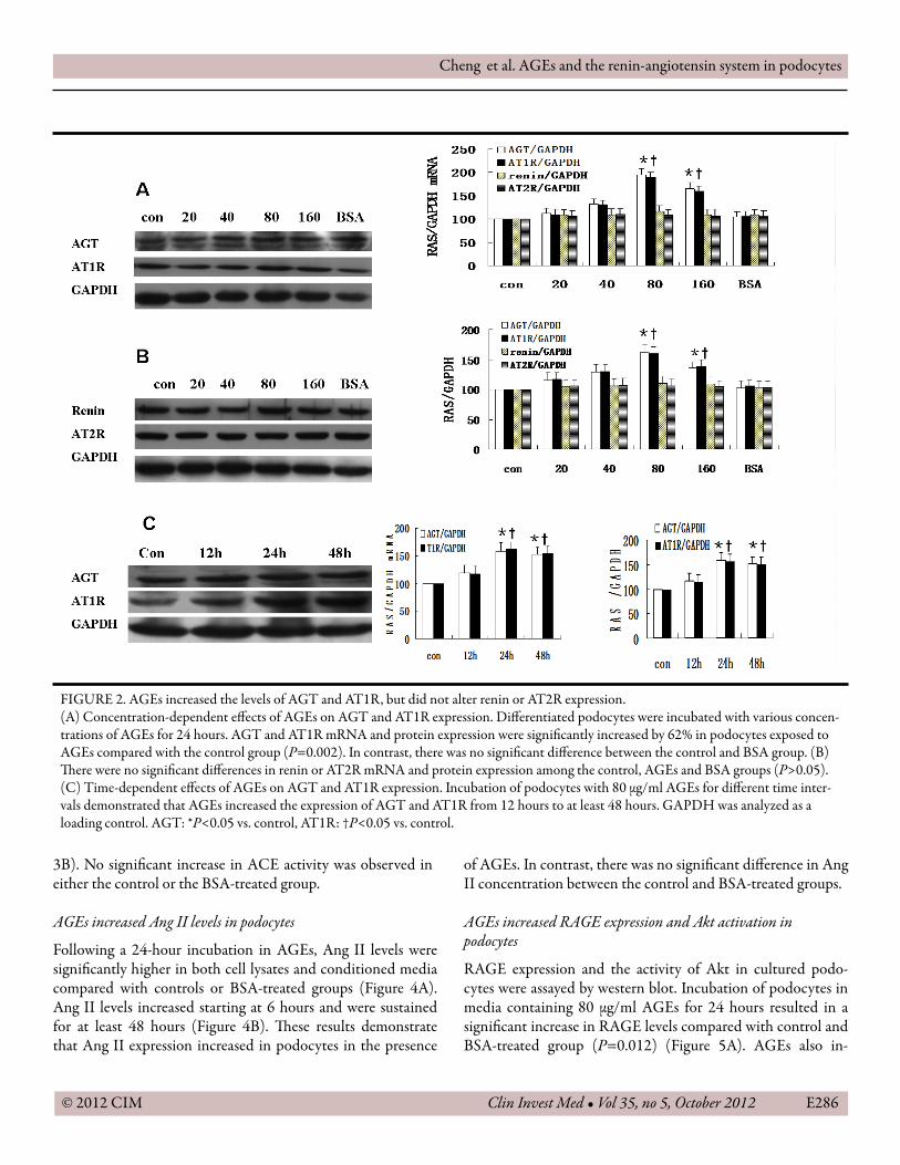
Incubation of podocytes in media containing 80 μg/ml AGEs produced a signi�cant increase in both AGT and AT1R mRNA and protein levels compared with controls (P<0.05); however, there were no changes in the levels of renin or AT2R (P>0.05). In contrast, incubation in 80 μg/ml BSA did not increase either AGT or AT1R expression (Figure 2A, 2B). �e increase in AGT and AT1R expressions was observed within 24 hours and was maintained for up to 48 hours (P<0.05) (Figure 2C).AGEs increased ACE activity
ACE in podocytes is present as a membrane-bound ectoen-zyme and in a secreted form; therefore, both cell lysates and conditioned media were analyzed for ACE activity. A�er a 24 hour incubation period in various concentration of AGEs, the activity of ACE in cell lysates and media increased in a dose-dependent manner (Figure 3A). ACE activity increased at 6 hours and the increase was sustained for up to 48 hours (Figure
Cheng et al. AGEs and the renin-angiotensin system in podocytes
© 2012 CIM Clin Invest Med • Vol 35, no 5, October 2012 E285
FIGURE 1. AGEs did not a�ect podocyte proliferation and metabolic activity. (A) �e e�ects of various concentrations of AGEs on podocyte proliferation were evaluated by the MTT assay. AGEs (80 μg/ml) did not a�ect podocyte proliferation compared with control and BSA-treated group (P>0.05). (B) Cytotoxicity was evaluated using the LDH release assay. LDH did not increase in the AGE-treated group compared with the control (P>0.05). Data are expressed as the mean ± SD of two individual experiments. *P<0.05 compared with the control at identical time points.
3B). No signi�cant increase in ACE activity was observed in either the control or the BSA-treated group.
AGEs increased Ang II levels in podocytes
Following a 24-hour incubation in AGEs, Ang II levels were signi�cantly higher in both cell lysates and conditioned media compared with controls or BSA-treated groups (Figure 4A). Ang II levels increased starting at 6 hours and were sustained for at least 48 hours (Figure 4B). �ese results demonstrate that Ang II expression increased in podocytes in the presence
of AGEs. In contrast, there was no signi�cant di�erence in Ang II concentration between the control and BSA-treated groups.
AGEs increased RAGE expression and Akt activation in podocytes
RAGE expression and the activity of Akt in cultured podo-cytes were assayed by western blot. Incubation of podocytes in media containing 80 μg/ml AGEs for 24 hours resulted in a signi�cant increase in RAGE levels compared with control and BSA-treated group (P=0.012) (Figure 5A). AGEs also in-
Cheng et al. AGEs and the renin-angiotensin system in podocytes
© 2012 CIM Clin Invest Med • Vol 35, no 5, October 2012 E286
FIGURE 2. AGEs increased the levels of AGT and AT1R, but did not alter renin or AT2R expression.(A) Concentration-dependent e�ects of AGEs on AGT and AT1R expression. Di�erentiated podocytes were incubated with various concen-trations of AGEs for 24 hours. AGT and AT1R mRNA and protein expression were signi�cantly increased by 62% in podocytes exposed to AGEs compared with the control group (P=0.002). In contrast, there was no signi�cant di�erence between the control and BSA group. (B) �ere were no signi�cant di�erences in renin or AT2R mRNA and protein expression among the control, AGEs and BSA groups (P>0.05). (C) Time-dependent e�ects of AGEs on AGT and AT1R expression. Incubation of podocytes with 80 μg/ml AGEs for di�erent time inter-vals demonstrated that AGEs increased the expression of AGT and AT1R from 12 hours to at least 48 hours. GAPDH was analyzed as a loading control. AGT: *P<0.05 vs. control, AT1R: †P<0.05 vs. control.

Cheng et al. AGEs and the renin-angiotensin system in podocytes
© 2012 CIM Clin Invest Med • Vol 35, no 5, October 2012 E287
FIGURE 4. AGEs increased Ang II levelsAng II concentrations in media and cell lysates were measured by ELISA. (A) AGEs increased Ang II levels: AGEs (80 μg/ml) increased Ang II levels by 65% in media (P= 0.01) and 70% in cell lysates (P= 0.018) when compared with control cells (P<0.05). (B) AGEs (80 μg/ml) increased Ang II levels in a time-dependent manner in both media and cell lysates; this increase started at 12 hours and was sustained until 48 hours. Data are expressed as the mean ± SD for three individual experiments. Media: *P<0.05 vs. control, cell lysates: †P<0.05 vs. control.
FIGURE 3. AGEs increased ACE activity(A) Concentration-dependent e�ects of AGEs on ACE activity in media and cell lysates. AGEs (80 μg/ml) increased ACE activity by 65% in conditioned media (P= 0.023) and by 68% in cell lysates (P= 0.035), when compared with control. In contrast, BSA had no e�ect on ACE activity. (B) Time-dependent e�ects of AGEs on ACE activity in media and cell lysates. AGEs increased ACE activity in conditioned media and cell lysates signi�cantly in 24 hours, and this increase persisted at 48 hours. Data were expressed as the mean ± SD of three individual experiments. Media: *P<0.05 vs. control, cell lysates: †P<0.05 vs. control.

creased the levels of the active form of Akt signi�cantly (P=0.001) (Fig 5B). Pre-treatment with anti-RAGE antibody alleviated the activated RAS
Podocytes were treated for 24 hours with BSA, AGEs (80 μg/ml) alone or AGEs plus pre-treatment with anti-RAGE anti-body (50 μg/ml) for 60 minutes. Pretreatment with anti-RAGE antibody signi�cantly inhibited the increase of AGT and AT1R protein expression by 37% and 34%, respectively (Figure 6A). ACE activity in cell lysates and media were re-duced by 39% and 45%, respectively (Figure 6B). Ang II con-centrations in cell lysates and media decreased by 52% and 53%, respectively, compared with AGEs group (Figure 6C). PI3K inhibitor attenuated the activated RAS
Podocytes were treated with BSA, AGEs (80 μg/ml) alone or AGEs in combination with the PI3K inhibitor LY294002 (10 μM), captopril (100 μM) or chymastatin (50 μM). �e AGEs-induced increases in the expression of AGT and AT1R were decreased by 64% and 56%, respectively (Figure 7A). ACE
activity in cell lysates and media were reduced by 50% and 39%, respectively (Figure 7B). Ang II concentrations in cell lysates and media were lowered by 44% and 44% in LY294002 treated group, respectively (Figure 7C). Captopril alleviated the levels of Ang II by 50% and 43% in cell lysates and condi-tioned media, respectively; however, chymastatin did not change the levels of Ang II concentrations in cell lysates or in conditioned media. �ese results indicate that the RAGE-PI3-K/Akt pathway might be involved in the activated RAS in po-docytes, and ACE is the main enzyme for the production of Ang II converted from Ang I.
AGEs induced podocyte apoptosis in an Ang II-dependent fusion
To investigate biologic signi�cance of AGEs-induced activation of local RAS, podocyte apoptosis was measured. As shown in Figure 8, AGEs resulted in a near three-fold increase in apopto-sis when compared with the control (23.4±4.6% vs. 7.8±3.8%, P=0.001). To con�rm the apoptosis induced by AGEs was due to activation of RAS, Ang II receptor antagonist losartan (10-5
Cheng et al. AGEs and the renin-angiotensin system in podocytes
© 2012 CIM Clin Invest Med • Vol 35, no 5, October 2012 E288
FIGURE 5. AGEs increased the expression of RAGE and Akt activityRAGE expression and Akt activity were tested by western blot of extracts of podocytes incubated with various concentrations of AGEs for di�erent periods of time. (A) AGEs increased RAGE expression by 50% compared with controls (P=0.012). GAPDH was used as a loading control. (B) AGEs increased Akt activation. AGEs increased the active form of Akt by 100% (P<0.001). �e expression of p-Akt was normal-ized to total Akt expression. �e values are the means ± SD from three experiments, *P<0.05 vs. control.

M) and ACE inhibitor captopril (100 μM) was added to cul-tured podocytes. Both losartan and captopril alleviated signi�-cantly the apoptotic podocytes induced by AGEs (13.6±4.9 % vs. 23.4±4.6%, P=0.021, 17.1±5.2% vs. 23.4±4.6%, P=0.025 respectively). Chymase inhibitor chymastatin (50 μM) failed to alter the rate of apoptosis (20.8±3.6 vs. 23.4±4.6%, P>0.05). �ese results show that AGEs-induced podocyte apoptosis occurs mainly through activation of RAS.
Discussion
AGEs concentrations increased signi�cantly in diabetes melli-tus. �e kidney is not only a target of AGEs but also a source, because declining renal function triggers a rapid increase in plasma concentrations of AGEs[16]. �e intrarenal RAS plays an important role not only in the regulation of glomerular he-
modynamics, but also in glomerular hypertrophy and sclero-sis[17]. Several pieces of evidence have shown that high glucose and mechanical strain activate a local RAS in podocytes[8.18]. Whether AGEs activate the local RAS in podocytes, and the mechanisms underlying these events, is still unclear.
�e present study has demonstrated that AGEs stimulated AGT and AT1R production in mouse podocytes. No signi�-cant changes in the expression of renin and AT2R were ob-served. Although some authors found an increase in renin con-tent in diabetic patients[5], other authors showed low-renin state in diabetic nephropathy[19]. �e levels of Ang !were higher in glomerular podocytes, which means a local RAS was activated through di�erent pathway. �e stimulatory e�ects of AGEs on AGT and AT1R in podocytes indicate that AGEs can increase both the substrate and the receptor for the genera-tion of Ang II. Some studies have demonstrated that di�erenti-
Cheng et al. AGEs and the renin-angiotensin system in podocytes
© 2012 CIM Clin Invest Med • Vol 35, no 5, October 2012 E289
FIGURE 6. Pretreatment with anti-RAGE antibody reduced the activated RASPodocytes were treated for 24 hours with BSA, AGEs (80 μg/ml) alone or AGEs plus pre-treatment with anti-RAGE antibody (50 μg/ml) for 60 minutes. (A) Cell lysates were subjected to western blot using anti-AGT or AT1R antibody. Treatment with anti-RAGE antibody sig-ni�cantly reduced AGT and AT1R expression by 37% (P=0.025) and 34%(P=0.031) respectively. (B) ACE activity in the media and cell lysates were lowered by 45% (P=0.017) and 39% (P=0.021) respectively. (C) Ang II concentrations in the media and cell lysates were re-duced by 53% (P=0.022) and 52% (P=0.019) respectively. Media: *P<0.05 vs. control, §P<0.05 vs. AGEs group; cell lysates: †P<0.05 vs. control, ‡P<0.05 vs. AGEs group.

ated podocytes predominantly express AT1R (about 75%) with a lesser amount of AT2R (about 25%)[20,21]. Our data suggest that the activation of AT2R is not signi�cantly in-volved in the functional responses to Ang II but that AT1R plays a more important role in mediating most of the physio-logical actions of Ang II. In fact, we speculate that the e�ects of Ang II on podocytes under physiological and pathophysiologi-cal conditions are predominantly mediated by the activation of the AT1R.
�e formation of Ang II is mediated by ACE and non-ACE enzymes, and ACE has been established as the most im-portant enzyme for the generation of Ang II[23]. Previous studies have reported ACE protein and activity in both glom-
erular and cultured podocytes. Our results demonstrated that AGEs increase ACE activity signi�cantly in cell lysates and conditioned media, as compared with controls. ACE activity increased from 6 hours and persisted at 48 hours and these results are consistent with previous reports in mesangial cells[23]. Pretreatment with the ACE inhibitor, captopril, rather than the chymase inhibitor, chymastatin, ameliorated the increase of Ang II levels in AGEs-stimulated podocytes and their conditioned media. �ese results imply the involvement of ACE rather than chymase pathways in AGEs-induced Ang II production in podocytes. �e results are consistent with previously published results showing that chymase expression was not detected in podocytes[24] and that the chymase in-
Cheng et al. AGEs and the renin-angiotensin system in podocytes
© 2012 CIM Clin Invest Med • Vol 35, no 5, October 2012 E290
FIGURE 7. PI3K inhibitor attenuated the activated RAS Podocytes were treated with BSA, AGEs (80 μg/ml) alone or AGEs in combination with LY294002 (10 μM), captopril (100 μM), or chy-mastatin (50 μM). (A) Pre-treatment with 10 μM LY294002 led to a decrease in AGT and AT1R expression by 64% (P=0.031) and 56% (P=0.028) respectively. (B) ACE activity in the media and cell lysates were reduced by 39% (P=0.024) and 50% (P=0.021) respectively. (C) Pretreatment with LY294002 resulted in a decrease in Ang II concentrations in the media and cell lysates were reduced by 44% (P=0.031) and 44% (P=0.027) respectively. Captopril alleviated the levels of Ang II by 50% in cell lysates (P=0.025) and 43% in conditioned media (P=0.019) ; however, chymastatin did not change the levels of Ang II concentrations in cell lysates or in conditioned media (P>0.05). Media: *P<0.05 vs. control, §P<0.05 vs. AGEs group; cell lysates: †P<0.05 vs. control, ‡P<0.05 vs. AGEs group.

hibitor chymastatin did not change Ang II secretion induced by mechanical stress[18,25].
Ang II, the �nal e�ector of the RAS, plays a key role in the pathogenesis of diabetic nephropathy, especially in podocyte injury. Podocytes possess the metabolic machinery necessary for autologous synthesis of Ang II. Mouse podocytes were sub-jected to various concentrations of AGEs for di�erent time intervals and the levels of Ang II in the media and cell lysates were determined using a competitive ELISA. As shown in Fig-ure 4A and B, there was a signi�cant increase in Ang II levels at 24 hours and these changes were still present at 48 hours. �e e�ects of local RAS activation on podocyte apoptosis were also observed by �ow cytometry. AGEs resulted in a near three-fold increase in apoptosis as compared with controls. Both losartan and captopril signi�cantly alleviated the apoptotic podocytes induced by AGEs; however, chymastatin did not signi�cantly alter the rate of apoptosis induced by AGEs .�ese results im-ply that AGEs induced podocyte injury via activation local RAS in podocytes.
Our results show that AGEs increased the expression of RAGE in a dose- dependent manner. �ese results are consis-
tent with published reports from endothelial cells[26], peri-cytes[27], mesangial cells[23] and podocytes[12]. RAGE is a multi-ligand, immunoglobulin superfamily, cell surface recep-tor that is expressed in podocytes and the glomerular endothe-lium in the kidney. Previous studies have shown that RAGE-overexpressing diabetic mice exhibit progressive glomeruloscle-rosis with renal dysfunction when compared with diabetic lit-ter mates lacking the RAGE transgene[28]. When pre-incubated with an anti-RAGE antibody, the AGEs-induced increases in the expression of AGT, AT1R , the levels of Ang II, and ACE activity decreased signi�cantly; therefore, the AGE-RAGE interaction in podocytes could be involved in the acti-vated RAS. Rüster found that Ang II upregulated RAGE ex-pression via AT2R in podocytes[29], and the upregulation of RAGE may further activate RAS .Our results show the in-creased levels of Ang II induce podocyte apoptosis mainly via the upregulated AT1R. �ese results show the increased Ang II may cause podocyte injury via both AT1R and AT2R, which aggravates the process of DN.
Our results show that AGEs induced the activation of the Akt kinase activity signi�cantly. To determine whether the ef-
Cheng et al. AGEs and the renin-angiotensin system in podocytes
© 2012 CIM Clin Invest Med • Vol 35, no 5, October 2012 E291
FIGURE 8. AGEs induced podocytes apoptosis in an angiotensin II-dependent fasionPodocytes were treated with BSA, AGEs (80 μg/ml) alone or AGEs in combination losartan (10-5 M), captopril (100 μM), chymastatin (50 μM). Podocyte apoptosis was measured by �ow cytometry. AGEs resulted in a near three-fold increase in apoptosis when compared with po-docytes treated with BSA. Both losartan and captopril signi�cantly reduced the podocyte apoptosis induced by AGEs; however, chymase inhibitor chymastatin did not signi�cantly alter the rate of apoptosis induced by AGEs II P<0.05 vs. control, † P<0.05 vs. AGEs.
fects of AGEs on the RAS were PI3-kinase-dependent, the PI3-K inhibitor, LY294002, was added. LY294002 signi�-cantly reduced the e�ects of AGEs on the expression levels of AGT, AT1R and Ang II as well as attenuating the AGEs-induced increase in ACE activity. �ese results indicate that the PI3-K/Akt pathway is involved in the activated RAS in-duced by AGEs.
Conclusion
In summary, podocytes not only respond to Ang II by activa-tion of AT1 receptors, but also are capable of directly produc-ing Ang II. Stimulation of Ang II production by AGEs pro-vides a direct link between AGEs and the RAS, which may be important in the pathogenesis of DN. �e following data from this study suggest that AGEs activated the RAS via the RAGE-PI3-K pathway in podocytes. First, AGEs led to the up-regulation of AGT and AT1R, the increase of Ang II produc-tion and the activity of ACE. Second, AGEs increased the ex-pression of RAGE and the active form of Akt kinase. �ird, pre-treatment with an anti-RAGE antibody or the PI3-K in-hibitor LY294002 signi�cantly inhibited the changes induced by AGEs. �us, therapies that block the ability of AGEs bind-ing to RAGE or inhibit the PI3-K pathway may alleviate the activated RAS and delay the progression of DN. �ese hy-potheses deserve further investigation in vivo.
Financial Support
�is work was supported by National Natural Science Founda-tion of China (81070581) and Science and Technology Plan-n i n g P r o j e c t o f G u a n g d o n g P r o v i n c e , C h i n a (2010B031600202, 2011B080701005). All the authors de-clared no competing interests.
References1. Makita Z, Rado� S, Ray�eld EJ, Yang Z, Skolnik E, Delaney V,
Friedman EA, Cerami A, Vlassara H . Advanced glycosylation end products in patients with diabetic nephropathy. N Engl J Med 1991;325:836–842.
2. Pavenstadt H, Kriz W, and Kretzler M. Cell biology of the glomerularpodocyte. Physiol Rev 2003 ;83: 253–307.
3. Brenner BM, Cooper ME, de Zeeuw D, Keane WF, Mitch WE, Parving HH, Remuzzi G, Snapinn SM, Zhang Z, Shahinfar S; RENAAL Study Investigators. E�ects of losartan on renal and cardiovascular outcomes in patients with type 2 diabetes and nephropathy. N Engl J Med 2001; 345: 861–869.
4. Gross ML, El-Shakmak A, Szábó A, Koch A, Kuhlmann A, Münter K, Ritz E, Amann K. ACE-inhibitors but not endothe-
linreceptor blockers prevent podocyte loss in early diabetic nephropathy. Diabetologia 2003; 46: 856–868
5. Singh R, Singh AK, Leehey DJ. A novel mechanism for angio-tensin II formation in streptozotocin-diabetic rat glomeruli. Am J Physiol Renal Physiol 2005;288: F1183–F1190.
6. Singh R, Singh AK, Alavi N, Leehey DJ. Mechanism of in-creased angiotensin II levels in glomerular mesangial cells cul-tured in high glucose. J Am Soc Nephrol 2003; 14: 873–880.
7. Zhang SL, To C, Chen X, Filep JG, Tang SS, Ingel�nger JR, Chan JS. Essential roles of the intrarenal renin–angiotensin sys-tem in transforming growth factor-beta1 gene expression and induction of hypertrophy of rat kidney proximal tubular cells in high glucose. J Am Soc Nephrol 2002; 13: 302–312.
8. Durvasula RV, Petermann AT, Hiromura K, Blonski M, Pippin J, Mundel P, Pichler R, Gri�n S, Couser WG, Shankland SJ Acti-vation of a local tissue angiotensin system in podocytes by me-chanical strain. Kidney Int 2004;65: 30–39.
9. Co�er PJ, Jin J, Woodgett JR. Protein kinase B (c-Akt): a multi-functional mediator of phosphatidylinositol 3-kinase activation. Biochem J 1998; 335(Pt 1):1–13
10. Fruman DA, Cantley LC . Phosphoinositide 3-kinase in immu-nological systems. Semin Immunol 2002;14:7–18
11. Tejada T, Catanuto P, IjazA, Santos JV, Xia X, Sanchez P, Sanab-ria N, Lenz O, Elliot SJ, Fornoni A. Failure to phosphorylate AKT in podocytes from mice with early diabetic nephropathy promotes cell death. Kidney Int 2008;73 (12) : 1385-1393
12. Tae-Sun Ha. High-glucose and advanced glycosylation end products increased podocyte permeability via PI3-K/Akt signal-ing. J Mol Med 2010; 88:391–400.
13. Kim SH, Jang YW, Hwang P, Kim HJ, Han GY, Kim CW. �e Reno-Protective E�ect of a PI3K Inhibitor (Wortmannin) on STZ-Induced Proteinuric Renal Disease Rats. Exp Mol Med. 2011 Nov 3. [Epub ahead of print]
14. Ye ZC, Wang C, Tang Y, Liu X, Peng H, Zhang H, Lou TQ. Serum IgA1 from patients with IgA nephropathy up-regulates integrin-linked kinase synthesis and inhibits adhesive capacity in podocytes through indirect pathways. Clin Invest Med 2009; 32(1):E20-7.
15. Cushman DW, Cheung HS. Spectrophotometric assay and properties of the angiotensin-converting enzyme of rabbit lung. Biochem Pharmacol 1971;20:1637–48.
16. Bohlender J M, Franke S, Stein G, et al. Advanced glycation end products and the kidney. Am J Physiol Renal Physiol 2005; 289: F645–F659.
17. Wolf G, Butzmann U, and Wenzel UO. �e renin-angiotensin system and progression of renal disease: from hemodynamics to cell biology. Nephron Physiol 2003;93: 3–13.
18. Yoo TH, Li JJ, Kim JJ, Jung DS, Kwak SJ, Ryu DR, Choi HY, Kim JS, Kim HJ, Han SH, Lee JE, Han DS, Kang SW. Activa-tion of the renin–angiotensin system within podocytes in diabe-tes. Kidney International 2007;71:1019–1027.
Cheng et al. AGEs and the renin-angiotensin system in podocytes
© 2012 CIM Clin Invest Med • Vol 35, no 5, October 2012 E292

19. Price DA, Porter LE, Gordon M, et al. �e paradox of the low-renin state in diabetic nephropathy J Am Soc Nephrol 1999 ;10(11):2382-91.
20. Wang L, Flannery PJ, Spurney RF. Characterization of angio-tensin II-receptor subtypes in podocytes. J Lab Clin Med 2003; 142: 313–321.
21. Liebau MC, Lang D, Böhm J, Endlich N, Bek MJ, Witherden I, Mathieson PW, Saleem MA, Pavenstädt H, Fischer KG. Func-tional expression of the renin angiotensin system in human po-docytes. Am J Physiol Renal Physiol 2006; 27: F710–F719.
22. Siragy HM. AT(1) and AT(2) receptors in the kidney: role in disease and treatment. Am J Kidney Dis 2000; 36: S4–S9.
23. Usui M, Egashira K, Kitamoto S, Koyanagi M, Katoh M, Ka-taoka C, Shimokawa H, Takeshita A. Pathogenic role of oxida-tive stress in vascular angiotensin-converting enzyme activation in longterm blockade of nitric oxide synthesis in rats. Hyperten-sion 1999;34:546–551.
24. Huang XR, Chen WY, Truong LD, Lan HY. Chymase is upregulated in diabetic nephropathy: implications for an alter-native pathway of angiotensin II-mediated diabetic renal and vascular disease. J Am Soc Nephrol 2003;14: 1738–1747.
25. Liebau MC, Lang D, Bohm J, Endlich N, Bek MJ, Witherden I, Mathieson PW, Saleem MA, Pavenstadt H, Fischer KG. Func-
tional expression of the renin-angiotensin system in human po-docytes. Am J Physiol Renal Physiol 2006; 290: F710–F719.
26. Tanaka N, Yonekura H, Yamagishi S, Fujimori H, Yamamoto Y, Yamamoto H. �e receptor for advanced glycation end products is induced by the glycation products themselves and tumor ne-crosis factor-alpha through nuclear factor kappa B, and by 17-beta-estradiol through Sp-1 in human vascular endothelial cells. J Biol Chem 2000;275:25781–25790.
27. Yamagishi S, Amano S, Inagaki Y, Okamoto T, Takeuchi M, Makita Z. Beraprost sodium, a prostaglandin I2 analogue, pro-tects against advanced glycation end products-induced injury in cultured retinal pericytes. Mol Med 2002;8:546–550.
28. Yamamoto Y, Kato I, Doi t, Yonekura H, Ohashi S, Takeuchi M, Watanabe T, Yamagishi S, Sakurai S, Takasawa S, Okamoto H, Yamamoto H. Development and prevention of advanced dia-betic nephropathy in RAGE-overexpressing mice. J Clin Invest 2001;108:261–268.
29. Rüster C, Franke S, Wenzel U, et al. Podocytes of AT2 receptor knockout mice are protected from angiotensin II-mediated RAGE induction. Am J Nephrol 2011;34(4):309-17.
Cheng et al. AGEs and the renin-angiotensin system in podocytes
© 2012 CIM Clin Invest Med • Vol 35, no 5, October 2012 E293