Embed Size (px)
Citation preview
�������� ����� ��
Recent advances in self-assembled peptides: Implications for targeted drugdelivery and vaccine engineering
Sharareh Eskandari, Thalia Guerin, Istvan Toth, Rachel J. Stephenson
PII: S0169-409X(16)30206-XDOI: doi: 10.1016/j.addr.2016.06.013Reference: ADR 13026
To appear in: Advanced Drug Delivery Reviews
Received date: 15 February 2016Revised date: 10 June 2016Accepted date: 21 June 2016
Please cite this article as: Sharareh Eskandari, Thalia Guerin, Istvan Toth, RachelJ. Stephenson, Recent advances in self-assembled peptides: Implications for targeteddrug delivery and vaccine engineering, Advanced Drug Delivery Reviews (2016), doi:10.1016/j.addr.2016.06.013
This is a PDF file of an unedited manuscript that has been accepted for publication.As a service to our customers we are providing this early version of the manuscript.The manuscript will undergo copyediting, typesetting, and review of the resulting proofbefore it is published in its final form. Please note that during the production processerrors may be discovered which could affect the content, and all legal disclaimers thatapply to the journal pertain.
ACC
EPTE
D M
ANU
SCR
IPT
ACCEPTED MANUSCRIPT
1
Recent advances in self-assembled peptides: implications for targeted drug delivery and
vaccine engineering
Sharareh Eskandaria, Thalia Guerina, Istvan Totha,b,c*, Rachel J. Stephensona*
a School of Chemistry and Molecular Biosciences, The University of Queensland, Brisbane, QLD 4072, Australia
b School of Pharmacy, The University of Queensland, Brisbane, QLD 4102, Australia
c Institute for Molecular Bioscience, The University of Queensland, Brisbane, QLD 4072, Australia
*Corresponding author address:
School of Chemistry and Molecular Biosciences, The University of Queensland, Brisbane, QLD 4072, Australia
Phone: (+61) 7 3346 9893
Fax: (+61) 7 3365 4273
E-mails: [email protected]

ACC
EPTE
D M
ANU
SCR
IPT
ACCEPTED MANUSCRIPT
2
Abstract
Self-assembled peptides have shown outstanding characteristics for vaccine delivery and
drug targeting. Peptide molecules can be rationally designed to self-assemble into specific
nanoarchitectures in response to changes in their assembly environment including: pH,
temperature, ionic strength, and interactions between host (drug) and guest molecules. The
resulting supramolecular nanostructures include nanovesicles, nanofibers, nanotubes,
nanoribbons, and hydrogels and have a diverse range of mechanical and physicochemical
properties. These molecules can be designed for cell-specific targeting by including adhesion
ligands, receptor recognition ligands, or peptide-based antigens in their design, often in a
multivalent display. Depending on their design, self-assembled peptide nanostructures have
advantages in biocompatibility, stability against enzymatic degradation, encapsulation of
hydrophobic drugs, sustained drug release, shear-thinning viscoelastic properties, and/or
adjuvanting properties. These molecules can also act as intracellular transporters and
respond to changes in the physiological environment. Furthermore, this class of
materials has shown sequence- and structure-dependent impacts on the immune system
that can be tailored to non-immunogenic for drug targeting, and immunogenic for vaccine
delivery. This review explores self-assembled peptide nanostructures (beta sheets, alpha
helices, peptide amphiphiles, amino acid pairing, elastin like polypeptides, cyclic peptides,
short peptides, Fmoc peptides, and peptide hydrogels) and their application in vaccine
delivery and drug targeting.
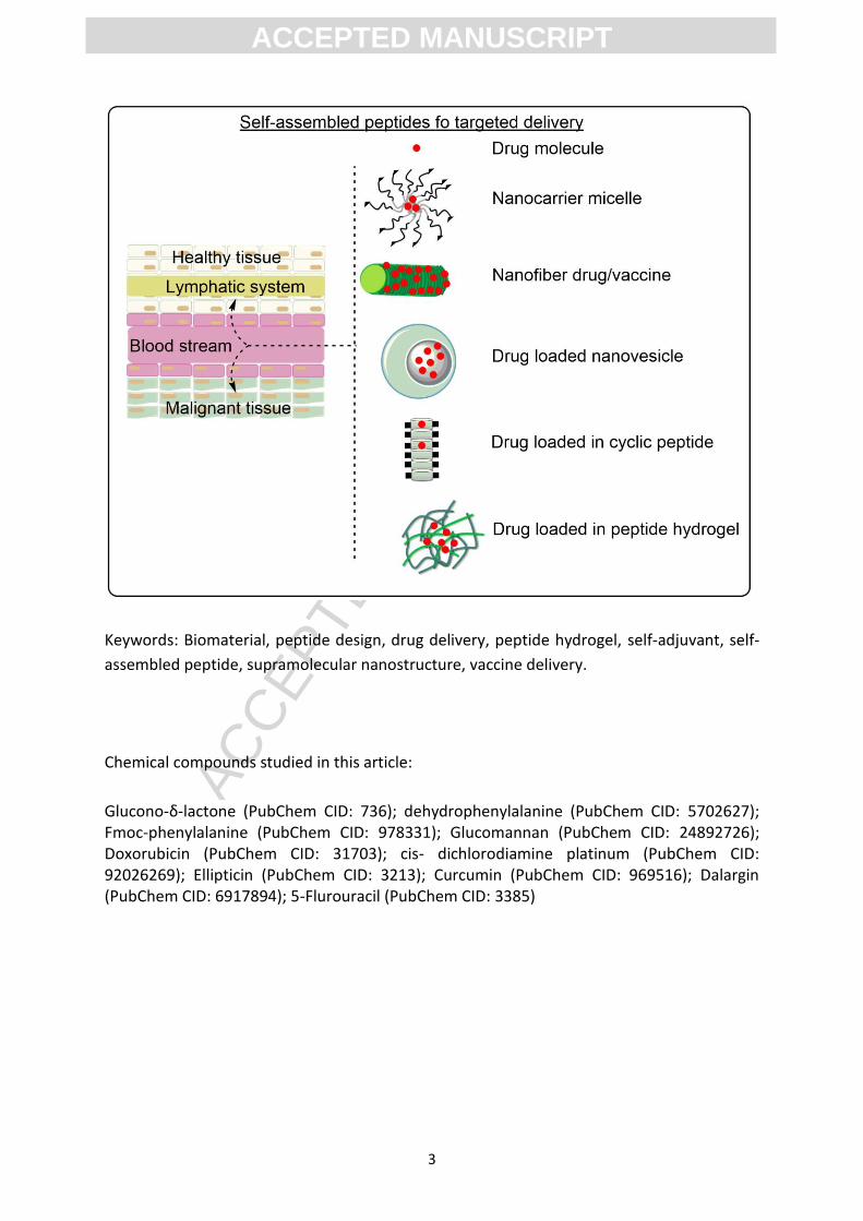
Graphical abstract
ACC
EPTE
D M
ANU
SCR
IPT
ACCEPTED MANUSCRIPT
3
Keywords: Biomaterial, peptide design, drug delivery, peptide hydrogel, self-adjuvant, self-
assembled peptide, supramolecular nanostructure, vaccine delivery.
Chemical compounds studied in this article:
Glucono-δ-lactone (PubChem CID: 736); dehydrophenylalanine (PubChem CID: 5702627); Fmoc-phenylalanine (PubChem CID: 978331); Glucomannan (PubChem CID: 24892726); Doxorubicin (PubChem CID: 31703); cis- dichlorodiamine platinum (PubChem CID: 92026269); Ellipticin (PubChem CID: 3213); Curcumin (PubChem CID: 969516); Dalargin (PubChem CID: 6917894); 5-Flurouracil (PubChem CID: 3385)
ACC
EPTE
D M
ANU
SCR
IPT
ACCEPTED MANUSCRIPT
4
CONTENTS
1. Introduction ................................................................................................................................ 5
2. Self-assembling peptide designs and hydrogel formation ......................................................... 6
2.1. Beta sheets ....................................................................................................................... 9
2.2. Alpha helices and coiled coils ......................................................................................... 11
2.3. Peptide amphiphiles ....................................................................................................... 12
2.4. Amino acid pairing .......................................................................................................... 13
2.5. Elastin-like polypeptides (ELPs) ...................................................................................... 14
2.6. Cyclic peptides ................................................................................................................ 16
2.7. Short peptides ................................................................................................................ 17
2.8. Fluorenylmethoxycarbonyl (Fmoc) peptides.................................................................. 18
3. Self-assembled peptide hydrogel characteristics and release kinetics .................................... 19
3.1. Peptide hydrogel characteristics ........................................................................................... 20
3.2. Hydrogel release kinetics ................................................................................................ 26
4. Drug delivery applications of self-assembled peptides ............................................................ 27
4.1. Delivering drugs to the central nervous system .................................................................... 27
4.2. Intra-ocular drug delivery ............................................................................................... 27
4.3. Cardiovascular drug delivery .......................................................................................... 28
4.4. Bone drug delivery .......................................................................................................... 29
4.5. Anticancer drug delivery................................................................................................. 29
5. Vaccine engineering .................................................................................................................. 32
5.1. Self-assembled peptides for vaccine design................................................................... 32
5.1.1. Vaccine design using peptides that form beta sheets…………………..…………………… 32
5.1.2. Vaccine design using lipidated peptide amphiphiles………………………..…………………34
6. Conclusion ................................................................................................................................. 38
ACC
EPTE
D M
ANU
SCR
IPT
ACCEPTED MANUSCRIPT
5
1. Introduction
Drug delivery systems can target the drug to specific tissues, minimise side effects,
overcome solubility problems and toxicity and be tailored to have the appropriate
immunogenicity and metabolic stability. Despite the use of rational design principles,
developing a drug delivery platform often relies on synthesising and screening a library of
drug derivatives to find a suitable candidate. The ability to predict the physicochemical and
biological properties of peptide nanomaterials from the sequence alone is a continuing field
of research. Ideal nanomaterials would allow the delivery of several active pharmaceutical
ingredients with different release profiles. Ongoing research aims to reformulate existing
drugs using smart materials to control the drug structure and function at the molecular level
to mimic the three-dimensional structure of biological proteins [1, 2]. In the absence of
predictable structure-activity outcomes, a wealth of research has been conducted to
develop platforms for a diverse range of drug and vaccine candidates on a case-by-case
basis.
Self-assembled peptide nanoparticles form part of a bottom-up strategy to create
reproducible nanosized delivery systems [3]. Molecular self-assembly is a practical approach
in which molecules are spontaneously organized into ordered structures using processes
driven by free-energy that include, Van der Waals, electrostatic, hydrogen bonding, and π-π
stacking interactions [4]. The balance of attractive and repulsive forces within (and
between) molecules affects their arrangement and is dependent on molecular composition,
assembly kinetics, and variation in assembly environment (pH, solvent, co-assembling
molecules, temperature, and ionic strength) [4, 5]. Peptide-based drug and vaccine carrier
molecules can deliberately be designed to spontaneously self-assemble under specific
environmental conditions [6]. For example, peptide scaffolds that spontaneously assembled
into a beta sheet structure were isolated from the yeast protein Zuotin and contained
alternating hydrophobic and chargedhydrophilic amino acids, lysine and glutamic acid, with
50% charged residues (EAK16-II, AEAEAKAKAEAEAKAK) [7]. Synthetic polypeptides derived
from large or natural amino acids are very useful building blocks for fabricating self-
assembling structures for medical and pharmaceutical applications due to their high
physicochemical stability, diversity in sequence and shape, suitability for large scale
synthesis, biodegradability, and bio-compatibility [4]. The self-assembly of various
amphiphilic molecules including micelles, polymeric vesicles, microemulsions, liposomes,
and nanoparticles has been widely studied for drug delivery applications. Nevertheless,
increased control over the structure and biofunctionality of self-assembled peptide delivery
systems would improve their application to the delivery of drug and vaccine candidates [8].
Peptide delivery systems have advantages over liposomes or nanoparticles because they can
be composed of amphiphilic prodrugs with high drug loading, low drug leakage,
biodegradability, and high permeability to bio-membranes of the target cells [3]. In addition,
the formation of stable colloidal suspensions allows for the stabilization of hydrophobic
compounds as micro- and nano-crystals more effectively than micelles [8]. These systems
efficiently adopted the desired stimuli responsiveness that resulted from their molecular
ACC
EPTE
D M
ANU
SCR
IPT
ACCEPTED MANUSCRIPT
6
design, biological environment, and interactions. For example, the pH triggered release of
an encapsulated drug or targeted delivery with a recognition pattern led to a more efficient
delivery system with less side effects [1]. Moreover, peptide delivery systems have been
shown to form a protective coat on the surface of hydrophobic compounds, resulting in
increased control of drug release, protecting the drug from exposure to degradation agents
and reducing exposure of normal tissues to toxic drugs [1, 8, 9]. Depending on the choice of
starting materials, self-assembled particles have been shown to display large numbers of
biologically active peptides on their surface enabling them to be recognized by cell surface
receptors, thus serving as both drug and drug delivery agents simultaneously without the
need to further encapsulate drugs within their nanostructure. The diversity of peptides that
can be employed in drug and vaccine delivery highlights the importance of ongoing research
in this field.
The effect of a peptide on the immune system is dependent on sequence and
physicochemical properties. Strategies for either avoiding or specifically inducing immune
response are the subject of continuing research. New delivery systems must be tailored to
elicit the desired immune response: non-immunogenic peptides for drug delivery
applications and immunogenic self-adjuvanting peptides for vaccine delivery. Therefore,
investigation into the correlation between the self-assembled peptide sequence and its
immunogenicity will facilitate the design of self-assembled peptides for both applications
[10-16]. This review outlines recent advances in self-assembled peptides for drug delivery,
focusing on peptide designs that produced nanostructures for the targeted delivery of
therapeutics. In addition, applications of peptide-based self-assembly for the development
of peptide subunit vaccines have also been outlined.
2. Self-assembling peptide designs and hydrogel formation
Amino acids provide the primary structure and site for chemical modification when
designing peptide nanomaterials [17]. Amino acid side chains have different charge,
hydrophobicity, size, and polarity. The number, type, and sequence of amino acids can be
manipulated to design unique self-assembled peptide nanostructures with specific
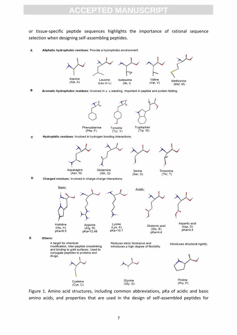
secondary structures and physicochemical properties [4]. Figure 1 summarises amino acid
properties and their role in self-assembly. Variation in peptide length, the percentage and
number of repeats of hydrophobic residues (i.e., alanine, valine, isoleucine, leucine,
tyrosine, phenylalanine, tryptophan) and the difference in charge distribution significantly
influences the mechanical properties of nanostructure scaffolds and the speed of self-
assembly [7, 18]. Known peptide ligands can be added to biologically inert substrates to
target the nanomaterial to the appropriate tissue or receptor in vivo [17, 19]. This strategy
includes adding pattern recognition receptor ligands to stimulate specific immune responses
through toll-like receptor pathways. Cellular adhesion ligands (e.g. RGD) can also be added
to promote cellular interaction through transmembrane receptors (integrin proteins) that
mediate cell-cell and extracellular matrix (ECM) interactions. The ability to include targeting
ACC
EPTE
D M
ANU
SCR
IPT
ACCEPTED MANUSCRIPT
7
or tissue-specific peptide sequences highlights the importance of rational sequence
selection when designing self-assembling peptides.
Figure 1. Amino acid structures, including common abbreviations, pKa of acidic and basic
amino acids, and properties that are used in the design of self-assembled peptides for
ACC
EPTE
D M
ANU
SCR
IPT
ACCEPTED MANUSCRIPT
8
biomedical applications [17]. Reproduced and adapted by permission from The Royal
Society of Chemistry.
Cell adhesion and integrin binding are an important component of drug targeting. Molecular
binding at one end of an integrin mediates interaction with intracellular signalling pathways,
including calcium channels, kinases, phosphatases and other binding proteins, and also
recruits intracellular non-receptor tyrosine kinases. These changes affect cell cycle
regulation, cell shape and motility, and can result in up-regulation of integrin receptors on
the cells surface [20]. ECM proteins are an important feature for development and
morphogenesis because they act as ligands for integrins, including:
fibronectin, vitronectin, collagen, and laminin [21, 22]. Peptide-based biomaterials can be
engineered to optimize the spatial organization, effective density, and accessibility of the
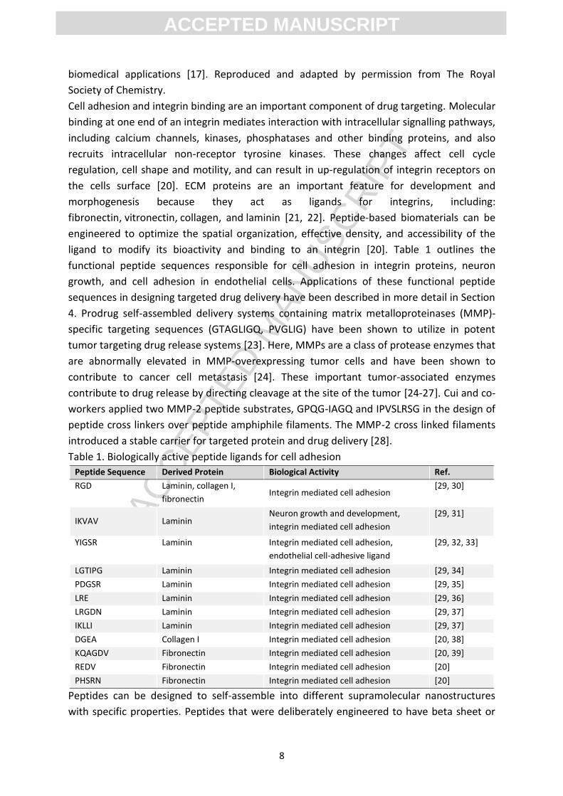
ligand to modify its bioactivity and binding to an integrin [20]. Table 1 outlines the
functional peptide sequences responsible for cell adhesion in integrin proteins, neuron
growth, and cell adhesion in endothelial cells. Applications of these functional peptide
sequences in designing targeted drug delivery have been described in more detail in Section
4. Prodrug self-assembled delivery systems containing matrix metalloproteinases (MMP)-
specific targeting sequences (GTAGLIGQ, PVGLIG) have been shown to utilize in potent
tumor targeting drug release systems [23]. Here, MMPs are a class of protease enzymes that
are abnormally elevated in MMP-overexpressing tumor cells and have been shown to
contribute to cancer cell metastasis [24]. These important tumor-associated enzymes
contribute to drug release by directing cleavage at the site of the tumor [24-27]. Cui and co-
workers applied two MMP-2 peptide substrates, GPQG-IAGQ and IPVSLRSG in the design of
peptide cross linkers over peptide amphiphile filaments. The MMP-2 cross linked filaments
introduced a stable carrier for targeted protein and drug delivery [28].
Table 1. Biologically active peptide ligands for cell adhesion
Peptide Sequence Derived Protein Biological Activity Ref.
RGD Laminin, collagen I,
fibronectin Integrin mediated cell adhesion
[29, 30]
IKVAV Laminin Neuron growth and development,
integrin mediated cell adhesion
[29, 31]
YIGSR Laminin Integrin mediated cell adhesion,
endothelial cell-adhesive ligand
[29, 32, 33]
LGTIPG Laminin Integrin mediated cell adhesion [29, 34]
PDGSR Laminin Integrin mediated cell adhesion [29, 35]
LRE Laminin Integrin mediated cell adhesion [29, 36]
LRGDN Laminin Integrin mediated cell adhesion [29, 37]
IKLLI Laminin Integrin mediated cell adhesion [29, 37]
DGEA Collagen I Integrin mediated cell adhesion [20, 38]
KQAGDV Fibronectin Integrin mediated cell adhesion [20, 39]
REDV Fibronectin Integrin mediated cell adhesion [20]
PHSRN Fibronectin Integrin mediated cell adhesion [20]
Peptides can be designed to self-assemble into different supramolecular nanostructures
with specific properties. Peptides that were deliberately engineered to have beta sheet or
ACC
EPTE
D M
ANU
SCR
IPT
ACCEPTED MANUSCRIPT
9
alpha helix secondary structure and amphiphilic peptide monomers have shown promise in
self-generating supramolecular assemblies, with applications in vaccine design and targeted
drug delivery. Peptide molecules that self-assembled in aqueous media to form cylindrical
nanofibers were shown to also assemble into a higher network arrangement of peptide
nanofiber-based hydrogels, where the bioactive peptide was displayed on the surface.
Hydrogels are polymeric networks with three-dimensional configurations which have the
capacity to absorb large quantities of water or biological fluids due to the presence of
hydrophilic groups (amine, hydroxyl, ether, sulphate, or carboxyl) in their structural
networks [40]. Peptide-based hydrogels have prominent advantages over traditional
polymeric hydrogels which include biodegradability, low bioaccumulation and toxicity,
spontaneous formation without the use of harmful reagents (such as chemical cross-
linkers), and the facile incorporation of cell-specific bioactive moieties. Peptide-based
hydrogels are also cost effective, easily synthesized and responsive to external stimuli,
including temperature, ionic strength, pH, light, enzyme and magnetic fields [41-47]. The
responsiveness of these peptide hydrogels to biological stimuli attributed to their ability to
sense changes to the local environment and release therapeutics in a controlled manner
[47].
The hydrogelation of peptide hydrogels is easily modified through the attachment of
chemical and biological moieties [48]. Peptide hydrogels are similar in scale to natural ECM
and provide an in vivo cell environment with properties that favour cellular adhesion [49].
These peptide hydrogels were engineered to encapsulate hydrophobic guest molecules,
target specific cell types, and improve stability, thus overcoming the peptides susceptibility
to enzymatic degradation in vivo [50, 51]. Hydrogel drug delivery vehicles can be designed
to entrap drug molecules through physical or covalent bonds.
The noncovalent interaction between drug and peptide structure with a net hydrophobicity
can be used to stabilise the entrapped therapeutic molecule in micelles or porous
nanoparticles. These systems must be rationally engineered and optimised to load and
release the drug appropriately [52, 53]. Chemical conjugation of the drug into the peptide
molecule via a biochemically cleavable linker has been shown to provide more control over
the triggered release of the drug. The bioactivity of the drug depends on how effectively it is
cleaved from the carrier at the target tissue or site of action [53, 54].
Therapeutic agents released from peptide hydrogels are controlled by the mesh size of the
network to diffuse encapsulated drugs, and the self-supporting properties of the hydrogel to
sustain itself and any active component. In the case of drug conjugation to polypeptides,
network degradation has been shown to control the release of a drug [55]. The optimal
design parameters of synthetic peptide hydrogels and their environment have been shown
to dictate their molecular structure (e.g, beta sheet secondary structure), biological
function, mechanical properties, and stability [55].
2.1. Beta sheets
A series of hydrogen bonds between residues in different polypeptide chains or between
residues in different sections of a folded polypeptide produced parallel or anti-parallel
ACC
EPTE
D M
ANU
SCR
IPT
ACCEPTED MANUSCRIPT
10
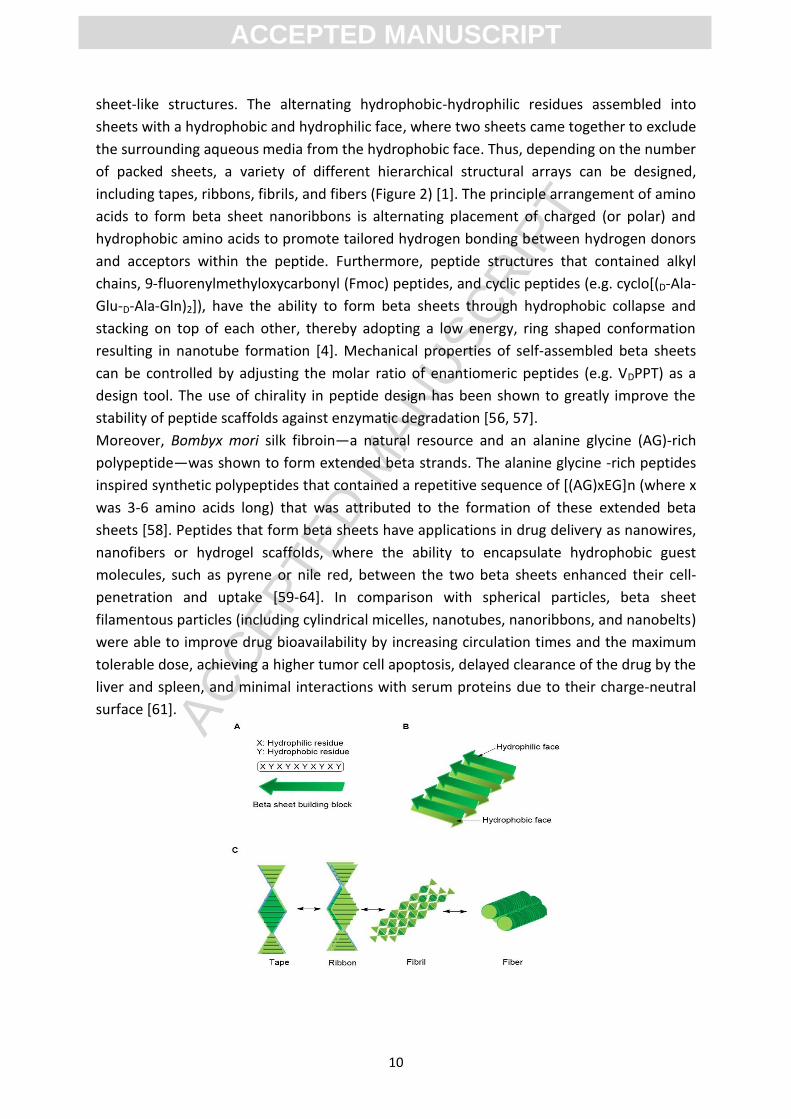
sheet-like structures. The alternating hydrophobic-hydrophilic residues assembled into
sheets with a hydrophobic and hydrophilic face, where two sheets came together to exclude
the surrounding aqueous media from the hydrophobic face. Thus, depending on the number
of packed sheets, a variety of different hierarchical structural arrays can be designed,
including tapes, ribbons, fibrils, and fibers (Figure 2) [1]. The principle arrangement of amino
acids to form beta sheet nanoribbons is alternating placement of charged (or polar) and
hydrophobic amino acids to promote tailored hydrogen bonding between hydrogen donors
and acceptors within the peptide. Furthermore, peptide structures that contained alkyl
chains, 9-fluorenylmethyloxycarbonyl (Fmoc) peptides, and cyclic peptides (e.g. cyclo[(D-Ala-
Glu-D-Ala-Gln)2]), have the ability to form beta sheets through hydrophobic collapse and
stacking on top of each other, thereby adopting a low energy, ring shaped conformation
resulting in nanotube formation [4]. Mechanical properties of self-assembled beta sheets
can be controlled by adjusting the molar ratio of enantiomeric peptides (e.g. VDPPT) as a
design tool. The use of chirality in peptide design has been shown to greatly improve the
stability of peptide scaffolds against enzymatic degradation [56, 57].
Moreover, Bombyx mori silk fibroin—a natural resource and an alanine glycine (AG)-rich
polypeptide—was shown to form extended beta strands. The alanine glycine -rich peptides
inspired synthetic polypeptides that contained a repetitive sequence of [(AG)xEG]n (where x
was 3-6 amino acids long) that was attributed to the formation of these extended beta
sheets [58]. Peptides that form beta sheets have applications in drug delivery as nanowires,
nanofibers or hydrogel scaffolds, where the ability to encapsulate hydrophobic guest
molecules, such as pyrene or nile red, between the two beta sheets enhanced their cell-
penetration and uptake [59-64]. In comparison with spherical particles, beta sheet
filamentous particles (including cylindrical micelles, nanotubes, nanoribbons, and nanobelts)
were able to improve drug bioavailability by increasing circulation times and the maximum
tolerable dose, achieving a higher tumor cell apoptosis, delayed clearance of the drug by the
liver and spleen, and minimal interactions with serum proteins due to their charge-neutral
surface [61].
ACC
EPTE
D M
ANU
SCR
IPT
ACCEPTED MANUSCRIPT
11
Figure 2. Schematic representation of peptides that form beta sheets and the self-
assembled structures that can be formed. A: a peptide sequence with alternating
hydrophilic (X) and hydrophobic (Y) residues. B: assembly of the beta sheet peptides into a
molecule that contains both a hydrophilic and hydrophobic face. C: self-assembly of the
beta sheet forming peptide into a tape, ribbon, fibril, and fiber based on their packing
density [65]. Reproduced and adapted with permission from Elsevier.
2.2. Alpha helices and c oiled coils
Alpha helix assemblies are used as components of coiled coils which are a basic folding
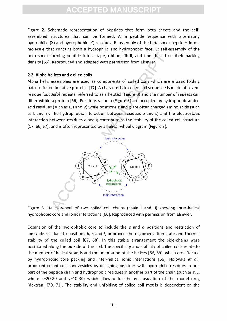
pattern found in native proteins [17]. A characteristic coiled coil sequence is made of seven-
residue (abcdefg) repeats, referred to as a heptad (Figure 3) and the number of repeats can
differ within a protein [66]. Positions a and d (Figure 3) are occupied by hydrophobic amino
acid residues (such as L, I and V) while positions e and g are often charged amino acids (such
as L and E). The hydrophobic interaction between residues a and d, and the electrostatic
interaction between residues e and g contribute to the stability of the coiled coil structure
[17, 66, 67], and is often represented by a helical-wheel diagram (Figure 3).
Figure 3. Helical-wheel of two coiled coil chains (chain I and II) showing inter-helical
hydrophobic core and ionic interactions [66]. Reproduced with permission from Elsevier.
Expansion of the hydrophobic core to include the e and g positions and restriction of
ionisable residues to positions b, c and f, improved the oligomerization state and thermal
stability of the coiled coil [67, 68]. In this stable arrangement the side-chains were
positioned along the outside of the coil. The specificity and stability of coiled coils relate to
the number of helical strands and the orientation of the helices [66, 69], which are affected
by hydrophobic core packing and inter-helical ionic interactions [66]. Holowka et al.,
produced coiled coil nanovesicles by designing peptides with hydrophilic residues in one
part of the peptide chain and hydrophobic residues in another part of the chain (such as KxLy
where x=20-80 and y=10-30) which allowed for the encapsulation of the model drug
(dextran) [70, 71]. The stability and unfolding of coiled coil motifs is dependent on the
ACC
EPTE
D M
ANU
SCR
IPT
ACCEPTED MANUSCRIPT
12
temperature, pH and ionic strength which is often used in the design of controlled release
delivery systems that respond to a specific stimulus [66]. Furthermore, the unique
association-dissociation of coiled coils make them an ideal candidate for physical cross-
linkers to form protein-based supramolecular fibrils [1, 72].
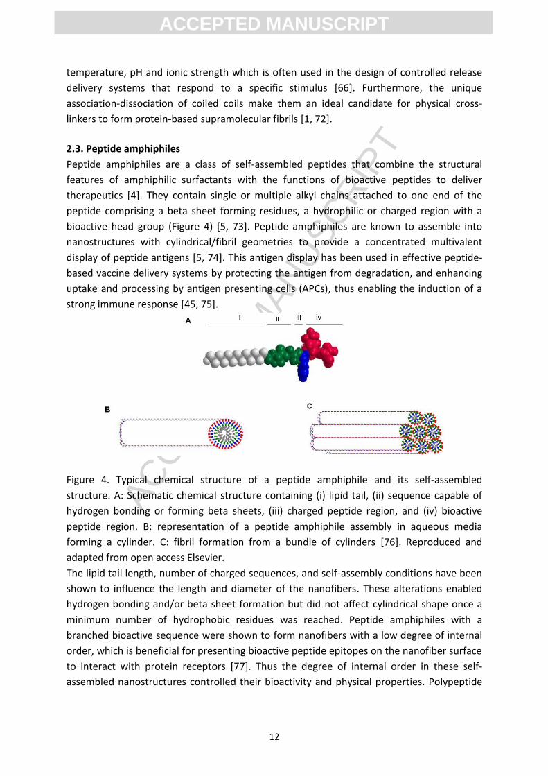
2.3. Peptide amphiphiles
Peptide amphiphiles are a class of self-assembled peptides that combine the structural
features of amphiphilic surfactants with the functions of bioactive peptides to deliver
therapeutics [4]. They contain single or multiple alkyl chains attached to one end of the
peptide comprising a beta sheet forming residues, a hydrophilic or charged region with a
bioactive head group (Figure 4) [5, 73]. Peptide amphiphiles are known to assemble into
nanostructures with cylindrical/fibril geometries to provide a concentrated multivalent
display of peptide antigens [5, 74]. This antigen display has been used in effective peptide-
based vaccine delivery systems by protecting the antigen from degradation, and enhancing
uptake and processing by antigen presenting cells (APCs), thus enabling the induction of a
strong immune response [45, 75].
Figure 4. Typical chemical structure of a peptide amphiphile and its self-assembled
structure. A: Schematic chemical structure containing (i) lipid tail, (ii) sequence capable of
hydrogen bonding or forming beta sheets, (iii) charged peptide region, and (iv) bioactive
peptide region. B: representation of a peptide amphiphile assembly in aqueous media
forming a cylinder. C: fibril formation from a bundle of cylinders [76]. Reproduced and
adapted from open access Elsevier.
The lipid tail length, number of charged sequences, and self-assembly conditions have been
shown to influence the length and diameter of the nanofibers. These alterations enabled
hydrogen bonding and/or beta sheet formation but did not affect cylindrical shape once a
minimum number of hydrophobic residues was reached. Peptide amphiphiles with a
branched bioactive sequence were shown to form nanofibers with a low degree of internal
order, which is beneficial for presenting bioactive peptide epitopes on the nanofiber surface
to interact with protein receptors [77]. Thus the degree of internal order in these self-
assembled nanostructures controlled their bioactivity and physical properties. Polypeptide
ACC
EPTE
D M
ANU
SCR
IPT
ACCEPTED MANUSCRIPT
13
dendrimers that contained multiple branches could mimic protein structure (due to their
nanoscale size), and produced stable micelles that demonstrated different phase behaviours
when compared to linear polypeptides. Additionally, their physicochemical properties could
be controlled by the functionalization of peripheral groups, which is important for their
application, e.g. acting as a drug carrier or targeted drug therapy [78-81].
The nanostructures of peptide amphiphiles have been shown to encapsulate and slowly release both hydrophilic and hydrophobic drugs. Micelles of peptide amphiphiles that bore an alkyl chain (C16) were shown to permeate and internalize in the endocytic vesicles of osteosarcoma cells (SJSA-1 cells) via adsorption-mediated, energy-dependent pathways [79]. Moreover, nanofibers of a peptide amphiphile from a collagen peptide that bore a C16 alkyl chain formed a hydrogel, stimulating collagen production and wound healing [83, 84]. Stupp and co-workers created a biopolymer membrane delivery system by self-assembling
negatively charged hyaluronic acid with positively charged peptide amphiphiles C16-
VVVAAAKKK-NH (K3 PA) and C16-VVVAAAGGKLAKLAKKLAKLAK-NH (KLAK PA). The latter,
KLAKL PA, was shown to be responsive to the cancerous environment by inducing MB-MDA-
231 breast cancer cell death and degradation of the tumor environment. Crosslinking of
tyramine-functionalized hyaluronic acid using horse radish peroxidase and H2O2 under mild
conditions following co-assembly of the peptide amphiphile membrane was shown to
stabilize the structure of these peptides against osmotic pressure and ionic degradation in
buffered medium. In addition, the morphology of the membrane was shown to be a
determinant in its functionality as a sustained release vehicle or localized surface cytotoxic
delivery system. Here, the KLAK PA and K3 PA mixed in a 50:50 ratio formed spherical
particles and presented a sustained release reservoir of the cytotoxic peptide following
enzymatic degradation by hyaluronidase over a 2 day period. However, when KLAK PA and
K3 PA were mixed in a 20:80 ratio over the course of 2 h, formation of a fibril membrane
with localized surface cytotoxicity was observed. In addition, this construct has less
cytotoxicity than the 50:50 ratio construct. The introduced biopolymer membrane has the
potential to be used as a localized adjuvant therapy post lumpectomy [85]. Peptide
amphiphile nanofibers have also been shown to perform as a molecular transporter in brain
delivery (discussed in further detail in Section 4.1) [86].
2.4. Amino acid pairing
Another method for designing self-assembled peptides is using an amino acid pairing
peptide (AAPP) strategy. This model used combinations of amino acid pairings that self-
assembled through weak interactions, including hydrogen bonds, hydrophobic interactions
and ionic bonds. For example, a peptide that contained the hydrogen bonding pair (Q-N),
one ionic-complementary pair (E-K) and two hydrophobic residue pairs (F-F) [87]. The
charge distribution along the backbone of this ionic-complementary peptide was a
determining factor in its self-assembly conformation and nanostructured stability. Three
types of charged distributions were studied extensively: type I (-+ or +-), type II (--++ or ++--)
and type III (----++++ or ++++----) where - and + refer to negatively and positively charged
amino acids respectively [6]. EAKA16-I (AEAKAEAKAEAKAEAK, type I, -+) and RADA 16-II
ACC
EPTE
D M
ANU
SCR
IPT
ACCEPTED MANUSCRIPT
14
(NH2-RARADADARARADADA-COOH, type II, ++--) are well-known ionic-complementary self-
assembled peptides that have been used in drug delivery applications [6, 88-90]. Here, the
hydrophobic residues encapsulated and stabilised hydrophobic drugs and enhanced self-
association of the peptide. Additionally, hydrogen bonds were found to stabilise peptide
assemblies with charged residues, increasing the solubility of the peptide [87]. It has been
shown that EAK16-II (AEAEAKAKAEAEAKAK) and EAK16-IV (AEAEAEAEAKAKAKAK) have
different charge distributions creating stabilized hydrophobic compounds in aqueous
solutions and releasing them into the cell in a controlled manner. Additionally, they have
been shown to self-assemble into stable beta sheet fibrils over a wide pH range (1.5-11) in
the presence of proteases and denaturing agents without an adverse immune response in
mice, rabbits, or goats [9]. This shows their potential as a drug delivery vehicle to maintain
therapeutic drug concentrations throughout the body or at a specific location in a cell.
Saadatmousavi et al., designed a peptide that contained FEFQFNFK and demonstrated
concentration-dependent peptide self-assembly that stabilised ellipticin, an antineoplastic
agent. The applications and characteristics of this delivery system are explained in more
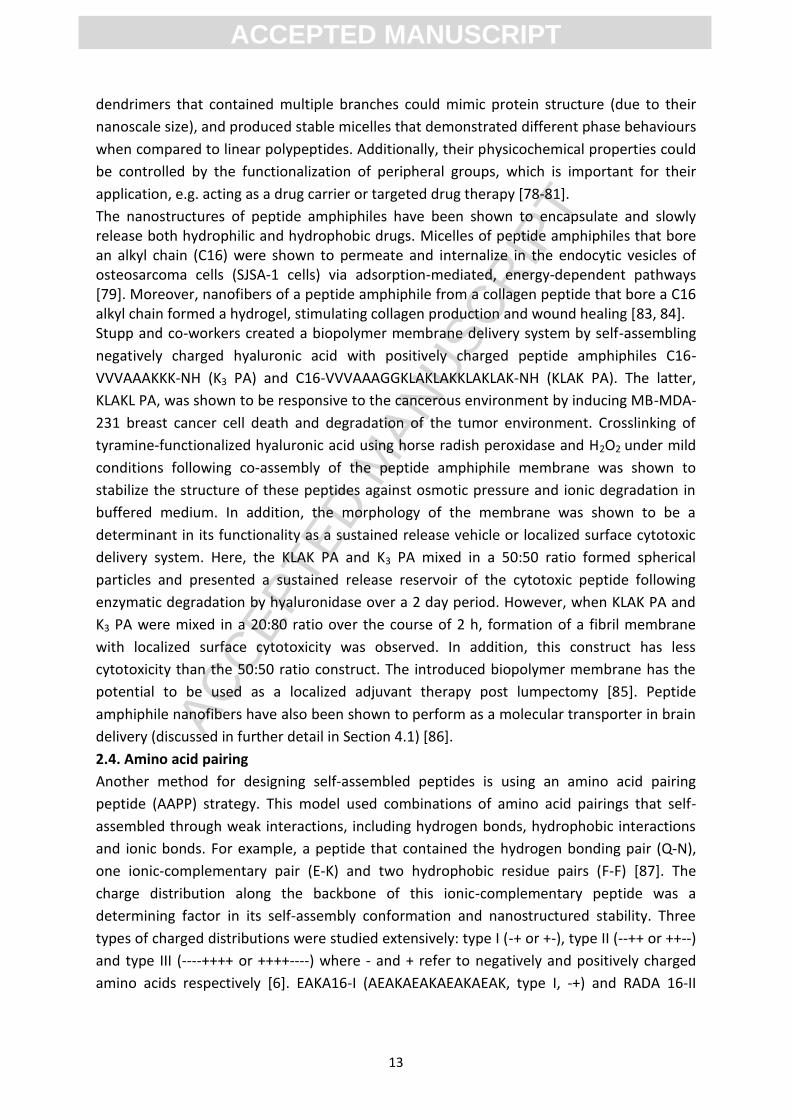
detail in Section 4.5 [6]. Figure 5 shows representative ionic pairing peptide hydrogels.
Figure 5. Schematic representation of ionic pairing in a peptide assembly. A: Hydrogel
scaffold with ordered aggregates of ionic-complementary self-assembled peptides, B:
Hydrogel scaffold composed of alternating hydrophilic and hydrophobic residues and
clusters of negative and positive charges on the N- and C-terminus, respectively. Adapted
from [87].
2.5. Elastin-like polypeptides (ELPs)
Elastin-like polypeptides (ELPs) are a group of biopolymers that self-assemble under
physiological conditions because of their behaviour at their lower critical solution
temperature (LCST) [91]. ELPs are protein polymers with [VPGXG]n amino acid repeats
(where X is a variable amino acid) derived from a highly conserved repeat sequence in
mammalian tropoelastin. These biopolymers display temperature dependent phase
behaviour and form secondary structure ranging from random coil to cylindrical micelles
made from beta sheets depending on the polarity of the residue [1, 92]. ELPs are
characterised by rubber-like elasticity, large extensibility before rupture, flexible
deformation without loss of energy, high resilience upon stretching, thermo-responsiveness,
and biodegradability. Elastin and resilin, highly cross-linked proteins, are examples of two
elastomeric biopolymers and recombinant polypeptides. Synthetic polypeptides that mimic
ACC
EPTE
D M
ANU
SCR
IPT
ACCEPTED MANUSCRIPT
15
elastin composition have been applied as macromolecular and nano-carriers in the form of
controlled release gels and drug-eluting films [93]. ELP-based carriers can be categorised
into soluble (transition temperature (Tt) above body temperature) and insoluble ELPs (Tt
below body temperature) [88]. Conjugation of doxorubicin with ELP[V1A8G7-160], which has
a Tt above 37 οC, self-assembled into 40 nm micelles and was shown to reduce tumor size in
a mouse model. This conjugate was five times more effective than free doxorubicin when
tested against E0771 murine breast tumors [94, 95]. Raucher and coworkers developed a
technique to fuse a cell penetrating peptide to the ELP carrier to facilitate cellular uptake of
doxorubicin [95]. This technique resulted in an effective cancer therapy in vitro, however, to
date no in vivo studies have been reported [91]. ELPs can also be used for the local delivery
of drugs by triggering coacervation in response to changes in body temperature, providing a
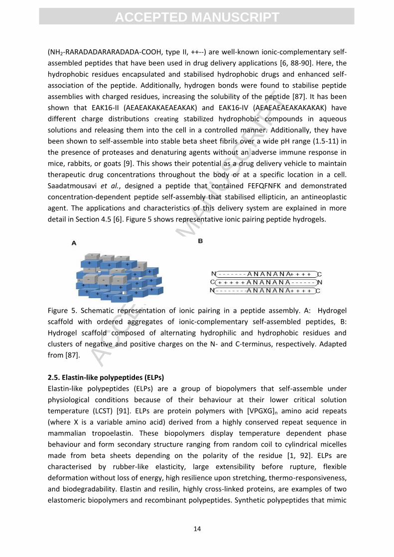
depot for prolonged release of the drug [96-97]. Here, fusion genes that contained a
hydrophilic, high Tt ELP[V1A8G7-n] gene at the N-terminus and a hydrophobic, low Tt ELP[V5-
n] gene was created to encode a diblock (contains ELP[V1A8G7-n] and ELP[V5-n]) ELP. This
ELP self-assembled into spherical micelles at 40 °C and presented multiple copies of the
targeting moiety attached to the N-terminal ELP and entrapped the drug within the core of
the micelle (Figure 6).
Figure 6. Schematic illustration of depot micelle formation at the tumor site in response to
coacervation of an elastin-like polypeptide with a Tt above 37 ◦C. Reprinted with permission
from [98]. Copyright {2008} American Chemical Society.
Ghandehari and co-workers applied silk ELP polypeptide hydrogels to the intra-tumoral
injection of adenoviruses in a head and neck cancer model in an attempt to overcome the
challenges of cancer gene therapy. They showed a ten-fold increase in β-galactosidase gene
expression in a head and neck cancer model, indicating a more efficient and localized
transfection compared to gene therapy with viral vectors (adenovirus carrying the β-
galactosidase gene) without the biopolymer [91, 99, 100]. ELP nanocarriers produced via
ACC
EPTE
D M
ANU
SCR
IPT
ACCEPTED MANUSCRIPT
16
genetic engineering techniques were shown to have no acute systemic toxicity or
immunogenicity in mice following intraperitoneal, intravenous and subcutaneous
administration, and no systemic antigenicity in guinea pigs following intravenous
administration [101]. However, further tests are required to demonstrate the safety of
these engineered nanocarriers in vivo [101]. Additionally, drug molecules with high
hydrophobicity and/or a large number of hydrogen-bond donors and acceptors showed high
ELP encapsulation efficiency. Therefore, drugs that are currently challenging to formulate
using more conventional delivery vehicles might be good candidates for delivery by
genetically engineered ELPs [92].
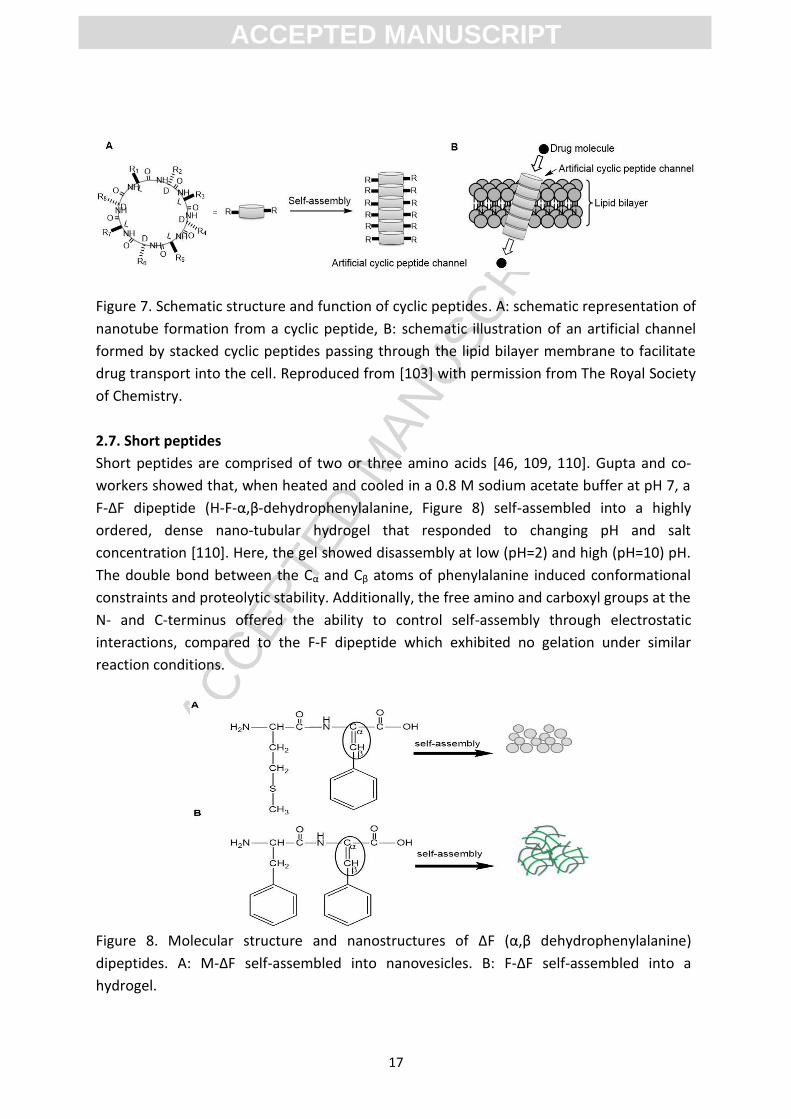
2.6. Cyclic peptides
Peptide cyclisation has been employed to impose rigidity to allow the molecule to adopt a
number of conformations that are not available to linear peptides. Increasing the rigidity of
the structure has been shown to enhance receptor binding affinity [102]. Cyclic peptides
have been shown to adopt flat conformations or stack via hydrogen bonding to form self-
assembled peptide nanotubes where the amino acid side chains of the peptide ring are
oriented outward (Figure 7) [103]. The diameter of the nanotube was controlled by the size
of the unit ring, and its external surface provided specific conformational properties by
modifying the interaction of the side chains. These nanostructures can be used in artificial
photosystems, biosensors, antimicrobials, electronic devices, photoresponsive materials,
selective transmembrane transport channels, and drug delivery [103-105]. A novel drug
delivery system comprised of alternating tryptophan and arginine in a cyclic octapeptide
[WR]4 was introduced by Parang and co-workers [102]. They showed that the optimal
balance between electrostatic and hydrophobic interactions of the cyclic peptides (drug
carrier) and phosphopeptides (transporters which give on/off signals to many enzymes
through interactions with protein kinases), led to the formation of circular vesicle-like
nanostructures (25-60 nm in diameter) with improved intracellular phosphopeptide
delivery. Compared to its linear counterpart this delivery system had higher enzymatic
stability, bypassed endosomal uptake, improved cell permeability, and allowed the nuclear
targeting and cellular delivery of impermeable phosphopeptides [102-106]. Furthermore, in
an aqueous solution of chloroaurate this cyclic peptide formed gold-capped nanoparticles
through the reducing activity of the tryptophan residue and attraction of chloroaurate
anions towards the positively charged arginine residues. This gold-capped cyclic peptide
delivery system was loaded with hydrophobic drugs (including doxorubicin, lamivudine,
emtricitabine, and stavudine) in an equal molar ratio and showed improved cellular uptake
and retention when used as a molecular transporter [107]. Additionally, cyclic peptide
nanotubes composed of (W-D-L)4-Q-D-L and applied to the delivery of the antitumor drug 5-
fluorouracil (5-FU) rapidly reached a high level of penetration into tumor cells where the
drug effect was strengthened as the dosage of the cyclic peptide increased. This increase
was associated with the cyclic peptide improving the transport of the drug into the target
cell [108].
ACC
EPTE
D M
ANU
SCR
IPT
ACCEPTED MANUSCRIPT
17
Figure 7. Schematic structure and function of cyclic peptides. A: schematic representation of
nanotube formation from a cyclic peptide, B: schematic illustration of an artificial channel
formed by stacked cyclic peptides passing through the lipid bilayer membrane to facilitate
drug transport into the cell. Reproduced from [103] with permission from The Royal Society
of Chemistry.
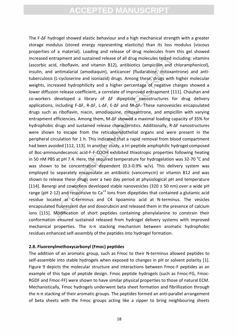
2.7. Short peptides
Short peptides are comprised of two or three amino acids [46, 109, 110]. Gupta and co-
workers showed that, when heated and cooled in a 0.8 M sodium acetate buffer at pH 7, a
F-ΔF dipeptide (H-F-α,β-dehydrophenylalanine, Figure 8) self-assembled into a highly
ordered, dense nano-tubular hydrogel that responded to changing pH and salt
concentration [110]. Here, the gel showed disassembly at low (pH=2) and high (pH=10) pH.
The double bond between the Cα and Cβ atoms of phenylalanine induced conformational
constraints and proteolytic stability. Additionally, the free amino and carboxyl groups at the
N- and C-terminus offered the ability to control self-assembly through electrostatic
interactions, compared to the F-F dipeptide which exhibited no gelation under similar
reaction conditions.
Figure 8. Molecular structure and nanostructures of ∆F (α,β dehydrophenylalanine)
dipeptides. A: M-∆F self-assembled into nanovesicles. B: F-∆F self-assembled into a
hydrogel.
ACC
EPTE
D M
ANU
SCR
IPT
ACCEPTED MANUSCRIPT
18
The F-∆F hydrogel showed elastic behaviour and a high mechanical strength with a greater
storage modulus (stored energy representing elasticity) than its loss modulus (viscous
properties of a material). Loading and release of drug molecules from this gel showed
increased entrapment and sustained release of all drug molecules tested including: vitamins
(ascorbic acid, riboflavin, and vitamin B12), antibiotics (ampicillin and chloramphenicol),
insulin, and antimalarial (amodiaquin), anticancer (fludarabine, mitoxantrone) and anti-
tuberculosis (L-cycloserine and isoniazid) drugs. Among these, drugs with higher molecular
weights, increased hydrophilicity and a higher percentage of negative charges showed a
lower diffusion release coefficient, a correlate of improved entrapment [111]. Chauhan and
co-workers developed a library of ∆F dipeptide nanostructures for drug delivery
applications, including F-∆F, R-∆F, L-∆F, E-∆F and M-∆F. These nanovesicles encapsulated
drugs such as riboflavin, niacin, amodiaquine, mitoxantrone, and ampicillin with varying
entrapment efficiencies. Among them, M-∆F showed a maximal loading capacity of 35% for
hydrophobic drugs and sustained release characteristics. Additionally, R-∆F nanostructures
were shown to escape from the reticuloendothelial organs and were present in the
peripheral circulation for 1 h. This indicated that a rapid removal from blood compartment
had been avoided [112, 113]. In another study, a tri-peptide amphiphilic hydrogel composed
of Boc-aminoundecanoic acid-F-F-COOH exhibited thixotropic properties following heating
in 50 nM PBS at pH 7.4. Here, the required temperature for hydrogelation was 32-70 οC and
was shown to be concentration dependent (0.3-0.9% w/v). This delivery system was
employed to separately encapsulate an antibiotic (vancomycin) or vitamin B12 and was
shown to release these drugs over a two day period at physiological pH and temperature
[114]. Banergi and coworkers developed stable nanovesicles (320 ± 50 nm) over a wide pH
range (pH 2-12) and responsive to Ca+2 ions from dipeptides that contained a glutamic acid
residue located at C-terminus and C4 lipoamino acid at N-terminus. The vesicles
encapsulated fluorescent dye and doxorubicin and released them in the presence of calcium
ions [115]. Modification of short peptides containing phenylalanine to constrain their
conformation ensured sustained released from hydrogel delivery systems with improved
mechanical properties. The π-π stacking mechanism between aromatic hydrophobic
residues enhanced self-assembly of the peptides into hydrogel formation.
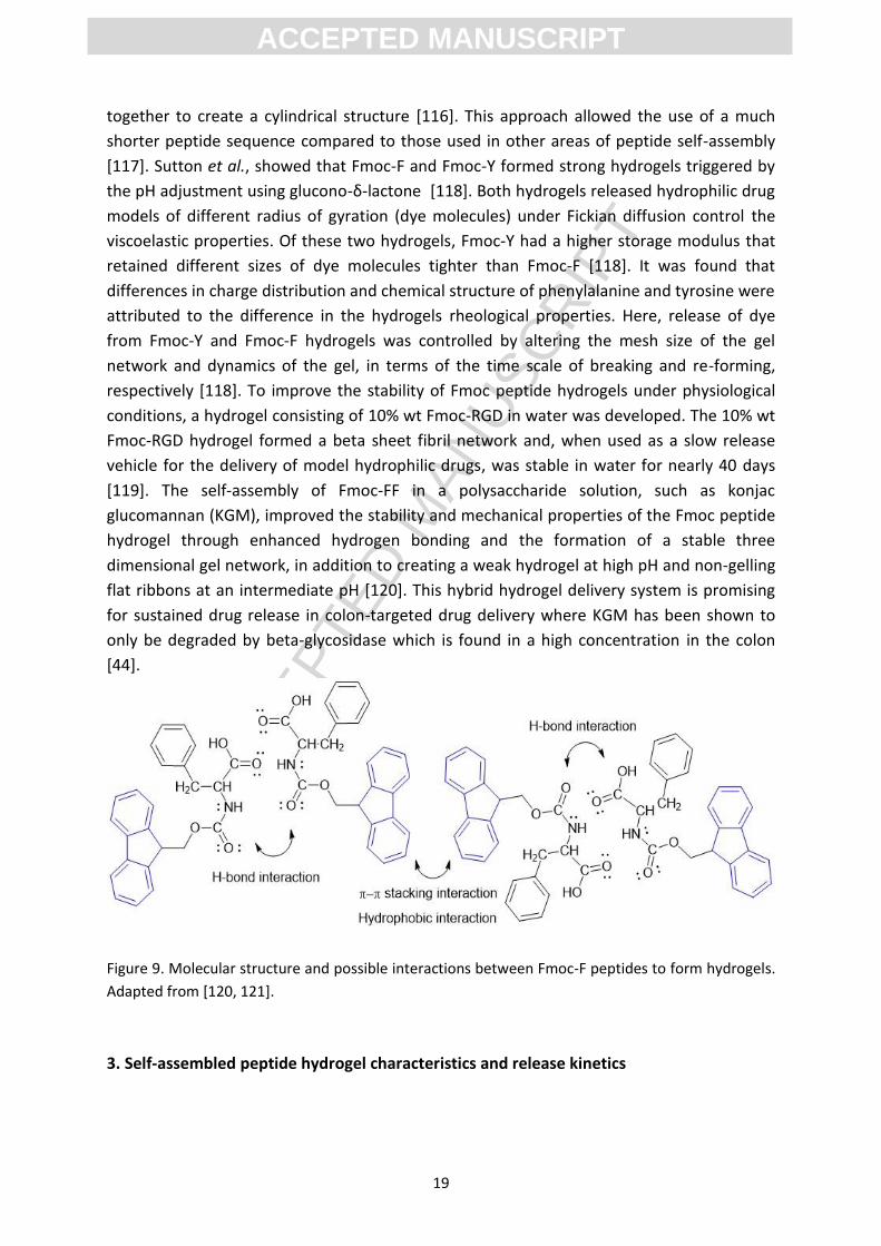
2.8. Fluorenylmethoxycarbonyl (Fmoc) peptides
The addition of an aromatic group, such as Fmoc to their N-terminus allowed peptides to
self-assemble into stable hydrogels when exposed to changes in pH or solvent polarity [1].
Figure 9 depicts the molecular structure and interactions between Fmoc-F peptides as an
example of this type of peptide design. Fmoc peptide hydrogels (such as Fmoc-FG, Fmoc-
RGDF and Fmoc-FF) were shown to have similar physical properties to those of natural ECM.
Mechanistically, Fmoc hydrogels underwent beta sheet formation and fibrillization through
the π-π stacking of their aromatic groups. The peptides formed an anti-parallel arrangement
of beta sheets with the Fmoc groups acting like a zipper to bring neighbouring sheets
ACC
EPTE
D M
ANU
SCR
IPT
ACCEPTED MANUSCRIPT
19
together to create a cylindrical structure [116]. This approach allowed the use of a much
shorter peptide sequence compared to those used in other areas of peptide self-assembly
[117]. Sutton et al., showed that Fmoc-F and Fmoc-Y formed strong hydrogels triggered by
the pH adjustment using glucono-δ-lactone [118]. Both hydrogels released hydrophilic drug
models of different radius of gyration (dye molecules) under Fickian diffusion control the
viscoelastic properties. Of these two hydrogels, Fmoc-Y had a higher storage modulus that
retained different sizes of dye molecules tighter than Fmoc-F [118]. It was found that
differences in charge distribution and chemical structure of phenylalanine and tyrosine were
attributed to the difference in the hydrogels rheological properties. Here, release of dye
from Fmoc-Y and Fmoc-F hydrogels was controlled by altering the mesh size of the gel
network and dynamics of the gel, in terms of the time scale of breaking and re-forming,
respectively [118]. To improve the stability of Fmoc peptide hydrogels under physiological
conditions, a hydrogel consisting of 10% wt Fmoc-RGD in water was developed. The 10% wt
Fmoc-RGD hydrogel formed a beta sheet fibril network and, when used as a slow release
vehicle for the delivery of model hydrophilic drugs, was stable in water for nearly 40 days
[119]. The self-assembly of Fmoc-FF in a polysaccharide solution, such as konjac
glucomannan (KGM), improved the stability and mechanical properties of the Fmoc peptide
hydrogel through enhanced hydrogen bonding and the formation of a stable three
dimensional gel network, in addition to creating a weak hydrogel at high pH and non-gelling
flat ribbons at an intermediate pH [120]. This hybrid hydrogel delivery system is promising
for sustained drug release in colon-targeted drug delivery where KGM has been shown to
only be degraded by beta-glycosidase which is found in a high concentration in the colon
[44].
Figure 9. Molecular structure and possible interactions between Fmoc-F peptides to form hydrogels.
Adapted from [120, 121].
3. Self-assembled peptide hydrogel characteristics and release kinetics
ACC
EPTE
D M
ANU
SCR
IPT
ACCEPTED MANUSCRIPT
20
3.1. Peptide hydrogel characteristics
An ideal hydrogel system should have good biocompatibility, biosafety, high stability,
optimal mechanical strength, and readily allow incorporation of bioactive ingredients with
controlled release at the required biological sites [122]. Altering the nano-architecture of
the peptide hydrogel network modulates hydrogel stiffness and porosity, effect the viscosity
and overall drug release profile [4].
In peptide hydrogel delivery systems, water soluble peptides undergo a transition from
liquid to a gel-like state (which is more commonly referred to as sol-gel transition) at the
target site, in response to changes in the ionic strength, temperature or pH of the medium
[44]. Physical gelation under biological conditions (pH-, ionic strength and thermo-
responsiveness) allows for the three dimensional, homogeneous encapsulation of desired
molecules and/or cells [48]. Moreover, peptide hydrogels exhibit mechano-responsive
properties. Thixotropic hydrogels induced a gel-sol transition by mechanical shaking and
quickly recovered into a gel-state after the stress was removed [123]. This self-healing
property is essential when designing a delivery system that has the ability to shear-thin
within a syringe needle and transform back to the gel after expulsion from the syringe.
Thixotropic hydrogels allow the drug to be injected directly into the targeted site without
surgical implantation [114]. For example, Schneider and co-workers developed a peptide
hydrogel comprised of 20 amino acids (VKVKVKVKVDPLPTKVKVKVKV-NH2) with sol-gel
transformation triggered by an increase in ionic strength in the presence of Dulbecco's
Modified Eagle's medium buffer. This gel-forming construct showed a shear-thin recovery
after injection to the targeted site, without syringe-clogging [48].
Veerman et al., showed that assembly time (tg) of the beta hairpin peptide
VKVKVKVKVDPLPTKVKVKVKV-NH2 decreased as the peptide concentration increased, from
approxmately 30 min for 0.05 wt % to 11 min for 0.15 wt %. They also demonstrated that
the relationship between gelation time and concentration followed power-law equation 1
where tg is gelation time and c is peptide concentration in weight percent [124, 125].
𝑡𝑔 = 𝑘𝑐−1 Eq.1
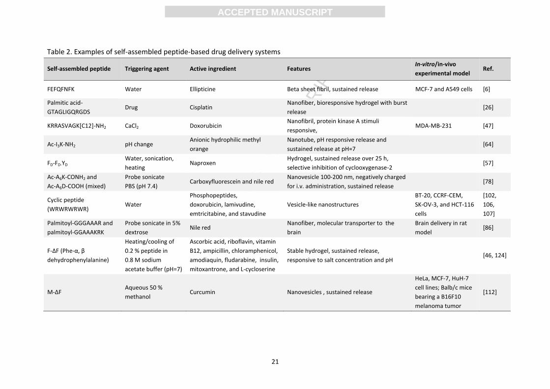
Examples of self-assembled peptide hydrogels are shown in Table 2.
ACC
EPTE
D M
ANU
SCR
IPT
ACCEPTED MANUSCRIPT
21
Table 2. Examples of self-assembled peptide-based drug delivery systems
Self-assembled peptide Triggering agent Active ingredient Features In-vitro/in-vivo
experimental model Ref.
FEFQFNFK Water Ellipticine Beta sheet fibril, sustained release MCF-7 and A549 cells [6]
Palmitic acid-
GTAGLIGQRGDS Drug Cisplatin
Nanofiber, bioresponsive hydrogel with burst
release [26]
KRRASVAGK[C12]-NH2 CaCl2 Doxorubicin Nanofibril, protein kinase A stimuli
responsive, MDA-MB-231 [47]
Ac-I3K-NH2 pH change Anionic hydrophilic methyl
orange
Nanotube, pH responsive release and
sustained release at pH=7 [64]
FD-FD-YD Water, sonication,
heating Naproxen
Hydrogel, sustained release over 25 h,
selective inhibition of cyclooxygenase-2 [57]
Ac-A6K-CONH2 and
Ac-A6D-COOH (mixed)
Probe sonicate
PBS (pH 7.4) Carboxyfluorescein and nile red
Nanovesicle 100-200 nm, negatively charged
for i.v. administration, sustained release [78]
Cyclic peptide
(WRWRWRWR) Water
Phosphopeptides,
doxorubicin, lamivudine,
emtricitabine, and stavudine
Vesicle-like nanostructures
BT-20, CCRF-CEM,
SK-OV-3, and HCT-116
cells
[102,
106,
107]
Palmitoyl-GGGAAAR and
palmitoyl-GGAAAKRK
Probe sonicate in 5%
dextrose Nile red
Nanofiber, molecular transporter to the
brain
Brain delivery in rat
model [86]
F-ΔF (Phe-α, β
dehydrophenylalanine)
Heating/cooling of
0.2 % peptide in
0.8 M sodium
acetate buffer (pH=7)
Ascorbic acid, riboflavin, vitamin
B12, ampicillin, chloramphenicol,
amodiaquin, fludarabine, insulin,
mitoxantrone, and L-cycloserine
Stable hydrogel, sustained release,
responsive to salt concentration and pH [46, 124]
M-ΔF Aqueous 50 %
methanol Curcumin Nanovesicles , sustained release
HeLa, MCF-7, HuH-7
cell lines; Balb/c mice
bearing a B16F10
melanoma tumor
[112]
ACC
EPTE
D M
ANU
SCR
IPT
ACCEPTED MANUSCRIPT
22
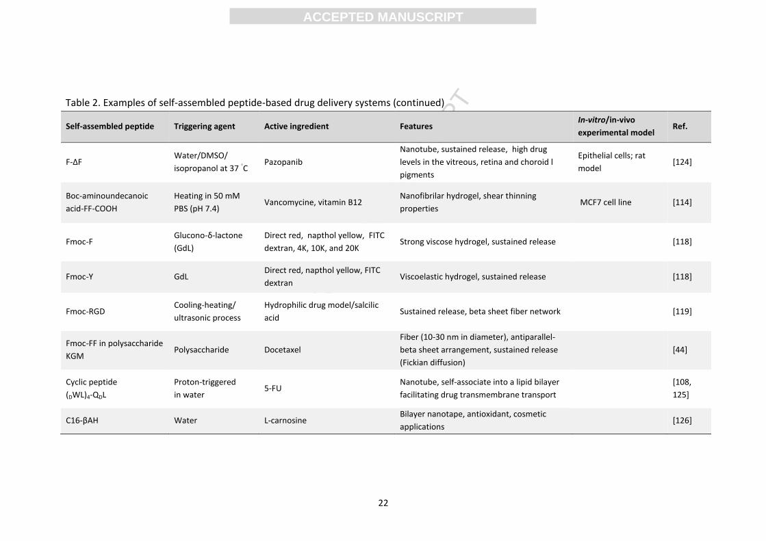
Table 2. Examples of self-assembled peptide-based drug delivery systems (continued)
Self-assembled peptide Triggering agent Active ingredient Features In-vitro/in-vivo
experimental model Ref.
F-ΔF Water/DMSO/
isopropanol at 37 ◦C
Pazopanib
Nanotube, sustained release, high drug
levels in the vitreous, retina and choroid l
pigments
Epithelial cells; rat
model [124]
Boc-aminoundecanoic
acid-FF-COOH
Heating in 50 mM
PBS (pH 7.4) Vancomycine, vitamin B12
Nanofibrilar hydrogel, shear thinning
properties MCF7 cell line [114]
Fmoc-F Glucono-δ-lactone
(GdL)
Direct red, napthol yellow, FITC
dextran, 4K, 10K, and 20K Strong viscose hydrogel, sustained release [118]
Fmoc-Y GdL Direct red, napthol yellow, FITC
dextran Viscoelastic hydrogel, sustained release [118]
Fmoc-RGD Cooling-heating/
ultrasonic process
Hydrophilic drug model/salcilic
acid Sustained release, beta sheet fiber network [119]
Fmoc-FF in polysaccharide
KGM Polysaccharide Docetaxel
Fiber (10-30 nm in diameter), antiparallel-
beta sheet arrangement, sustained release
(Fickian diffusion)
[44]
Cyclic peptide
(DWL)4-QDL
Proton-triggered
in water 5-FU
Nanotube, self-associate into a lipid bilayer
facilitating drug transmembrane transport
[108,
125]
C16-βAH Water L-carnosine Bilayer nanotape, antioxidant, cosmetic
applications [126]
ACC
EPTE
D M
ANU
SCR
IPT
ACCEPTED MANUSCRIPT
23
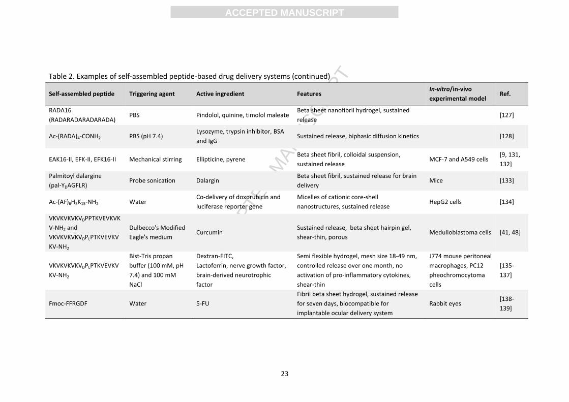
Table 2. Examples of self-assembled peptide-based drug delivery systems (continued)
Self-assembled peptide Triggering agent Active ingredient Features In-vitro/in-vivo
experimental model Ref.
RADA16
(RADARADARADARADA) PBS Pindolol, quinine, timolol maleate
Beta sheet nanofibril hydrogel, sustained
release [127]
Ac-(RADA)4-CONH2 PBS (pH 7.4) Lysozyme, trypsin inhibitor, BSA
and IgG Sustained release, biphasic diffusion kinetics [128]
EAK16-II, EFK-II, EFK16-II Mechanical stirring Ellipticine, pyrene Beta sheet fibril, colloidal suspension,
sustained release MCF-7 and A549 cells
[9, 131,
132]
Palmitoyl dalargine
(pal-YDAGFLR) Probe sonication Dalargin
Beta sheet fibril, sustained release for brain
delivery Mice [133]
Ac-(AF)6H5K15-NH2 Water Co-delivery of doxorubicin and
luciferase reporter gene
Micelles of cationic core-shell
nanostructures, sustained release HepG2 cells [134]
VKVKVKVKVDPPTKVEVKVK
V-NH2 and
VKVKVKVKVDPLPTKVEVKV
KV-NH2
Dulbecco's Modified
Eagle's medium Curcumin
Sustained release, beta sheet hairpin gel,
shear-thin, porous Medulloblastoma cells [41, 48]
VKVKVKVKVDPLPTKVEVKV
KV-NH2
Bist-Tris propan
buffer (100 mM, pH
7.4) and 100 mM
NaCl
Dextran-FITC,
Lactoferrin, nerve growth factor,
brain-derived neurotrophic
factor
Semi flexible hydrogel, mesh size 18-49 nm,
controlled release over one month, no
activation of pro-inflammatory cytokines,
shear-thin
J774 mouse peritoneal
macrophages, PC12
pheochromocytoma
cells
[135-
137]
Fmoc-FFRGDF Water 5-FU
Fibril beta sheet hydrogel, sustained release
for seven days, biocompatible for
implantable ocular delivery system
Rabbit eyes [138-
139]
ACC
EPTE
D M
ANU
SCR
IPT
ACCEPTED MANUSCRIPT
24
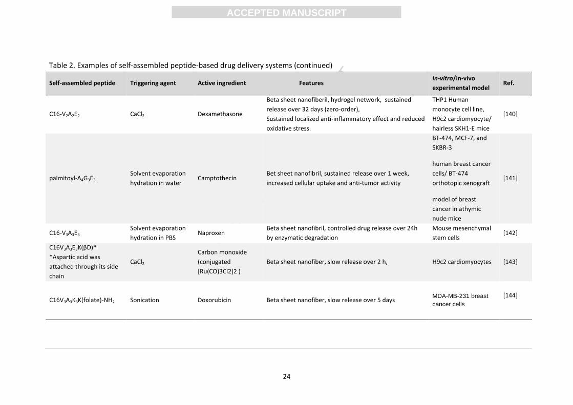
Table 2. Examples of self-assembled peptide-based drug delivery systems (continued)
Self-assembled peptide Triggering agent Active ingredient Features In-vitro/in-vivo
experimental model Ref.
C16-V2A2E2 CaCl2 Dexamethasone
Beta sheet nanofiberil, hydrogel network, sustained
release over 32 days (zero-order),
Sustained localized anti-inflammatory effect and reduced
oxidative stress.
THP1 Human
monocyte cell line,
H9c2 cardiomyocyte/
hairless SKH1-E mice
[140]
palmitoyl-A4G3E3 Solvent evaporation
hydration in water Camptothecin
Bet sheet nanofibril, sustained release over 1 week,
increased cellular uptake and anti-tumor activity
BT-474, MCF-7, and
SKBR-3
human breast cancer
cells/ BT-474
orthotopic xenograft
model of breast
cancer in athymic
nude mice
[141]
C16-V3A3E3 Solvent evaporation
hydration in PBS Naproxen
Beta sheet nanofibril, controlled drug release over 24h
by enzymatic degradation
Mouse mesenchymal
stem cells [142]
C16V3A3E3K(βD)*
*Aspartic acid was
attached through its side
chain
CaCl2
Carbon monoxide
(conjugated
[Ru(CO)3Cl2]2 )
Beta sheet nanofiber, slow release over 2 h, H9c2 cardiomyocytes [143]
C16V3A3K3K(folate)-NH2 Sonication Doxorubicin Beta sheet nanofiber, slow release over 5 days MDA-MB-231 breast
cancer cells
[144]
ACC
EPTE
D M
ANU
SCR
IPT
ACCEPTED MANUSCRIPT
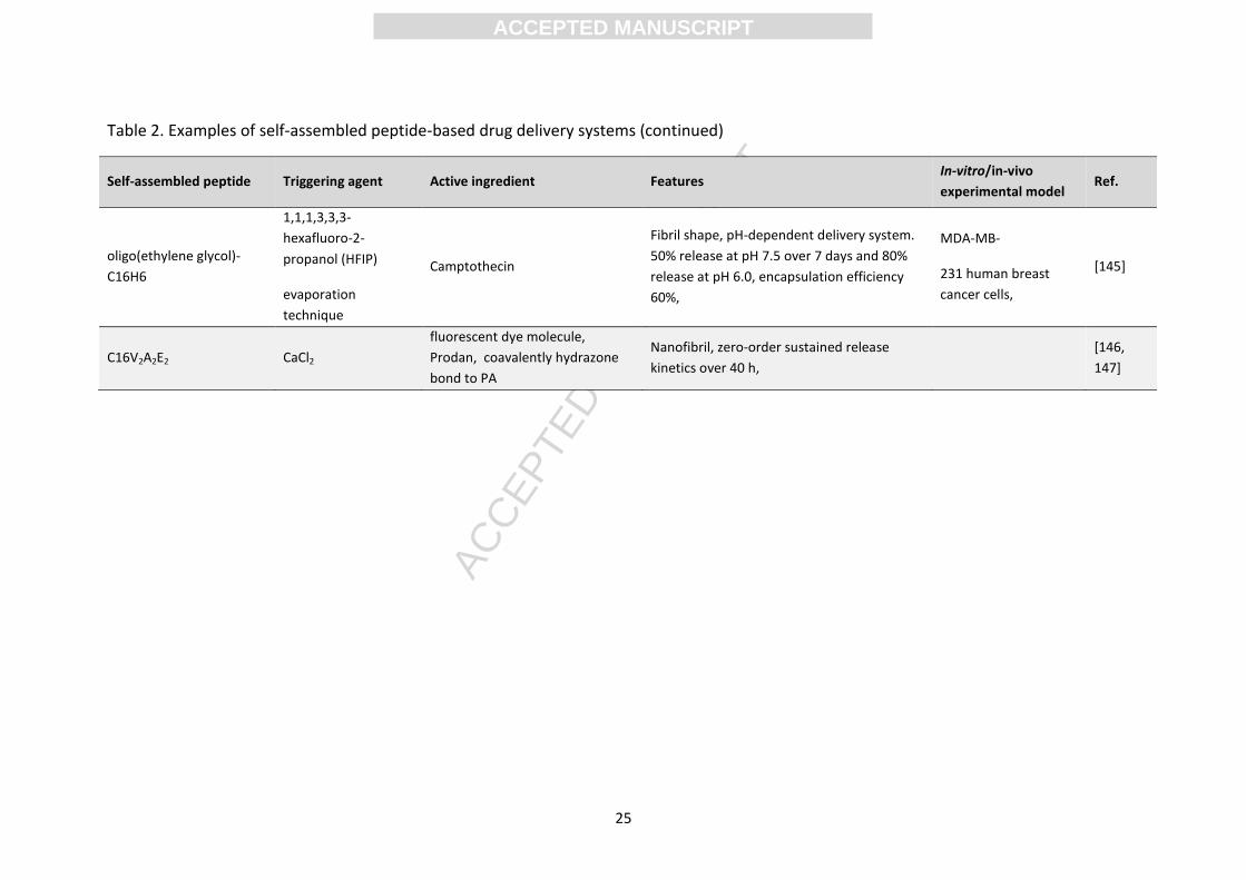
25
Table 2. Examples of self-assembled peptide-based drug delivery systems (continued)
Self-assembled peptide Triggering agent Active ingredient Features In-vitro/in-vivo
experimental model Ref.
oligo(ethylene glycol)-
C16H6
1,1,1,3,3,3-
hexafluoro-2-
propanol (HFIP)
evaporation
technique
Camptothecin
Fibril shape, pH-dependent delivery system.
50% release at pH 7.5 over 7 days and 80%
release at pH 6.0, encapsulation efficiency
60%,
MDA-MB-
231 human breast
cancer cells,
[145]
C16V2A2E2 CaCl2
fluorescent dye molecule,
Prodan, coavalently hydrazone
bond to PA
Nanofibril, zero-order sustained release
kinetics over 40 h,
[146,
147]
ACC
EPTE
D M
ANU
SCR
IPT
ACCEPTED MANUSCRIPT
26
3.2. Hydrogel release kinetics
Drug release through a peptide hydrogel matrix can be controlled by many factors, including
the network mesh size, surface area to volume ratio, the properties and concentration of
the gelator, salt concentration, pH, and interactions between the matrix and the entrapped
molecules [118]. Briuglia et al., showed that the drug release kinetics were dependent on
the chemical properties of the drug (Log P, pKa, isoelectric point, presence of aromatic rings,
and steric hindrance) and the medium chosen for the release study. For instance, controlled
release of small hydrophobic drug molecules (quinine and pindolol) from the RADA16
peptide hydrogel caused specific binding between the aromatic groups and peptide matrix,
or sterically hindered the drug molecule, which reduced the drugs diffusion capacity [129].
This system enabled release of the drug over a seven day period without changes to the
drug morphology [129].
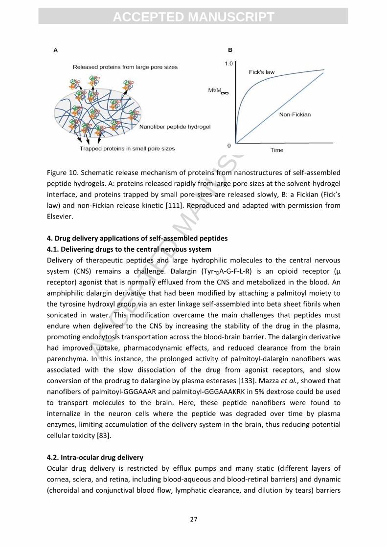
The release kinetics of proteins from peptide hydrogels was predominantly dependent on
the density of the peptide hydrogel, protein size and charge [46, 111, 118]. In a study by
Nagai et al., the release of four proteins (lysozyme, trypsin inhibitor, BSA and IgG) from the
peptide hydrogel (Ac-(RADA)4-CONH2) were investigated separately. All four proteins
released from this delivery system showed a biphasic diffusion with burst release in the first
hour (related to the escape of the proteins located in the solvent-hydrogel interface which
had a larger pore size, Figure 10A), and obeyed Fickian diffusion (Figure 10B). The remaining
protein drug was released in a hyperbolic manner up to 100% over 30-50 hours. Small pore
size or other obstacles to diffusion restricted the release of these proteins to a non-Fickian,
anomalous diffusion (Figure 10). Increased peptide hydrogel density and increased protein
drugs size were associated with a decrease in the release rate. Drug release from Fmoc-
amino acid hydrogels was controlled by the dynamics of the gel network and followed a
Fickian diffusion model [118].
Another factor that affected the kinetics of protein-release from the nanofiber hydrogel was
the protein charge under physiological conditions [46, 111, 148]. Here, the conformational
properties and functionality of the proteins did not change before or after release from this
system, indicating that minimal interactions occurred between the folded proteins and the
nanofiber hydrogel [130]. The effect of a peptide hydrogels pore size, density and
electrostatic interactions between the drug and peptide(s) upon drug release from the beta
hairpin, VKVKVKVKVDPLPTKVEVKVKV-NH2 was also studied by Branco et al. [135, 137, 148].
Another feature of peptide hydrogels is that they do not swell after formation, even when
exposed to bodily fluids, supporting consistent diffusion characteristics [41]. Moreover, the
shape of the peptide hydrogel can be maintained during the release process without
shrinkage [111, 119].
ACC
EPTE
D M
ANU
SCR
IPT
ACCEPTED MANUSCRIPT
27
Figure 10. Schematic release mechanism of proteins from nanostructures of self-assembled
peptide hydrogels. A: proteins released rapidly from large pore sizes at the solvent-hydrogel
interface, and proteins trapped by small pore sizes are released slowly, B: a Fickian (Fick’s
law) and non-Fickian release kinetic [111]. Reproduced and adapted with permission from
Elsevier.
4. Drug delivery applications of self-assembled peptides
4.1. Delivering drugs to the central nervous system
Delivery of therapeutic peptides and large hydrophilic molecules to the central nervous
system (CNS) remains a challenge. Dalargin (Tyr-DA-G-F-L-R) is an opioid receptor (µ
receptor) agonist that is normally effluxed from the CNS and metabolized in the blood. An
amphiphilic dalargin derivative that had been modified by attaching a palmitoyl moiety to
the tyrosine hydroxyl group via an ester linkage self-assembled into beta sheet fibrils when
sonicated in water. This modification overcame the main challenges that peptides must
endure when delivered to the CNS by increasing the stability of the drug in the plasma,
promoting endocytosis transportation across the blood-brain barrier. The dalargin derivative
had improved uptake, pharmacodynamic effects, and reduced clearance from the brain
parenchyma. In this instance, the prolonged activity of palmitoyl-dalargin nanofibers was
associated with the slow dissociation of the drug from agonist receptors, and slow
conversion of the prodrug to dalargine by plasma esterases [133]. Mazza et al., showed that
nanofibers of palmitoyl-GGGAAAR and palmitoyl-GGGAAAKRK in 5% dextrose could be used
to transport molecules to the brain. Here, these peptide nanofibers were found to
internalize in the neuron cells where the peptide was degraded over time by plasma
enzymes, limiting accumulation of the delivery system in the brain, thus reducing potential
cellular toxicity [83].
4.2. Intra-ocular drug delivery
Ocular drug delivery is restricted by efflux pumps and many static (different layers of
cornea, sclera, and retina, including blood-aqueous and blood-retinal barriers) and dynamic
(choroidal and conjunctival blood flow, lymphatic clearance, and dilution by tears) barriers
ACC
EPTE
D M
ANU
SCR
IPT
ACCEPTED MANUSCRIPT
28
[149]. Ocular targeting and maintaining therapeutic drug levels over time present major
delivery challenges [150]. Nanotechnology-based formulations, such as nanomicelles,
liposomes and poly-lysine dendrimers (200-2000 nm), have been developed to overcome
these barriers and deliver drugs to the posterior segment of the eye with a long term
release profile [150, 151]. Recent studies found that the Fmoc-FFRGDF peptide self-
assembled into beta sheet fibrils with a width of 20 nm. This transparent hydrogel exhibited
biocompatibility in rabbit eyes as an implantable delivery system for the treatment of ocular
anterior diseases such as glaucoma and keratopathy [139]. When loaded with 5-fluorouracil
(5-FU, 5% wt), an anti-proliferative agent, this hydrogel demonstrated a sustained release
profile without swelling, shrinking or erosion of the gel in the aqueous media. Additionally,
no burst release was observed. Here, the peptide hydrogel released the drug with a Fickian
diffusion controlled mechanism of (Figure 10B) [152]. This implantable delivery system
significantly lowered the intraocular pressure of the rabbits eyes within the 28 day
postoperative period [138]. Furthermore, Panda et al., used nanotubes generated from a F-
ΔF dipeptide for the intravitreal delivery of pazopanib to treat choroidal neovascularisation.
An in vitro study showed 25% drug loading, 55% loading efficiency and a profile of sustained
release over 35 days. Moreover high drug levels were detected in vitreous, retina and
choroidal pigment epithelial cells, compared to the plain drug, with a sustained release over
15 days after intravitreal injection in an in vivo rat model [124].
4.3. Cardiovascular drug delivery
Growth factors (GFs), hormones, and other proteins can promote myocardial regeneration
and enhance the survival of grafted cells. To successfully deliver these drugs to the
myocardial tissue a novel and promising strategy that combined self-assembling peptides
with functional motifs to design noncytotoxic, biodegradable, porous, permeable, and
flexible scaffolds. Here, prolonged cardiogenesis was stimulated at the myocardial scars by
the sustained delivery of multiple GFs with distinct release kinetics [153]. The peptide
amphiphile RADA16 was found to adsorb GFs through noncovalent interactions and induced
angiogenesis with sustained release over a 14 day period [153]. In another study, a peptide
scaffold was designed by attaching the heparin-binding sequence domain LRKKLGKA to the
RADA16 peptide, which self-assembled into nanofibers under physiological conditions. This
delivery system provided sustained release of a vascular endothelial growth factor (VEGF)
for at least one month after transplantation when delivered by ventricular myocardial
injection in a murine model. Here, improvement of cardiac function and reduction of scar
size and collagen deposition was observed [154]. Moreover, a nitric oxide (NO) releasing
peptide amphiphile that mimicked the native endothelial ECM was designed to coat
cardiovascular implants. Two different lipidated peptide amphiphiles were mixed with
deionized water (9:1 molar ratio) forming nanofibrous scaffolds and then subsequently
reacted with pure NO under high pressure. Peptide amphiphiles contained either an
endothelial cell-adhesive ligand (GTAGLIGQ-YIGSR) or a poly-lysine (GTAGLIGQ-KKKKK) NO
donor. Burst release of NO was detected within 48 hours followed by 30 days of sustained
ACC
EPTE
D M
ANU
SCR
IPT
ACCEPTED MANUSCRIPT
29
release. The self-assembled nanofiber matrix coating significantly enhanced the initial
adhesion and proliferation of endothelial cells, but limited proliferation of smooth muscle
cells. Additionally, the matrix limited adhesion of platelets, used to correlate the risk of
thrombosis, was 150-fold less than the standard, a collagen-coated surface [155].
4.4. Bone drug delivery
Targeting drugs to bone tissue for disorders such as osteoporosis or osteosarcoma is very
challenging due to the complex mineralized micro- and nano-structure of bone [156]. Short
repetitive peptide sequences of aspartic acid were shown to bind to hydroxyapatite in vitro
and in vivo, promoting accumulation of small drug molecules in the bone [156, 157].
Hydroxyapatite is a naturally occurring mineral form of calcium apatite [Ca5(PO4)3] found in
bone and dentin. Proteins that naturally bind calcium phosphate are rich in phosphorylated
serine [157, 158]. Stupp and co-workers showed that hydroxyapatite nucleated on the
surface of peptide amphiphile nanofibers that contained phosphorylated serine [158, 160].
Self-assembled peptide hydrogels have been modified by adding phosphoserine or RGD
residues to improve mineralisation and cell adhesion, respectively. Peptide sequences P(S-
PO4-F)5-S-PO4-P, C15H31C(O)-C4G3SARGD-COOH and P(Y-PO4-F)5-Y-PO4-P are examples of self-
assembled peptide hydrogels used in bone tissue drug delivery [118, 161]. These peptide
sequences can be formulated into hydrogels, membranes, solid matrices and mineral-
peptide composites, to deliver therapeutic agents, such as anti-resorptive drugs, to bone or
adjacent tissues [118, 162]. Peptide hydrogels composed of PD(FD)5P and loaded with
tricalcium phosphate increased alkaline phosphatase activity (an early osteogenic marker)
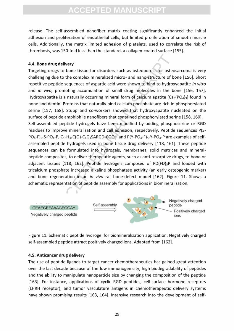
and bone regeneration in an in vivo rat bone-defect model [162]. Figure 11. Shows a
schematic representation of peptide assembly for applications in biomineralization.
Figure 11. Schematic peptide hydrogel for biomineralization application. Negatively charged
self-assembled peptide attract positively charged ions. Adapted from [162].
4.5. Anticancer drug delivery
The use of peptide ligands to target cancer chemotherapeutics has gained great attention
over the last decade because of the low immunogenicity, high biodegradability of peptides
and the ability to manipulate nanoparticle size by changing the composition of the peptide
[163]. For instance, applications of cyclic RGD peptides, cell-surface hormone receptors
(LHRH receptor), and tumor vasculature antigens in chemotherapeutic delivery systems
have shown promising results [163, 164]. Intensive research into the development of self-
ACC
EPTE
D M
ANU
SCR
IPT
ACCEPTED MANUSCRIPT
30
assembled peptide delivery vehicles for active or passive targeting of chemotherapeutics
has accelerated due to the peptides’ desirable physicochemical properties and potential for
tailoring to suit specific biological applications [26]. Successful applications include: design
of nanodelivery systems for targeted therapy, enhancing the efficiency of existing delivery
systems, and optimizing peptide-drug formulations to increase stability, loading efficiency
and control the release of the chemotherapeutic cargo [6]. The RGD sequence in the
peptide amphiphile is known to have cell adhesion properties, the ability to mimic
characteristics of the ECM, and is able to specifically bind to the upregulated αvβ3 and αvβ5
integrins (transmembrane glycoproteins) during tumor growth and metastasis, thus gaining
entry into the cells and enabling endocytosis [165]. This makes RGD an important sequence
in the design of peptide amphiphile for targeted anticancer drug delivery that aim to release
clinically significant levels of cytotoxic drugs into the localized tumor region. To this end, a
bioresponsive gel delivery system formed by the self-assembly of cisplatin (cis-
dichlorodiamine platinum(II)) in a 4% solution of palmitic acid-GTAGLIGQRGDS in water at
37 οC was designed [158]. This delivery system assembled into a nanofiber gel via the
formation of complexes between platinium and the carboxylic acid groups on the adjacent
nanofiber. The resultant gel displayed RGD ligands on the surface, which were directed at
the integrin receptors that are overexpressed on cancer cells. Upon increasing
concentration of matrix metalloproteinase-2 (MMP2) and biodegradation of the gel by
MMP2, the drug was released at the tumor site [26].
Stupp and co-workers developed a protein kinase A (PKA, an extracellular biomarker for
cancer) stimuli responsive peptide hydrogel to deliver doxorubicin to cancer cells. The
peptide amphiphile KRRASVAGK[C12]-NH2 contained the specific consensus substrate
(RRXSO, X: any residue, O: hydrophobic) for PKA that formed nanofibril structures. This
substrate was phosphorylated and dephosphorylated at the serine position by PKA and
alkaline phosphatase (AP), respectively. Treatment of the nanofibrils using PKA diminished
fibril morphology while subsequent treatment with AP restored fibril morphology. This
substrate was specific for PKA rather than other protein kinases. Loading of doxorubicin into
this delivery system showed a 40% release rate over 7 hours. Treatment of the breast
cancer cell line MDA-MB-231 with this drug loaded peptide amphiphile resulted in the death
of all cancer cells [47]. Delivery of therapeutics using dynamic assemblies is a promising
method for targeted drug delivery.
Fung et al., showed that EAK16-II and EAK16-IV formed a colloidal suspension in the
presence of ellipticine in an aqueous environment. The in vitro anticancer activity of these
formulations was dependent on the molecular state (protonated, crystal or neutral) of
ellipticine that was present during formulation [132]. Interestingly, this complex was formed
in a time-dependent manner by mechanically stirring (not sonication or shaking) the
solution, and was independent of peptide concentration over the tested range (0.01-0.5
mg/ml) [9]. When the peptide concentration was close to its critical aggregation
concentration (CAC, 0.1 mg/ml), the equilibration time was minimal (5 hours). Peptide
concentrations higher than the CAC conserved the protonated ellipticine for a longer time
ACC
EPTE
D M
ANU
SCR
IPT
ACCEPTED MANUSCRIPT
31
40 hours [131]. Nevertheless, the rate at which the drug was released from this system was
dependent on the peptide concentration, and the loading capacity was dependent on the
drug concentration [9]. In vitro toxicity assays indicated that the complexes (>5:1 ratio of
peptide:drug) formed with protonated ellipticine were effective at killing both human breast
cancer cells (MCF-7) and adenocarcinomic human alveolar basal epithelial cells (A549),
although their toxicity decreased significantly at lower concentrations [131]. Further,
increasing the hydrophobicity of the EFK16-II peptide increased hydrophobic interactions
between the drug and peptide, significantly increasing the stability of the formulation. The
original desired cytotoxic effect of the EFK16-II peptide against cancer cells was low due to
the formation of complexes with the crystal state of ellipticine [132]. Saadatmousavi et al.,
showed that the complementary peptide AC8 (FEFQFNFK) self-assembled in aqueous
solution with a CMC of 10-15 µM (0.01-0.015 mg/ml) and could encapsulate ellipticine (0.05
mg/ml) in its neutral molecular state in aqueous media at 0.1 mg/ml (above CMC) to form
100 nm particles. This complex (>2:1 ratio of peptide:drug) with neutral ellipticine was
effective at killing MCF-7 and A-549 cell lines after 48 hours [6]. The peptide sequence
affects properties of peptide-drug assemblies and their cellular toxicities, however, these
peptide-drug formulations need to be optimized to enhance the therapeutic effect and the
efficacy of the delivery system.
Haines-Butterick et al., designed a curcumin delivery peptide hydrogel
(VKVKVKVKVDPLPTKVKVKVKV-NH2) that gelated in response to an increase in the ionic
strength of the medium. Curcumin is a hydrophobic polyphenol drug derived from the
turmeric root and has been shown to have antioxidant, anti-inflammatory and anti-
tumorigenic properties. In a low strength ionic buffer at pH 7.4, electrostatic repulsion
between the positively charged lysine residues prevented the folding of the peptide. The
lysine-associated charges were neutralised by adding Dulbecco's Modified Eagle's medium
containing 160 mM of inorganic salts. This induced self-assembly of the peptide, both
laterally (intermolecular H-bond and van der Waals attractions) and facially (hydrophobic
face of the peptide), which led to a beta sheet hairpin structure (Figure 2) [48]. This peptide
hydrogel succeeded as a reservoir for the sustained release of curcumin (4 mM) over a two-
week period. Incorporating the drug in a peptide scaffold stiffened the gels. This peptide
hydrogel showed shear-thinning properties, increased drug stability under physiologic
conditions, and retained its bioactivity [41]. Liu et al., encapsulated curcumin in a tumor-
targeting self-assembled peptide nanofiber formed by Nap-GFFYGRGD with a 32%
encapsulation efficiency (0.5 mM curcumin encapsulated by the peptide at 5mg/mL). This
delivery system released 60% of the drug in a sustained manner over two days with high
cellular uptake in αvβ3 integrin-positive HepG2 liver carcinoma cells and was found to
accumulate in tumor cells after intravenous administration in a mouse model [166].
Panda and co-workers created nanovesicles that permitted the sustained release of
cucurmin over a 36 hour period. The ~40 nm nanovesicles were generated from the self-
assembly of M-∆F in methanol followed by methanol evaporation resulting in a drug loading
of 30% and a loading efficiency of 92%. The high drug loading may be related to the π-π
ACC
EPTE
D M
ANU
SCR
IPT
ACCEPTED MANUSCRIPT
32
stacking interaction between curcumin and ∆F. The cytotoxic effect of the cucurmin
nanovesicle system was higher than native cucurmin when tested in different cancerous cell
lines including human cervical cancer cell line (HeLa), MCF-7 and human hepatocarcinoma
cell line (HuH-7). Moreover, this formulation inhibited tumor growth in Balb/c mice that
bore a B16F10 melanoma tumor [112]. Future work for self-assembled anti-cancer delivery
systems requires the formulation of stable complexes and investigation into their
immunogenicity and in vivo therapeutic properties.
5. Vaccine engineering
The engineering of immunomodulatory materials to act as delivery vehicles and gain a
better understanding of the immune system is a challenging field. Self-assembled
nanomaterials can be designed to induce or prevent complement activation and can be
used as an adjuvant or adjuvant carrier. Also, these nanomaterials provide
microenvironments that may aid interaction with antigen presenting cells (APCs), thus
contributing to both prophylactic and immunotherapeutic vaccines [167].
5.1. Self-assembled peptides for vaccine design
Self-assembled peptides have shown various effects on the immune system. Many peptides
stimulate no detectable antibody responses while others elicit potent responses without the
addition of supplementary adjuvants. Strategies for avoiding or inducing an immune
response using these materials has not been well characterised [167]. Developing peptide-
based vaccines has been hampered by the need to display antigens in their native
conformation and the use of immuno-adjuvants that only exhibit adjuvanting properties
under specific circumstances [168]. Recently, several patents have been registered for the
development of self-adjuvanting vaccines from self-assembled peptides [164, 165]. These
materials provide scaffolds that can accommodate multivalency and molecular specificity,
and enable anable control over epitope position [169].
Self-assembled nanomaterials were shown to display high densities of antigens on their
surface and elicited high-titre, high affinity, and neutralizing IgG responses as well as CD8+ T
cell mediated protection [172, 173]. Nanomaterials have an important role in vaccine
delivery due to their intrinsic adjuvanticity influenced by the properties of particle,
enhancing the immune response [174, 175]. Most attempts at engineering a vaccine that
use this class of biomaterials include peptides that form beta sheets and lipidated peptide
amphiphiles (Sections 5.1.1 and 5.1.2). Another approach used repeats of the
immunodominant peptide that self-assembled into nanoparticles. Kaba et al. designed a
self-assembled peptide nanovaccine composed of approximately 25 nm nanoparticles with
high antigen display on their surface. Here, a nanovaccine comprised of the tandem repeats
of the B cell immunodominant malaria parasite, Plasmodium berghei circumsporozite
protein [(DPPPPNPN)2D], stimulated a long-lived protective immune response without an
additional adjuvant. Mice that received the vaccine candidates (three times at two weeks
intervals) were protected 6 months after the last immunization and up to 15 months after
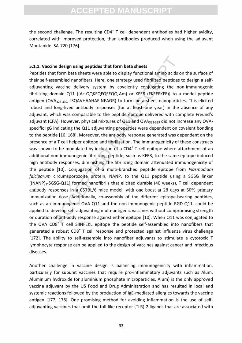
ACC
EPTE
D M
ANU
SCR
IPT
ACCEPTED MANUSCRIPT
33
the second challenge. The resulting CD4+ T cell dependent antibodies had higher avidity,
correlated with improved protection, than antibodies produced when using the adjuvant
Montanide ISA-720 [176].
5.1.1. Vaccine design using peptides that form beta sheets
Peptides that form beta sheets were able to display functional amino acids on the surface of
their self-assembled nanofibers. Here, one strategy used fibrilized peptides to design a self-
adjuvanting vaccine delivery system by covalently conjugating the non-immunogenic
fibrilizing domain Q11 [(Ac-QQKFQFQFEQQ-Am) or KFE8 (FKFEFKFE)] to a model peptide
antigen (OVA323-339, ISQAVHAAHAEINEAGR) to form beta sheet nanoparticles. This elicited
robust and long-lived antibody responses (for at least one year) in the absence of any
adjuvant, which was comparable to the peptide epitope delivered with complete Freund’s
adjuvant (CFA). However, physical mixtures of Q11 and OVA323-339 did not increase any OVA-
specific IgG indicating the Q11 adjuvanting properties were dependent on covalent bonding
to the peptide [10, 168]. Moreover, the antibody response generated was dependent on the
presence of a T cell helper epitope and fibrilization. The immunogenicity of these constructs
was shown to be modulated by inclusion of a CD4+ T cell epitope where attachment of an
additional non-immunogenic fibrilizing peptide, such as KFE8, to the same epitope induced
high antibody responses, diminishing the fibrilizing domain attenuated immunogenicity of
the peptide [10]. Conjugation of a multi-branched peptide epitope from Plasmodium
falciparum circumsporozoite protein, NANP, to the Q11 peptide using a SGSG linker
[(NANP)3-SGSG-Q11] formed nanofibrils that elicited durable (40 weeks), T cell dependent
antibody responses in a C57BL/6 mice model, with one boost at 28 days at 50% primary
immunization dose. Additionally, co-assembly of the different epitope-bearing peptides,
such as an immunogenic OVA-Q11 and the non-immunogenic peptide RGD-Q11, could be
applied to develop self-adjuvanting multi-antigenic vaccines without compromising strength
or duration of antibody response against either epitope [10]. When Q11 was conjugated to
the OVA CD8+ T cell SIINFEKL epitope the peptide self-assembled into nanofibers that
generated a robust CD8+ T cell response and protected against influenza virus challenge
[172]. The ability to self-assemble into nanofiber adjuvants to stimulate a cytotoxic T
lymphocyte response can be applied to the design of vaccines against cancer and infectious
diseases.
Another challenge in vaccine design is balancing immunogenicity with inflammation,
particularly for subunit vaccines that require pro-inflammatory adjuvants such as Alum.
Aluminium hydroxide (or aluminium phosphate microparticles, Alum) is the only approved
vaccine adjuvant by the US Food and Drug Administration and has resulted in local and
systemic reactions followed by the production of IgE-mediated allergies towards the vaccine
antigen [177, 178]. One promising method for avoiding inflammation is the use of self-
adjuvanting vaccines that omit the toll-like receptor (TLR)-2 ligands that are associated with
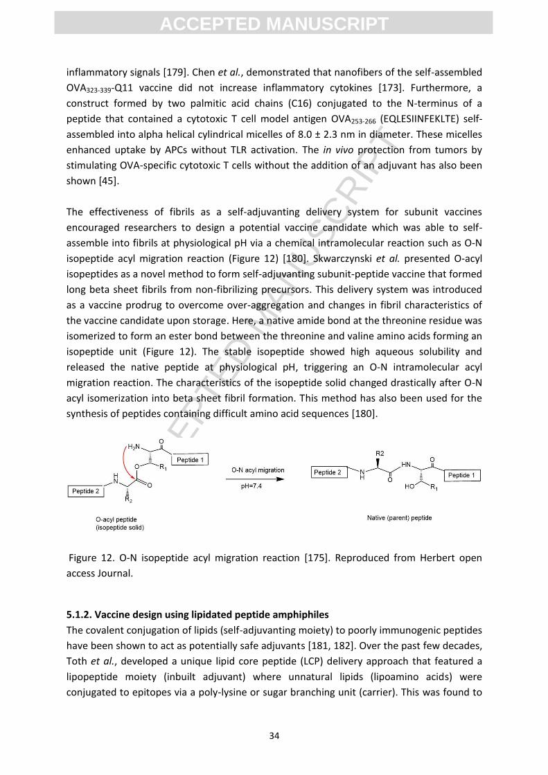
ACC
EPTE
D M
ANU
SCR
IPT
ACCEPTED MANUSCRIPT
34
inflammatory signals [179]. Chen et al., demonstrated that nanofibers of the self-assembled
OVA323-339-Q11 vaccine did not increase inflammatory cytokines [173]. Furthermore, a
construct formed by two palmitic acid chains (C16) conjugated to the N-terminus of a
peptide that contained a cytotoxic T cell model antigen OVA253-266 (EQLESIINFEKLTE) self-
assembled into alpha helical cylindrical micelles of 8.0 ± 2.3 nm in diameter. These micelles
enhanced uptake by APCs without TLR activation. The in vivo protection from tumors by
stimulating OVA-specific cytotoxic T cells without the addition of an adjuvant has also been
shown [45].
The effectiveness of fibrils as a self-adjuvanting delivery system for subunit vaccines
encouraged researchers to design a potential vaccine candidate which was able to self-
assemble into fibrils at physiological pH via a chemical intramolecular reaction such as O-N
isopeptide acyl migration reaction (Figure 12) [180]. Skwarczynski et al. presented O-acyl
isopeptides as a novel method to form self-adjuvanting subunit-peptide vaccine that formed
long beta sheet fibrils from non-fibrilizing precursors. This delivery system was introduced
as a vaccine prodrug to overcome over-aggregation and changes in fibril characteristics of
the vaccine candidate upon storage. Here, a native amide bond at the threonine residue was
isomerized to form an ester bond between the threonine and valine amino acids forming an
isopeptide unit (Figure 12). The stable isopeptide showed high aqueous solubility and
released the native peptide at physiological pH, triggering an O-N intramolecular acyl
migration reaction. The characteristics of the isopeptide solid changed drastically after O-N
acyl isomerization into beta sheet fibril formation. This method has also been used for the
synthesis of peptides containing difficult amino acid sequences [180].
Figure 12. O-N isopeptide acyl migration reaction [175]. Reproduced from Herbert open
access Journal.
5.1.2. Vaccine design using lipidated peptide amphiphiles
The covalent conjugation of lipids (self-adjuvanting moiety) to poorly immunogenic peptides
have been shown to act as potentially safe adjuvants [181, 182]. Over the past few decades,
Toth et al., developed a unique lipid core peptide (LCP) delivery approach that featured a
lipopeptide moiety (inbuilt adjuvant) where unnatural lipids (lipoamino acids) were
conjugated to epitopes via a poly-lysine or sugar branching unit (carrier). This was found to
ACC
EPTE
D M
ANU
SCR
IPT
ACCEPTED MANUSCRIPT
35
induce self-assembly and provide self-adjuvanting properties to vaccine constructs [183,
184]. Toth et al., tested the LCP vaccine delivery system by developing vaccine candidates
that contained antigens from Group A Streptococcus (GAS) strains that were endemic to
northern Australia. The native alpha helical secondary structure of the minimal B cell
epitope from a GAS membrane protein (M protein) was maintained by flanking the antigen
with regions of the yeast GCN4 protein to create the J14 sequence (KQAEDKVK-
ASREAKKQVEKALE-QLEDKVK). Molecules that contained multiple copies of J14 conjugated to
the LCP delivery system self-assembled into large nanoparticles (200-1000 nm in diameter)
and stimulated J14-specific IgG antibody response in a B10.Br (H-2K) mice model more
immunogenic than the negative control PBS. This study showed attachment of J14 epitope
from its N- or C-terminus to the LCP (Figure 13A) system produced similar physicochemical
properties and antibody response [185].
The J14-like epitope dJ14i, contained two repeats of the minimal B cell epitope and a triple-
alanine spacer (AAA-ASREAKKQVEKALE-ASREAKKQVEKALE), and self-assembled into smaller
nanoparticles (15-20 nm) than J14 when used in an LCP system (Figure 13A) while retaining
a helical secondary structure. The dJ14i nanoparticles induced a similar immune response to
the J14 epitope when administered subcutaneously with CFA in a mouse model. However,
the antibodies did not recognize the native GAS M protein epitope, p145 post second boost
[186]. A further study investigated the efficacy of the self-adjuvanting lipopeptide-based
GAS vaccine, LP-88/30-J14, which contained the conserved C-terminal J14 epitope, the N-
terminal GAS epitope (88/30, DNGKAIYERARERALQELGP), a universal T cell helper peptide
epitope (P25, KLIPNASLIENCTKAEL), and two lipoamino acids (C16). Here, LP-88/30-J14
(Figure 13B) self-assembled into nanoparticles of 5 nm, enhanced APC uptake, maturation
and antibody response without additional adjuvant. Furthermore, incorporation of N-
terminus 88/30 epitope into the construct did not diminish J14-specific antibody response.
The IgG antibodies were capable of binding to endemic GAS strains in a
immunofluorescence microscopy assay [187]. This study found that the immune response
correlated with particle size and the structural arrangement of the vaccine components in
the chimeric molecule. Azmi et al. designed vaccine constructs where J14 was conjugated to
the fibrilizing peptide Q11 (Figure 13C) with or without lipoamino acids were characterized
and tested in a B10.BR mice model. The Q11 peptide failed to induce fibril formation when
conjugated to J14, instead resulting in polydisperse nanoparticles of 20-250 nm. These
nanoparticles failed to stimulate a high antibody titre, a result that was inconsistent with
the earlier findings of Rudra et al. [168, 188]. Azmi et al., hypothesized that the poor
immune response may have resulted from disruption of fibril formation or administering
one tenth of the peptide dose compared to the high immunogenicity results previously
obtained from Q11 peptide conjugates[10, 189]. Nevertheless, the J14-Q11-lipoamino acid
constructs (10-250 nm in diameter) induced a high antibody response in a non-size
dependent manner, emphasising the importance of lipoamino acids as adjuvanting moieties
in this delivery system [188].
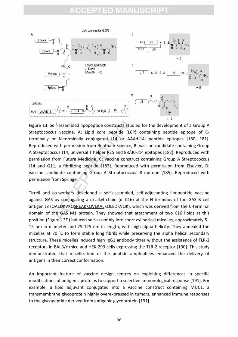
ACC
EPTE
D M
ANU
SCR
IPT
ACCEPTED MANUSCRIPT
36
Figure 13. Self-assembled lipopeptide constructs studied for the development of a Group A
Streptococcus vaccine. A: Lipid core peptide (LCP) containing peptide epitope of C-
terminally or N-terminally conjugated J14 or AAAdJ14i peptide epitopes [180, 181].
Reproduced with permission from Bentham Science, B: vaccine candidate containing Group
A Streptococcus J14, universal T helper P25 and 88/30-J14 epitopes [182]. Reproduced with
permission from Future Medicine, C: vaccine construct containing Group A Streptococcus
J14 and Q11, a fibrilizing peptide [183]. Reproduced with permission from Elsevier, D:
vaccine candidate containing Group A Streptococcus J8 epitope [185]. Reproduced with
permission from Springer.
Tirrell and co-workers developed a self-assembled, self-adjuvanting lipopeptide vaccine
against GAS by conjugating a di-alkyl chain (di-C16) at the N-terminus of the GAS B cell
antigen J8 (QAEDKVKQSREAKKQVEKALKQLEDKVQK), which was derived from the C-terminal
domain of the GAS M1 protein. They showed that attachment of two C16 lipids at this
position (Figure 13D) induced self-assembly into short cylindrical micelles, approximately 5–
15 nm in diameter and 25-125 nm in length, with high alpha helicity. They annealed the
micelles at 70 ◦C to form stable long fibrils while preserving the alpha helical secondary
structure. These micelles induced high IgG1 antibody titres without the assistance of TLR-2
receptors in BALB/c mice and HEK-293 cells expressing the TLR-2 receptor [190]. This study
demonstrated that micellization of the peptide amphiphiles enhanced the delivery of
antigens in their correct conformation.
An important feature of vaccine design centres on exploiting differences in specific
modifications of antigenic proteins to support a selective immunological response [191]. For
example, a lipid adjuvant conjugated into a vaccine construct containing MUC1, a
transmembrane glycoprotein highly overexpressed in tumors, enhanced immune responses
to the glycopeptide derived from antigenic glycoprotein [191].
ACC
EPTE
D M
ANU
SCR
IPT
ACCEPTED MANUSCRIPT
37
In 2007, Boons and co-workers developed a MUC1-based tricomponent therapeutic vaccine
comprised of (i) a decapeptide fragment of the variable-number tandem repeat (VNTR) of
the tumor-associated mucin 1 (MUC1) glycopeptide, (ii) a promiscuous T cell helper epitope,
and (iii) the immunoadjuvant Pam3CysSer(Lys)4. High humoral and cellular immune
responses in a BALB/c mice model, and enhanced cellular binding and uptake in MUC1-
expressing MCF7 human breast cancer and HEK 293T cells lines was observed [191]. This
concept led Apostolopoulos and co-workers to synthesise a self-assembled and self-
adjuvanting therautic peptide cancer vaccine candidate with isotropic particles of uniform
size 20 nm [186]. Apostolopoulos vaccine consisted of a complete copy of the VNTR eptiope
from the MUC1 glycopeptide, the universal T cell helper peptide epitope (PADRE), and the
lipidic immunoadjuvant, (Pam3CysSer). Here, strong IgG1 antibodies in C57BL/6 mice was
elicited and were found to bind tumor cells over-expressing the MUC1 glycoprotein,
including MCF7 and melanoma B16.MUC1 [192]. This study showed suitable design features
required for self-assembly of the molecularly defined vaccine into nanostructures to
generate a high antibody response. Robinson and co-workers designed lipopeptide vaccine
constructs by conjugation of a phospholipid to the N-terminus of a coiled coil peptide
sequence from the heptad repeat region 1 of respiratory syncytial virus and used for antigen
attachment at its C-terminus via a cysteine linker. This delivery system self-assembled into
homogenous 20 nm nanoparticles which displayed multiple copies of the antigen, inducing a
high antigen-specific antibody titre in New Zealand white rabbits without the need for an
additional adjuvant [193]. This study highlighted promising results for the development of
self-adjuvanting vaccine constructs using self-assembled peptides. Vaccine engineering
using these materials is seen as a way to systematically explore the impact of structure and
particle characterization on immune response.
The importance of particle size and mono dispersity has previously been reviewed and plays
an important role in vaccine development because it affects the mechanism by which APCs
take up the vaccine candidate. However, there is no consensus on the optimal particle size
to achieve this goal [194-196]. The higher immunogenicity of smaller particles (20-200 nm)
is thought to result from increased facile transport into the lymphatic system and improved
uptake by dendritic cells. Other particle characteristics such as shape, stability and the
ability to display multiple antigens on their surfaces have shown an important impact on the
immunogenicity of nanoparticles. Nevertheless, a more detailed understanding of the
fundamental effect of the physical properties of a nanoparticle on cellular interaction and
biodistribution within diverse tissues is required. Rational design and synthesis of
nanovaccines in combination with the development of reproducible nanoparticle synthesis
methods will provide vaccines with desired immunity properties.
ACC
EPTE
D M
ANU
SCR
IPT
ACCEPTED MANUSCRIPT
38
6. Conclusion
There is increasing demand to design efficient and biodegradable delivery systems to target
and control the release of therapeutics and decrease drug side effects. Supramolecular self-
assembled peptide nanostructures offer a promising alternative to existing delivery vehicles
due to their low toxicity, high biocompatibility and ability to target therapeutic small drug
molecules and proteins. These materials are able to improve cellular permeability and
regulate biological action of other pharmaceutical agents [197]. Peptide hydrogels also have
favourable mechanical properties: shear-thinning for convenient delivery and long release
profiles to enhance drug uptake and minimize the total drug dose. Self-assembled peptide
nanostructures have shown promising outcomes when used to deliver therapeutics to the
brain, eye, cardiovascular system, bone tissue, and cancer cells. However, these delivery
systems require further investigation before they will be suitable candidates for clinical
studies.
Several challenges remain in the design, functionalization and development of peptide
nanostructures as drug delivery vehicles. To successfully transition to clinical use, peptide
nanostructures must have increased stability across a broader pH range and the ability to be
manipulated to produce nanoparticles of a specific size. The relationship between the
morphology of the self-assembled peptides, their physicochemical properties, and biological
activity must also be explored in more detail [4]. Future experiments that employ rational
systematic design will provide a deeper understanding of peptide self-assembly and allow
the production of stable self-assembled peptides for drug encapsulation. This class of
materials has shown a varying impact on the immune system, ranging from non-
immunogenic (which is beneficial for drug delivery designs) to self-adjuvanting (which is
favourable for vaccine delivery). Therefore, the immunogenicity of the self-assembled
nanostructures designed for drug delivery needs to be investigated more thoroughly in vivo
[4]. Peptide designs that generated nanofibrils and lipidated peptide amphiphiles
conjugated to antigenic epitopes have shown promising results in the development of
subunit vaccines. Peptides play a valuable role in the delivery and targeting of future drug
and vaccine candidates. Further research is needed before the potential of this growing field
can be translated to clinical application.
References:
[1] P. Kumar, V. Pillay, G. Modi, Y. E Choonara, L. C du Toit, D. Naidoo, Self-assembling peptides: implications for patenting in drug delivery and tissue engineering, Recent Pat. Drug Delivery Formul. , 5 (2011) 24-51.
[2] S. Maude, L. Tai, R. Davies, B. Liu, S. Harris, P. Kocienski, A. Aggeli, Peptide synthesis and self-assembly, Peptide-based materials, Springer, NY, 2012, pp. 27-69.
[3] D.d. Bruyn Ouboter, Rational design of purely peptidic amphiphiles for drug delivery applications, University of Basel, 2011.
[4] D. Mandal, A.N. Shirazi, K. Parang, Self-assembly of peptides to nanostructures, Org. Biomol. Chem. , 12 (2014) 3544-3561.
ACC
EPTE
D M
ANU
SCR
IPT
ACCEPTED MANUSCRIPT
39
[5] H. Cui, M.J. Webber, S.I. Stupp, Self‐assembly of peptide amphiphiles: From molecules to nanostructures to biomaterials, Pept. Sci. , 94 (2010) 1-18.
[6] P. Sadatmousavi, Peptide-mediated anticancer drug delivery, University of Waterloo, 2009. [7] F. Gelain, A. Horii, S. Zhang, Designer self‐assembling peptide scaffolds for 3‐D tissue cell cultures
and regenerative medicine, Macromol. Biosci. , 7 (2007) 544-551. [8] C. Keyes-Baig, J. Duhamel, S.-Y. Fung, J. Bezaire, P. Chen, Self-assembling peptide as a potential
carrier of hydrophobic compounds, J. Am. Chem. Soc. , 126 (2004) 7522-7532. [9] S. Fung, H. Yang, P. Chen, Formation of colloidal suspension of hydrophobic compounds with an
amphiphilic self-assembling peptide, Colloids Surf. B., 55 (2007) 200-211. [10] J.S. Rudra, T. Sun, K.C. Bird, M.D. Daniels, J.Z. Gasiorowski, A.S. Chong, J.H. Collier, Modulating
adaptive immune responses to peptide self-assemblies, ACS Nano. , 6 (2012) 1557-1564. [11] X. Xu, Y. Jian, Y. Li, X. Zhang, Z. Tu, Z. Gu, Bio-inspired supramolecular hybrid dendrimers self-
assembled from low-generation peptide dendrons for highly-efficient gene delivery and biological tracking, ACS Nano. , (2014).
[12] L. Liu, X. Liu, Q. Xu, P. Wu, X. Zuo, J. Zhang, H. Deng, Z. Wu, A. Ji, Self-assembled nanoparticles based on the c (RGDfk) peptide for the delivery of siRNA targeting the VEGFR2 gene for tumor therapy, Int. J. Nanomed. , 9 (2014) 3509.
[13] J.X. Chen, X.D. Xu, S. Yang, J. Yang, R.X. Zhuo, X.Z. Zhang, Self‐assembled bola‐like amphiphilic peptides as viral‐mimetic gene vectors for cancer cell targeted gene delivery, Macromol. Biosci., 13 (2013) 84-92.
[14] J.J. Panda, A. Varshney, V.S. Chauhan, Self-assembled nanoparticles based on modified cationic dipeptides and DNA: novel systems for gene delivery, J.Nanobiotechnol. , 11 (2013) 18.
[15] N.V. Berezhnoy, N. Korolev, L. Nordenskiöld, Principles of electrostatic interactions and self-assembly in lipid/peptide/DNA systems: Applications to gene delivery, Adv.Colloid Interface Sci., 205 (2014) 221-229.
[16] J.H. Jeong, T.G. Park, S.H. Kim, Self-assembled and nanostructured siRNA delivery systems, Pharm. Res. , 28 (2011) 2072-2085.
[17] R.V. Ulijn, A.M. Smith, Designing peptide based nanomaterials, Chem. Soc. Rev. , 37 (2008) 664-675.
[18] S. Jun, Y. Hong, H. Imamura, B.-Y. Ha, J. Bechhoefer, P. Chen, Self-assembly of the ionic peptide EAK16: the effect of charge distributions on self-assembly, Biophys. J. , 87 (2004) 1249-1259.
[19] T. Hayashi, T. Nakamura, A. Takaoka, [Pattern recognition receptors], Nihon rinsho men'eki gakkai kaishi= Japanese journal of clinical immunology, 34 (2010) 329-345.
[20] I. Wheeldon, A. Farhadi, A.G. Bick, E. Jabbari, A. Khademhosseini, Nanoscale tissue engineering: spatial control over cell-materials interactions, Nanotechnol. , 22 (2011) 212001.
[21] R.O. Hynes, K.M. Yamada, Fibronectins: multifunctional modular glycoproteins, J. Cell Biol. , 95 (1982) 369-377.
[22] D.A. Lauffenburger, J.J. Linderman, Receptors: models for binding, trafficking, and signaling, Oxford University Press NY, 1993.
[23] E.C. Wu, S. Zhang, C.A. Hauser, Self‐assembling peptides as cell‐interactive scaffolds, Adv. Funct. Mater. , 22 (2012) 456-468.
[24] H. Nagase, R. Visse, G. Murphy, Structure and function of matrix metalloproteinases and TIMPs, Cardiovasc. Res. , 69 (2006) 562-573.
[25] D. Hua, W. Kong, X. Zheng, Z. Zhou, B. Yu, Y. Li, Y. Wang, X. Yang, C. Liu, L. Tang, Potent tumor targeting drug release system comprising MMP-2 specific peptide fragment with self-assembling characteristics, Drug Des. Dev. Ther. , 8 (2014) 1839.
[26] J.-K. Kim, J. Anderson, H.-W. Jun, M.A. Repka, S. Jo, Self-assembling peptide amphiphile-based nanofiber gel for bioresponsive cisplatin delivery, Mol. Pharmaceutics, 6 (2009) 978-985.
[27] J.F. Woessner Jr, MMPs and TIMPs, in: I.M. Clark (Ed.) Matrix metalloproteinase protocols, Humana press, USA, 2001, pp. 1-23.
ACC
EPTE
D M
ANU
SCR
IPT
ACCEPTED MANUSCRIPT
40
[28] Y.-A. Lin, Y.-C. Ou, A.G. Cheetham, H. Cui, Rational design of MMP degradable peptide-based supramolecular filaments, Biomacromolecules, 15 (2014) 1419-1427.
[29] D.A. Harrington, E.Y. Cheng, M.O. Guler, L.K. Lee, J.L. Donovan, R.C. Claussen, S.I. Stupp, Branched peptide‐amphiphiles as self‐assembling coatings for tissue engineering scaffolds, Biomed. Mater. Res., Part A. , 78 (2006) 157-167.
[30] A. Barnard, D.K. Smith, Self‐assembled multivalency: dynamic ligand arrays for high‐affinity binding, Angew. Chem., Int. Ed. , 51 (2012) 6572-6581.
[31] S. Sur, C.J. Newcomb, M.J. Webber, S.I. Stupp, Tuning supramolecular mechanics to guide neuron development, Biomaterials. , 34 (2013) 4749-4757.
[32] S. Massia, J. Hubbell, Human endothelial cell interactions with surface‐coupled adhesion peptides on a nonadhesive glass substrate and two polymeric biomaterials, J. Biom.l Mater. Res. , 25 (1991) 223-242.
[33] S.P. Massia, J.A. Hubbell, Convalent surface immobilization of Arg-Gly-Asp-and Tyr-Ile-Gly-Ser-Arg-containing peptides to obtain well-defined cell-adhesive substrates, Anal.l Biochem. , 187 (1990) 292-301.
[34] M.J. Humphries, The molecular basis and specificity of integrin-ligand interactions, J. Cell Sci. , 97 (1990) 585-592.
[35] J. Zhu, J. Hu, R. Marchant, Biomimetic hydrogels as scaffolds for tissue-engineering applications, in: A.J. Ruys (Ed.) Biomimetic biomaterials: structure and applications, Woodhead Publisher, UK, 2013, pp. 238-275.
[36] D. Hunter, N. Cashman, R. Morris-Valero, J. Bulock, S. Adams, J. Sanes, An LRE (leucine-arginine-glutamate)-dependent mechanism for adhesion of neurons to S-laminin, J. Neurosci. , 11 (1991) 3960-3971.
[37] M.B. Rahmany, M. Van Dyke, Biomimetic approaches to modulate cellular adhesion in biomaterials: A review, Acta Biomater. , 9 (2013) 5431-5437.
[38] W. Staatz, K. Fok, M. Zutter, S. Adams, B. Rodriguez, S. Santoro, Identification of a tetrapeptide recognition sequence for the alpha 2 beta 1 integrin in collagen, J. Biol. Chem. , 266 (1991) 7363-7367.
[39] J.J. Calvete, W. Schafer, K. Mann, A. Henschen, J. Gonzalez-Rodriguez, Localization of the cross‐linking sites of RGD and KQAGDV peptides to the isolated fibrinogen receptor, the human platelet integrin glicoprotein IIb/IIIa, Eur. J. Biochem. , 206 (1992) 759-765.
[40] C. Gonçalves, P. Pereira, M. Gama, Self-assembled hydrogel nanoparticles for drug delivery applications, Mater. , 3 (2010) 1420-1460.
[41] A. Altunbas, S.J. Lee, S.A. Rajasekaran, J.P. Schneider, D.J. Pochan, Encapsulation of curcumin in self-assembling peptide hydrogels as injectable drug delivery vehicles, Biomaterials., 32 (2011) 5906-5914.
[42] H. Tsutsumi, H. Mihara, Soft materials based on designed self-assembling peptides: from design to application, Mol. BioSyst., 9 (2013) 609-617.
[43] G. Verma, P. Hassan, Self assembled materials: design strategies and drug delivery perspectives, Phys. Chem. Chem. Phys. , 15 (2013) 17016-17028.
[44] R. Huang, W. Qi, L. Feng, R. Su, Z. He, Self-assembling peptide–polysaccharide hybrid hydrogel as a potential carrier for drug delivery, Soft Matter. , 7 (2011) 6222-6230.
[45] M. Black, A. Trent, Y. Kostenko, J.S. Lee, C. Olive, M. Tirrell, Self‐assembled peptide amphiphile micelles containing a cytotoxic T‐cell epitope promote a protective immune response in-vivo, Adv. Mater. , 24 (2012) 3845-3849.
[46] J.J. Panda, A. Mishra, A. Basu, V.S. Chauhan, Stimuli responsive self-assembled hydrogel of a low molecular weight free dipeptide with potential for tunable drug delivery, Biomacromolecules. , 9 (2008) 2244-2250.
[47] M.J. Webber, C.J. Newcomb, R. Bitton, S.I. Stupp, Switching of self-assembly in a peptide nanostructure with a specific enzyme, Soft Matter., 7 (2011) 9665-9672.
ACC
EPTE
D M
ANU
SCR
IPT
ACCEPTED MANUSCRIPT
41
[48] L. Haines-Butterick, K. Rajagopal, M. Branco, D. Salick, R. Rughani, M. Pilarz, M.S. Lamm, D.J. Pochan, J.P. Schneider, Controlling hydrogelation kinetics by peptide design for three-dimensional encapsulation and injectable delivery of cells, Proc. Natl. Acad. Sci. , 104 (2007) 7791-7796.
[49] L. Liang, J. Yang, Q. Li, M. Huo, F. Jiang, X. Xu, X. Zhang, A novel targeting drug delivery system based on self-assembled peptide hydrogel, J. Biomater. Nanobiotechnol. , 2 (2011) 622.
[50] J. Castillo, L. Sasso, W.E. Svendsen, Self-assembled peptide nanostructures: Advances and applications in nanobiotechnology, CRC Press, FL, 2012.
[51] J.J. Panda, V.S. Chauhan, Short peptide based self-assembled nanostructures: implications in drug delivery and tissue engineering, Polym. Chem. , 5 (2014) 4418-4436.
[52]. M. Liu, J.M. Fréchet, Designing dendrimers for drug delivery, Pharmaceutical science &
technology today, 2 (1999) 393-401.
[53]. E.J. Chung, Y. Cheng, R. Morshed, K. Nord, Y. Han, M.L. Wegscheid, B. Auffinger, D.A. Wainwright, M.S. Lesniak, M.V. Tirrell, Fibrin-binding, peptide amphiphile micelles for targeting glioblastoma, Biomaterials, 35 (2014) 1249-1256.
[54]. Y. Shiose, H. Kuga, H. Ohki, M. Ikeda, F. Yamashita, M. Hashida, Systematic Research of Peptide Spacers Controlling Drug Release from Macromolecular Prodrug System, Carboxymethyldextran Polyalcohol− Peptide− Drug Conjugates, Bioconjugate chemistry, 20 (2008) 60-70.
[55] A. Altunbas, D.J. Pochan, Peptide-based and polypeptide-based hydrogels for drug delivery and tissue engineering, Peptide-based materials, Springer2012, pp. 135-167.
[56] K.J. Nagy, M.C. Giano, A. Jin, D.J. Pochan, J.P. Schneider, Enhanced mechanical rigidity of hydrogels formed from enantiomeric peptide assemblies, J. Am. Chem. Soc., 133 (2011) 14975-14977.
[57] J. Li, Y. Kuang, Y. Gao, X. Du, J. Shi, B. Xu, d-Amino acids boost the selectivity and confer supramolecular hydrogels of a nonsteroidal anti-inflammatory drug (NSAID), Journal of the American Chemical Society, 135 (2012) 542-545.
[58] J. Zhang, R. Hao, L. Huang, J. Yao, X. Chen, Z. Shao, Self-assembly of a peptide amphiphile based on hydrolysed Bombyx mori silk fibroin, Chem. Commun., 47 (2011) 10296-10298.
[59] E.K. Chung, E. Lee, Y.b. Lim, M. Lee, Cyclic peptide facial amphiphile preprogrammed to self‐assemble into bioactive peptide capsules, Chem. - Eur. J. , 16 (2010) 5305-5309.
[60] Y.-b. Lim, K.-S. Moon, M. Lee, Recent advances in functional supramolecular nanostructures assembled from bioactive building blocks, Chem. Soc. Rev. , 38 (2009) 925-934.
[61] J.-h. Lee, Y.J. Choi, Y.-b. Lim, Self-assembled filamentous nanostructures for drug/gene delivery applications, Expert opin. Drug Delivery. , 7 (2010) 341-351.
[62] E. Mastrobattista, M.A. Van Der Aa, W.E. Hennink, D.J. Crommelin, Artificial viruses: a nanotechnological approach to gene delivery, Nat. Rev. Drug Discovery. , 5 (2006) 115-121.
[63] M.M. Henricus, K.T. Johnson, I.A. Banerjee, Investigation of insulin loaded self-assembled microtubules for drug release, Bioconjugate Chem. , 19 (2008) 2394-2400.
[64] Q. Wang, X. Zhang, J. Zheng, D. Liu, Self-assembled peptide nanotubes as potential nanocarriers for drug delivery, RSC Adv. , (2014).
[65] Y. Loo, S. Zhang, C.A. Hauser, From short peptides to nanofibers to macromolecular assemblies in biomedicine, Biotechnol. Adv. , 30 (2012) 593-603.
[66] Y.B. Yu, Coiled-coils: stability, specificity, and drug delivery potential, Adv. Drug Delivery rev. , 54 (2002) 1113-1129.
[67] N.L. Fletcher, C.V. Lockett, A.F. Dexter, A pH-responsive coiled-coil peptide hydrogel, Soft Matter. , 7 (2011) 10210-10218.
[68] Y. Deng, J. Liu, Q. Zheng, D. Eliezer, N.R. Kallenbach, M. Lu, Antiparallel four-stranded coiled coil specified by a 3-3-1 hydrophobic heptad repeat, Struct. , 14 (2006) 247-255.
ACC
EPTE
D M
ANU
SCR
IPT
ACCEPTED MANUSCRIPT
42
[69] M.G. Ryadnov, D. Papapostolou, D.N. Woolfson, The leucine zipper as a building block for self-assembled protein fibers, Nanostructure Design, Hmana press, NJ, 2008, pp. 35-51.
[70] A. Lomander, W. Hwang, S. Zhang, Hierarchical self-assembly of a coiled-coil peptide into fractal structure, Nano Lett., 5 (2005) 1255-1260.
[71] E.P. Holowka, D.J. Pochan, T.J. Deming, Charged polypeptide vesicles with controllable diameter, J. Am. Chem. Soc. , 127 (2005) 12423-12428.
[72] C. Xu, V. Breedveld, J. Kopecek, Reversible hydrogels from self-assembling genetically engineered protein block copolymers, Biomacromolecules. , 6 (2005) 1739-1749.
[73] I. Hamley, Self-assembly of amphiphilic peptides, Soft Matter. , 7 (2011) 4122-4138. [74] D.V. Krogstad, Investigating the structure-property relationships of aqueous self-assembled
materials, University of California, Santa Barbara, 2013. [75] S.G. Reed, S. Bertholet, R.N. Coler, M. Friede, New horizons in adjuvants for vaccine
development, Trends Immunol. , 30 (2009) 23-32. [76] A. Tan, J. Rajadas, A.M. Seifalian, Biochemical engineering nerve conduits using peptide
amphiphiles, J. Controlled Release. , 163 (2012) 342-352. [77] H. Jiang, M.O. Guler, S.I. Stupp, The internal structure of self-assembled peptide amphiphiles
nanofibers, Soft Matter. , 3 (2007) 454-462. [78] D.G. Fatouros, D.A. Lamprou, A.J. Urquhart, S.N. Yannopoulos, I.S. Vizirianakis, S. Zhang, S.
Koutsopoulos, Lipid-like self-assembling peptide nanovesicles for drug delivery, ACS Appl. Mater. Interfaces. , 6 (2014) 8184-8189.
[79] S.J. Son, M.A. Brimble, S. Yang, P.W. Harris, T. Reddingius, B.W. Muir, O.E. Hutt, L. Waddington, J. Guan, G.P. Savage, Synthesis and self-assembly of a peptide–amphiphile as a drug delivery vehicle, Aust. J. Chem. , 66 (2013) 23-29.
[80] T. Moyer, Self-assembling peptide amphiphiles for targeted drug delivery, Northwestern University, 2013.
[81] X. Xu, Y. Li, H. Li, R. Liu, M. Sheng, B. He, Z. Gu, Smart nanovehicles based on pH‐triggered disassembly of supramolecular peptide‐amphiphiles for efficient intracellular drug delivery, Small. , 10 (2014) 1133-1140.
[82] D. Missirlis, H. Khant, M. Tirrell, Mechanisms of Peptide Amphiphile Internalization by SJSA-1 Cells in Vitro†, Biochem., 48 (2009) 3304-3314.
[83] V. Castelletto, I.W. Hamley, J. Perez, L. Abezgauz, D. Danino, Fibrillar superstructure from extended nanotapes formed by a collagen-stimulating peptide, Chem. Commun. , 46 (2010) 9185-9187.
[84] I. Hamley, A. Dehsorkhi, V. Castelletto, Coassembly in binary mixtures of peptide amphiphiles containing oppositely charged residues, Langmuir. , 29 (2013) 5050-5059.
[85] R.H. Zha, S. Sur, S.I. Stupp, Self‐assembly of cytotoxic peptide amphiphiles into supramolecular membranes for cancer therapy, Adv. Healthcare Mater., 2 (2013) 126-133.
[86] M. Mazza, A. Patel, R. Pons, C. Bussy, K. Kostarelos, Peptide nanofibres as molecular transporters: from self-assembly to in vivo degradation, Faraday discuss. , 166 (2013) 181-194.
[87] S.Y. Fung, H. Yang, P. Sadatmousavi, Y. Sheng, T. Mamo, R. Nazarian, P. Chen, Amino Acid Pairing for de novo design of self‐assembling peptides and their drug delivery potential, Adv. Funct. Mater. , 21 (2011) 2456-2464.
[88] S. Zhang, D.M. Marini, W. Hwang, S. Santoso, Design of nanostructured biological materials through self-assembly of peptides and proteins, Curr.Opin.Chem. Biol., 6 (2002) 865-871.
[89] S. Zhang, C. Lockshin, R. Cook, A. Rich, Unusually stable β‐sheet formation in an ionic self‐complementary oligopeptide, Biopolymers., 34 (1994) 663-672.
[90] X. Wang, A. Horii, S. Zhang, Designer functionalized self-assembling peptide nanofiber scaffolds for growth, migration, and tubulogenesis of human umbilical vein endothelial cells, Soft Matter. , 4 (2008) 2388-2395.
[91] T. Deming, Peptide-based materials, Springer, NY, 2012.
ACC
EPTE
D M
ANU
SCR
IPT
ACCEPTED MANUSCRIPT
43
[92] P. Shi, J.A. Gustafson, J.A. MacKay, Genetically engineered nanocarriers for drug delivery, Int. J. Nanomed., 9 (2014) 1617.
[93] R. Herrero-Vanrell, A. Rincon, M. Alonso, V. Reboto, I. Molina-Martinez, J. Rodriguez-Cabello, Self-assembled particles of an elastin-like polymer as vehicles for controlled drug release, J. Controlled Release., 102 (2005) 113-122.
[94] J.R. McDaniel, S.R. MacEwan, M. Dewhirst, A. Chilkoti, Doxorubicin-conjugated chimeric polypeptide nanoparticles that respond to mild hyperthermia, J. Controlled Release., 159 (2012) 362-367.
[95] S. Moktan, E. Perkins, F. Kratz, D. Raucher, Thermal targeting of an acid-sensitive doxorubicin conjugate of elastin-like polypeptide enhances the therapeutic efficacy compared with the parent compound in vivo, Mol. Cancer Ther. , 11 (2012) 1547-1556.
[96] J.R. McDaniel, D.J. Callahan, A. Chilkoti, Drug delivery to solid tumors by elastin-like polypeptides, Adv. Drug Delivery Rev. , 62 (2010) 1456-1467.
[97] M. Amiram, K.M. Luginbuhl, X. Li, M.N. Feinglos, A. Chilkoti, Injectable protease-operated depots of glucagon-like peptide-1 provide extended and tunable glucose control, Proc. Natl. Acad. Sci., 110 (2013) 2792-2797.
[98] M.R. Dreher, A.J. Simnick, K. Fischer, R.J. Smith, A. Patel, M. Schmidt, A. Chilkoti, Temperature triggered self-assembly of polypeptides into multivalent spherical micelles, J. Am. Chem. Soc. , 130 (2008) 687-694.
[99] J.A. Gustafson, H. Ghandehari, Silk-elastinlike protein polymers for matrix-mediated cancer gene therapy, Adv. Drug Delivery Rev. , 62 (2010) 1509-1523.
[100] J. Chang, X.-F. Peng, K. Hijji, J. Cappello, H. Ghandehari, S.D. Solares, J. Seog, Nanomechanical stimulus accelerates and directs the self-assembly of silk-elastin-like nanofibers, J. Am. Chem. Soc. , 133 (2011) 1745-1747.
[101] S.R. MacEwan, A. Chilkoti, Applications of elastin-like polypeptides in drug delivery, J. Controlled Release., (2014).
[102] A. Nasrolahi Shirazi, R.K. Tiwari, D. Oh, A. Banerjee, A. Yadav, K. Parang, Efficient delivery of cell impermeable phosphopeptides by a cyclic peptide amphiphile containing tryptophan and arginine, Mol. Pharmaceutics. , 10 (2013) 2008-2020.
[103] R.J. Brea, C. Reiriz, J.R. Granja, Towards functional bionanomaterials based on self-assembling cyclic peptide nanotubes, Chem. Soc. Rev. , 39 (2010) 1448-1456.
[104] N. Ashkenasy, W.S. Horne, M.R. Ghadiri, Design of self‐assembling peptide nanotubes with delocalized electronic states, Small. , 2 (2006) 99-102.
[105] W.S. Horne, C.D. Stout, M.R. Ghadiri, A heterocyclic peptide nanotube, J. Am. Chem. Soc. , 125 (2003) 9372-9376.
[106] A. Nasrolahi Shirazi, D. Oh, R.K. Tiwari, B. Sullivan, A. Gupta, G.D. Bothun, K. Parang, Peptide amphiphile containing arginine and fatty acyl chains as molecular transporters, Mol. Pharmaceutics. , 10 (2013) 4717-4727.
[107] A. Nasrolahi Shirazi, R.K. Tiwari, D. Oh, B. Sullivan, K. McCaffrey, D. Mandal, K. Parang, Surface decorated gold nanoparticles by linear and cyclic peptides as molecular transporters, Mol. Pharmaceutics. , 10 (2013) 3137-3151.
[108] H. Liu, J. Chen, Q. Shen, W. Fu, W. Wu, Molecular insights on the cyclic peptide nanotube-mediated transportation of antitumor drug 5-Fluorouracil, Mol. Pharmaceutics. , 7 (2010) 1985-1994.
[109] A.T. Petkova, R.D. Leapman, Z. Guo, W.-M. Yau, M.P. Mattson, R. Tycko, Self-propagating, molecular-level polymorphism in Alzheimer's ß-amyloid fibrils, Science. , 307 (2005) 262-265.
[110] M. Gupta, A. Bagaria, A. Mishra, P. Mathur, A. Basu, S. Ramakumar, V.S. Chauhan, Self‐assembly of a dipeptide‐containing conformationally restricted dehydrophenylalanine residue to form ordered nanotubes, Adv. Mater. , 19 (2007) 858-861.
[111] Y. Nagai, L.D. Unsworth, S. Koutsopoulos, S. Zhang, Slow release of molecules in self-assembling peptide nanofiber scaffold, J. Controlled Release. , 115 (2006) 18-25.
ACC
EPTE
D M
ANU
SCR
IPT
ACCEPTED MANUSCRIPT
44
[112] S. Alam, J.J. Panda, V.S. Chauhan, Novel dipeptide nanoparticles for effective curcumin delivery, Int. J. Nanomed. , 7 (2012) 4207.
[113] J.J. Panda, A. Kaul, S. Alam, A.K. Babbar, A.K. Mishra, V.S. Chauhan, Designed peptides as model self-assembling nanosystems: characterization and potential biomedical applications, Ther. Delivery. , 2 (2011) 193-204.
[114] A. Baral, S. Roy, A. Dehsorkhi, I.W. Hamley, S. Mohapatra, S. Ghosh, A. Banerjee, Assembly of an injectable noncytotoxic peptide-based hydrogelator for sustained release of drugs, Langmuir. , 30 (2014) 929-936.
[115] J. Naskar, S. Roy, A. Joardar, S. Das, A. Banerjee, Self-assembling dipeptide-based nontoxic vesicles as carriers for drugs and other biologically important molecules, Org. Biomol. Chem. , 9 (2011) 6610-6615.
[116] A.M. Smith, R.J. Williams, C. Tang, P. Coppo, R.F. Collins, M.L. Turner, A. Saiani, R.V. Ulijn, Fmoc‐diphenylalanine self assembles to a hydrogel via a novel architecture based on π–π interlocked β‐sheets, Adv. Mater. , 20 (2008) 37-41.
[117] J.P. Jung, J.Z. Gasiorowski, J.H. Collier, Fibrillar peptide gels in biotechnology and biomedicine, Pept. Sci. , 94 (2010) 49-59.
[118] S. Sutton, N.L. Campbell, A.I. Cooper, M. Kirkland, W.J. Frith, D.J. Adams, Controlled release from modified amino acid hydrogels governed by molecular size or network dynamics, Langmuir. , 25 (2009) 10285-10291.
[119] V. Castelletto, I.W. Hamley, C. Stain, C. Connon, Slow-release RGD-peptide hydrogel monoliths, Langmuir. , 28 (2012) 12575-12580.
[120] C. Tang, A.M. Smith, R.F. Collins, R.V. Ulijn, A. Saiani, Fmoc-diphenylalanine self-assembly mechanism induces apparent pK a shifts, Langmuir. , 25 (2009) 9447-9453.
[121] V. Singh, K. Snigdha, C. Singh, N. Sinha, A.K. Thakur, Understanding the self-assembly of Fmoc-phenylalanine to hydrogel formation, Soft Matter., (2015).
[122] J.L. Drury, D.J. Mooney, Hydrogels for tissue engineering: scaffold design variables and applications, Biomaterials. , 24 (2003) 4337-4351.
[123] C.H. Lee, V. Moturi, Y. Lee, Thixotropic property in pharmaceutical formulations, Journal of Controlled Release, 136 (2009) 88-98.
[124]. C. Veerman, K. Rajagopal, C.S. Palla, D.J. Pochan, J.P. Schneider, E.M. Furst, Gelation kinetics of β-hairpin peptide hydrogel networks, Macromolecules, 39 (2006) 6608-6614.
[125]. C. Yan, D.J. Pochan, Rheological properties of peptide-based hydrogels for biomedical and other applications, Chemical Society Reviews, 39 (2010) 3528-3540.
[126] J.J. Panda, S. Yandrapu, R.S. Kadam, V.S. Chauhan, U.B. Kompella, Self-assembled phenylalanine-α, β-dehydrophenylalanine nanotubes for sustained intravitreal delivery of a multi-targeted tyrosine kinase inhibitor, J. Controlled Release. , 172 (2013) 1151-1160.
[127] A. Nangia, Encyclopaedia of supramolecular chemistry, Taylor &Francis, NY, 2007. [128] V. Castelletto, G. Cheng, C. Stain, C. Connon, I. Hamley, Self-assembly of a peptide amphiphile
containing l-carnosine and its mixtures with a multilamellar vesicle forming lipid, Langmuir., 28 (2012) 11599-11608.
[129] M.-L. Briuglia, A.J. Urquhart, D.A. Lamprou, Sustained and controlled release of lipophilic drugs
from a self-assembling amphiphilic peptide hydrogel, Int. J. Pharm., (2014). [130] S. Koutsopoulos, L.D. Unsworth, Y. Nagai, S. Zhang, Controlled release of functional proteins
through designer self-assembling peptide nanofiber hydrogel scaffold, Proc. Natl. Acad. Sci. , 106 (2009) 4623-4628.
[131] S.Y. Fung, H. Yang, P.T. Bhola, P. Sadatmousavi, E. Muzar, M. Liu, P. Chen, Self‐assembling peptide as a potential carrier for hydrophobic anticancer drug ellipticine: complexation, release and in vitro delivery, Adv. Funct. Mater. , 19 (2009) 74-83.
[132] S.Y. Fung, H. Yang, P. Chen, Sequence effect of self-assembling peptides on the complexation and in vitro delivery of the hydrophobic anticancer drug ellipticine, PLoS One., 3 (2008) e1956.
ACC
EPTE
D M
ANU
SCR
IPT
ACCEPTED MANUSCRIPT
45
[133] M. Mazza, R. Notman, J. Anwar, A. Rodger, M. Hicks, G. Parkinson, D. McCarthy, T. Daviter, J. Moger, N. Garrett, Nanofiber-based delivery of therapeutic peptides to the brain, ACS Nano. , 7 (2013) 1016-1026.
[134] N. Wiradharma, Y.W. Tong, Y.-Y. Yang, Self-assembled oligopeptide nanostructures for co-delivery of drug and gene with synergistic therapeutic effect, Biomaterials., 30 (2009) 3100-3109.
[135] M.C. Branco, D.J. Pochan, N.J. Wagner, J.P. Schneider, Macromolecular diffusion and release from self-assembled β-hairpin peptide hydrogels, Biomaterials, 30 (2009) 1339-1347.
[136] L.A. Haines-Butterick, D.A. Salick, D.J. Pochan, J.P. Schneider, In vitro assessment of the pro-inflammatory potential of β-hairpin peptide hydrogels, Biomaterials, 29 (2008) 4164-4169.
[137] S. Lindsey, J.H. Piatt, P. Worthington, C. Sonmez, S. Satheye, J.P. Schneider, D.J. Pochan, S.A. Langhans, Beta hairpin peptide hydrogels as an injectable solid vehicle for neurotrophic growth factor delivery, Biomacromolecules., 16 (2015) 2672-2683.
[138] X.-D. Xu, L. Liang, C.-S. Chen, B. Lu, N.-l. Wang, F.-G. Jiang, X.-Z. Zhang, R.-X. Zhuo, Peptide hydrogel as an intraocular drug delivery system for inhibition of postoperative scarring formation, ACS Appl. Mater. Interfaces., 2 (2010) 2663-2671.
[139] L. Liang, X.D. Xu, C.S. Chen, J.H. Fang, F.G. Jiang, X.Z. Zhang, R.X. Zhuo, Evaluation of the biocompatibility of novel peptide hydrogel in rabbit eye, J. Biomed. Mater. Res. , Part B. , 93 (2010) 324-332.
[140] M.J. Webber, J.B. Matson, V.K. Tamboli, S.I. Stupp, Controlled release of dexamethasone from peptide nanofiber gels to modulate inflammatory response, Biomaterials., 33 (2012) 6823-6832.
[141] S. Soukasene, D.J. Toft, T.J. Moyer, H. Lu, H.-K. Lee, S.M. Standley, V.L. Cryns, S.I. Stupp, Antitumor activity of peptide amphiphile nanofiber-encapsulated camptothecin, ACS Nano., 5 (2011) 9113-9121.
[142] M. Conda-Sheridan, S.S. Lee, A.T. Preslar, S.I. Stupp, Esterase-activated release of naproxen from supramolecular nanofibres, Chem. Commun., 50 (2014) 13757-13760.
[143] J.B. Matson, M.J. Webber, V.K. Tamboli, B. Weber, S.I. Stupp, A peptide-based material for therapeutic carbon monoxide delivery, Soft Matter, 8 (2012) 6689-6692.
[144] J. Boekhoven, R.H. Zha, F. Tantakitti, E. Zhuang, R. Zandi, C.J. Newcomb, S.I. Stupp, Alginate–peptide amphiphile core–shell microparticles as a targeted drug delivery system, RSC Adv., 5 (2015) 8753-8756.
[145] Y. Chen, H.X. Gan, Y.W. Tong, pH-controlled hierarchical self-assembly of peptide amphiphile, Macromolecules., 48 (2015) 2647-2653.
[146] J.B. Matson, C.J. Newcomb, R. Bitton, S.I. Stupp, Nanostructure-templated control of drug release from peptide amphiphile nanofiber gels, Soft Matter., 8 (2012) 3586-3595.
[147] J.B. Matson, S.I. Stupp, Drug release from hydrazone-containing peptide amphiphiles, Chem. Commun., 47 (2011) 7962-7964.
[148] M.C. Branco, D.J. Pochan, N.J. Wagner, J.P. Schneider, The effect of protein structure on their controlled release from an injectable peptide hydrogel, Biomaterials., 31 (2010) 9527-9534.
[149] R. Gaudana, H.K. Ananthula, A. Parenky, A.K. Mitra, Ocular drug delivery, AAPS J. , 12 (2010) 348-360.
[150] A. Patel, K. Cholkar, V. Agrahari, A.K. Mitra, Ocular drug delivery systems: An overview, World J. Pharmacol. , 2 (2013) 47-64.
[151] M. Sikandar, P. Sharma, S. Visht, Ocular drug delivery system: an overview, Int. J. Pharm. Sci. Res., 2 (2011) 1168-1175.
[152] J. Siepmann, N. Peppas, Modeling of drug release from delivery systems based on hydroxypropyl methylcellulose (HPMC), Adv. Drug Delivery Rev. , 48 (2001) 139-157.
[153] V.A. Puig-Sanvicens, C.E. Semino, Self-assembling peptide scaffolds as innovative platforms for drug and cell delivery systems in cardiac regeneration, Drug Delivery Transl. Res. , 3 (2013) 330-335.
ACC
EPTE
D M
ANU
SCR
IPT
ACCEPTED MANUSCRIPT
46
[154] H.-d. Guo, G.-h. Cui, J.-j. Yang, C. Wang, J. Zhu, L.-s. Zhang, J. Jiang, S.-j. Shao, Sustained delivery of VEGF from designer self-assembling peptides improves cardiac function after myocardial infarction, Biochem. Biophys. Res. commun. , 424 (2012) 105-111.
[155] M. Kushwaha, J.M. Anderson, C.A. Bosworth, A. Andukuri, W.P. Minor, J.R. Lancaster Jr, P.G. Anderson, B.C. Brott, H.-W. Jun, A nitric oxide releasing, self assembled peptide amphiphile matrix that mimics native endothelium for coating implantable cardiovascular devices, Biomaterials. , 31 (2010) 1502-1508.
[156] T. Jiang, X. Yu, E.J. Carbone, C. Nelson, H.M. Kan, K.W.-H. Lo, Poly aspartic acid peptide-linked PLGA based nanoscale particles: potential for bone-targeting drug delivery applications, Int. J. Pharm. , 475 (2014) 547-557.
[157] L. Ouyang, W. Huang, G. He, L. Guo, Bone targeting prodrugs based on peptide dendrimers, synthesis and hydroxyapatite binding in vitro, Lett. Org. Chem. , 6 (2009) 272-277.
[158] E.D. Spoerke, S.G. Anthony, S.I. Stupp, Enzyme directed templating of artificial bone mineral, Adv. Mater., 21 (2009) 425.
[159] L.C. Palmer, C.J. Newcomb, S.R. Kaltz, E.D. Spoerke, S.I. Stupp, Biomimetic systems for hydroxyapatite mineralization inspired by bone and enamel, Chem. Rev., 108 (2008) 4754-4783.
[160] E.D. Sone, S.I. Stupp, Semiconductor-encapsulated peptide-amphiphile nanofibers, J. Am. Chem. Soc., 126 (2004) 12756-12757.
[161] H. Rapaport, Amphiphilic peptides and hydrogel matrices thereof for bone repair, US Patent, US8658763 B2, 2014.
[162] L. Polo-Corrales, M. Latorre-Esteves, J.E. Ramirez-Vick, Scaffold design for bone regeneration, J. Nanosci. Nanotechnol. , 14 (2014) 15.
[163] F.X. Gu, R. Karnik, A.Z. Wang, F. Alexis, E. Levy-Nissenbaum, S. Hong, R.S. Langer, O.C. Farokhzad, Targeted nanoparticles for cancer therapy, Nano Today. , 2 (2007) 14-21.
[164] R. Stephenson, P. Varamini, N. Butcher, R. Minchin, I. Toth, Effect of lipidated gonadotropin-releasing hormone peptides on receptor mediated binding and uptake into prostate cancer cells in vitro, Nanomed.: Nanotechnol., Biol. Med. , (2014).
[165] K. Temming, R.M. Schiffelers, G. Molema, R.J. Kok, RGD-based strategies for selective delivery of therapeutics and imaging agents to the tumour vasculature, Drug Resist. Updates., 8 (2005) 381-402.
[166] J. Liu, J. Liu, H. Xu, Y. Zhang, L. Chu, Q. Liu, N. Song, C. Yang, Novel tumor-targeting, self-assembling peptide nanofiber as a carrier for effective curcumin delivery, Int. J. Nanomed. , 9 (2014) 197.
[167] J.A. Hubbell, S.N. Thomas, M.A. Swartz, Materials engineering for immunomodulation, Nature. , 462 (2009) 449-460.
[168] J.S. Rudra, Y.F. Tian, J.P. Jung, J.H. Collier, A self-assembling peptide acting as an immune adjuvant, Proc. Natl. Acad. Sci. , 107 (2010) 622-627.
[169] X.S. Sun, J. Shi, Vaccine adjuvants from self-assembling peptides, US Patent, US20140086952 A1, 2012.
[170] P. Burkhard, C. Kulangara, Self-assembling peptide nanoparticles as vaccines against infection with norovirus, US Patent, 20140242104 A1, 2012.
[171] J.H. Collier, Modular self-assembling biomaterials for directing cellular responses, Soft Matter. , 4 (2008) 2310-2315.
[172] C.B. Chesson, E.J. Huelsmann, A.T. Lacek, F.J. Kohlhapp, M.F. Webb, A. Nabatiyan, A. Zloza, J.S. Rudra, Antigenic peptide nanofibers elicit adjuvant-free CD8+ T cell responses, Vaccine., 32 (2014) 1174-1180.
[173] J. Chen, R.R. Pompano, F.W. Santiago, L. Maillat, R. Sciammas, T. Sun, H. Han, D.J. Topham, A.S. Chong, J.H. Collier, The use of self-adjuvanting nanofiber vaccines to elicit high-affinity B cell responses to peptide antigens without inflammation, Biomaterials., 34 (2013) 8776-8785.
ACC
EPTE
D M
ANU
SCR
IPT
ACCEPTED MANUSCRIPT
47
[174] B.S. Zolnik, A. Gonzalez-Fernandez, N. Sadrieh, M.A. Dobrovolskaia, Minireview: nanoparticles and the immune system, Endocrinology., 151 (2010) 458-465.
[175] A. Hajizade, F. Ebrahimi, A.-H. Salmanian, A. Arpanae, J. Amani, Nanoparticles in vaccine development, J. Appl. Biotechnol. Rep. , 1 (2015) pp. 125-134.
[176] S.A. Kaba, C. Brando, Q. Guo, C. Mittelholzer, S. Raman, D. Tropel, U. Aebi, P. Burkhard, D.E. Lanar, A nonadjuvanted polypeptide nanoparticle vaccine confers long-lasting protection against rodent malaria, J. Immunol., 183 (2009) 7268-7277.
[177] J. Aguilar, E. Rodriguez, Vaccine adjuvants revisited, Vaccine., 25 (2007) 3752-3762. [178] R.K. Gupta, Aluminum compounds as vaccine adjuvants, Adv. Drug Delivery Rev. , 32 (1998)
155-172. [179] L. Huntimer, A.E. Ramer‐Tait, L.K. Petersen, K.A. Ross, K.A. Walz, C. Wang, J. Hostetter, B.
Narasimhan, M.J. Wannemuehler, Evaluation of biocompatibility and administration site reactogenicity of polyanhydride‐particle‐based platform for vaccine delivery, Adv. Healthcare Mater. , 2 (2013) 369-378.
[180] M. Skwarczynski, J. Kowapradit, Z.M. Ziora, I. Toth, pH-triggered peptide self-assembly into fibrils: a potential peptide-based subunit vaccine delivery platform, Biochem. Compd. , 1 (2013) 2.
[181] P.M. Moyle, C. Olive, M.F. Good, I. Toth, Method for the synthesis of highly pure vaccines using the lipid core peptide system, J. Pept. Sci., 12 (2006) 800-807.
[182] B.Y. Chua, E.M. Eriksson, L.E. Brown, W. Zeng, E.J. Gowans, J. Torresi, D.C. Jackson, A self-adjuvanting lipopeptide-based vaccine candidate for the treatment of hepatitis C virus infection, Vaccine., 26 (2008) 4866-4875.
[183] K. El Bissati, Y. Zhou, D. Dasgupta, D. Cobb, J.P. Dubey, P. Burkhard, D.E. Lanar, R. McLeod, Effectiveness of a novel immunogenic nanoparticle platform for Toxoplasma peptide vaccine in HLA transgenic mice, Vaccine., 32 (2014) 3243-3248.
[184] M. Skwarczynskim, M. Zaman, I. Toth, Lipo-peptides/saccharides in peptide vaccine delivery, in: A. Kastin (Ed.) Handbook of biologically active peptides, Academic Press, USA, 2013, pp. 571-579.
[185] M. Skwarczynski, A. AH Ahmad Fuaad, L. Rustanti, Z. M Ziora, M. Aqil, M. R Batzloff, M. F Good, I. Toth, Group A streptococcal vaccine candidates based on the conserved conformational epitope from M protein, Drug Delivery Lett. , 1 (2011) 2-8.
[186] M. Skwarczynski, K. A Kamaruzaman, S. Srinivasan, M. Zaman, I. Lin, M. R Batzloff, M. F Good, I. Toth, M-protein-derived conformational peptide epitope vaccine candidate against Group A Streptococcus, Curr. Drug Delivery. , 10 (2013) 39-45.
[187] M. Zaman, S. Chandrudu, A.K. Giddam, J. Reiman, M. Skwarczynski, V. McPhun, P.M. Moyle, M.R. Batzloff, M.F. Good, I. Toth, Group A Streptococcal vaccine candidate: contribution of epitope to size, antigen presenting cell interaction and immunogenicity, Nanomedicine., 9 (2014) 2613-2624.
[188] F. Azmi, A.A.H.A. Fuaad, A.K. Giddam, M.R. Batzloff, M.F. Good, M. Skwarczynski, I. Toth, Self-adjuvanting vaccine against group A streptococcus: Application of fibrillized peptide and immunostimulatory lipid as adjuvant, Bioorg. Med. Chem. , 22 (2014) 6401-6408.
[189] J.S. Rudra, S. Mishra, A.S. Chong, R.A. Mitchell, E.H. Nardin, V. Nussenzweig, J.H. Collier, Self-assembled peptide nanofibers raising durable antibody responses against a malaria epitope, Biomaterials. , 33 (2012) 6476-6484.
[190] A. Trent, B.D. Ulery, M.J. Black, J.C. Barrett, S. Liang, Y. Kostenko, N.A. David, M.V. Tirrell, Peptide amphiphile micelles self-adjuvant group A streptococcal vaccination, AAPS J., (2014) 1-9.
[191] S. Ingale, M.A. Wolfert, J. Gaekwad, T. Buskas, G.-J. Boons, Robust immune responses elicited by a fully synthetic three-component vaccine, Nat. Chem. Biol. , 3 (2007) 663-667.
ACC
EPTE
D M
ANU
SCR
IPT
ACCEPTED MANUSCRIPT
48
[192] B.L. Wilkinson, S. Day, R. Chapman, S. Perrier, V. Apostolopoulos, R.J. Payne, Synthesis and immunological evaluation of self‐assembling and self‐adjuvanting tricomponent glycopeptide cancer‐vaccine candidates, Chem. , Eur. J. , 18 (2012) 16540-16548.
[193] F. Boato, R.M. Thomas, A. Ghasparian, A. Freund‐Renard, K. Moehle, J.A. Robinson, Synthetic virus‐like particles from self‐assembling coiled‐coil lipopeptides and their use in antigen display to the immune system, Angew. Chem. , 119 (2007) 9173-9176.
[194] C. Foged, Subunit vaccines of the future: the need for safe, customized and optimized particulate delivery systems, Ther. Delivery. , 2 (2011) 1057-1077.
[195] S.D. Xiang, A. Scholzen, G. Minigo, C. David, V. Apostolopoulos, P.L. Mottram, M. Plebanski, Pathogen recognition and development of particulate vaccines: does size matter?, Methods. , 40 (2006) 1-9.
[196] D.J. Irvine, M.A. Swartz, G.L. Szeto, Engineering synthetic vaccines using cues from natural immunity, Nat. Mater. , 12 (2013) 978-990.
[197] T. Uhlig, T. Kyprianou, F.G. Martinelli, C.A. Oppici, D. Heiligers, D. Hills, X.R. Calvo, P. Verhaert, The emergence of peptides in the pharmaceutical business: From exploration to exploitation, EuPA Open Proteomics. , 4 (2014) 58-69.


![Advanced Drug Delivery Reviews - NuvOx Pharma · Cardiovascular drug delivery with ultrasound and microbubbles ... e.g. N10 ... [40]. Microspheres can be used to load relatively high](https://img.pdfslide.us/doc/110x75/5b39a18f7f8b9a40428ebce4/advanced-drug-delivery-reviews-nuvox-cardiovascular-drug-delivery-with-ultrasound.jpg)