Embed Size (px)
Citation preview
Page 1 of 5
Research study
Licensee OA Publishing London 2013. Creative Commons Attribution License (CC-BY)
For citation purposes: Clauser L, Tieghi R, Palmieri A, Carinci F. Adipose-derived stem cells secrete neurotrophic factors. Annals of Oral & Maxillofacial Surgery 2013 Mar 01;1(2):12.
Com
petin
g in
tere
sts:
non
e de
clar
ed. C
onfli
ct o
f int
eres
ts: n
one
decl
ared
. A
ll au
thor
s co
ntrib
uted
to c
once
ption
and
des
ign,
man
uscr
ipt p
repa
ratio
n, re
ad a
nd a
ppro
ved
the
final
man
uscr
ipt.
All
auth
ors
abid
e by
the
Ass
ocia
tion
for M
edic
al E
thic
s (A
ME)
eth
ical
rule
s of
dis
clos
ure.
Onco
logy
& R
econ
stru
ctio
n
Adipose-derived stem cells secrete neurotrophic factorsL Clauser1, R Tieghi1, A Palmieri2, F Carinci1*
AbstractIntroductionStructural fat grafting is a widely approved technique in craniomaxil-lofacial reconstructive surgery used to treat several types of diseases including congenital and post-trau-matic deformities, and to fill out tis-sue depression due to orbital and periorbital surgery, scars or cancer resections.
Adipose tissue is considered a secreting organ that produces spe-cial proteins such as adipokine and neurotrophic factors such as nerve growth factor and brain-derived neu-rotrophic factor.
Given their role in stimulating re-pair of peripheral nerves, their ex-pression could influence the good outcome of a structural fat grafting. This study discusses the ability of adipose-derived stem cells to secrete neurotrophic factors.Materials and methodsIn this study, we compared the abil-ity of adipose-derived stem cells and adipocytes, obtained from the same patient, to secrete these two neuro-trophic factors. The expression levels of nerve growth factor and brain-derived neurotrophic factor were measured using quantitative real-time polymerase chain reaction.
ResultsStem cells showed up-regulation of brain-derived neurotrophic factor
and nerve growth factor with respect to adipocytes.DiscussionThese result demonstrated that adi-pose tissue is an abundant and eas-ily accessible wealth of mesenchymal stem cells and that these cells are re-sponsible for the production of nerve growth factor and brain-derived neu-rotrophic factor, which are involved in tissue and nerve repair.ConclusionAdipose tissue is easily obtainable by lipoaspiration with minimal discom-fort to the patient, and its MSC content is adequate for clinical-grade cell ma-nipulation in regenerative medicine.
IntroductionStructural fat grafting is a widely ap-proved technique in craniomaxillo-facial reconstructive surgery. It can be used to treat several types of dis-eases including congenital and post-traumatic deformities, and to fill out tissue depression due to orbital and periorbital surgery, scars or cancer resections1–4.
To restore the normal shape and vol-ume of soft tissue, regenerative medi-cine exploits a natural resource that is human adipose tissue. For years, sev-eral methods followed one another, but the one introduced by Coleman in 19915 is still successfully employed. Briefly, it provides the aspiration, pu-rification and subsequent reinjection of autologous fat. Each step must be performed carefully to prevent fatty structure damage. The importance and usefulness of this approach are due to its high yield in terms of sur-vival rate of fat grafting (of up to 90%).
Adipose tissue is an abundant and easily accessible wealth of mesen-chymal stem cells (MSC), which are able to differentiate into different types of cell lines, thus restoring soft
tissues. It is considered a secreting organ which produces special pro-teins such as adipokine6 and neuro-trophic factors such as nerve growth factor (NGF)7 and brain-derived neu-rotrophic factor (BDNF)8.
NGF is a target-derived neurotro-phin that plays a key role in growth and maintenance of sympathetic in-nervation of tissues such as the adi-pose tissue. NGF is also involved in immune and inflammatory respons-es9. In many studies, it was shown that different tissues including adi-pose tissue secreted NGF7,10.
BDNF is a potent anti-diabetic and anorexigenic factor11, with an impor-tant role in differentiation and plas-ticity of the central nervous system, body weight control and energetic homeostasis12.
Given their role in stimulating re-pair of peripheral nerves, their ex-pression could influence the good outcome of a structural fat grafting.
In this study we compared the ability of adipose-derived stem cells (ADSCs) and adipocytes, derived from the same patient, to secrete these two neurotrophic factors. The expression levels of NGF and BDNF were measured using the quantita-tive real-time polymerase chain reac-tion (qRT-PCR).
Materials and methodsThis study conforms to the values laid down in the Declaration of Helsinki (1964). The protocol of this study has been approved by the relevant ethical committee related to our in-stitution in which it was performed. All subjects gave full informed con-sent to participate in this study.
Stem isolationHuman adipose tissue was obtained by liposuction of adult volunteer
* Corresponding author Email: [email protected]; francesco.carinci@ unife.it1 Department of Morphology, Surgery and
Experimental Medicine, University of Ferrara, Ferrara, Italy
2 Department of Experimental, Diagnostic and Specialized Medicine, University of Bologna, Bologna, Italy
Page 2 of 5
Research study
Licensee OA Publishing London 2013. Creative Commons Attribution License (CC-BY)
For citation purposes: Clauser L, Tieghi R, Palmieri A, Carinci F. Adipose-derived stem cells secrete neurotrophic factors. Annals of Oral & Maxillofacial Surgery 2013 Mar 01;1(2):12
Com
petin
g in
tere
sts:
non
e de
clar
ed. C
onfli
ct o
f int
eres
ts: n
one
decl
ared
. A
ll au
thor
s co
ntrib
uted
to c
once
ption
and
des
ign,
man
uscr
ipt p
repa
ratio
n, re
ad a
nd a
ppro
ved
the
final
man
uscr
ipt.
All
auth
ors
abid
e by
the
Ass
ocia
tion
for M
edic
al E
thic
s (A
ME)
eth
ical
rule
s of
dis
clos
ure.
patients. Fat was finely minced with sterile scissors and transferred to a tube containing digestive solution (DMEM containing 1 mg/ml of colla-genase type II). The tube was placed in 37°C water bath for 60 min, swirl-ing occasionally.
The sample was centrifuged at 1300 rpm for 5 min. Then, it was removed from centrifuge, shaken vigorously (to complete separation of stromal cells from primary adi-pocytes) and centrifuged again for 5 min. The oil on the top of the tube (which includes primary adipocytes) was aspirated and discarded, while the stromal fraction at the bottom was washed three times with 10 ml of PBSA 1× and centrifuged again for 5 min. After the last wash, the pellet was resuspended in 10 ml of Alphamem medium (Sigma Aldrich, Inc., St. Louis, MO), supplemented with 10% foetal calf serum, antibiot-ics (penicillin 100 U/ml and strepto-mycin 100 µg/ml—Sigma, Chemical Co., St. Louis, MO) and amino acids (l-glutamine—Sigma, Chemical Co., St. Louis, MO). The medium was changed after 2–3 days. Cells were characterized for staminality by flow cytometric analyses.
Flow cytometric analysesThe purity of cell cultures was deter-mined by analysis of different anti-gens after staining with fluorochrome (FITC- or PE-) conjugated mAbs anti-human CD14-FITC, CD14-PE, CD34-FITC, CD45-FITC, CD90-PE, CD105-PE (Immunotech, Marseille, France), and analysed by FACScan. The non-spe-cific mouse IgG was used as isotype control (Immunotech). To avoid non-specific fluorescence from dead cells, live cells were gated tightly using for-ward and side scatter (Figure 1).
Primary human dermal fibroblast (HFb) cells cultureFragments of dermal tissue of healthy volunteers were collected during op-eration. The pieces were transferred to 75 cm2 culture flasks containing
11,0000g for 1 min, to permit RNA to bind at the membrane. Contaminat-ing DNA was removed using rDNase solution, which was directly applied onto the silica membrane.
Samples were washed with two dif-ferent buffers to remove salts and me-tabolites. Pure RNA was finally eluted with RNase-free water and quantified at NanoDrop (Thermo Scientific).
cDNA synthesisTotal RNA (500 ng) was mixed with 50 ng of random nonamers primer, dNTP mix (10 mM each) and RNase-free water, in a final volume of 12 µl.
The reaction was heated to 65°C for 5 min and quick-chilled on ice. Then the First-Strand Buffer (Invitro-gen) and 0.1 M DTT were added 5×. After incubation at 25°C for 2 min, 1 µl (200 units) of SuperScript™ II en-zyme (Invitrogen) was added.
DMEM medium (Sigma Aldrich, Inc., St. Louis, MO) supplemented with 20% foetal calf serum and antibiotics (penicillin 100 U/ml and streptomy-cin 100 µg/ml—Sigma Aldrich, Inc., St Louis, MO).
Cells were incubated in a humidi-fied atmosphere of 5% CO2 at 37°C. Medium was changed the next day and twice a week. After 15 days, the pieces of dermal tissue were removed from the culture flask. Cells were har-vested after 24 days of incubation.
RNA processingTotal RNA was isolated from ADSCs, HFb and adipose tissue using Nucle-ospin RNA tissue (Machery Nagel).
Cells and tissues were lysed by incubation in the lysis buffer (RA1). After addition of 70% ethanol, the lysate was loaded onto the Nucle-ospin column and centrifuged at
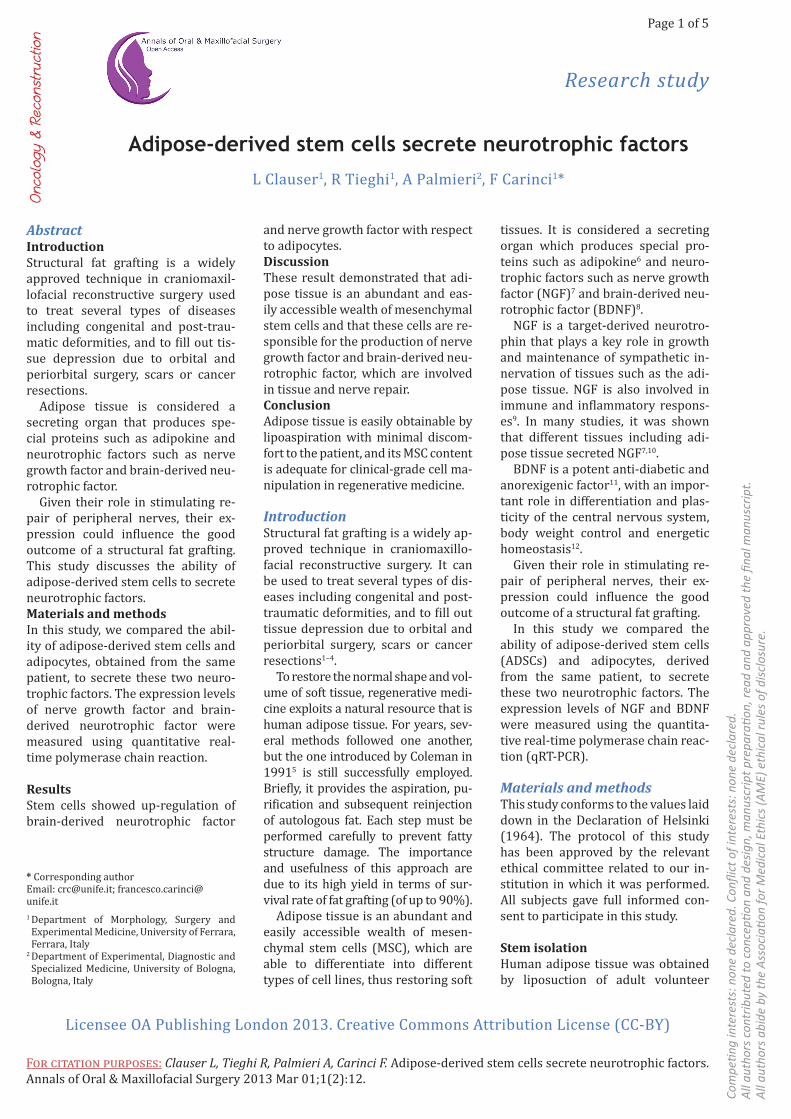
Figure 1: Surface antigene profile of ADSCs. Phenotypic characterization by flow cytometry of cell cultures derived from adipose tissue by staining with the indicated mAb. Representative dot plots documenting the purity of cell preparations and the homogenously CD105+, CD90+, CD34−, CD45, CD14− surface antigen profile, are shown. Irr., irrelevant, isotype control Ab.
Page 3 of 5
Research study
Licensee OA Publishing London 2013. Creative Commons Attribution License (CC-BY)
For citation purposes: Clauser L, Tieghi R, Palmieri A, Carinci F. Adipose-derived stem cells secrete neurotrophic factors. Annals of Oral & Maxillofacial Surgery 2013 Mar 01;1(2):12
Com
petin
g in
tere
sts:
non
e de
clar
ed. C
onfli
ct o
f int
eres
ts: n
one
decl
ared
. A
ll au
thor
s co
ntrib
uted
to c
once
ption
and
des
ign,
man
uscr
ipt p
repa
ratio
n, re
ad a
nd a
ppro
ved
the
final
man
uscr
ipt.
All
auth
ors
abid
e by
the
Ass
ocia
tion
for M
edic
al E
thic
s (A
ME)
eth
ical
rule
s of
dis
clos
ure.
ResultsADSCs were phenotypically char-acterized by flow cytometric analy-ses. Cell preparations derived from adipose tissue were homogenously CD105+, CD90+, CD34−, CD45−, CD14−, which is a typical MSC surface anti-gen profile (Figure 1).
The expression level of two growth factors, NGF and BDFN, was com-pared in three cell types: adipocytes, HFb and ADSCs.
Gene expression was quantified using qRT-PCR and normalized to the expression of the housekeeping gene RPL13.
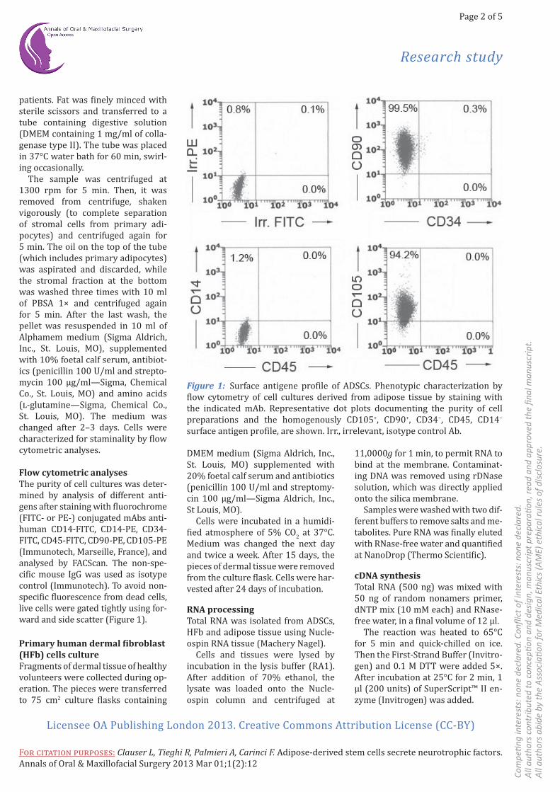
Comparing expression levels of ADSCs and HFb, we found that NGF gene was over-expressed in stem cells. Conversely BDFN was down-regulated (Figure 2).
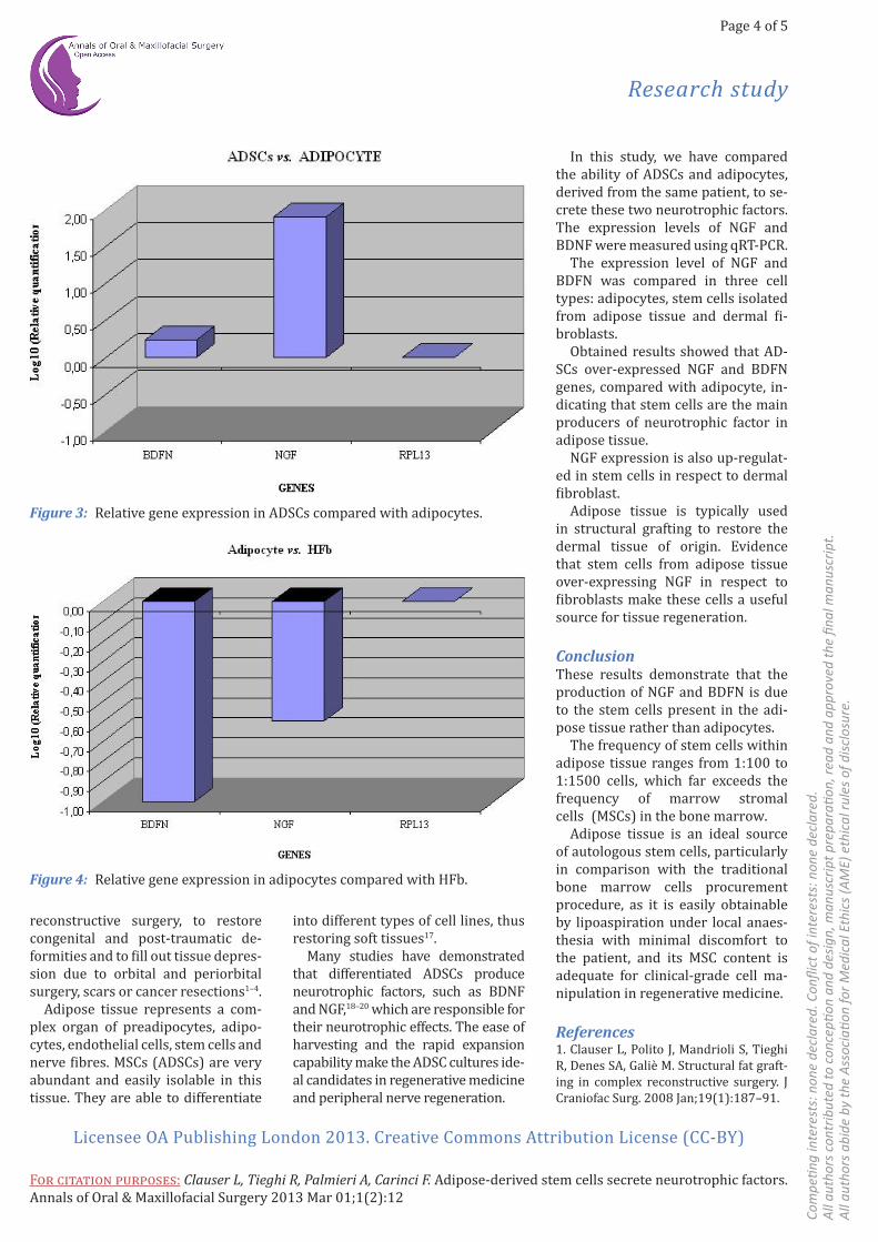
These two genes were down-regu-lated in adipocyte when compared with fibroblast (Figure 3) and stem cells.
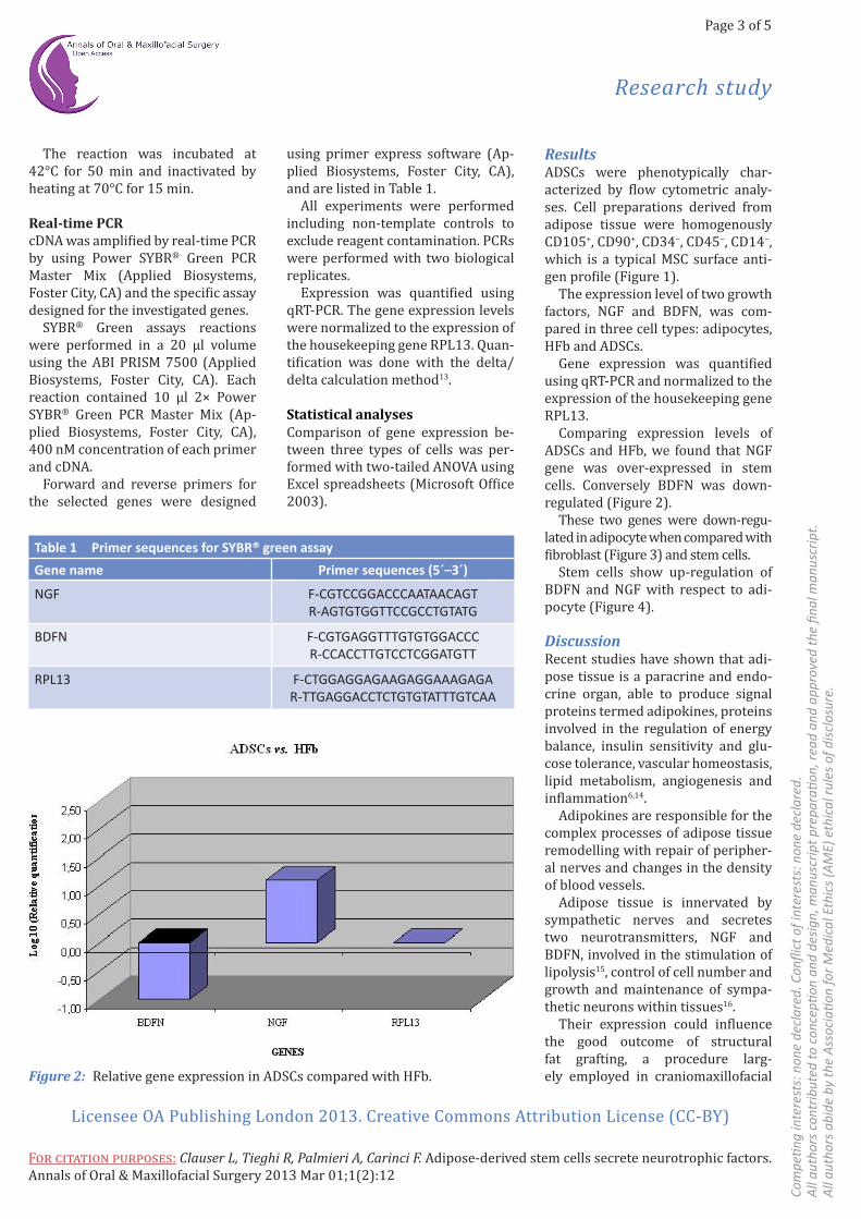
Stem cells show up-regulation of BDFN and NGF with respect to adi-pocyte (Figure 4).
DiscussionRecent studies have shown that adi-pose tissue is a paracrine and endo-crine organ, able to produce signal proteins termed adipokines, proteins involved in the regulation of energy balance, insulin sensitivity and glu-cose tolerance, vascular homeostasis, lipid metabolism, angiogenesis and inflammation6,14.
Adipokines are responsible for the complex processes of adipose tissue remodelling with repair of peripher-al nerves and changes in the density of blood vessels.
Adipose tissue is innervated by sympathetic nerves and secretes two neurotransmitters, NGF and BDFN, involved in the stimulation of lipolysis15, control of cell number and growth and maintenance of sympa-thetic neurons within tissues16.
Their expression could influence the good outcome of structural fat grafting, a procedure larg- ely employed in craniomaxillofacial
The reaction was incubated at 42°C for 50 min and inactivated by heating at 70°C for 15 min.
Real-time PCRcDNA was amplified by real-time PCR by using Power SYBR® Green PCR Master Mix (Applied Biosystems, Foster City, CA) and the specific assay designed for the investigated genes.
SYBR® Green assays reactions were performed in a 20 µl volume using the ABI PRISM 7500 (Applied Biosystems, Foster City, CA). Each reaction contained 10 µl 2× Power SYBR® Green PCR Master Mix (Ap-plied Biosystems, Foster City, CA), 400 nM concentration of each primer and cDNA.
Forward and reverse primers for the selected genes were designed
using primer express software (Ap-plied Biosystems, Foster City, CA), and are listed in Table 1.
All experiments were performed including non-template controls to exclude reagent contamination. PCRs were performed with two biological replicates.
Expression was quantified using qRT-PCR. The gene expression levels were normalized to the expression of the housekeeping gene RPL13. Quan-tification was done with the delta/delta calculation method13.
Statistical analysesComparison of gene expression be-tween three types of cells was per-formed with two-tailed ANOVA using Excel spreadsheets (Microsoft Office 2003).
Figure 2: Relative gene expression in ADSCs compared with HFb.
Table 1 Primer sequences for SYBR® green assay
Gene name Primer sequences (5´–3´)
NGF F-CGTCCGGACCCAATAACAGTR-AGTGTGGTTCCGCCTGTATG
BDFN F-CGTGAGGTTTGTGTGGACCCR-CCACCTTGTCCTCGGATGTT
RPL13 F-CTGGAGGAGAAGAGGAAAGAGAR-TTGAGGACCTCTGTGTATTTGTCAA
Page 4 of 5
Research study
Licensee OA Publishing London 2013. Creative Commons Attribution License (CC-BY)
For citation purposes: Clauser L, Tieghi R, Palmieri A, Carinci F. Adipose-derived stem cells secrete neurotrophic factors. Annals of Oral & Maxillofacial Surgery 2013 Mar 01;1(2):12
Com
petin
g in
tere
sts:
non
e de
clar
ed. C
onfli
ct o
f int
eres
ts: n
one
decl
ared
. A
ll au
thor
s co
ntrib
uted
to c
once
ption
and
des
ign,
man
uscr
ipt p
repa
ratio
n, re
ad a
nd a
ppro
ved
the
final
man
uscr
ipt.
All
auth
ors
abid
e by
the
Ass
ocia
tion
for M
edic
al E
thic
s (A
ME)
eth
ical
rule
s of
dis
clos
ure.
In this study, we have compared the ability of ADSCs and adipocytes, derived from the same patient, to se-crete these two neurotrophic factors. The expression levels of NGF and BDNF were measured using qRT-PCR.
The expression level of NGF and BDFN was compared in three cell types: adipocytes, stem cells isolated from adipose tissue and dermal fi-broblasts.
Obtained results showed that AD-SCs over-expressed NGF and BDFN genes, compared with adipocyte, in-dicating that stem cells are the main producers of neurotrophic factor in adipose tissue.
NGF expression is also up-regulat-ed in stem cells in respect to dermal fibroblast.
Adipose tissue is typically used in structural grafting to restore the dermal tissue of origin. Evidence that stem cells from adipose tissue over-expressing NGF in respect to fibroblasts make these cells a useful source for tissue regeneration.
ConclusionThese results demonstrate that the production of NGF and BDFN is due to the stem cells present in the adi-pose tissue rather than adipocytes.
The frequency of stem cells within adipose tissue ranges from 1:100 to 1:1500 cells, which far exceeds the frequency of marrow stromal cells (MSCs) in the bone marrow.
Adipose tissue is an ideal source of autologous stem cells, particularly in comparison with the traditional bone marrow cells procurement procedure, as it is easily obtainable by lipoaspiration under local anaes-thesia with minimal discomfort to the patient, and its MSC content is adequate for clinical-grade cell ma-nipulation in regenerative medicine.
References1. Clauser L, Polito J, Mandrioli S, TieghiR, Denes SA, Galiè M. Structural fat graft-ing in complex reconstructive surgery. J Craniofac Surg. 2008 Jan;19(1):187–91.
into different types of cell lines, thus restoring soft tissues17.
Many studies have demonstrated that differentiated ADSCs produce neurotrophic factors, such as BDNF and NGF,18–20 which are responsible for their neurotrophic effects. The ease of harvesting and the rapid expansion capability make the ADSC cultures ide-al candidates in regenerative medicine and peripheral nerve regeneration.
reconstructive surgery, to restore congenital and post-traumatic de-formities and to fill out tissue depres-sion due to orbital and periorbital surgery, scars or cancer resections1–4.
Adipose tissue represents a com-plex organ of preadipocytes, adipo-cytes, endothelial cells, stem cells and nerve fibres. MSCs (ADSCs) are very abundant and easily isolable in this tissue. They are able to differentiate
Figure 3: Relative gene expression in ADSCs compared with adipocytes.
Figure 4: Relative gene expression in adipocytes compared with HFb.
Page 5 of 5
Research study
Licensee OA Publishing London 2013. Creative Commons Attribution License (CC-BY)
For citation purposes: Clauser L, Tieghi R, Palmieri A, Carinci F. Adipose-derived stem cells secrete neurotrophic factors. Annals of Oral & Maxillofacial Surgery 2013 Mar 01;1(2):12
Com
petin
g in
tere
sts:
non
e de
clar
ed. C
onfli
ct o
f int
eres
ts: n
one
decl
ared
. A
ll au
thor
s co
ntrib
uted
to c
once
ption
and
des
ign,
man
uscr
ipt p
repa
ratio
n, re
ad a
nd a
ppro
ved
the
final
man
uscr
ipt.
All
auth
ors
abid
e by
the
Ass
ocia
tion
for M
edic
al E
thic
s (A
ME)
eth
ical
rule
s of
dis
clos
ure.
16. Bowers RR, Festuccia WT, Song CK,Shi H, Migliorini RH, Bartness TJ. Sympa-thetic innervation of white adipose tissue and its regulation of fat cell number. Am J Physiol Regul Integr Comp Physiol. 2004 Jun;286(6):R1167–75.17. Yoshimura H, Muneta T, Nimura A,Yokoyama A, Koga H, Sekiya I. Comparison of rat mesenchymal stem cells derived from bone marrow, synovium, perioste-um, adipose tissue, and muscle. Cell Tis-sue Res. 2007 Mar;327(3):449–62.18. Kingham PJ, Kalbermatten DF, MahayD, Armstrong SJ, Wiberg M, Terenghi G. Adipose-derived stem cells differenti-ate into a Schwann cell phenotype and promote neurite outgrowth in vitro. Exp Neurol. 2007 Oct;207(2):267–74.19. Kalbermatten DF, Schaakxs D, Kingham PJ, Wiberg M. Neurotrophic activity of human adipose stem cells isolated from deep and superficial layers of abdominal fat. Cell Tissue Res. 2011 May;344(2):251–60.20. Locke M, Windsor J, Dunbar PR. Human adipose-derived stem cells: iso-lation, characterization and applications in surgery. ANZ J Surg. 2009 Apr;79(4): 235–44.
autoimmune diseases. Autoimmunity. 1994;19(2):141–50.10. Chaldakov GN, Fiore M, Stankulov IS,Manni L, Hristova MG, Antonelli A. Neu-rotrophin presence in human coronary atherosclerosis and metabolic syndrome: a role for NGF and BDNF in cardiovascu-lar disease? Prog Brain Res. 2004;146: 279–89.11. Geroldi D, Minoretti P, Emanuele E.Brain-derived neurotrophic factor and the metabolic syndrome: more than just a hypothesis. Med Hypotheses. 2006;67(1):195–6.12. Cao L, Lin EJ, Cahill MC, Wang C, LiuX, During MJ. Molecular therapy of obe-sity and diabetes by a physiological au-toregulatory approach. Nat Med. 2009 Apr;15(4):447–54.13. Livak KJ, Schmittgen TD. Analysis ofrelative gene expression data using real-time quantitative PCR and the 2(-Delta Delta C(T)) Method. Methods. 2001 Dec;25(4):402–8.14. Trayhurn P, Beattie JH. Physi-ological role of adipose tissue: white adipose tissue as an endocrine and secretory organ. Proc Nutr Soc. 2001 Aug;60(3):329–39.15. Bartness TJ, Bamshad M. Innerva-tion of mammalian white adipose tissue: implications for the regulation of total body fat. Am J Physiol. 1998 Nov;275(5 pt 2):R1399–411.
2. Clauser L, Tieghi R. New mini-osteot-omy of the infraorbital nerve in bony de-compression for endocrine orbitopathy. J Craniofac Surg. 2010 Jan;21(1):222–4.3. Clauser L, Tieghi R, Consorti G. Parry-Romberg syndrome: volumetric regeneration by structural fat grafting technique. J Craniomaxillofac Surg. 2010 Dec;38(8):605–9.4. Clauser L, Tieghi R, Galie M, Carinci F.Structural fat grafting: facial volumet-ric restoration in complex reconstruc-tive surgery. J Craniofac Surg. 2011 Sep;22(5):1695–701.5. Coleman SR. Facial recontouring withlipostructure. Clin Plast Surg. 1997 Apr;24(2):347–67.6. Trayhurn P, Wood IS. Adipokines: in-flammation and the pleiotropic role of white adipose tissue. Br J Nutr. 2004 Sep;92(3):347–55.7. Ryan VH, German AJ, Wood IS, HunterL, Morris P, Trayhurn P. NGF gene ex-pression and secretion by canine adipo-cytes in primary culture: upregulation by the inflammatory mediators LPS and TNFalpha. Horm Metab Res. 2008 Dec;40(12):861–8.8. Sornelli F, Fiore M, Chaldakov GN, AloeL. Brain derived neurotrophic factor: a new adipokine. Biomed Rev. 2007; 18:85–8.9. Aloe L, Skaper SD, Leon A, Levi-Montalcini R. Nerve growth factor and


![STEM CELLS EMBRYONIC STEM CELLS/INDUCED PLURIPOTENT STEM CELLS Stem Cells.pdf · germ cell production [2]. Human embryonic stem cells (hESCs) offer the means to further understand](https://img.pdfslide.us/doc/110x75/6014b11f8ab8967916363675/stem-cells-embryonic-stem-cellsinduced-pluripotent-stem-cells-stem-cellspdf.jpg)

![Human Amniotic Fluid Mesenchymal Stem Cells in Combination … · 2018. 5. 16. · stem cells, and administration of neurotrophic factors [4– 7]. Recently, cell transplantation](https://img.pdfslide.us/doc/110x75/5fd57e80171a4858402613c4/human-amniotic-fluid-mesenchymal-stem-cells-in-combination-2018-5-16-stem-cells.jpg)