Embed Size (px)
Citation preview
Laura E. Hand,1 Paola Usan,2 Garth J.S. Cooper,2,3 Lance Y. Xu,3 Basil Ammori,4
Peter S. Cunningham,1 Reza Aghamohammadzadeh,4 Handrean Soran,4 Adam Greenstein,4
Andrew S.I. Loudon,1 David A. Bechtold,1 and David W. Ray2
Adiponectin Induces A20Expression in Adipose Tissue toConfer Metabolic BenefitDiabetes 2015;64:128–136 | DOI: 10.2337/db13-1835
Obesity is a major risk factor for metabolic disease, withwhite adipose tissue (WAT) inflammation emerging asa key underlying pathology. We detail that mice lackingReverba exhibit enhanced fat storage without the pre-dicted increasedWAT inflammation or loss of insulin sen-sitivity. In contrast to most animal models of obesity andobese human patients, Reverba2/2 mice exhibit elevatedserum adiponectin levels and increased adiponectin se-cretion fromWAT explants in vitro, highlighting a potentialanti-inflammatory role of this adipokine in hypertrophicWAT. Indeed, adiponectin was found to suppress primarymacrophage responses to lipopolysaccharide and proin-flammatory fatty acids, and this suppression dependedon glycogen synthase kinase 3b activation and inductionof A20. Attenuated inflammatory responses in Reverba2/2
WAT depots were associated with tonic elevation of A20protein and ex vivo shown to depend on A20. We alsodemonstrate that adipose A20 expression in obesehuman subjects exhibits a negative correlation with mea-sures of insulin sensitivity. Furthermore, bariatric surgery–induced weight loss was accompanied by enhanced WATA20 expression, which is positively correlated with in-creased serum adiponectin and improved metabolicand inflammatory markers, including C-reactive protein.The findings identify A20 as a mediator of adiponectinanti-inflammatory action in WAT and a potential targetfor mitigating obesity-related pathology.
Obesity is a major public health issue, with the principalcause of morbidity a result of metabolic dysfunction (e.g.,
type 2 diabetes, atherosclerosis). Progression of clinicalpathology has been strongly linked to chronic inflammationin white adipose tissue (WAT) (1,2), where hypertrophicadipocytes fail to efficiently store excess energy, leading toadipose tissue dysfunction, dyslipidemia, and insulin resis-tance. Increased tissue inflammation through adipocyte re-lease of cytokines [e.g., tumor necrosis factor-a (TNF-a)(3,4)], chemokines [e.g., monocyte chemoattractant protein1 (MCP1/CCL2) (5)], and proinflammatory saturated fattyacids (FAs) (6) drives monocyte recruitment and differenti-ation to M1-polarized macrophages, thereby expanding theinflammatory environment within adipose tissue beds.Proinflammatory factors produced by activated macro-phages act reciprocally on adipocytes, thereby perpetuatingadipose tissue inflammation and dysfunction (7).
WAT secretes several adipokines that not only areimportant hormonal regulators of systemic metabolismbut also possess either pro- or anti-inflammatory proper-ties. Adipokine production is disrupted in hypertrophicadipose tissue, resulting in obesity-associated inflammation(8). For example, circulating levels of adiponectin are con-sistently decreased in obese human subjects and experi-mental animals. A protective role for adiponectin inmodels of diabetes, dyslipidemia, and atherosclerosis hasbeen reported (9), and in obese human subjects, plasmaadiponectin concentrations are inversely correlated withcirculating inflammatory markers, including C-reactive pro-tein (CRP) and TNF-a (8,10). In line with these findings,direct anti-inflammatory activity of adiponectin has beendemonstrated. Adiponectin inhibits transformation of
1Faculty of Life Sciences, University of Manchester, Manchester, U.K.2Faculty of Medical and Health Sciences, University of Manchester, Manchester,U.K.3School of Biological Sciences, University of Auckland, Auckland, New Zealand4Centre for Advanced Discovery and Experimental Therapeutics, University ofManchester, Manchester, U.K.
Corresponding author: David W. Ray, [email protected], or David A.Bechtold, [email protected].
Received 5 December 2013 and accepted 10 August 2014.
This article contains Supplementary Data online at http://diabetes.diabetesjournals.org/lookup/suppl/doi:10.2337/db13-1835/-/DC1.
© 2015 by the American Diabetes Association. Readers may use this article aslong as the work is properly cited, the use is educational and not for profit, andthe work is not altered.
128 Diabetes Volume 64, January 2015
OBESITY
STUDIES

macrophages into foam cells (11) and reduces macrophagemigration and chemokine production (12,13). Adiponectinalso promotes macrophage polarization toward an anti-inflammatory M2 phenotype (14) and desensitizes macro-phages to Toll-like receptor (TLR) signaling (15,16).
The nuclear hormone receptor Reverba is a ligand-sensitive transcription factor that negatively regulatesthe expression of core clock proteins (17,18). Reverba haspreviously been implicated in adipocyte differentiation(19), lipid metabolism (20), and regulation of the inflam-matory response (21). In the current study, we identify anobese phenotype in Reverba2/2 mice characterized bya lack of the predicted M1 macrophage infiltration ofWAT depots, a paradoxical increase in adiponectin produc-tion, and preservation of insulin sensitivity. We furtherdemonstrate that adiponectin suppression of inflammatorysignaling in macrophages and adipose tissue depots is me-diated through the cytoplasmic ubiquitin-modifying en-zyme and a negative regulator of TLR signaling, A20,which is enhanced in WAT from Reverba2/2 mice. Wealso demonstrate that A20 expression in WAT from obesehuman subjects is correlated significantly with measures ofinsulin sensitivity, and in subjects after bariatric surgery,we observed an increase in WAT A20 expression, whichwas positively correlated with elevated serum adiponectinlevels and associated with improved levels of metabolic andinflammatory markers, such as CRP. Taken together, thesefindings identify a novel target pathway for modulatingadipose tissue inflammation in obesity.
RESEARCH DESIGN AND METHODS
Animal MaintenanceExperimental procedures were licensed under the AnimalsAct, 1986, and local animal welfare committee. Reverba2/2
mice were provided by Ueli Schibler (University of Geneva),as described (22). These mice were subsequently bred ontothe Per2:Luciferase reporter line generated by Joe Takahashi(University of Texas Southwestern Medical Center, Dallas,TX) (23) and maintained as an inbred line at the Univer-sity of Manchester. The control and knockout (KO) micefor all experiments were littermates because the line wasbred using heterozygotic parent mice. Adult male micewere maintained in a 12-h light, 12-h dark lighting sched-ule; housed at an ambient temperature of 20–22°C; andfed standard rodent chow and water supplied ad libitum(except for the study shown in Fig. 1C–E and H, where theanimals received diet-induced obesity [DIO] rodent puri-fied diet with 60% energy from fat [International ProductSupplies Ltd]). Glucose tolerance tests were carried out asin Bechtold et al. (24).
Cell CulturePeritoneal exudates cells were isolated and the CD11b+ mac-rophage population positively selected using anti-CD11bMicroBeads and MS autoMACS Columns (both MiltenyiBiotec) according to the manufacturer’s specifications. Cellswere resuspended in RPMI medium and plated.
ReagentsRecombinant human adiponectin was produced at theUniversity of Auckland as described previously (25,26).Multimeric adiponectin forms in the purified proteinwere confirmed by native SDS-PAGE. Mouse TNF-a, lipo-polysaccharide (LPS), SB216763, and FAs (sodium stea-rate, sodium palmitate, sodium myristate, and sodiumdodecanoate) were purchased from Sigma. Antibodies toA20 (D13H3) and glucose synthase kinase 3b (GSK3b)(27C10) were from Cell Signaling, to b-actin (AC-15)from Abcam, to TNF-a (MP6-XT22) from R&D Systems,to laminin B (C20) from Santa Cruz, to tubulin fromSigma, and to inducible nitric oxide synthase (iNOS)from Abcam. Secondary antibodies were horseradishperoxidase–conjugated sheep anti-mouse or donkey anti-rabbit IgGs (GE Healthcare) and fluorescein isothiocyanate–conjugated goat anti-rabbit (Jackson ImmunoResearch).
Preparation of FAsFAs were solubilized in ethanol stock solutions of 100mmol/L and stored at 220°C. FA-albumin complex solu-tions were freshly prepared before each experiment. Fivepercent FA-free and low-endotoxin BSA (27) (Sigma) wasdissolved in RPMI medium and filtered with 0.22 mmol/Llow-protein–binding filter (Millipore). Stock solutions ofFAs were added to the BSA medium to achieve an FA:BSAmolar ratio of 3:1 and incubated at 40°C for 1 h. Cellswere treated with individual FAs, whereas control cellsreceived BSA only.
Quantitative Real-Time PCRTissue was homogenized in TRIzol reagent (Invitrogen)using lysing matrix D tubes (MP Biomedicals), and totalRNA was isolated according to the manufacturer’s speci-fications. RNA from macrophages was extracted with theRNeasy Mini Kit (QIAGEN). After reverse-transcriptionusing the High-Capacity RNA-to-cDNA Kit (Applied Bio-systems), quantitative RT-PCR was performed usingPower SYBR Green PCR Master Mix and StepOnePlusReal-Time PCR System (both Applied Biosystems). Mousehousekeeping gene 18S rRNA was used as an internalcontrol.
RNA InterferenceA20-specific small interfering RNA (siRNA) and non-targeting scrambled control siRNA were designed andpurchased from Eurofins MWG Operon (siMAX siRNA).siRNAs were transfected into freshly isolated primaryCD11b+ murine macrophages with the Amaxa Nucleofectordevice set to program Y001 using the Mouse MacrophageNucleofector Kit. Cells were washed once 1 h after trans-fection and cultured for an additional 2 days beforeadiponectin and LPS stimulation.
Protein AnalysisAnalysis of adiponectin isoform expression was as pre-viously described (25,26). ELISAs were performed withpaired antibody sets as recommended by the manufac-turer (R&D Systems). For immunoblot analysis, tissue
diabetes.diabetesjournals.org Hand and Associates 129

was homogenized in Tissue Protein Extraction Reagent(Thermo Scientific) using lysing matrix D tubes (MP Bio-medicals). Cytoplasmic and nuclear cell extracts were pre-pared by lysing cells in buffer 1 (30 mmol/L Tris-HCl [pH7.4], 0.5 mmol/L EDTA, 150 mmol/L NaCl, 0.2% vol/volNP40), centrifuging nuclear pellets and resuspending thesein buffer 2 (30 mmol/L Tris-HCl [pH 7.4], 0.5 mmol/LEDTA, 400 mmol/L NaCl, 1% vol/vol Triton X-100, 0.1%wt/vol SDS). Protein concentration was measured with
a BCA protein quantification kit (Pierce Biotechnology).Proteins were subjected to SDS-PAGE using a Mini-PROTEAN 3 apparatus (Bio-Rad) according to the man-ufacturer’s instructions before transfer to nitrocellulosemembrane using an iBlot gel transfer system (Invitrogen).Immunoblots were incubated with the appropriate anti-body and imaged using enhanced chemiluminescence(ECL Prime detection system; GE Healthcare) and quan-tified using ImageJ software. Immunohistochemistry
Figure 1—Phenotype of Reverba2/2 mice. A and B: Body weight (A) and visceral WAT (B) mass of 12-week-old male Reverba2/2 mice andWT littermates maintained on an NC diet (n = 16–20/group). C: Histological sections of WAT from WT and Reverba2/2 mice fed NC and WTmice fed HFD to induce DIO. iNOS immunoreactivity demonstrates characteristic macrophage cuffs in WAT of WT DIO mice, which arevirtually absent in Reverba2/2 mice despite their obese phenotype. D and E: Body weight (D) and intraperitoneal glucose tolerance testresults (E) of WT and Reverba2/2 mice fed NC or HFD for 10 weeks (n = 5–6/group). F and G: Serum leptin (F ) and adiponectin (G)concentrations of 12-week-old Reverba2/2 mice were significantly higher than in WT controls (n = 18–21/group) and positively correlatedwith adipose tissue mass (P < 0.05, Pearson correlation). H: ELISA analysis of serum adiponectin in WT and Reverba2/2 mice fed NC vs.HFD. I: Immunoblot analysis of monomeric (top) and multimeric (bottom) adiponectin in sera of WT and Reverba2/2 mice (black arrowhead,high molecular weight; white arrowhead, hexamer; gray arrowhead, trimer). J: ELISA analysis of high–molecular weight adiponectin in seraof WT and Reverba2/2 mice expressed as a ratio of total adiponectin. Data are mean6 SEM; statistical significance was determined usingStudent t test or one-way ANOVA with Bonferroni post hoc test (C). *P < 0.05, **P < 0.01, ***P < 0.001. Adn, adiponectin; BW, bodyweight; HMW, high molecular weight.
130 Adiponectin Limits Adipose Inflammation Via A20 Diabetes Volume 64, January 2015

was carried out as previously described (24) usingparaformaldehyde-fixed and paraffin-embedded tissueepididymal fat pads.
Human Adipose Tissue Biopsies and BiochemicalAnalysesPatients with severe obesity (BMI .35 kg/m2, n = 12)who were awaiting gastric bypass surgery were recruitedafter full informed written consent in accordance withlocal research ethics committee approval. Participantswere invited to return for a follow-up assessment 6months after bariatric surgery. On both occasions,patients provided gluteal subcutaneous adipose samples(0.125 cm3) by undergoing a surgical biopsy under localanesthesia (28). The biopsy samples were immediatelyfrozen in liquid nitrogen. Fasting venous blood sampleswere also collected for the measurement of CRP, adipo-nectin, and leptin levels as previously described (28).
Statistical AnalysisData are presented as mean 6 SEM. The statistical testused is specified in each data section. Student t test wasused when only two groups were studied; one-wayANOVA was used when more than two groups were stud-ied and only one factor investigated, followed by post hocanalysis; and two-way ANOVA was used when more thantwo factors were analyzed, followed by Bonferroni posthoc analysis.
RESULTS
Obesity in Reverba2/2 Mice Is Not Associated WithWAT Inflammation and Insulin ResistanceOn normal chow (NC), 12-week-old male Reverba2/2
mice have comparable body weight to wild-type (WT) lit-termates (Fig. 1A) but have significantly increased WATmass (WT: 0.8 6 0.13 g; KO: 1.6 6 0.14; n = 16–20/group; P , 0.01, Student t test) (Fig. 1B) and adipocytehypertrophy (Fig. 1C). WT mice were fed a high-fat diet(HFD) to promote a comparable degree of WAT tissuemass (2.3 6 0.15 g) and adipocyte morphology toReverba2/2 mice maintained on NC. Despite showingsimilar WAT hypertrophy, obese WT mice exhibited char-acteristic inflammatory cuffs surrounding adipocytes,which were rarely observed in the NC-fed Reverba2/2
animals (Fig. 1C). Maintenance of the Reverba2/2 miceon HFD resulted in a profound increase in body weightand adiposity compared with HFD-fed WT mice (Fig. 1D).Despite this, Reverba2/2 mice did not exhibit obesity-related deterioration in glucose tolerance compared withWT littermates, suggesting preservation of insulin sensi-tivity (Fig. 1E). As expected, WT mice exhibited a signifi-cant increase in adipose inflammatory markers inresponse to 16 weeks of HFD feeding (TNFa: NC 1.1 60.1 vs. HFD 3.0 6 0.6 relative mRNA expression, P ,0.01; IL6: NC 1.2 6 0.3 vs. HFD 2.0 6 0.3, P = 0.06;MCP1: NC 1.2 6 0.3 vs. 5.8 6 0.7, P , 0.001; n = 6/group). In contrast, expression of inflammatory markers(TNFa, Il6, MCP1) were not increased by HFD feeding in
the Reverba2/2 mice (TNFa: NC 1.0 6 0.2 vs. HFD 1.8 60.3 relative mRNA expression; IL6: NC 1.3 6 0.3 vs. HFD1.0 6 0.3; MCP1: NC 1.1 6 0.5 vs. 1.4 6 0.5; n = 6/group). The absence of adipose inflammation in theReverba2/2 mice was not due to a general lack of inflam-matory response, as we have previously demonstratedthat these animals exhibit robust responses to systemicendotoxin administration (21). Furthermore, Reverba2/2
mice exhibited a pronounced elevation in serum free FAconcentrations (WT NC: 165.5 6 20.7 mmol/L; KO NC:274.0 6 17.6; P , 0.05, Student t test) indicative of in-creased adipose tissue lipolysis, a known proinflammatorysignal.
In line with increased adiposity, NC-fed Reverba2/2
mice exhibited increased circulating leptin concentrations(Fig. 1F). Unexpectedly, these animals also showed anelevation in circulating adiponectin (Fig. 1G), whichremained significantly elevated in HFD-fed Reverba2/2
mice compared with WT littermates (Fig. 1H). This find-ing contrasts many animal models of obesity and obesehuman subjects in whom circulating levels of adiponectinare reduced. The oligomerization state is critical to adipo-nectin function, with the high–molecular weight formsbeing the most bioactive (29). Circulating levels of thehigh–molecular weight form of adiponectin were signifi-cantly increased in Reverba2/2 mice compared with WTlittermates (Fig. 1I and J).
Adiponectin Drives GSK3b-Mediated Induction of A20to Attenuate Macrophage InflammationA number of studies have highlighted the ability ofadiponectin to improve aspects of metabolic disturbance,including insulin resistance and vascular dysfunction (8),Furthermore, mice lacking adiponectin expression showedelevated WAT inflammation in response to DIO (30). Wetherefore investigated the anti-inflammatory actions ofadiponectin using multimeric, appropriately posttransla-tionally modified human adiponectin (25).
In agreement with previous work (31), adiponectinpretreatment (6–18 h, 3 mg/mL) rendered CD11b+ pri-mary murine macrophages refractory to subsequent stim-ulation by the TLR4 agonist LPS as assessed bydiminished inflammatory gene induction of IL6, TNFa(Fig. 2A), iNOS, MCP1, CCL5, and IL10 (SupplementaryFig. 1). In the context of obesity-related inflammation,TLR4 activation can result from elevated production ofsaturated medium-chain FAs (e.g., C18:0, C16:0) releasedfrom hypertrophic adipocytes (32). Importantly, adipo-nectin pretreatment (18 h, 3 mg/mL) significantly reducedmacrophage inflammatory responses to C18:0 and C16:0(Supplementary Fig. 2).
Attenuated nuclear factor (NF)-kB signaling has beenpreviously implicated in models of induced immunotoler-ance (e.g., tolerance to subsequent LPS stimulation fol-lowing an initial proinflammatory stimulus of TNF-a orLPS) (33). Therefore, we measured the expression of neg-ative regulators of NF-kB signaling, including A20, SOCS1,
diabetes.diabetesjournals.org Hand and Associates 131

SOCS3, IRAKM, and SHIP1, following adiponectin admin-istration. Treatment of macrophages with adiponectin (3mg/mL) caused a rapid and pronounced induction of thecytoplasmic ubiquitin-modifying enzyme A20 (Supplemen-tary Fig. 3). SOCS1, SOCS3, and IRAKM mRNAs were alsoinduced to a lesser extent, whereas SHIP1 expression was
not affected (Supplementary Fig. 3). Because of its partic-ularly strong induction in response to adiponectin, wecharacterized further the role of A20 in adiponectin anti-inflammatory responses. Constitutive expression of A20protein in murine primary macrophages was minimal; how-ever, adiponectin treatment caused a strong induction of
Figure 2—Adiponectin-induced cross-tolerance to endotoxin is mediated by A20. A: Quantitative RT-PCR analysis of IL6 and TNFainduction in macrophages pretreated with adiponectin for the indicated times. B: Immunoblot analysis of A20 expression in murine primarymacrophages cultured for the indicated times with 3 mg/mL adiponectin. C: Quantitative RT-PCR analysis of A20 expression in primarymurine macrophages pretreated with adiponectin (3 mg/mL, 6 h) and challenged with LPS (100 ng/mL, 4 h). D: Quantitative RT-PCR andimmunoblot analyses of A20 expression of primary murine macrophages transfected with control or A20-specific siRNA. E: QuantitativeRT-PCR analysis of macrophages transfected with control or A20-specific siRNA and subject to adiponectin (3 mg/mL, 18 h) pretreatment,followed by LPS challenge (100 ng/mL, 4 h). F: Immunoblot analysis of cytosolic (left) and nuclear (right) extracts of primary macrophagestreated with 3 mg/mL adiponectin for the indicated times. G–I: Quantitative RT-PCR analysis of primary murine macrophages treated withvehicle control [dimethyl sulfoxide] or 50 mmol/L SB216763 and stimulated with 3 mg/mL adiponectin (G and H) or pretreated withadiponectin (3 mg/mL, 18 h) before LPS challenge (100 ng/mL, 4 h) (I). Data are mean 6 SEM from three independent experimentsnormalized to mouse 18S rRNA control and fold change relative to control untreated cells. Statistical significance was determined usingthe Student t test (D: ***P < 0.001), two-way ANOVA (E: *P < 0.05, **P < 0.01, ***P < 0.001 A20 siRNA vs. control siRNA; ###P < 0.001adiponectin pretreatment vs. no adiponectin treatment), or one-way ANOVA (A, F, and I: *P < 0.05, **P < 0.01, ***P < 0.001 adiponectinpretreatment vs. no adiponectin treatment) with Bonferroni post hoc test. Adn, adiponectin; Nucl., nuclear; SB, SB216763; siA20, A20siRNA; siCtrl, control siRNA.
132 Adiponectin Limits Adipose Inflammation Via A20 Diabetes Volume 64, January 2015

A20 within 3 h of stimulation, which remained elevatedbeyond 24 h posttreatment (Fig. 2B). A20 expression is alsoinduced following stimulation of macrophages with LPS(Fig. 2C). Of note, the induction of A20 in macrophagesto a secondary LPS challenge was not attenuated by pre-treatment with adiponectin (Fig. 2C), a time at which in-duction of IL6 and TNFa expression have becomeunresponsive to LPS stimulation, suggesting that A20 isnot subject to prestimulation-induced tolerance.
A direct role for A20 in adiponectin-induced macro-phage quiescence was next demonstrated by targetingA20 by siRNA (Fig. 2D, Supplementary Fig. 4). Knock-down of A20 enhanced the induction of IL6 and TNFain naïve macrophages treated with LPS, demonstratinga constitutive role of A20 in limiting the magnitude ofTLR-driven inflammatory responses in macrophages. Im-portantly, knockdown of A20 expression also blocked theability of adiponectin pretreatment to attenuate IL6 andTNFa expression in macrophages following secondary LPSchallenge (Fig. 2E).
GSK3b is a critical enzyme upstream of A20 expression(34), and in line with this, immunoblot analysis revealedthat adiponectin treatment increased nuclear localization ofGSK3b in macrophages (Fig. 2F). Preincubation of murinemacrophages with the specific GSK3b inhibitor SB216763blunted the induction of A20 mRNA in macrophages inresponse to adiponectin treatment (Fig. 2G) and attenuatedthe adiponectin impairment of TLR4 activation (Fig. 2I),demonstrating a role for GSK3b in adiponectin-inducedinflammatory tolerance. Of note, SB216763 treatment didnot diminish acute induction of TNFa by adiponectin (Fig.2H), demonstrating that adiponectin-induced tolerance wasnot secondary to TNF-a induction. Therefore, adiponectinsignaling stimulates GSK3b activity to drive an A20-mediatedanti-inflammatory program.
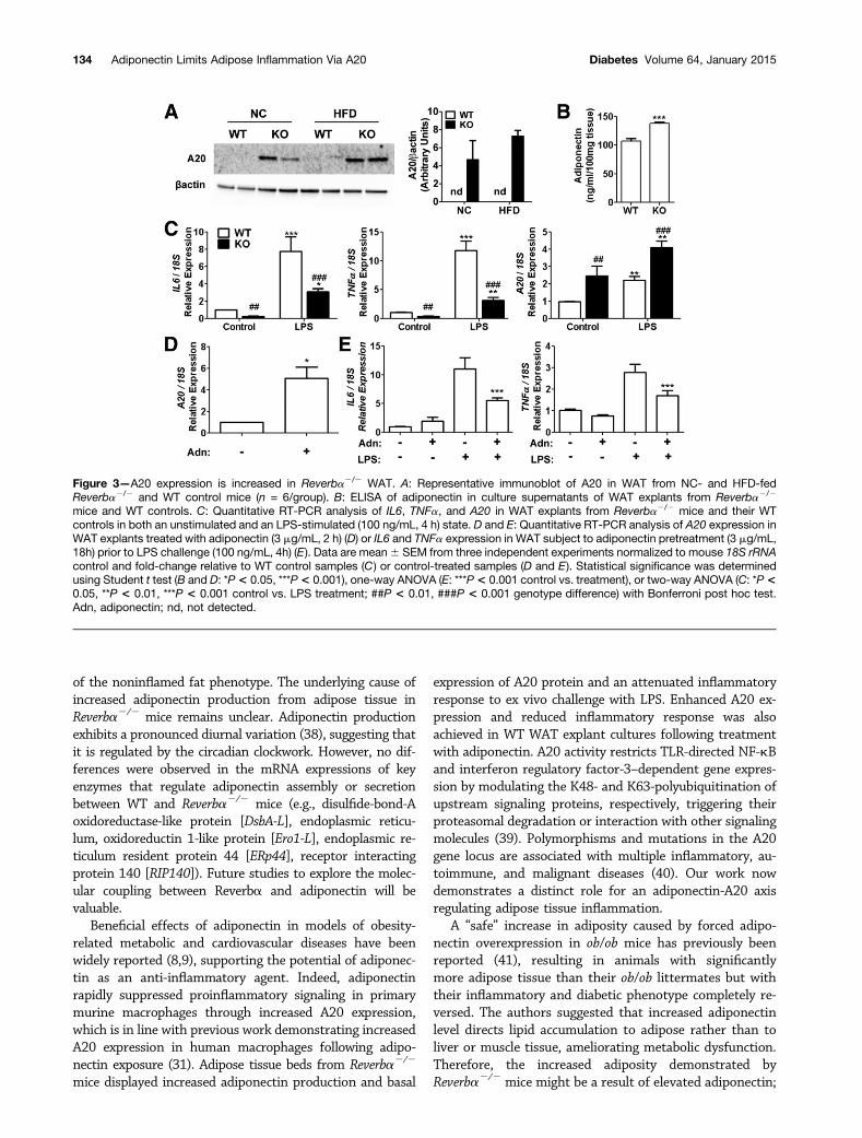
Enhanced Expression of A20 Is Associated With anAttenuation of WAT Inflammation in Reverba2/2 Miceand Improved Metabolic Profile in Bariatric SurgeryPatientsWe next examined the potential role of A20 in regulatingWAT inflammation in Reverba2/2 mice. Reverba2/2 miceexhibited significantly higher expression of A20 protein inWAT beds compared with WT controls, which was main-tained in profoundly obese KO mice maintained on HFD(Fig. 3A). WAT explants derived from Reverba2/2 micealso produced more adiponectin than WT explants (Fig.3B) and in either basal or LPS-stimulated states, exhibitedan attenuated expression of IL6 and TNFa (Fig. 3C). Fur-thermore, adiponectin treatment of WT WAT explant tis-sue significantly increased A20 expression (Fig. 3D) andreduced induction of both TNFa and IL6 in response toLPS challenge (Fig. 3E). Adiponectin was also effective atinducing the expression of A20 in differentiated 3T3-L1adipocytes (vehicle treated: 1.0 6 0.02 relative A20 ex-pression; adiponectin treated: 5.1 6 1.03; P , 0.01), in-dicating that its anti-inflammatory actions are not limited
to macrophages. These results identify adiponectin andA20 as important modulators of WAT inflammation invitro and in vivo.
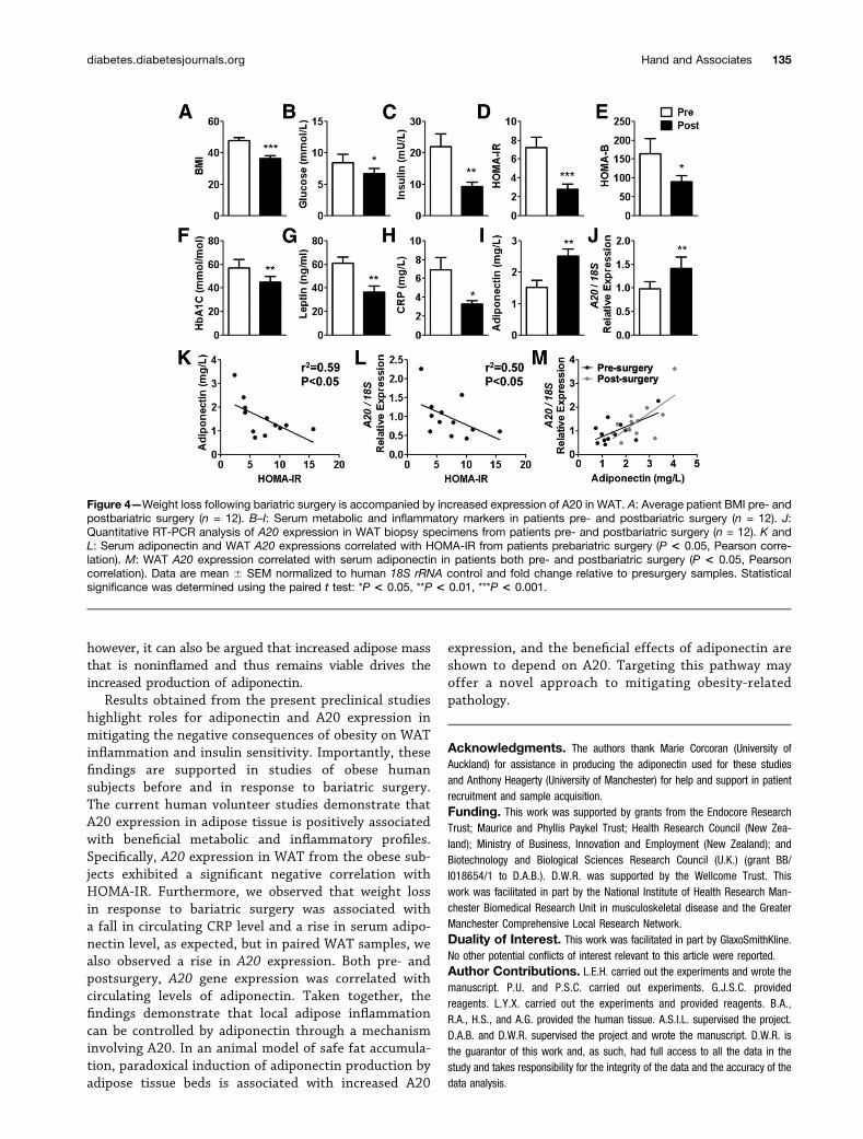
Our studies predict that hypertrophic human adiposetissue depots will show loss of adiponectin action,resulting in reduced A20 protein in both macrophagesand adipocytes, permitting unrestrained inflammatorysignaling to progress. To investigate this, we studieda cohort of obese subjects before and after bariatricsurgery. In support of the animal data revealing a bene-ficial effect of A20 in obesity, presurgery circulatingadiponectin levels and WAT expression of A20 in theobese subjects exhibited a significant negative correlationwith insulin resistance (HOMA-IR: n = 12, P , 0.05,Pearson correlation) (Fig. 4K and L). Furthermore, A20expression in patient WAT samples was positively corre-lated with serum adiponectin both pre- and postsurgery(P , 0.05, Pearson correlation) (Fig. 4M) and exhibiteda significant rise postsurgery (Fig. 4J), concomitant withsignificant weight loss (Fig. 4A), and improved levels ofserum markers of metabolism (i.e., adiponectin, leptin)(Fig. 4G and I) and inflammation (e.g., CRP) (Fig. 4H).
DISCUSSION
A major pathological characteristic of obesity is adiposetissue inflammation, which drives local dysfunction as wellas systemic consequences, such as insulin resistance. Weidentify a novel role for A20 in controlling adipose tissueinflammation and obesity-related pathology. The resultsindicate that rapid and prolonged induction of A20 byadiponectin can drive macrophage quiescence and reducedWAT inflammation. Furthermore, using a murine model ofobesity and, importantly, obese human subjects pre- andpostbariatric surgery, we confirm beneficial effects of in-creased circulating adiponectin levels and enhanced WATA20 expression associated with attenuated adipose tissueinflammation and improvement of metabolic diseasemarkers and insulin sensitivity.
Reverba is a key link between the circadian clockworkand metabolic pathways (35–37) and has been strongly im-plicated in adipocyte differentiation (19) and lipid handling(20). We show in the current study that mice lackingReverba demonstrated enhanced fat storage yet withoutthe expected loss of insulin sensitivity or WAT inflamma-tion. Delezie et al. (20) reported a similar phenotype, al-though the inflammatory status of the mice was notexplored. The findings imply that obesity in Reverba2/2
mice lacks typical proinflammatory stimuli and is accompa-nied by enhanced anti-inflammatory signaling. The absenceof adipose inflammation in the Reverba2/2 mice is not a re-sult of a general lack of inflammatory response becausethese animals exhibit robust responses to systemic endo-toxin administration, as we have previously demonstrated(21). Furthermore, Reverba2/2 mice exhibit profoundly hy-pertrophic adipocytes and elevated production of free FAssimilar to obese WT mice. Therefore, we focused on theenhanced production of adiponectin as a mediating factor
diabetes.diabetesjournals.org Hand and Associates 133
of the noninflamed fat phenotype. The underlying cause ofincreased adiponectin production from adipose tissue inReverba2/2 mice remains unclear. Adiponectin productionexhibits a pronounced diurnal variation (38), suggesting thatit is regulated by the circadian clockwork. However, no dif-ferences were observed in the mRNA expressions of keyenzymes that regulate adiponectin assembly or secretionbetween WT and Reverba2/2 mice (e.g., disulfide-bond-Aoxidoreductase-like protein [DsbA-L], endoplasmic reticu-lum, oxidoreductin 1-like protein [Ero1-L], endoplasmic re-ticulum resident protein 44 [ERp44], receptor interactingprotein 140 [RIP140]). Future studies to explore the molec-ular coupling between Reverba and adiponectin will bevaluable.
Beneficial effects of adiponectin in models of obesity-related metabolic and cardiovascular diseases have beenwidely reported (8,9), supporting the potential of adiponec-tin as an anti-inflammatory agent. Indeed, adiponectinrapidly suppressed proinflammatory signaling in primarymurine macrophages through increased A20 expression,which is in line with previous work demonstrating increasedA20 expression in human macrophages following adipo-nectin exposure (31). Adipose tissue beds from Reverba2/2
mice displayed increased adiponectin production and basal
expression of A20 protein and an attenuated inflammatoryresponse to ex vivo challenge with LPS. Enhanced A20 ex-pression and reduced inflammatory response was alsoachieved in WT WAT explant cultures following treatmentwith adiponectin. A20 activity restricts TLR-directed NF-kBand interferon regulatory factor-3–dependent gene expres-sion by modulating the K48- and K63-polyubiquitination ofupstream signaling proteins, respectively, triggering theirproteasomal degradation or interaction with other signalingmolecules (39). Polymorphisms and mutations in the A20gene locus are associated with multiple inflammatory, au-toimmune, and malignant diseases (40). Our work nowdemonstrates a distinct role for an adiponectin-A20 axisregulating adipose tissue inflammation.
A “safe” increase in adiposity caused by forced adipo-nectin overexpression in ob/ob mice has previously beenreported (41), resulting in animals with significantlymore adipose tissue than their ob/ob littermates but withtheir inflammatory and diabetic phenotype completely re-versed. The authors suggested that increased adiponectinlevel directs lipid accumulation to adipose rather than toliver or muscle tissue, ameliorating metabolic dysfunction.Therefore, the increased adiposity demonstrated byReverba2/2 mice might be a result of elevated adiponectin;
Figure 3—A20 expression is increased in Reverba2/2 WAT. A: Representative immunoblot of A20 in WAT from NC- and HFD-fedReverba2/2 and WT control mice (n = 6/group). B: ELISA of adiponectin in culture supernatants of WAT explants from Reverba2/2
mice and WT controls. C: Quantitative RT-PCR analysis of IL6, TNFa, and A20 in WAT explants from Reverba2/2 mice and their WTcontrols in both an unstimulated and an LPS-stimulated (100 ng/mL, 4 h) state. D and E: Quantitative RT-PCR analysis of A20 expression inWAT explants treated with adiponectin (3 mg/mL, 2 h) (D) or IL6 and TNFa expression in WAT subject to adiponectin pretreatment (3 mg/mL,18h) prior to LPS challenge (100 ng/mL, 4h) (E). Data are mean6 SEM from three independent experiments normalized to mouse 18S rRNAcontrol and fold-change relative to WT control samples (C) or control-treated samples (D and E). Statistical significance was determinedusing Student t test (B and D: *P< 0.05, ***P< 0.001), one-way ANOVA (E: ***P< 0.001 control vs. treatment), or two-way ANOVA (C: *P<0.05, **P < 0.01, ***P < 0.001 control vs. LPS treatment; ##P < 0.01, ###P < 0.001 genotype difference) with Bonferroni post hoc test.Adn, adiponectin; nd, not detected.
134 Adiponectin Limits Adipose Inflammation Via A20 Diabetes Volume 64, January 2015
however, it can also be argued that increased adipose massthat is noninflamed and thus remains viable drives theincreased production of adiponectin.
Results obtained from the present preclinical studieshighlight roles for adiponectin and A20 expression inmitigating the negative consequences of obesity on WATinflammation and insulin sensitivity. Importantly, thesefindings are supported in studies of obese humansubjects before and in response to bariatric surgery.The current human volunteer studies demonstrate thatA20 expression in adipose tissue is positively associatedwith beneficial metabolic and inflammatory profiles.Specifically, A20 expression in WAT from the obese sub-jects exhibited a significant negative correlation withHOMA-IR. Furthermore, we observed that weight lossin response to bariatric surgery was associated witha fall in circulating CRP level and a rise in serum adipo-nectin level, as expected, but in paired WAT samples, wealso observed a rise in A20 expression. Both pre- andpostsurgery, A20 gene expression was correlated withcirculating levels of adiponectin. Taken together, thefindings demonstrate that local adipose inflammationcan be controlled by adiponectin through a mechanisminvolving A20. In an animal model of safe fat accumula-tion, paradoxical induction of adiponectin production byadipose tissue beds is associated with increased A20
expression, and the beneficial effects of adiponectin areshown to depend on A20. Targeting this pathway mayoffer a novel approach to mitigating obesity-relatedpathology.
Acknowledgments. The authors thank Marie Corcoran (University ofAuckland) for assistance in producing the adiponectin used for these studiesand Anthony Heagerty (University of Manchester) for help and support in patientrecruitment and sample acquisition.Funding. This work was supported by grants from the Endocore ResearchTrust; Maurice and Phyllis Paykel Trust; Health Research Council (New Zea-land); Ministry of Business, Innovation and Employment (New Zealand); andBiotechnology and Biological Sciences Research Council (U.K.) (grant BB/I018654/1 to D.A.B.). D.W.R. was supported by the Wellcome Trust. Thiswork was facilitated in part by the National Institute of Health Research Man-chester Biomedical Research Unit in musculoskeletal disease and the GreaterManchester Comprehensive Local Research Network.Duality of Interest. This work was facilitated in part by GlaxoSmithKline.No other potential conflicts of interest relevant to this article were reported.Author Contributions. L.E.H. carried out the experiments and wrote themanuscript. P.U. and P.S.C. carried out experiments. G.J.S.C. providedreagents. L.Y.X. carried out the experiments and provided reagents. B.A.,R.A., H.S., and A.G. provided the human tissue. A.S.I.L. supervised the project.D.A.B. and D.W.R. supervised the project and wrote the manuscript. D.W.R. isthe guarantor of this work and, as such, had full access to all the data in thestudy and takes responsibility for the integrity of the data and the accuracy of thedata analysis.
Figure 4—Weight loss following bariatric surgery is accompanied by increased expression of A20 in WAT. A: Average patient BMI pre- andpostbariatric surgery (n = 12). B–I: Serum metabolic and inflammatory markers in patients pre- and postbariatric surgery (n = 12). J:Quantitative RT-PCR analysis of A20 expression in WAT biopsy specimens from patients pre- and postbariatric surgery (n = 12). K andL: Serum adiponectin and WAT A20 expressions correlated with HOMA-IR from patients prebariatric surgery (P < 0.05, Pearson corre-lation). M: WAT A20 expression correlated with serum adiponectin in patients both pre- and postbariatric surgery (P < 0.05, Pearsoncorrelation). Data are mean 6 SEM normalized to human 18S rRNA control and fold change relative to presurgery samples. Statisticalsignificance was determined using the paired t test: *P < 0.05, **P < 0.01, ***P < 0.001.
diabetes.diabetesjournals.org Hand and Associates 135

References1. Odegaard JI, Chawla A. Alternative macrophage activation and metabolism.Ann Rev Pathol 2011;6:275–2972. Dalmas E, Clément K, Guerre-Millo M. Defining macrophage phenotype andfunction in adipose tissue. Trends Immunol 2011;32:307–3143. Hotamisligil GS, Shargill NS, Spiegelman BM. Adipose expression of tumornecrosis factor-alpha: direct role in obesity-linked insulin resistance. Science1993;259:87–914. Kern PA, Saghizadeh M, Ong JM, Bosch RJ, Deem R, Simsolo RB. The ex-pression of tumor necrosis factor in human adipose tissue. Regulation by obesity,weight loss, and relationship to lipoprotein lipase. J Clin Invest 1995;95:2111–21195. Kanda H, Tateya S, Tamori Y, et al. MCP-1 contributes to macrophageinfiltration into adipose tissue, insulin resistance, and hepatic steatosis in obesity.J Clin Invest 2006;116:1494–15056. Nguyen MT, Favelyukis S, Nguyen AK, et al. A subpopulation of macro-phages infiltrates hypertrophic adipose tissue and is activated by free fatty acidsvia Toll-like receptors 2 and 4 and JNK-dependent pathways. J Biol Chem 2007;282:35279–352927. Baker RG, Hayden MS, Ghosh S. NF-kB, inflammation, and metabolicdisease. Cell Metab 2011;13:11–228. Ouchi N, Parker JL, Lugus JJ, Walsh K. Adipokines in inflammation andmetabolic disease. Nat Rev Immunol 2011;11:85–979. Lara-Castro C, Fu Y, Chung BH, Garvey WT. Adiponectin and the metabolicsyndrome: mechanisms mediating risk for metabolic and cardiovascular disease.Curr Opin Lipidol 2007;18:263–27010. Arita Y, Kihara S, Ouchi N, et al. Paradoxical decrease of an adipose-specificprotein, adiponectin, in obesity. Biochem Biophys Res Commun 1999;257:79–8311. Ouchi N, Kihara S, Arita Y, et al. Adipocyte-derived plasma protein, adipo-nectin, suppresses lipid accumulation and class A scavenger receptor expression inhuman monocyte-derived macrophages. Circulation 2001;103:1057–106312. Yokota T, Oritani K, Takahashi I, et al. Adiponectin, a new member of the familyof soluble defense collagens, negatively regulates the growth of myelomonocyticprogenitors and the functions of macrophages. Blood 2000;96:1723–173213. Okamoto Y, Folco EJ, Minami M, et al. Adiponectin inhibits the production ofCXC receptor 3 chemokine ligands in macrophages and reduces T-lymphocyterecruitment in atherogenesis. Circ Res 2008;102:218–22514. Lumeng CN, Bodzin JL, Saltiel AR. Obesity induces a phenotypic switch inadipose tissue macrophage polarization. J Clin Invest 2007;117:175–18415. Yamaguchi N, Argueta JG, Masuhiro Y, et al. Adiponectin inhibits Toll-likereceptor family-induced signaling. FEBS Lett 2005;579:6821–682616. Tsatsanis C, Zacharioudaki V, Androulidaki A, et al. Adiponectin inducesTNF-alpha and IL-6 in macrophages and promotes tolerance to itself and otherpro-inflammatory stimuli. Biochem Biophys Res Commun 2005;335:1254–126317. Yin L, Lazar MA. The orphan nuclear receptor Rev-erbalpha recruits the N-CoR/histone deacetylase 3 corepressor to regulate the circadian Bmal1 gene. MolEndocrinol 2005;19:1452–145918. Raghuram S, Stayrook KR, Huang P, et al. Identification of heme as theligand for the orphan nuclear receptors REV-ERBalpha and REV-ERBbeta. NatStruct Mol Biol 2007;14:1207–121319. Wang J, Lazar MA. Bifunctional role of Rev-erbalpha in adipocyte differ-entiation. Mol Cell Biol 2008;28:2213–222020. Delezie J, Dumont S, Dardente H, et al. The nuclear receptor REV-ERBa isrequired for the daily balance of carbohydrate and lipid metabolism. FASEB J2012;26:3321–333521. Gibbs JE, Blaikley J, Beesley S, et al. The nuclear receptor REV-ERBamediates circadian regulation of innate immunity through selective regulation ofinflammatory cytokines. Proc Natl Acad Sci U S A 2012;109:582–587
22. Preitner N, Damiola F, Lopez-Molina L, et al. The orphan nuclear receptorREV-ERBalpha controls circadian transcription within the positive limb of themammalian circadian oscillator. Cell 2002;110:251–26023. Yoo SH, Yamazaki S, Lowrey PL, et al. PERIOD2:LUCIFERASE real-timereporting of circadian dynamics reveals persistent circadian oscillations in mouseperipheral tissues. Proc Natl Acad Sci U S A 2004;101:5339–534624. Bechtold DA, Sidibe A, Saer BR, et al. A role for the melatonin-relatedreceptor GPR50 in leptin signaling, adaptive thermogenesis, and torpor. Curr Biol2012;22:70–7725. Wang Y, Xu A, Knight C, Xu LY, Cooper GJ. Hydroxylation and glycosylationof the four conserved lysine residues in the collagenous domain of adiponectin.Potential role in the modulation of its insulin-sensitizing activity. J Biol Chem2002;277:19521–1952926. Xu A, Chan KW, Hoo RL, et al. Testosterone selectively reduces the highmolecular weight form of adiponectin by inhibiting its secretion from adipocytes.J Biol Chem 2005;280:18073–1808027. Schmid T, Bajer MM, Blees JS, et al. Inflammation-induced loss of Pdcd4 ismediated by phosphorylation-dependent degradation. Carcinogenesis 2011;32:1427–143328. Greenstein AS, Khavandi K, Withers SB, et al. Local inflammation andhypoxia abolish the protective anticontractile properties of perivascular fat inobese patients. Circulation 2009;119:1661–167029. Pajvani UB, Hawkins M, Combs TP, et al. Complex distribution, not absoluteamount of adiponectin, correlates with thiazolidinedione-mediated improvementin insulin sensitivity. J Biol Chem 2004;279:12152–1216230. Maeda N, Shimomura I, Kishida K, et al. Diet-induced insulin resistance inmice lacking adiponectin/ACRP30. Nat Med 2002;8:731–73731. Folco EJ, Rocha VZ, López-Ilasaca M, Libby P. Adiponectin inhibits pro-inflammatory signaling in human macrophages independent of interleukin-10.J Biol Chem 2009;284:25569–2557532. Shi H, Kokoeva MV, Inouye K, Tzameli I, Yin H, Flier JS. TLR4 links innateimmunity and fatty acid-induced insulin resistance. J Clin Invest 2006;116:3015–302533. Biswas SK, Lopez-Collazo E. Endotoxin tolerance: new mechanisms, mol-ecules and clinical significance. Trends Immunol 2009;30:475–48734. Park SH, Park-Min KH, Chen J, Hu X, Ivashkiv LB. Tumor necrosis factorinduces GSK3 kinase-mediated cross-tolerance to endotoxin in macrophages.Nat Immunol 2011;12:607–61535. Bechtold DA, Gibbs JE, Loudon AS. Circadian dysfunction in disease. TrendsPharmacol Sci 2010;31:191–19836. Yin L, Wu N, Lazar MA. Nuclear receptor Rev-erbalpha: a heme receptorthat coordinates circadian rhythm and metabolism. Nucl Recept Signal 2010;8:e00137. Duez H, Staels B. Rev-erb-alpha: an integrator of circadian rhythms andmetabolism. J Appl Physiol (1985) 2009;107:1972–198038. Scheer FA, Chan JL, Fargnoli J, et al. Day/night variations of high-molecular-weight adiponectin and lipocalin-2 in healthy men studied under fedand fasted conditions. Diabetologia 2010;53:2401–240539. Verstrepen L, Verhelst K, van Loo G, Carpentier I, Ley SC, Beyaert R. Ex-pression, biological activities and mechanisms of action of A20 (TNFAIP3). Bio-chem Pharmacol 2010;80:2009–202040. Vereecke L, Beyaert R, van Loo G. The ubiquitin-editing enzyme A20(TNFAIP3) is a central regulator of immunopathology. Trends Immunol 2009;30:383–39141. Kim JY, van de Wall E, Laplante M, et al. Obesity-associated improvementsin metabolic profile through expansion of adipose tissue. J Clin Invest 2007;117:2621–2637
136 Adiponectin Limits Adipose Inflammation Via A20 Diabetes Volume 64, January 2015


![Inflammatory Adipokines Decrease Expression of Two High ... · Inflammatory Adipokines Decrease Expression of Two High Molecular Weight ... [17] in which adipose tissue macro-phages](https://img.pdfslide.us/doc/110x75/5e77ac9b964f7c77b05a368e/inflammatory-adipokines-decrease-expression-of-two-high-inflammatory-adipokines.jpg)