Embed Size (px)
Citation preview
The Consensus Meeting drew up a set of recommen-dations which, under the aegis of the society, could con-stitute a useful tool in the management of the dysphagicpatient. The method for drafting the document was basedupon the manual: “How to produce, distribute and updaterecommendations for clinical practice”, published inMay 2002 by the Italian Superior Institute for Health(Istituto Superiore di Sanità), reproduced here in the fol-lowing sections.
Selection of topics
The epidemiological method (relevance in terms of inci-dence, mortality, etc.), the availability of evidence (data-base analysis of the availability of the latest informationon the effectiveness of actions on the identified healthproblems) and the opinions of health-care workers havebeen combined in a semistructured way.
Selection of the work group
The choice was made according to experts’ specific com-petencies, their curriculum vitae and the representative-ness of the topic.
Definition of the topics treated
- Diagnostic framework and clinical tests- Endoscopy
Introduction(S.G. Sukkar)
In November 2006 at the Azienda Ospedaliera Uni -versitaria San Martino, Genoa, Italy, a ConsensusMeeting on “Nutritional Recommendations for Dy spha -gia” took place. It was organised by the Artificial Nutri -tion section of the Italian Dietetics and Clinical NutritionAssociation (Associazione Italiana di Dietetica eNutrizione Clinica), which gathered a multidisciplinarygroup of national experts on the subject. Official partici-pants of the Consensus Meeting were the ItalianAssociation of Speech Therapists (Associazione Italianadei Logopedisti) as well as the study group for Cere -brovascular Disease, which is affiliated to the ItalianNeurology Society (Società Italiana di Neurologia, SIN)
Mediterr J Nutr Metab (2009) 2:49–80DOI 10.1007/s12349-009-0043-9
P O S I T I O N P A P E R
ADI nutritional recommendations for dysphagia
F. Barbiera · A. Bosetti · M.G. Ceravolo · F. Cortinovis ·A. Crippa · N. Facchin · C. Flosi · C. Gandolfo ·E. Juliani · F. Leonardi · P. Nanni · P. Pallini · M. Petrelli ·F. Raganini · G. Ravera · U. Raiteri · S. Riso · L. Rovera ·G. Ruoppolo · A. Schindler · O. Schindler · A. Seneghini ·M.P. Sormani · S.G. Sukkar · B. Travalca Cupillo · M.T. Van Lint · D. Vassallo
Published online: 7 April 2009
© Springer-Verlag 2009
S.G. Sukkar (�)U.O. di Dietetica e Nutrizione ClinicaAzienda Ospedaliera Universitaria S. MartinoUniversità degli Studi di Genova, Genoa, Italye-mail: [email protected]
*See the end of the text for the complete list of affiliations
- ECGD (Diagnostic and Therapeutic Guidelines inNeurology from the German Neurology Society) 2005
- Joanna Briggs Institute. Identification and nursing ofdysphagia in adults with neurological impairment.(Best practice 4(2), 2000)
- College of Audiologists and Speech LanguagePathologists of Ontario. Preferred practice guidelinesfor dysphagia, 2000
- Scottish Intercollegiate Guidelines Network (SIGN).Management of patients with stroke: identificationand management of dysphagia.
- SPREAD (Stroke Prevention and EducationalAwareness Diffusion): Italian Guidelines, March 2005
- Reviews, clinical case studies from Pubmed
Drafting and development of the recommendations
Once the evidence had been gathered and evaluated (syn-thesised on specific charts regarding the assigned topic)the work group proceeded with the first draft of the rec-ommendations.
Assignment grading
Recommendations were classified according to a specif-ic evidence level and strength of recommendation,respectively referred to Roman numerals (from I to VI)and in letters (from A to E) (Tables 1 and 2). Evidencelevels refer to the probability that a given amount ofknowledge is derived from the planned trial, which isconducted in such a way as to produce valid data with-out systematic errors. The strength of recommendation
50 Mediterr J Nutr Metab (2009) 2:49–80
- Video fluoroscopy (VFS)- Nutritional indications during oncological dysphagia- Nutritional indications during chronic neurological
dysphagia- Nutritional indications during acute neurological dys-
phagia- Nutritional indications during functional dysphagia- Nutritional indications in diabetic dysphagic patients- Predictive factors in neurological dysphagia- Speech therapy rehabilitation and indications for re-
feeding in neurogenic dysphagia after brain damage- Speech therapy rehabilitation and indications for re-
feeding in dysphagia after surgery- Nutritional management of the patient with penetra-
tion, silent aspiration and ingestion pneumonia
Gathering of evidence
A hierarchical structure was used for research and assess-ment of the evidence. Proofs of efficacy and safety weremainly looked for in systematic reviews (CDSR) pro-duced by the Cochrane Groups. Alternatively other refer-ence databases were consulted (Medline, Medscape, etc.)in order to identify single trials and/or non-randomisedcomparative studies.
In particular, the following guidelines were taken intoconsideration:- ACCP (evidence-based clinical practice guidelines) 2006- AHPR (Agency for Health Care Policy and Research)
2001- ASHA (Medical Review Guidelines for dysphagia
services – American Speech Language HearingAssociation) 2004
Table 1 Recommendation evidence levels
Level Quality of evidence
I Evidence obtained from randomised controlled clinical trials and/or systematic reviews of randomised trialsII Evidence obtained from a single randomised trial of adequate designIII Evidence obtained from non-randomised cohort trials with concurrent or historic controls or their meta-analysisIV Evidence obtained from retrospective trials such as case-controlled trials or their meta-analysisV Evidence obtained from case studies (“series of cases”) without a control groupVI Evidence based on authoritative expert opinions or committees of experts as shown in the guidelines or consensus conference
Table 2 Strength of recommendations
Grade Definition
A The specific procedure or diagnostic test is strongly recommended. Indicates a particular recommendation supported by goodquality scientific evidence even if not of type I or II.
B Doubts exist as to whether a particular procedure or operation should always be recommended, but it is thought that carryingit out should be carefully considered.
C There is substantial doubt as too whether the procedure or operation can be recommended.D The procedure is not recommended.E The procedure is highly inadvisable.
refers to the probability that the practical application ofa recommendation brings about an improvement in thestate of health of the general population, which is theaim of the recommendation. There are several gradingsystems to prove efficacy and strength of recommenda-tions reported in literature. The system adopted for thedrafting of the recommendation was based on the re-elaboration, finely tuned by the Centre for theEvaluation of the Effectiveness of Health Care(CeVEAS). The main characteristic of this system is thatthe strength of the recommendation is not just based onthe design of the study but also considers other factorssuch as feasibility, acceptability and the economic sus-tainability of the operation.
The EBM scores were elaborated according to theNHS Research and Development Centre for Evidence-Based Medicine (CEBM – Phillips R, Ball C, Sackett D,Badenoch D, Strauss S, Haynes B, Dawes M) [1].
Graphic layout of recommendations
The recommendations were graphically laid out to allow arapid and easy consultation as well as learning and memo-risation. The overall structure of the documents and thecharacteristics of form and style (regarding the texts andgraphics) were streamlined, despite changes required bythe various topics, as in the structure presented below.
The main text must be preceded by some essentialinformation:1 The authors’ recommendations and their qualifications
specified together with their role in the collaboration.2. The date when the document was written and, where
possible, an indication as to the foreseeable durationof validity before updating.
3. Title, topic and the area of the recommendations.4. Structure of the document. This starts with a specific
question with an explicit identification of the keydecisions posed in clinical practice concerning thecategory of patients and the actions under examina-tion. The recommendations formulated by theauthors, on the basis of critical evaluation using thebest empirical evidence available, are associated witheach relevant decisional question. Each recommenda-tion is accompanied by the evidence level and thestrength of the recommendation.
5. The document ends with the references.
Publication and distribution of the recommendations
The recommendations will be published on a predeter-mined date through the Associazione Italiana di
Dietetica e Nutrizione Clinica – Italian Dietary andClinical Nutrition Association (via the sitewww.ADIITALIA.com), journals belonging to otherscientific societies and other hard-copy media or train-ing courses for health personnel.
Updating the recommendations
The recommendations will be updated every 2 years,except in those cases where there are errors, significantomissions or where the basis of evidence has changed insuch a manner as to render the recommendations or theirgrading obsolete.
Clinical assessment of dysphagia(O. Schindler, A. Schindler)
The following text does not meet the criteria of the ADIRecommendations being an abstract from the “ComitatoPromotore Logopedisti Italiani. Guidelines on phonicand speech therapy management of adult patients withdysphagia, Turin, 29 January 2007”.
When should dysphagia be evaluated?
1. The risk of dysphagia should be determined in allstroke patients before the administration of food ordrink (SIGN 2) [1].
2. Screening and evaluation must be carried out in allthose patients with suspected dysphagia (signs orsymptoms, start of complications) prior to adminis-tering food or drink.In general, patients with both acute and chronic neu-
rological diseases, neurodegenerative disorders or thosewith dysphagia secondary to other causes such as vas-cular disease, trauma, etc. may show a dysphagic pic-ture relative to one or more phases of swallowing suchas the silent type [2–7]. Appropriate management ofdysphagia reduces the risk of complications and theassociated costs.
A considerable number of studies, particularly bet -ween 1980 and 2000, describing specific tests dedicatedto the management of dysphagia, were found on Medlineand Cinhal using the keyword “dysphagia” [8–10].
How to evaluate dysphagia: screening procedures
3. The water bolus test should be part of screening for therisk of aspiration in stroke patients (SIGN 2.1.1.) [1].
51Mediterr J Nutr Metab (2009) 2:49–80
4. The water bolus test should be part of screening forthe risk of aspiration in patients with the diseases.
5. The screening procedures for swallowing shouldinclude:- initial observation of the patient’s level of aware-
ness and- observation of the level of postural control.If the patient is capable of actively collaborating andis able to maintain the trunk in an upright position,the procedure should include:- observation of oral hygiene,- observation of the control of oral secretions and- if appropriate, water bolus test (SIGN 2.2.1.) [1].Screening protocols must provide clear indications of
action (i.e., further specialist examination, nil by mouth,possible oral feeding) regarding all possible outcomes(SIGN 2.2.1.) [1].
Screening is strongly recommended to identifypatients at risk of dysphagia and to put early care intoplace so as to prevent symptoms of dysphagia and reducethe risks [11].
Only some studies suggest that screening for dyspha-gia (i.e., the set of simple methodologies of which theprincipal diagnostic references are represented by thesigns of dysphagia) in stroke patients can result in areduction in pneumonia, hospital stay, costs and care ofthe patient [12–15]. There are not many studies on thebenefits of screening in patients with other causes of dys-phagia, but one study found that patients can generallybenefit from screening procedures (including screeningfor dysphagia) [16].
In clinical practice, screening can be carried out byappropriately trained personnel (e.g., nursing staff) inorder to identify patients who need to undergo specialistcare (audiology, speech therapy).
Screening procedures must take into consideration, asprerequisites to swallowing, some basic clinical character-istics (awareness, attention, orientation). Among thescreening procedures to be carried out is the water bolustest (sensitivity >70%, specificity 22–66%) [13–17]. Thewater bolus test is contra-indicated in patients in whomaspiration is probable or marked on the basis of other signs.
How to evaluate dysphagia: clinical evaluation
1. A standard clinical evaluation (bedside assessment)should be carried out by a competent professional inthe management of dysphagia (usually the speechtherapist) (SIGN 3.1) [1].
2. Standard clinical evaluation (bedside assessment) isrecommended according to the Logemann Protocol orsimilar codified protocols (SIGN 3.1) [1].
3. Communication skills, cognitive functions and deci-sional ability must always be assessed in patients withdysphagia (SIGN 6.5) [1].Examination without using instruments usually
includes four aspects: general and specific medical his-tory, observation of the patient and the clinical examina-tion of swallowing. General and specific medical histo-ry must include information regarding clinical diagno-sis, history of the beginning and progress of swallowingdifficulties, current drug treatment, nutritional status,respiratory function (with particular attention to possiblepulmonary complications), communicational and cogni-tive abilities [18–20].
Clinical observation and assessment are the mostcommonly used methods to evaluate dysphagia, whichmust adhere to specific protocols [21, 22]. These proto-cols must include the noting of the prerequisites (aware-ness, attention and orientation), assessment of sensitivi-ty of the oropharyngeal and laryngeal motor and praxicstructures, and performance of swallowing tests withsubstances and/or food of different consistencies.Finally the presence or absence of pathological signs(manifestation of reflexive cough associated with the actof swallowing, a wet or gargle-like voice, traces of boluspresent from stoma or the tracheal channel, index ofinhalation).
The clinical evaluation tests used by clinicians are notoften supported by scientific evidence [23, 24]. There -fore it is advisable to refer to codified tools or check lists[1, 25] and to complete the diagnosis through instrumen-tal examination.
Necessary training to carry out screening and assessment
Screening
Standard training for nursing staff necessary to carry outscreening for dysphagia must include:- identification of risk factors,- identification of early warning signs,- observation of eating habits (including the way in
which the meal is eaten),- water bolus test,- monitoring of level of hydration and- monitoring of weight and the risk of malnutrition
(SIGN 4.1) [1].
Results
1. All qualified personnel involved in the discoveryand management of dysphagia should be trained
52 Mediterr J Nutr Metab (2009) 2:49–80
on the basis of the criteria identified by theScottish Intercollegiate Guidelines Network (SIGN4.2.4) [1].
2. If the management or continuity of care of a patientcannot be guaranteed by qualified personnel, thencommunication among clinicians and the exchange ofknowledge and support tools are crucial.
3. Standard criteria must be established for the interpre-tation of results of radiological and fibreoptic endo-scopic examinations (SIGN 4.2.4) [1].Personnel involved in the clinical care of patients
should be ready to recognise and identify swallowingdifficulties early in order to contact specific personnel(audiologists and speech therapists). A systematicreview of descriptive studies indicates that the theoreti-cal and practical training of nursing staff must includethe identification of risk factors, the observation of eat-ing habits, monitoring of hydration and weight, evalua-tion of the risk of malnutrition and knowledge of thebolus water test. Specific theoretical and practical train-ing will allow early referral to specialized personnel andthe appropriate steps to be taken [26, 27]. Theoreticaland practical training must include all personnelinvolved in the management of dysphagia and mustmeet the criteria identified by the representative of therelevant professional association. Radiological andfibreoptic endoscopic evaluations must provide resultsaccording to standard criteria for unequivocal interpre-tation [28].
The principal assessment parameters for endoscopycan be codified [29, 30] and are such as to adopt flexibleendoscopic evaluation of swallowing (FEES) as a routinemethodical tool in the diagnosis and treatment of dyspha-gia. These parameters are identifiable on VFS. FEES andVFS allow the documentation of:- Spillage: direct evaluation VFS/FEES.- Penetration/inhalation prior to swallowing: direct
evaluation VFS/FEES.- Inhalation while swallowing: direct evaluation
VFS/indirect evaluation FEES.- Inhalation after swallowing: direct evaluation
VFS/FEES.- Assessment of stagnation: direct evaluation VFS/FEES.
Quantitative parameters for the evaluation of radio-logical and endoscopic methods have not yet beenagreed.
Where the continuity of treatment of a patient is notpossible by qualified personnel, due to geographical orenvironmental limitations or the lack of resources orother factors, communication among clinicians is crucial.Those personnel with greater experience should super-vise and support those personnel with less experience(CASLPA, 2002).
Fibreoptic endoscopic investigation of swallowing(B. Travalca Cupillo)
Introduction
The current guidelines suggest the formulation of bestclinical practice recommendations regarding the stan-dardisation of some criteria adopted in flexible endo-scopic evaluation of swallowing (FEES).
FEES was introduced into clinical practice more than10 years ago. It is minimally invasive and is usually car-ried out on an outpatient basis.
Using a flexible rhinolarygoscope the pharyngeal cavi-ty and epiglottic tract can be reached through the nasalfossa. Once the instrument is in position, examination ofthe anatomofunctionality of the palate, pharynx and larynxis carried out, including assessment of the sensitivity of thelarynx and subsequently an examination of swallowingwith the administration of food according to the needs ofeach case. With respect to this last aspect, FEES allowsevaluation of spillage of bolus, penetration, inspiration(before and after swallowing), efficacy of the voluntary andreflex cleansing cough and stagnation after swallowing,and allows compensative posture to be verified and somepharyngeal and laryngeal trigger zones to be elicited.
This study is also valid for the remedial programmesince the decision whether to feed the patient orally ornot is based on the outcome. If the patient can be nour-ished orally, how this can be achieved (alone or withsupervision, which foods, the way the food should beadministered and in what position, etc.), whether othernutritional means, for example nasogastric tube, percuta-neous endoscopic gastrostomy (PEG), are necessary,whether double nutrition is feasible (i.e., PEG plus oral),whether speech therapy is necessary and how drugs are tobe administered are indicated. Finally, through the use ofinformed counselling, information will be gathered forthe care of the patient (precautionary behaviour, mannerof oral hygiene, dentures and prosthesis, the use of aids).
Some critical points are as follows:- When to use this type of investigation compared to
VFS of swallowing.- Who should carry out the fibre endoscopic examina-
tion.- The need to use FEES to monitor weaning from enter-
al nutrition.- The possibility of complications.
What can we learn from FEES?
FEES with sensory testing (FEESST) allows the follow-ing to be examined: the morphology and functionality of
53Mediterr J Nutr Metab (2009) 2:49–80
the nose, pharynx, base of the tongue and larynx, pharyn-geal reflex, spillage, stagnation after swallowing, pene-tration, aspiration prior to and after swallowing, somepharyngeal and laryngeal trigger zones. Furthermore, theefficacy of the voluntary and reflex cleansing cough canbe evaluated, signs of fatigue noted and compensativeposture verified.
Instrumental assessment (e.g., modified barium studyof swallowing, FEESST, modified barium study of swal-lowing with manual fluorographics) is advisable forpatients with suspected dysphagia or those at high risk ofdysphagia.
Video-recorded endoscopic evaluation of swallowing(i.e., FEES) involves fibreoptic rhinopharyngoscopy toevaluate the pharyngeal phase. Detailed information isobtained on swallowing and the relative functions of thesuperior aerodigestive tract. During this examination, ther-apeutic manoeuvres are carried out to achieve the intake ofa safe diet and to improve the efficacy of swallowing.
Video recorded endoscopic evaluation of swallowingwhich tests the sensitivity of the larynx (also calledFEESST) involves fibreoptic rhinopharyngoscopy to eval-uate endoscopic functionality of swallowing and simulta-neously sensitivity (FEESST). Sensitivity testing is com-pleted by rhythmically administering air with sequencedpressure in order to elicit abduction of the larynx. Thus alevel of sensitivity is established. Swallowing tests usingthe laryngoscope provide motor and sensory data on swal-lowing which are both thorough and objective [31].Fibreoptic laryngoscopy is a valid method of evaluatingdysphagia [32]. FEES is an additional examination to clin-ical assessment and is used to directly visualise the struc-ture and physiology of swallowing in the oral cavity, phar-ynx and larynx in order to determine alterations and com-pensative or therapeutic strategies that increase the safetyand efficiency of swallowing [33].
Strength of recommendation: This is a synthesis andnot a recommended behaviour.
Can FEES be adopted to monitor dysphagia in weaningfrom enteral nutrition, and as a diagnostic tool and meansof orientating procedures for remedial speech therapy ornutrition?
Compensatory procedures (changes in posture, swallow-ing techniques) in certain dysphagias are very effective.Equally effective are dietary procedures, for example thethickening of liquids, providing that they are monitoredwith VFS and/or fibreoptic endoscopy [34] (Grade A).
It is important to closely evaluate weaning from enter-al nutrition in patients with favourable prognostic indexes.Weaning should be carried out in a standard manner
including clinical monitoring, VFS and/or endoscopy byqualified personnel [35]. Repeated FEES allows the objec-tive monitoring of dysphagia symptoms and the timelyimplementation of changes to diet and/or therapeuticstrategies for the safe continuation of oral nutrition and themaintenance of an optimal quality of life [36].
Progressive chronic conditions
A clinical and instrumental evaluation may be recom-mended to investigate the risk of inspiration by the patientand possible need to make dietary changes or institutealternative nutritional approaches [37]. FEES reveals dys-phagia in more than 50% of patients intubated for morethan 48 h, many of whom show silent aspiration. Dietaryrecommendations based on the results of FEES preventclinically significant cases of aspiration [38].
FEES is also recommended to objectively monitor,over a period of time, the progress of dysphagia symp-toms as well as the remedial treatments undertaken withregard to both compensatory and dietary procedures(including any necessary changes to diet) in order tomonitor weaning from enteral nutrition.
Strength of recommendation: B
Can FEES be carried out by any health-care staff?
The guidelines of the Royal College of Speech andLanguage Therapists and the opinion of other expertsagree that fibreoptic endoscopy should be carried out byappropriately trained qualified health-care personnel [39].All personnel involved in determining and managing dys-phagia should be trained in accordance with the recom-mendations of a relevant professional institution [1].
It is advisable to closely assess weaning from enteralnutrition in those patients with a favourable prognosisand such assessment should be carried out in a standardfashion including clinical monitoring, VFS and/orendoscopy by qualified personnel [35]. Audiologists whouse FEES must in any case carry out the procedure inaccordance with the rules of the relevant professionalinstitution [18]. FEESST is a safe and well tolerated pro-cedure to objectively assess patients with dysphagiawhen carried out by a qualified audiologist [40].Endoscopic evaluation of swallowing should ideally becarried out by an audiologist [41]. It is recommended thatFEES is carried out by an audiologist or alternatively byan otorhinolaryngologist, both of whom must have fullknowledge of the physiopathology of swallowing andrehabilitation criteria
Strength of recommendation: B
54 Mediterr J Nutr Metab (2009) 2:49–80
Are FEES and VFS of swallowing both valid? Is one bet-ter than the other?
The diagnostic procedures of VFS and transnasalendoscopy of swallowing are complementary in theirdiagnostic potential (Grade A); besides endoscopy, atleast upon initial diagnosis, VFS should be carried out soas not to neglect the (frequent) dysfunctions of the supe-rior oe sophageal sphincter [34]. Probably fluoroscopyand en doscopy are equally valid in achieving the aims ofmonito red swallowing [34]. VFS and endoscopic evalua-tion of swallowing are both valid methods for the assess-ment of dysphagia. Clinicians must consider which one ismore ap propriate for each patient in different settings [1].There is no evidence that FEES is superior or inferior toVFS of swallowing with regard to reliability and validity[33]. From the available literature it appears that FEESand VFS of swallowing are both valid diagnostic exami-nations that are best considered complementary ratherthan alternative [42].
If a dysphagic outpatient has management and behav-iour guided by the results of VFS of swallowing andFEESST, the results concerning the incidence of inges-tion pneumonia and the healthy intervals betweenepisodes are essentially the same [43]. FEES and VFS ofswallowing can be considered valid and complementaryinstrumental evaluations. It is recommended in clinicalpractice that the clinician assesses which is the moreappropriate.
Strength of recommendation: B
Does FEES induce any complications?
FEESST is a safe and well tolerated procedure to objec-tively evaluate patients with dysphagia when carried outby an expert audiologist [40]. The audiologist who usesFEES must be well aware of the risks, which includeepistaxis, mucosal damage, retching, allergic reactionsto the local anaesthetic, laryngeal spasm, vasovagalresponse, etc. [18]. FEESST is a safe method for assess-ing dysphagia in a third-level care setting and it can alsohave an application in a chronic care setting [32].
Aviv et al. [44] have reported the experience of variousauthors regarding possible complications. Reported com-plications included epistaxis, vasovagal syncope, laryngealspasm and allergic reactions to lidocaine. The incidence ofcomplications is extremely low; epistasis is the most fre-quent; ingestion reactions, in some cases more complex(vasovagal syncope, laryngeal spasm, allergic reactions tolidocaine), have an incidence lower that 1/1000.
The use of blue dye to make food used in swallowingtests visible must be avoided in patients who show a
deficit in glucose-6-phosphate dehydrogenase (favism):the possibility of triggering a haemolytic crisis is signif-icant from a clinical point of view [45].
The incidence of complications is extremely low, butit is advisable not to submit patients to FEES who showclinical states that could induce complex reactions suchas vasovagal crisis, laryngeal spasm, allergic reactions tolocal anaesthetics and allergies to dairy products. Inpatients with favism, blue dye should be avoided as atracer in the bolus test.
Strength of recommendation: A
VFS and other imaging techniques(F. Barbiera, E. Juliani)
Which instrumental examinations are useful in the evalu-ation of patients with oropharyngeal dysphagia?
The most suitable instrumental examinations in patientswith oropharyngeal dysphagia are VFS and rhinoendosco -py, which is the gold standard for the evaluation of the struc-tural causes (organic lesions) of oropharyngeal dysphagia.
Evidence level: IStrength of recommendation: A
What is the role of VFS in evaluating patients withoropharyngeal dysphagia?
VFS is considered the gold-standard method in evaluat-ing oropharyngeal dysphagia as it reveals the presence ofsilent aspirations in a more accurate manner than clinicalevaluation (20–40% of undetected inspirations) [46].
Evidence level: IStrength of recommendation: A
Who should evaluate the dysphagic patient?
Evaluation is necessarily multidisciplinary, but in the firstinstance it is the audiologist who, after clinical assessmentand if necessary endoscopy, decides if VFS should be used.
Evidence level: VIStrength of recommendation: A
Is VFS a screening tool?
VFS cannot be used in all patients, it cannot be carriedout with the patient in bed and specific equipment is nec-essary. Thus the test with water appears to be the best andmost simple screening tool.
55Mediterr J Nutr Metab (2009) 2:49–80
The risk of dysphagia is therefore definable in a suf-ficiently accurate manner with the use of simple clinicalevaluation such as the Bedside Swallowing Assessment(BSA). Furthermore, clinical evaluation cannot be avoid-ed, but VFS, because of the risks associated with expo-sure to ionising radiation, should ONLY be performed ifthere are genuine clinical indications for its use (ItalianLegislative Decree 187/2000).
Evidence level: IStrength of recommendation: A
Is there a correlation between the presence of inspirationand pulmonary infection?
Numerous studies have shown a significant associationbetween fluoroscopic evidence of inspiration of bari-um and the risk of developing pulmonary complica-tions [35].
A recent study has shown that VFS can predict therisk of respiratory infection. In particular, of 381 pa -tients, those with laryngeal penetration, tracheal-bron -chial inspiration or silent inspiration had, respectively, 4times (p = 0.008), 10 times (p < 0.00019) and 13 times(p < 0.0001) the risk of developing pneumonia comparedto patients with normal swallowing [47, 48].
Evidence level: IStrength of recommendation: A
When should VFS be carried out?
VFS should be used when clinical and endoscopic eval-uation are insufficient to determine or exclude swal-lowing dysfunctions with the risk of inhalation. Thereis no doubt when there is dysfunction in the pharyngealphase (i.e., disease of the superior oesophageal sphinc-ter), for which VFS represents the diagnostic “goldstandard”.Evidence level: IStrength of recommendation: A
What are the most common cases for the use of VFS?
- In suspected inspiration including silent or penetra-tion inspiration: complementary to clinical evalua-tion and endoscopy, VFS is indicated to identify thealtered swallowing phase that is caused by theinspiration.
- In order to establish the manner of feeding(oral or non-oral) and the consistency of foodsallowed.
- In guiding nutritional rehabilitation as well as usingthe most adequate compensatory postures to evaluateefficacy.Evidence level: IStrength of recommendation: A
What is the most appropriate method of monitoringspeech therapy?
Laryngoscopy and VFS are complementary examina-tions. The above-mentioned considerations are relevantfor radiological examination with regard to the diagnos-tic use of ionising radiation.
Evidence level: IVStrength of recommendation: A
Recommendations
In carrying out radiological investigation, the radiolo-gist’s choice of consistency of barium enema depends onthe clinical question and the type of dysphagia. Thuseach case must be evaluated individually.
Evidence level: VIStrength of recommendation: A
When carrying out a radiological assessmentof thepatient with oropharyngeal dysphagia, the radiologistmust consider all the causes of oesophageal dysphagia,completing the examination, when considered appropri-ate, with a study of the oesophagus.
Evidence level: VIStrength of recommendation: A
In suspected dysfunctional oesophageal pathologies,radiological examination can be considered validas an initial examination providing that it is carriedout with excellent technique (in ortho- and clinosta-tism).
Evidence level: VIStrength of recommendation: A
Nutritional indications during oncological dysphagia(S. Riso, N. Facchin)
What criteria should be adopted in evaluating indicationsfor a modified diet or artificial nutrition?
In the presence of sufficiently safe swallowing, oralnutrition (with a modified diet consistency) representsthe first choice [49]. Artificial nutrition is advisable in
56 Mediterr J Nutr Metab (2009) 2:49–80
cases of serious dysphagia (when oral nutrition is con-traindicated).
Evidence level: VIStrength of recommendation: A
When is artificial nutrition advisable in patients beingnourished orally?
Artificial nutrition is recommended in patients who arebeing nourished orally but whose intake is <60% of calo-rie-protein requirements [50].
When should nutritional support be adopted?
The use of nutritional support (thickened if necessary) isadvisable when intake is <60% of calorie-proteinrequirements (in practice, if nutritional support of up to600 kcal/day is needed).
Evidence level: VIStrength of recommendation: B
Level of nutritional support
In the absence of specific data it would seem reasonableto recommend, for non-obese cancer patients, the follow-ing support [51]:- Calories: 30–35 kcal/kg ideal weight in a mobile patient;
20–25 kcal/kg ideal weight in a bedridden patient.- Protein: 1.2–1.5 g/kg of ideal weight.- Water requirements: 30–35 cm3/kg of current weight.Evidence level: VIStrength of recommendation: B
What type of diet should be given in a patient undergoingradiotherapy?
Radiotherapy to the head and neck or thorax can causedysphagia, mucositis, xerostomia, hypo-/dysgeusia andoesophagitis.
It may be necessary from time to time to modify theconsistency of the diet (from semiliquid/fine purée tolight/soft), avoid food and drinks which can cause/ag -gravate pain (e.g., fruit juices, acidic fruit), consider sen-sitivity to the various temperatures of food, add moistfoods such as sauces/condiments and drink during meals,and use artificial aromas, spices and strong-tasting foods(bitter, sour, spicy) [52, 53].
Evidence level: VIStrength of recommendation: B
What type of diet should be given to patients with anoesophageal prosthesis?
In order to avoid the risk of occlusion, a diet with amodified consistency is advisable, from fine soft tosemiliquid purée. The patency of the lumen can bemaintained by drinking fizzy drinks during meals. Somefoods may be best avoided (e.g., large portions of fruitand fresh vegetables). Meals must be eaten sitting upstraight [52].
Evidence level: VIStrength of recommendation: B
Which patients should receive nutritional support?
Patients undergoing radiotherapy for oesophagus or headand neck cancer [54, 55], where advisable, shouldreceive nutritional support and nutritional counselling,which are effective in increasing calorie-protein intakeand in preventing loss of weight [56] and the interruptionof therapy [57].
Evidence level: IIStrength of recommendation: A
When should nutritional support begin?
Nutritional support must begin in the presence of malnu-trition or when a period of serious dysphagia of morethan 7 days or the ingestion of <60% of requirements ofat least 10 days is foreseen [50].
Evidence level: VIStrength of recommendation: B
Which route of administration should be adopted?
In patients with oesophagus or head and neck cancer,where the gastrointestinal tract is accessible and func-tioning, enteral nutrition is the route of choice. Enteralnutrition is effective in minimising weight loss [58, 59],preventing dehydration and the interruption of radio-/chemotherapy [58], reducing the frequency of hospitali-sation [58] and improving the quality of life [59, 60].
Evidence level: IIStrength of recommendation: B
Which access route of administration should be used?
In the presence of oesophageal or head and neck cancer,enteral nutrition may be administered through a nasogas-
57Mediterr J Nutr Metab (2009) 2:49–80
tric tube or, if the duration of nutritional support foreseenis >3–4 weeks, through a PEG (or percutaneous fluoro-scopic gastrostomy) [61–63].
Evidence level: VIStrength of recommendation: B
When is prophylactic gastrostomy tube insertion advis-able in patients selected for radio-/chemotherapy?
In those patients at a high risk of serious dysphagia (e.g.,if high-dose radiotherapy or integrated radio-/chemotherapy is planned, or in those with stage II–IVtumours), prophylactic gastrostomy (or jejunostomy)insertion is recommended. Prophylactic placement ofthe enteral access significantly reduces the likelihood ofsubsequent hospitalisation for malnutrition and dehydra-tion [59, 64–67].
Evidence level: IIIStrength of recommendation: B
Which nutritional parameters should be assessed at thebeginning of nutritional treatment and during follow-up?
The following parameters must be evaluated at the begin-ning of nutritional treatment and during follow-up: oralingestion (calories, protein, liquids), type of diet (consis-tency and division of meals), state of hydration (skintone, mucus hydration, water balance, haematocrit, aver-age corpuscular volume, azotaemia, sodium loss),anthropometric parameters (weight, or in patients whosebody weight cannot be evaluated, plical triceps and armcircumference), biochemical (albumin and, if possible,vitamins and minerals) and functional (total lympho-cytes, handgrip) [68].
Evidence level: VIStrength of recommendation: A
When should nutritional support or diet be suspended?
In patients who recommence oral feeding, enteral nutri-tion must be continued until about 75% of nutritionalrequirements are met by oral feeding [69].
The intake of liquids must also be carefully evaluat-ed. It would appear reasonable to suggest maintenanceof the enteral nutrition access (sometimes only neces-sary for the administration of liquids) for a sufficienttime (some mon ths or more) after the recommencementof oral nutrition.
Evidence level: VIStrength of recommendation: B
Nutritional indications during mucositis associatedwith hematopoietic stem cell transplantation(S.G. Sukkar, M.T. Van Lint)
What recommendations should be made during dyspha-gia due to mucositis in stem cell transplant?
Dysphagia and odynophagia with grade III–IV mucositisoccur in 67–98% of patients who have undergone het-erologous stem cell transplantation and completed theconditioning period [70].
For this reason a change in diet and resort to artificialnutrition are mandatory in the majority of patients with atreatment duration of approximately 30 days.
The aim of nutritional support is to prevent and curemalnutrition, thus limiting nutritional sequelae fromconditioning regimens, complications from the trans-plant itself or rejection (GVHD) and venoocclusive dis-ease (VOD) and from the increased needs linked tocatabolic stress and haematological reconstruction[70–73]. Nutritional support is also associated with anincrease by the time of relapse and by disease-free sur-vival [74].
Evidence level: IStrength of recommendation: A
Which route of access should be used for artificial nutri-tion?
Numerous clinical studies have evaluated the use ofparenteral compared to enteral nutrition in patientsundergoing haematopoietic stem cell transplantation.Con sidering the high occurrence of grade III–IVmucositis in heterologous stem cell transplantation,artificial parenteral nutrition is recommended througha central venous access, which allows better modula-tion in administering liquids, ions and macronutrients[75–77].
Evidence level: IStrength of recommendation: A
During autotransplantation the use of the enteral routeprovides numerous potential advantages compared to theparenteral route: recovery of intestinal function, a reduc-tion in cholestasis by stimulation of the gallbladder and areduction in bacteraemia as a result of reduced bacterialtranslocation [78].
With regard to the route of access, the nasojejunaltube is preferable to the nasogastric tube. It is inserted forprophylactic purposes during the transplant, prior to theoccurrence of mucositis, which would make placementdifficult. Mo reover, it should be associated with prophy-
58 Mediterr J Nutr Metab (2009) 2:49–80
lactic and important antiemetic treatment to prevent itsdislodgement [79].
Evidence level: IIIStrength of recommendation: B
When should nutritional support begin?
There is no consensus on the criteria for starting par-enteral nutritional support, which in any case must bebased upon the general condition of the patient, the gradeof mucositis and the effective weight loss. Furthermore,it should be remembered that metabolic/nutritional inter-vention must be considered as an integral part of the sup-portive treatment for patients undergoing haematopoieticstem cell transplantation.
Evidence level: VIStrength of recommendation: B
What are the nutritional requirements?
With regard to energy requirements, estimates are ataround 130–150% (30–35 kcal/kg/day) of the basic theo-retical energy expenditure [75, 80, 81].
The contribution of protein via the enteral route isestimated at about 0.2 g/kg unless the patient hasa BMI of <30 kg/m2, in which case it must be reducedto 75%.
The parenteral contribution of protein, in standardamino acidic solution form, is stabilised at 1.4–1.5g/kg/day [82–84].
Long-chain or long-/medium-chain triglyceridesshould supply 30–40% of non-protein energy, which isparticularly useful in those patients in whom iatrogenichyperglycaemia develops or infections arise [75, 80].
Evidence level: IIStrength of recommendation: B
What role does glutamine play in enteral or parenteralpharmaconutrition?
Effects of oral integration of glutamine vs. placebo
As regards the use of oral glutamine, taken in the form ofeye drops, a significant reduction in the time necessaryfor the normalisation of the level of circulating neu-trophils (6.82 days, 95% CI 1.67–11.98; p = 0.0009) hasbeen observed. In contrast, there do not appear to be sig-nificant results regarding hospitalisation, changes inbody weight, the duration of nutritional support, thenumber of positive haemocultures and survival at 100
days [85]. Therefore indications on the use of enteral glu-tamine do not exist.
Evidence level: IIStrength of recommendation: D
Effects of parenteral integration with glutamine vs.placebo
Regarding parenteral glutamine, one of the most signifi-cant results is a reduction in hospital stay of 6.62 days(95% CI −9.77 to 3.47; p = 0.0004). The odds ratio forthese patients to develop positive haemocultures is sig-nificantly lower than for patients treated with standardparenteral nutrition (OR 0.23; 95% CI 0.08–0.65; p =0.006) [86].
In contrast, significant differences have not beenreported with regard to the severity of mucositis, changesin body weight, duration of nutritional support, GVHDincidence >2, duration of neutropenia and survival at 100days.
Thus, on the basis of data in the currently availableliterature and from a practical point of view, it is advis-able to use glutamine routinely in patients who are goingto undergo bone marrow transplantation whereas pro-longed intestinal insufficiency is foreseen [87].
Evidence level: IStrength of recommendation: B
Nutritional indications during acute neurological dys-phagia(L. Rovera, P. Nanni)
Which nutritional parameters should be monitored?
The following should be carried out [35, 88]:- Screening for malnutrition 24–48 h after hospitalisa-
tion using the Nutritional Risk Screening (NRS) toolor the Malnutrition Universal Screening Tool(MUST), to be repeated once a week in not at-riskpatients.
- Assessment of dietary intake once a week if intakemeets 75–100% of protein-calorie requirements andtwice a week if intake is <75% of daily requirements.In at-risk patients, a more thorough nutritional evalu-
ation is necessary, which involves:1. Clinical assessment to identify possible conditions
that could alter nutritional requirements.2. Essential anthropometric measurements and indexes
(body weight, BMI, weight loss, brachial circumfer-ence in order to evaluate body fat). Weight measure-ments are recommended once a week.
59Mediterr J Nutr Metab (2009) 2:49–80
3. Essential biochemical assessment (albumin, plasmaprotein at slowest half-life, lymphocyte count).Weekly checks of plasma protein at slowest half-life.Evidence level: VIStrength of recommendation: A
Which criteria should be used to evaluate recommenda-tions for a modified diet or artificial nutrition?
Criteria to evaluate recommendations for a modified con-sistency diet or artificial nutrition are the state of aware-ness, the severity of dysphagia and the presence of mal-nutrition.
The modified consistency diet must be used in pres-ence of sufficiently safe swallowing. Artificial nutrition(enteral) is recommended in coma, severe dysphagia andin association with a modified consistency diet if thisdoes not guarantee meeting daily requirements (<60% ofrequirements for 3 days) and in non-dysphagic patientswho have insufficient dietary intake confirmed over aperiod of time [35].
Evidence level: VIStrength of recommendation: A
What type of nourishment?
In non-dysphagic patients nourishment should be in accor-dance with the Healthy Eating Guidelines, possiblyenriched with nutritional support in malnourished patients.
In dysphagic patients the diet must be, where possible,pleasant and well presented and include different levels ofconsistency (from the “puréed” diet to a normal one)according to the ability to swallow solids and liquids. Thedivision of meals, posture and behaviour suitable for safeswallowing can favour dietary intake [1, 89].
In those patients unable to consume adequate vol-umes of food, the use of high-caloric density foods isnecessary.
Evidence level: VIStrength of recommendation: A
Which nutritional requirements?
Energy requirements can be measured using an indirectcalorimeter, but in the absence of specific data, energyrequirements can be obtained using the factorial method(predicted metabolic base↔1.1).
Protein requirement in uncomplicated cases is 1g/kg/day and in the presence of hypercatabolic condi-tions or bed sores 1.2–1.5 g/kg/day.
Vitamin and mineral requirements for normally nour-ished patients overlap with those of the general popula-tion in terms of age, sex and body weight, while for mal-nourished patients estimates must be made on an individ-ual basis.
Water requirement where there are losses not relatedto disease is 30–35 ml/kg/day and in the elderly 25–30ml/kg/day, but this should be reassessed where disease-related losses or environmental conditions change or onthe basis of specific clinical situations [66].
Evidence level: VIStrength of recommendation: B
When should nutritional support be used?
Oral nutritional support must be used when intake is 75%of requirements, preferably in malnourished non-dys-phagic patients [90].
Evidence level: IIStrength of recommendation: A
Which thickeners are preferable? When should jelliedwater be adopted?
There is no agreement on the use of supplements in dys-phagic patients [1].
In the presence of dysphagia, “commercially” avail-able fluid thickeners effectively and in a stable mannermodify viscosity, making the administration of fluidspossible as well as improving swallowing functions andreducing the risk of inhalation [89, 91].
Thickeners and jellied water do not meet fluid require-ments, but jellied water can favour greater intake [92].
Evidence level: VIStrength of recommendation: B
When should artificial nutrition begin?
Artificial nutrition of choice is enteral. Starting enteralnutrition no later than 5–7 days in normally nourishedpatients and no later than 24–48 h in malnourishedpatients has a favourable effect on survival [35, 93].
Evidence level: IIStrength of recommendation: B
Which route?
Parenteral nutrition is recommended if enteral nutritioncannot be achieved, is contraindicated or is unable to
60 Mediterr J Nutr Metab (2009) 2:49–80
meet daily nutritional requirements (in this case bothmethods can be used simultaneously) [35].
Evidence level: VIStrength of recommendation: A
Which access?
The nasogastric tube, if there are no contraindications toits placement, is the preferred route of access [93] in thefirst 2–3 weeks after stroke in order to achieve the bestimpact on outcome; insertion of the PEG must not be car-ried out earlier than 4 weeks after the stroke enteral nutri-tion of a duration of more than 2 months is foreseeable.
Evidence level: IIStrength of recommendation: A
Where evident risk of aspiration exists, the placement ofa jejunal tube is recommended [66].
Evidence level: VIStrength of recommendation: A
What should be monitored?
In those patients receiving a modified consistency diet itis necessary to monitor dysphagia, the risk of inspiration,the contribution of foods (macro- and micronutrients)and fluids, whether daily nutritional requirements arebeing met and the progress of the diet on the basis ofswallowing capacity.
In patients receiving enteral nutrition, the contribu-tion and whether daily nutritional requirements are beingmet should be monitored together with the position of thepatient, the presence of gastrooesophageal reflux, gastricstagnation and the speed of administration.
All patients should be monitored for nutritional status(weekly weighing and biochemical parameters) and stateof hydration (hydration of mucus, water balance, azo-taemia, natraemia, haematocrit).
Evidence level: VIStrength of recommendation: B
How long should treatment last? When should diet orartificial nutrition be suspended?
The suspension of the modified consistency diet and theintake of a normal diet depend on the progressive recov-ery of safe and effective swallowing [89]. The suspensionof enteral nutrition is recommended when oral intake isadequate (meeting approximately 75% of nutritionalrequirements) and is possible without complications [61].
Nutritional indications during chronic neurologicaldysphagia(A. Crippa, A. Bosetti)
Introduction
By chronic neurological dysphagia we mean compro-mised swallowing which manifests itself during a pro-gressive neurological disease, for example Parkinson’sdisease, Parkinsonism, amyotrophic lateral sclerosis(ALS), multiple sclerosis, neurodegenerative dementiaand dementia secondary to chronic cerebral vasculopa-thy. Also included in this framework is dysphagia inthe elderly, which comprises both difficulty swallowingsecondary to primary alterations in ageing or pres-byphagia and dysphagia with a multifactorial aetiolo-gy, which are often difficult to insert into a diagnosticstructure and have a neurodegenerative aetiopatho-genicity.
Common characteristics of these conditions aredeceptive onset of swallowing difficulties with a slowand progressive evolution of symptoms; difficulty inearly diagnosis due to a subtle and unclear appearancein the complexity of symptomatology of the diseasethat caused it; nutritional rehabilitation influenced bythe progression of symptomatology, which aims tomaintain safe oral nutrition, even partial, as long aspossible.
The absence of specific guidelines in the literatureregarding nutritional treatment of chronic dysphagia inprogressive neurological diseases must be highlighted.
What role do nursing staff play in recognising and treat-ing swallowing difficulties in neurologically impairedadults?
The literature that attributes recognition and treatment ofdysphagia to non-medical professionals is not basedupon clinical trials or the opinion of experts [94]. Thusthere is no evidence of its efficacy. This review discussesthe role of the nurse in recognising and evaluating dys-phagia, interventions and treatment of the dysphagicpatient, and the role of education of relatives and non-professional carers.
Nursing staff have an important role in recognisingand treating dysphagia. It is necessary that informationon risk factors, strategies for the early discovery andmonitoring of symptoms, safe and effective strategies,posture, diet, how to present and position food, and otherimportant behavioural and environmental factors is madeavailable to carers [95].
Strength of recommendation: B
61Mediterr J Nutr Metab (2009) 2:49–80
Which patients should be checked for dysphagia?
All patients suffering from multiple sclerosis should under-go a close evaluation of their swallowing functions, espe-cially those with bulbar compromise and elevated levels ofdisability [96, 97]. Symptoms must be looked for aspatients often do not complain about them. Dysphagia ismore severe and frequent in advanced forms of multiplesclerosis but it is also present in less severe disease [97].
Evidence level: VStrength of recommendation: B
In other cases where there are no specific indications, thefollowing recommendations can be made:
Dysphagia should be considered in all patients suffer-ing from chronic neurological disease and in the elderlyin general who show a weight loss >5% in 3–6 months,probably secondary to hypophagia and not otherwiseexplainable and/or those with repeated lower respiratorytract infections.
How to intervene with dysphagia?
Evidence in the literature is scarce. In multiple sclerosisthe frequency of other concomitant problems makes auseful multidisciplinary approach. The personnel andrespective roles are [98]:- Speech pathologist, to define the ability to swallow.- Physiotherapist, to define posture during meals.- Occupational therapist, to define the need for auxil-
iary tools (cutlery).- Physician, to identify nutritional requirements.- Physician/pharmacist, to evaluate possible drug inter-
ference with appetite and nutritional status.- Dietician, to formulate an appetising diet that respects
the nutritional requirements, swallowing ability andthe patient’s tastes. A homogeneous diet can beinsipid and boring if it is not given particular atten-tion, thus favouring weight loss.In Parkinson’s disease there is no evidence from con-
trolled clinical trials that non-pharmacological treatmentof swallowing is effective in preventing inspiration. The201 protocol is currently underway to study the effects ofpostural treatment and treatment with thickened fluids inpreventing episodes of inspiration [99].
In neurodegenerative dementia and dementia second-ary to chronic cerebral vasculopathy, there is no suffi-cient evidence to suggest that rehabilitative interventionmay be effective in improving dysphagia. In patients withHuntington’s chorea some preliminary evidence suggeststhat exercise (speech therapy) may reduce the risk ofinspiration in patients with low-grade disease [100].
When should non-oral or modified oral nutrition be intro-duced?
Specific indications are lacking and as weight loss inthese patients is partly due to the clinical progression ofthe disease itself, the difficulty in providing indicationsregarding nutritional parameters is obvious.
According to the guidelines of the American Gastroen -terological Association (AGA), indications for enteral nu -trition for dysphagia must meet the following criteria [100].
After discovering dysphagia the first decision to betaken is in relation to the introduction of non-oral feed-ing. This decision depends on the probability that thepatient can maintain safe orally feeding and on the(unproven but reasonable) grounds that non-oral feedingprobably reduces the risk of pneumonia from aspiration.This decision should be taken together with the speechtherapist who can, on the basis of the VFS examinationof swallowing and therapeutic manoeuvres, estimate thechances of reducing the risks from oral feeding andimproving the efficacy of swallowing with such manoeu-vres (posture or compensatory strategies). The choicebetween oral and non-oral feeding will also be influ-enced by the natural progression and prognosis of theunderlying disease as well as the patient’s cognitivecapability.
At this stage the introduction of dietary changes andspecific therapies for swallowing is advisable. Accordingto AGA, the treatment of oropharyngeal dysphagia is notan exact science. The quality of the evidence in supportof much that is generally accepted, representing currentbest practice, is not particularly high but is sustained byreasonable evidence of the biological plausibility and theweight of clinical opinion.
Evidence level: VIStrength of recommendation: B
When is the best time to change to enteral nutrition?
In patients with ALS there is no evidence of the besttime to move to tube feeding (PEG placement).Generally, in equally serious conditions (the principalpredictive significance seems to be linked to respiratoryconditions, however the factors involved are numerous),survival appears to be correlated with the early place-ment of a PEG. Studies on whether enteral support canimprove or stabilise maximum vital capacity, and there-fore research on the different nutritional conditions, arenecessary [101].
In the absence of evidence, the American Academy ofNeurology’s recommendation is still valid, even though itis not well substantiated, i.e., that PEG is indicated
62 Mediterr J Nutr Metab (2009) 2:49–80
“when dysphagia is significant and there is a loss ofweight” [102].
In multiple sclerosis there is no evidence concerningthe best time to change to tube feeding (PEG placement).As a general rule, if weight decreases, with or withoutdysphagia, despite dietary intervention then enteral nutri-tion should be considered using a nasogastric tube inthose in whom the intervention is an interim or tempo-rary measure, and PEG in those undergoing continualnutrition [102]. Whereas percutaneous radiological gas-trostomy appears to be safer than PEG in patients withALS with moderate or severe respiratory impairment,and has been followed by a longer survival in a non-ran-domised study, Chiò et al. [cited in 102] and otherauthors did not find differences between the two methods(Desport et al. and Shaw et al., cited in 102]).
In elderly patients the guidelines of the mainScientific Societies for clinical nutrition, including theSocietà Italiana di Nutrizione Parenterale ed Enterale(SINPE), European Society of Parenteral and EnteralNutrition (ESPEN) and American Society of Parenteraland Enteral Nutrition (ASPEN), give indications for arti-ficial nutrition in the following cases:- Serious or moderate protein-energy malnutrition
(weight loss >10% in the last 6 months) with a dietarycontribution foreseen or estimated as insufficient(<50% of requirement) for a period of more than 5days. In such cases the aim of artificial nutrition is tocorrect existing malnutrition.
- Normal nutritional status with an estimate or predic-tion of insufficient oral nutrition for at least 10 days.In such cases the aim of artificial nutrition is to pre-vent malnutrition [103].These conditions also apply to dysphagic patients.In general, the route of administration of the nutrition
varies on the basis of the capacity to meet the nutritionalrequirements and the risk of aspiration according to thefollowing progression:- Oral feeding: using strategies that deliver incremental
dietary adjustments of specific food consistency withnatural characteristics to be re-evaluated over time.
- Mixed nutrition: from oral feeding as the principalsource+nutritional integration/water via a tube (some-times intravenously for hydration only) up to enteralnutrition as the principal source+secondary oral inte-gration.
- Total enteral nutrition.
Is enteral nutrition nutritionally advantageous?
In patients with ALS there is weak but positive evidence.It appears obvious that patients who are unable to swal-
low could benefit from PEG placement, but evidence islacking in favour of nutritional improvement in thosepatients who lose weight for reasons not related to dys-phagia [104].
Evidence level: IIIStrength of recommendation: B
Prospective studies are needed to compare the outcomein patients who have undergone PEG at different nutri-tional stages and malnutrition. Enteral nutrition in multi-ple sclerosis can improve the nutritional status, reducethe risk of pneumonia from aspiration, reduce the risk ofulcer from pressure and reduce to a minimum tirednessassociated with feeding. Oral feeding can often be con-tinued and in some cases a complete recovery of totaloral nutrition can be achieved [98].
In Alzheimer’s disease it is not clear whether the lossof weight which accompanies the advanced stages of thedisease can be completely prevented by optimising thetreatment of dysphagia [105].
Is enteral nutrition advantageous to survival?
In patients suffering from ALS there is some evidencethat radiologic gastrostomy placement may be advanta-geous vs. PEG [104, 106].
Evidence level: IIIStrength of recommendation: B
Further trials are necessary as there are many factors,apart from PEG placement, which influence survival.
Pneumonia in Alzheimer’s disease is a common causeof morbidity and death. The risk of pneumonia is not onlycorrelated with dysphagia and the risk of aspiration butalso with motor independence, nutritional status andimmune response. Prevention of pneumonia through theappropriate treatment of dysphagia is not supported byempirical evidence. The potential role of tube feeding inpatients with advanced Alzheimer’s disease is small [105].
Evidence level: VIStrength of recommendation: C
Which nutritional parameters should be monitored?
There is no available evidence. We are dealing withpatients who lose weight for reasons that are unrelated todysphagia; these include sarcopaenia, immobility, possi-ble metabolic alterations, altered organ function, reducedfunctional reserve up to organ insufficiency for the dis-ease itself or for concomitant diseases.
Evidence level: VI
63Mediterr J Nutr Metab (2009) 2:49–80
What impact does PEG have on the quality of life?
In patients with ALS there is no evidence of an improve-ment in the quality of life after PEG placement (two tri-als) [104].
Evidence level: III
The performance of this procedure in order to improvequality of life is doubtful [106].
Strength of recommendation: C
Patients with multiple sclerosis may see artificial nutri-tion as frightening, representing the last resort.Artificial nutrition also has an effect on the level ofsupport for the carer when the patient leaves hospital.After careful consideration of the needs of the patient,artificial nutrition should be discussed with sensitivityand a treatment plan agreed upon by both the patientand carer [98].
Nutritional indications during “functional” dysphagia(F. Cortinovis, P. Pallini)
Introduction
The term “functional dysphagia” is used to define differ-ent frameworks:1. The area of functional gastrointestinal disorders (dif-
ferent groups and variable combinations of chronic orrecurrent gastrointestinal symptoms which are notattributable to organic, structural or biochemicalalterations, and do not recognise an identified phys-iopathological mechanism).Dysphagia is defined as an abnormal sensation when
passing bolus via the oesophagus. In order to make a diag-nosis morphological lesions must be excluded as well asgastroesophageal reflux and oesophageal motor diseases.
Criteria must be present for at least 3 months andsymptoms must have arisen at least 6 months prior todiagnosis [105].1. The area of motility disorders as a cause of dysphagia:
Primary motility disorders- achylia- diffuse oesophageal spasm [107]- nutcracker-like oesophagus- nonspecific motility disorders
2. Secondary motility disorders- GER (gastroesophageal reflux) [108, 109]- eosinophilic oesophagitis [110]- collagen diseases (particularly scleroderma)- neuromuscular diseases- endometabolic diseases
3. The area of psychological–psychiatric diseases. Inthis field dysphagia is characterised by:- avoidance of food- fear of suffocation or vomiting- absence of cognitive distortions regarding weight
and/or body shape- absence of morbid preoccupations regarding
weight and/or body shape- absence of organic diseases or psychosis.It is recommended that all patients with functional
dysphagia undergo a thorough investigation of their eat-ing history, which assesses food intake. This couldunderline possible self-imposed limitations potentiallycapable of creating nutritional deficiencies.
The diseases from the first area normally do notrequire to change the usual oral diet. If the patient plansto exclude some foods from the diet, the possible conse-quences over time must be evaluated [103, 105].
Only in some diseases in the second area (particular-ly collagen diseases and neuromuscular diseases) maythe use of modified consistency diets or artificial nutri-tion be necessary. These patients have a reduced foodintake. Variations in body weight are an indicator thatconstitutes the best ratio between reliability and low cost.For a more thorough and precise diagnosis, the patientmust undergo an exhaustive nutritional assessment com-prising anthropometric information (as a minimum:determination of the plica and circumference of the cen-tral third of the arm) and blood chemistry (at leasthaemochrome with the leucocytic formula, protein elec-trophoresis, transferrin, pre-albumin). In order to obtaina complete evaluation of a patient’s nutritional state it isalso advisable to perform instrumental examinations,including impedance testing and DEXA.
Nutritional requirements do not vary from the stan-dard provided on the basis of energy expenditure for thevarious types of individuals described by LARN(Italian Recommended Dietary Allowances). The mod-ified diet should be suggested jointly among the variousprofessionals involved in the clinical management ofthe patient: speech pathologist, speech therapist, nutri-tionist and dietician. Each professional prescribes thecharacteristics on the basis of his or her professionalcompetence (the speech pathologist and speech thera-pist will contribute with the consistency, while thenutritionist and dietician will define the nutrient com-position).
The use of nutritional support is advisable where natu-ral foods do not meet the nutritional requirements. Whendysphagia is such that a modified diet does not exclude therisk of penetration and inspiration then artificial nutritionis instituted starting with enteral nutrition with the creationof an artificial stoma (gastric or jejunal); the latter is
64 Mediterr J Nutr Metab (2009) 2:49–80
preferable in cases of gastric reflux. Parenteral nutritionshould be reserved for those cases where the digestive tractis not accessible. During enteral nutrition patients mustundergo the monitoring examinations provided for in theSINPE and/or ESPEN guidelines.
Resorting to a modified diet or artificial nutrition isnot necessary and can be counterproductive in diseasesfrom the third area [111].
In which functional dysphagia-related diseases is a mod-ified diet or artificial nutrition indicated?
It is not a matter of disease! The intensity of dysphagiamust be assessed together with the aetiology, the proba-bility of therapeutic success and the time necessary tocarry out treatment, and with these criteria the actual andpotential consequences can be evaluated, and a therapeu-tic strategy and/or a strategy to prevent malnutritionestablished. It must be remembered that in the case ofdysphagia from diseases of the third area, a modified dietcan be counterproductive.
Evidence level: VIStrength of recommendation: B
Which nutritional parameters should be monitored (start-up and follow-up)?
Body weight (or rather the change in weight) and foodactually eaten (evaluation of left-overs) should be moni-tored. If necessary pre-albumin as rapid response param-eter to the variation in food intake should also be deter-mined.
Evidence level: VIStrength of recommendation: A
What are the nutritional requirements?
These should be estimated or rather measured for everypatient (indirect calorimetry) (see also the Introduction).
Evidence level: VIStrength of recommendation: A
Which type of nutrition should be adopted in the variousfunctional disorders?
The aetiological and physiopathological diagnosis of dyspha-gia together with the associated disorders must be evaluated.
Evidence level: VIStrength of recommendation: B
When should nutritional support be used
Nutritional support should be instituted when a reduction innutritional intake in the last week is equal to or more than10% of the energy required to balance requirements.
Evidence level: VIStrength of recommendation: B
When should artificial nutrition begin?
Artificial nutrition should begin when dysphagia is suchthat a reduction in nutritional intake in the last week isequal to or more than 20% and the risks of penetrationand aspiration are no longer balanced by an advantage tothe patient (psychological, nutritional, social).
Evidence level: VIStrength of recommendation: B
Which route should be adopted?
The ideal route for enteral nutrition is through a PEG, pro-vided it is not contraindicated for clinical or age-related rea-sons (see ESPEN guidelines). The ideal route for parenteralnutrition is through a central venous catheter. Total parenter-al nutrition via a peripheral route is useful for very short peri-ods and if the intention is to provide nutritional support.
Evidence level: VIStrength of recommendation: A
What should be monitored?
Weight, glycaemia, pre-albumin, electrolytes, stool fre-quency.
Evidence level: VIStrength of recommendation: A
How long should the patient undergo treatment?
This depends on the cause of dysphagia and the thera-peutic aim: prevention or cure of malnutrition?Evidence level: VIStrength of recommendation: B
How to carry out enteral nutrition during serious gastroe-sophageal reflux?
When there is a lack of scientific evidence enteral nutri-tion should be carried out via a jejunal tube with contin-
65Mediterr J Nutr Metab (2009) 2:49–80
uous administration. In persistent gastroesophagealreflux a jejunostomy is preferable.Evidence level: VIStrength of recommendation: A
When should enteral nutrition or diet be suspended?
When dysphagia has been cured and when the patient isable to meet his or her own requirements with sponta-neous feeding without the risk of penetration or inspira-tion of foods.Evidence level: VIStrength of recommendation: A
Predictive factors for neurogenic dysphagia(C. Gandolfo, G. Ravera, M.P. Sormani, F. Leonardi)
Introduction
Neurological dysphagia can arise in a whole number ofdiverse diseases that somehow negatively interfere withthe swallowing mechanism. Alterations to the superiorneurological functions (awareness and cognition) can bepresent as well as alterations to motor coordination (par-alytic disorders) or alterations to automatic and reflexmechanisms of swallowing. The muscles assigned toswallowing can be altered in a temporary or chron-ic/progressive manner. In a general sense then, we canhave chronic-degenerative diseases or acute-subacutediseases which affect the central nervous system (CNS)or the peripheral nervous system (PNS) or both, as wellas diseases or syndromes in which the dysfunction is pri-marily muscular.
Among the acute-subacute diseases of the CNS,encephalic vascular disorders predominate due to boththeir high incidence and prevalence and to the greatfrequency with which they determine transitory or pro-
longed dysphagia, which is sometimes indefinite. Thusthey are associated with important nutritional concernsas they are the basis of potentially significant compli-cations.
Dysphagia is often seen in acute stroke. Availabledata greatly vary on the basis of a whole series of fac-tors, some of which are easily understood while othersare harder to perceive. They concern, on the one hand,the characteristics of the patient (age, sex, neurologicalseverity, pre-existing disabilities, comorbidities) andon the other, the time of observation (hyperacute phase,first days or weeks, neurologically stabilised patients).The scientific evidence from hospital cases (which aremore common) may be very different from the evi-dence from population studies (which are extremelyrare).
Clinical severity (usually evaluated using a neurologi-cal “impairment” or disability or dependence scale) is con-sidered the most relevant factor in the occurrence of pro-longed dysphagia in stroke patients. Analysis of the litera-ture is complicated by the different methodologies adopt-ed, the different settings in which the various authors haveworked and the different selection procedures for casestudies.
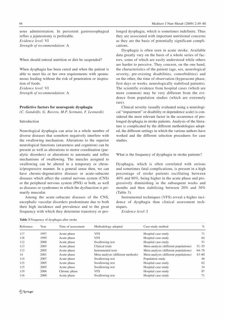
What is the frequency of dysphagia in stroke patients?
Dysphagia, which is often correlated with seriousand sometimes fatal complications, is present in a highpercentage of stroke patients oscillating between40% and 80%, being higher in the acute phase and pro-gressively diminishing in the subsequent weeks andmonths and then stabilising between 20% and 30%(Table 3).
Instrumental techniques (VFS) reveal a higher inci-dence of dysphagia than clinical assessment tech-niques.
Evidence level: I
66 Mediterr J Nutr Metab (2009) 2:49–80
Table 3 Frequency of dysphagia after stroke
Reference Year Time of assessment Methodology adopted Case-study method %
117 1997 Acute phase VFS Hospital case-study 71118 1999 Acute phase VFS Hospital case-study 64112 2000 Acute phase Swallowing test Hospital case-study 51113 2005 Acute phase Clinical trials Meta-analysis (different populations) 51–55113 2005 Acute phase Instrumental tests Meta-analysis (different populations) 64–7814 2001 Acute phase Meta-analysis (different methods) Meta-analysis (different populations) 43–80114 2007 Acute phase Swallowing test Population study 48115 2005 Acute phase Swallowing test Hospital case-study 62115 2005 Acute phase Swallowing test Hospital case-study 34119 2006 Chronic phase VFS Hospital case-study 87116 2000 Acute phase Swallowing test Hospital case-study 71
In about 3% of acute stroke victims chronic dysphagia isso serious as to render PEG necessary.
Evidence level: IStrength of recommendation: B
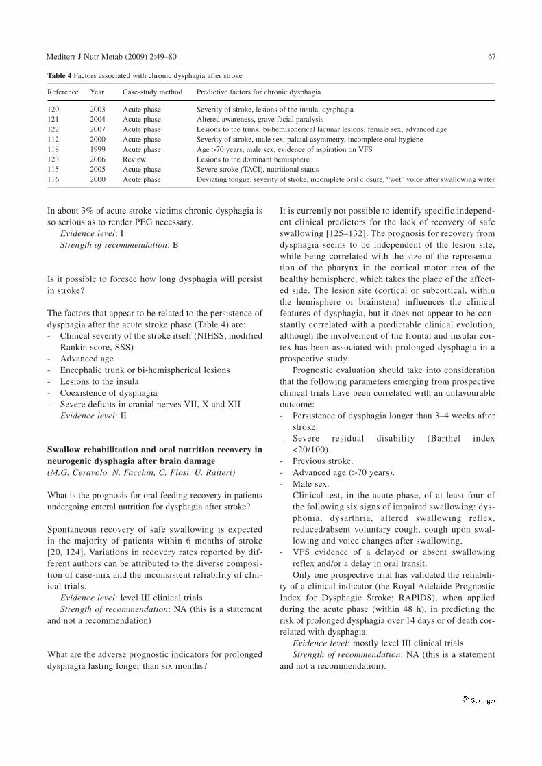
Is it possible to foresee how long dysphagia will persistin stroke?
The factors that appear to be related to the persistence ofdysphagia after the acute stroke phase (Table 4) are:- Clinical severity of the stroke itself (NIHSS, modified
Rankin score, SSS)- Advanced age- Encephalic trunk or bi-hemispherical lesions- Lesions to the insula- Coexistence of dysphagia- Severe deficits in cranial nerves VII, X and XII
Evidence level: II
Swallow rehabilitation and oral nutrition recovery inneurogenic dysphagia after brain damage(M.G. Ceravolo, N. Facchin, C. Flosi, U. Raiteri)
What is the prognosis for oral feeding recovery in patientsundergoing enteral nutrition for dysphagia after stroke?
Spontaneous recovery of safe swallowing is expectedin the majority of patients within 6 months of stroke[20, 124]. Variations in recovery rates reported by dif-ferent authors can be attributed to the diverse composi-tion of case-mix and the inconsistent reliability of clin-ical trials.
Evidence level: level III clinical trialsStrength of recommendation: NA (this is a statement
and not a recommendation)
What are the adverse prognostic indicators for prolongeddysphagia lasting longer than six months?
It is currently not possible to identify specific independ-ent clinical predictors for the lack of recovery of safeswallowing [125–132]. The prognosis for recovery fromdysphagia seems to be independent of the lesion site,while being correlated with the size of the representa-tion of the pharynx in the cortical motor area of thehealthy hemisphere, which takes the place of the affect-ed side. The lesion site (cortical or subcortical, withinthe hemisphere or brainstem) influences the clinicalfeatures of dysphagia, but it does not appear to be con-stantly correlated with a predictable clinical evolution,although the involvement of the frontal and insular cor-tex has been associated with prolonged dysphagia in aprospective study.
Prognostic evaluation should take into considerationthat the following parameters emerging from prospectiveclinical trials have been correlated with an unfavourableoutcome:- Persistence of dysphagia longer than 3–4 weeks after
stroke.- Severe residual disability (Barthel index
<20/100).- Previous stroke.- Advanced age (>70 years).- Male sex.- Clinical test, in the acute phase, of at least four of
the following six signs of impaired swallowing: dys-phonia, dysarthria, altered swallowing reflex,reduced/absent voluntary cough, cough upon swal-lowing and voice changes after swallowing.
- VFS evidence of a delayed or absent swallowingreflex and/or a delay in oral transit.Only one prospective trial has validated the reliabili-
ty of a clinical indicator (the Royal Adelaide PrognosticIndex for Dysphagic Stroke; RAPIDS), when appliedduring the acute phase (within 48 h), in predicting therisk of prolonged dysphagia over 14 days or of death cor-related with dysphagia.
Evidence level: mostly level III clinical trialsStrength of recommendation: NA (this is a statement
and not a recommendation).
67Mediterr J Nutr Metab (2009) 2:49–80
Table 4 Factors associated with chronic dysphagia after stroke
Reference Year Case-study method Predictive factors for chronic dysphagia
120 2003 Acute phase Severity of stroke, lesions of the insula, dysphagia121 2004 Acute phase Altered awareness, grave facial paralysis122 2007 Acute phase Lesions to the trunk, bi-hemispherical lacunar lesions, female sex, advanced age 112 2000 Acute phase Severity of stroke, male sex, palatal asymmetry, incomplete oral hygiene118 1999 Acute phase Age >70 years, male sex, evidence of aspiration on VFS123 2006 Review Lesions to the dominant hemisphere115 2005 Acute phase Severe stroke (TACI), nutritional status116 2000 Acute phase Deviating tongue, severity of stroke, incomplete oral closure, “wet” voice after swallowing water
Which patients undergoing enteral nutrition for dyspha-gia after stroke are candidates for weaning procedures?
There are no shared predictors for the recovery from dys-phagia in patients undergoing enteral nutrition after braindamage [133–148].
Considering the value represented by oral feeding it isrecommended that attempts should be made to wean allpatients where the following safety criteria are guaranteed:- Low risk of aspiration or alternatively a low risk of
aspiration pneumonia- A body mass index that is not indicative of malnutrition- Ability to maintain alertness during meals- Adequate cooperation
Evidence level: the most representative level is IVStrength of recommendation: B
What resources (human, instrumental, structural) arerequired to start weaning from enteral nutrition?
There are no indications with respect to the most appro-priate setting where the weaning from enteral nutritionshould be performed.
Considering the need to monitor risk indicators formalnutrition and aspiration pneumonia during weaning,it is recommended that the procedure be carried out in asetting that guarantees a multidisciplinary approach(involving the neurologist, speech therapist, physiothera-pist, nurse, nutritionist and dietician).
In particular, the assessment of the risks correlatedwith the reintroduction of oral feeding and the monitor-ing of functional swallowing ability must be assigned tospecialised professionals (i.e., speech and swallow thera-pist). Likewise the monitoring of the nutritional statusand nutritional support should be the responsibility of thenutritionist and dietician [35, 149].
Evidence level: the most representative level is IVStrength of recommendation: B
What are the most effective and efficient weaningstrategies?
In general terms, the therapeutic approaches duringweaning fall into two categories: compensatory strategies(conservative treatment) and rehabilitative strategies(restorative treatment). The compensatory strategies areaimed at guaranteeing the prevention of risks linked tooral feeding; the rehabilitative strategies are aimed atspeeding up the functional recovery process [150–156].
Recourse to formal rehabilitative approaches is notrecommended compared to conservative ones in order to
achieve a faster recovery of functional swallowing.Evidence level: the most representative level is IStrength of recommendation: B
In candidates suitable for weaning from enteral nutrition,a conservative approach is recommended which aims atminimising the risk of respiratory infections while await-ing swallow recovery.
Evidence level: the most representative level is IVStrength of recommendation: B
What is the appropriate schedule for rehabilitativeapproaches?
Early and high-intensity rehabilitative interventions arerecommended [157–159].
Evidence level: only one level II trialStrength of recommendation: B
What are the most effective compensatory strategies?
The use of compensatory strategies, including both themodification of the texture and viscosity of foods andpostural strategies, is recommended in order to reducethe risk of complications during weaning [160, 161].
There are four levels of achievable combinations offood texture and viscosity when modifying diet to pre-vent aspiration pneumonia due to dysphagia. The use offood thickeners is required for this. Even though theinternational literature distinguishes between four andfive dysphagia severity levels with correspondingdietary models, there is no agreement based on medicalevidence concerning the use of the above-mentionedmodels. Roughly, two levels of intervention may be pro-posed for the management of neurogenic dysphagia,which are assigned to different patterns of dysphagia,respectively identified as type I (dysphagia for liquids)and type II (partial defect in bolus preparation,decreased pharyngeal transit or problems at a pharyn-geal transit level).
Evidence level: Homogeneous trials IIa–IIb–III Strength of recommendation: B
How can the adaptation of the rheological componentof foods during weaning from enteral nutrition be opti-mised?
Instruments for measuring food density are recom-mended in order to guarantee the formulation of lessdense consistencies while supplying a more adequate
68 Mediterr J Nutr Metab (2009) 2:49–80
intake of fluids without increasing the risk of aspiration[162, 163].
Evidence level: a few trials, mostly level IIStrength of recommendation: B
Which rehabilitative techniques are indicated to comple-ment conservative approaches?
The adoption of either exercises to open theoesophageal sphincter or electric stimulation are recom-mended as useful complements to compensatory strate-gies. There is not sufficient evidence to support the useof tactile or thermal stimulation or biofeedback tech-niques [164–171].Evidence level: a few trials, mostly level III–IVStrength of recommendation: C
Are there effective drugs to promote weaning from enter-al nutrition?
The effectiveness of drugs such as nifedipine and ACEinhibitors in reducing the risk of aspiration is only sup-ported by cohort studies and case series. Furthermore,their use in clinical practice does not currently appear tobe recommended [172, 173].Evidence level: few trials, mostly level IV–VStrength of recommendation: D
Which nutritional indicators should undergo monitoringduring weaning?
It is recommended that nutritional status is monitoredvia the repeated Nutritional Risk Score (NRS) whichcovers weight assessment and previous habits of foodintake [174–176]. The NRS must be carried out at thebeginning of the nutritional intervention and thenrepeated by scheduling follow-up intervals according tothe established individual risk. The measurement ofweight in the presence of protein energy malnutritionmust be repeated three times a week. When a hoistweighing scale is not available, it is advisable to deter-mine the central arm circumference and to measure thetriceps plica twice a week in bedridden patients.Assessment of dietary intake and the recording of foodeaten by the patient in collaboration with a nurse per-mits the verification of the adequacy of contributions tonutritional requirements, the need for nutritional sup-port or a change in the nutritional route. If protein ener-gy malnutrition is present this must be done on a dailybasis.
When oral feeding is possible but intake is insuffi-cient, nutritional support with a caloric density of 1kcal/ml with or without a dietary indication is recom-mended. The choice of support with a polymer module isevaluated according to the patient’s nutritional require-ments and the palatability of the supplement.
Evidence level: few trials, mostly level VStrength of recommendation: B
Speech rehabilitation and indications for re-nourish-ment in postsurgical dysphagia(M. Petrelli, G. Ruoppolo)
Should the placement of a feeding tube be planned priorto head and neck surgery?
Advanced planning for the placement of a feeding tube isnecessary in patients undergoing surgery that will alterswallowing (calorie intake <60% of requirements formore than 10 days) or compromise swallowing (for morethan 7 days) [177]. A PEG should be placed if dysphagiais foreseen for >4 weeks (otherwise a nasogastric tube)and the patient has a normally functioning gastrointesti-nal tract. Parenteral nutrition via a central route is recom-mended only if the gastrointestinal tract is not function-ing so that the caloric quota administered enterally is lessthan 60% [178, 179].
Evidence level: IIIStrength of recommendation: A
When should feeding begin?
In patients with a high nutritional risk (easily identifiedby at least one of these situations:- Weight loss >10–15% in 6 months- BMI <18.5 kg/m2
- Serum albumin <30 g/l without other causesNutritional support must begin 10–14 days prior to
surgery and continue afterwards [179, 180]. In patientswith a good nutritional status, nutritional support mustbegin immediately until improvement/resolution of dys-phagia [181].
Evidence level: IIStrength of recommendation: A
Which patients are suitable candidates for weaning fromenteral nutrition?
Patients suitable for weaning from enteral nutrition arethose who have undergone surgery which has not removed
69Mediterr J Nutr Metab (2009) 2:49–80
or permanently altered the anatomical structures for chew-ing and swallowing and those who have undergone plasticsurgery (maxillofacial, ENT) aimed at restructuring orrepairing anatomical structures governing chewing andswallowing [1]. Postsurgical radiotherapy is not an imped-iment to weaning, but it can slow the process [182, 183].
Evidence level: IVStrength of recommendation: B
In which phase after head and neck surgery should wean-ing from enteral nutrition begin?
Two weeks after surgery is an appropriate time to carry outa clinical-instrumental examination of the patient’s swal-lowing ability [184]. It will be possible to decide whetherto start weaning with creamy foods without the risk ofaspiration or delay this phase on the basis of the assess-ment results [1, 185]. Radiotherapy after surgery does notimpede weaning, but it can slow the process [186].
Evidence level: IIIStrength of recommendation: B
Which personnel are involved in the preliminary clinical-functional assessment for weaning?
The surgeon, speech therapist, radiologist, nutritionist,dietician, nurse and care-giver [1, 187] are involved.
Evidence level: VIStrength of recommendation: B
Which clinical and instrumental indicators predict thesuccess of the re-nourishment procedures?
Patients with dysphagia should be reassessed on a regularbasis by a professional competent in the management ofdysphagia; the frequency is relative to the clinical charac-teristics of the dysphagia and nutritional status [178, 188].VFS with a modified barium study or a fibreoptic endo-scopic study of swallowing are both valid methods toaccurately evaluate the possibility of re-nourishment [43,189]. Patient age, gastroenteric function and complianceare factors to take into consideration [188, 190].
Evidence level: IIStrength of recommendation: A
In which setting (hospital, home) can weaning start?
Ideally it is appropriate to begin weaning in a hospitalsetting (prior to discharge or in day-hospital) in order to
carefully assess the effective adherence to the proposednutritional protocol and avoid the risks of aspiration [1,191]. After this initial phase, the setting is moved tohome where the patient can continue to follow theschemes and protocols provided. Integration between thehospital and local structures is desirable.
Evidence level: VIStrength of recommendation: B
What role does speech rehabilitation play during weaning?
At the end of a complete appraisal of swallowing indica-tions must be provided regarding relative dietary changesand compensatory techniques (posture and manoeuvre)[1, 187]. In some dysphagic frameworks, compensatoryprocedures should be used (compensative posture, swal-lowing techniques) in order to significantly reduce therisk of aspiration [192].
The adoption of a modified diet must be included inthe treatment plan, providing that its effect is monitoredwith VFS and/or fibreoptic endoscopy [1, 187].
Treatment of the buccal structures (sensitivity, motil-ity, praxia) is indicated in association with other types ofapproaches (dietary changes, compensatory techniques,postural changes) [193].
Evidence level: VStrength of recommendation: B
Which indicators must be monitored during weaning?
All of the following [186, 194, 195]:- Body weight- Postural changes- Choice of foods and dietary changes- Appetite and food preparation management- Management of behaviour and environmental factors- Oral hygiene- Psychological state
Evidence level: IIStrength of recommendation: A
Clinical nutrition management in the patient withpenetration, silent aspiration and ingestion bronchop-neumonia(D. Vassallo, A. Seneghini, F. Raganini)
Introduction
According to some authors, ingestion pneumonia is theprincipal cause of death in stroke [196]. Treatment con-
70 Mediterr J Nutr Metab (2009) 2:49–80
sists of maintaining vital functions, controlling arterialhypertension and hyperglycaemia, suspending oral feed-ing if unsafe and the preference for postpyloric parenter-al or enteral nutrition [197].
Classically, dysphagia is diagnosed with VFS, a diag-nostic examination that highlights oropharyngeal motili-ty, duration of the laryngeal phase, laryngeal penetrationand aspiration. With respect to the last two severe com-plications, according to Schmidt et al. [198] “laryngealpenetration” is present when the contrast medium entersthe vestibule and “aspiration” is present when the con-trast medium goes beyond the vestibule and below thetrue vocal cords. The SPREAD guidelines (Stroke Pre -vention and Education Awareness Diffusion [35]) advisethat upon hospital admission or rehabilitation, strokepatients must be assessed for the risk of malnutritionusing the NRS or the MUST recommendation 11.15,grade D [35]. This recommendation is derived fromstrong evidence that protein-energy malnutrition inpatients affected by acute stroke is very frequent and hasa considerable impact upon the possible functionalrecovery and overall management of the patient.
Assessment of the patient’s nutritional status is fun-damental to rapidly identifying malnutrition and to main-taining or re-establishing an adequate nutritional status.Adequate nutrition is important in order to avoid compli-cations, reduce hospital stay, improve the quality of lifeand to make treatment more simple and effective (synthe-sis 11.6 [35]). Assessment of nutritional status and nutri-tional intervention are thus indicated as essential compo-nents in clinicotherapeutic protocols for stroke both dur-ing the acute phase and during rehabilitation (recommen-dation 11.15, grade D [35]).
The placement of a nasogastric tube may not elimi-nate the risk of inhalation in those with delayed gastricemptying, particularly in patients with more serious cere-bral lesions. In these patients the risk of inhalation isreduced if the bolus is inserted far from the pylorus,beyond Treitz corner [35, 47, 199].
The use of jejunostomy is indicated where there is arisk of bronchopulmonary aspiration (and in those withoesophageal reflux, gastroparalysis in the presence ofinoperable gastric, or pancreatic cancer with subocclu-sion or occlusion). The advantage of this access is thedecreased risk of aspiration. The technique is carried outsurgically under general anaesthesia. Also feasible is apercutaneous endoscopic jejunostomy (PEJ) which usesthe PEG technique with the final passage of a duodenaltube and endoscopic control [200].
Practical recommendations [201, 202] are as follows:1. PEG is the preferred technique for patients who will
undergo long-term enteral nutrition that can be pre-ceded by a period of nasogastric nutrition; the latter
can be adopted definitively when PEG is not suitable.2. During enteral nutrition via a nasogastric tube it is
advisable to:- prevent coughing, regurgitation and vomiting
(even pharmacologically),- make sure the patient does not fiddle with or dis-
lodge the tube and- wash the tube regularly in order to avoid obstruc-
tion.PEJ and jejunostomy are indicated in cases where
there is a risk of ingestion pneumonia, oesophagitis fromreflux, gastroparalysis, gastrectomy, and gastric and pan-creatic cancer.
Which dietary treatment should be adopted in suspectedpenetration, aspiration or ingestion pneumonia?
Prior to commencing oral feeding in neurologicalpatients, swallowing ability and the risk of aspirationmust be evaluated (level B according to SINPE 2002guidelines) [63]. The following strategies are considereduseful and recommended in order to compensate forslight swallowing defects:- Sitting straight when eating.- Sitting with head and neck supported.- The use of semisolid foods.- The use of boluses smaller than a teaspoon.- Restriction of liquid foods.- Use of a cup or spoon as opposed to a straw for fluids.- A gentle cough after swallowing.- Swallow several times, even for small boluses, in
order to completely empty the oesophagus.The recommendations in paragraph 11 [35] note that:
- In patients in whom oral feeding is possible the use ofroutine nutritional support is unnecessary as it is notassociated with an improvement in prognosis. The useof nutritional support must be guided by the results ofthe nutritional status evaluation (grade B).
- Standard monitoring of swallowing function is advis-able in order to prevent secondary complications todysphagia (grade D).
- Clinical assessment of the risk of dysphagia should becarried out using the BSA and a simple test of swal-lowing water, which in view of evidence-based medi-cine are the most appropriate in acute stroke patients.
- In specialised Centres, more sophisticated approachescan be adopted, such as an examination by the speechtherapist or a VFS.Functionally aimed swallowing treatment is a funda-
mental point in dietary treatment. Swallowing is theresponsibility of the speech therapist with specialisttraining. It is improved further by a multidisciplinary
71Mediterr J Nutr Metab (2009) 2:49–80
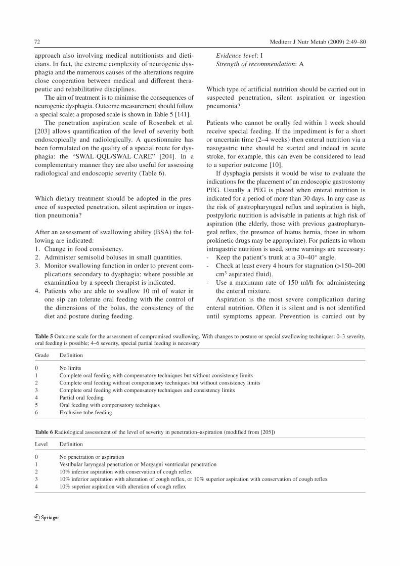
approach also involving medical nutritionists and dieti-cians. In fact, the extreme complexity of neurogenic dys-phagia and the numerous causes of the alterations requireclose cooperation between medical and different thera-peutic and rehabilitative disciplines.
The aim of treatment is to minimise the consequences ofneurogenic dysphagia. Outcome measurement should followa special scale; a proposed scale is shown in Table 5 [141].
The penetration aspiration scale of Rosenbek et al.[203] allows quantification of the level of severity bothendoscopically and radiologically. A questionnaire hasbeen formulated on the quality of a special route for dys-phagia: the “SWAL-QQL/SWAL-CARE” [204]. In acomplementary manner they are also useful for assessingradiological and endoscopic severity (Table 6).
Which dietary treatment should be adopted in the pres-ence of suspected penetration, silent aspiration or inges-tion pneumonia?
After an assessment of swallowing ability (BSA) the fol-lowing are indicated:1. Change in food consistency.2. Administer semisolid boluses in small quantities.3. Monitor swallowing function in order to prevent com-
plications secondary to dysphagia; where possible anexamination by a speech therapist is indicated.
4. Patients who are able to swallow 10 ml of water inone sip can tolerate oral feeding with the control ofthe dimensions of the bolus, the consistency of thediet and posture during feeding.
Evidence level: IStrength of recommendation: A
Which type of artificial nutrition should be carried out insuspected penetration, silent aspiration or ingestionpneumonia?
Patients who cannot be orally fed within 1 week shouldreceive special feeding. If the impediment is for a shortor uncertain time (2–4 weeks) then enteral nutrition via anasogastric tube should be started and indeed in acutestroke, for example, this can even be considered to leadto a superior outcome [10].
If dysphagia persists it would be wise to evaluate theindications for the placement of an endoscopic gastrostomyPEG. Usually a PEG is placed when enteral nutrition isindicated for a period of more than 30 days. In any case asthe risk of gastropharyngeal reflux and aspiration is high,postpyloric nutrition is advisable in patients at high risk ofaspiration (the elderly, those with previous gastropharyn-geal reflux, the presence of hiatus hernia, those in whomprokinetic drugs may be appropriate). For patients in whomintragastric nutrition is used, some warnings are necessary:- Keep the patient’s trunk at a 30–40° angle.- Check at least every 4 hours for stagnation (>150–200
cm3 aspirated fluid).- Use a maximum rate of 150 ml/h for administering
the enteral mixture.Aspiration is the most severe complication during
enteral nutrition. Often it is silent and is not identifieduntil symptoms appear. Prevention is carried out by
72 Mediterr J Nutr Metab (2009) 2:49–80
Table 5 Outcome scale for the assessment of compromised swallowing. With changes to posture or special swallowing techniques: 0–3 severity,oral feeding is possible; 4–6 severity, special partial feeding is necessary
Grade Definition
0 No limits1 Complete oral feeding with compensatory techniques but without consistency limits2 Complete oral feeding without compensatory techniques but without consistency limits3 Complete oral feeding with compensatory techniques and consistency limits4 Partial oral feeding5 Oral feeding with compensatory techniques 6 Exclusive tube feeding
Table 6 Radiological assessment of the level of severity in penetration–aspiration (modified from [205])
Level Definition
0 No penetration or aspiration1 Vestibular laryngeal penetration or Morgagni ventricular penetration2 10% inferior aspiration with conservation of cough reflex3 10% inferior aspiration with alteration of cough reflex, or 10% superior aspiration with conservation of cough reflex4 10% superior aspiration with alteration of cough reflex
minimising the risk of regurgitation and vomiting.Guide lines agree on advising frequent monitoring ofgastric stagnation, especially during the initial phasesof the infusion and in all at-risk patients. Should gastricstagnation be >200 ml, enteral nutrition should be sus-pended or the rate of infusion reduced, possible causeseliminated and stagnation rechecked after 2 h. If afterrechecking stagnation is <200 ml, the infusion can berecommenced with reduced volumes and rate. If repeat-ed episodes of gastric stagnation of >200 ml persist,then it might be useful to consider a postpyloric or jeju-nal infusion.
During enteral nutrition the patient should be main-tained where possible in the Fowler position or in theright lateral decubitus position.
In acute stroke patients the artificial nutrition treat-ment of choice is enteral nutrition. It is recommendedthat enteral nutrition be started early on and in any caseafter no more than 5–7 days in normally nourishedpatients and no more than 24–72 h in malnourishedpatients (recommendation 11.17, grade B [35]).
Parenteral nutrition is exclusively indicated whereenteral nutrition is not feasible, contraindicated or as asupplement to enteral nutrition whenever the latter doesnot allow the administration of adequate nutrients (rec-ommendation 11.17b, grade D [35, 63]).
In the case of ingestion pneumonia enteral nutritionshould be stopped and corrective medical measures takentogether with the commencement of parenteral nutrition.
Evidence level: IIStrength of recommendation: A
Which EN route should be considered?
The placement of a nasogastric tube is recommendedwhen dysphagia occurs, assessed according to the previ-ously described methods. The timing for the insertion ofa nasogastric tube is theoretical. Early treatment is advis-able (between 24 and 72 h) for malnourished patients.
PEG is the preferred technique for those patientsundergoing long-term enteral nutrition, possibly precededby a period of nasogastric feeding. This latter route maybe chosen definitively when PEG is no longer feasible.
The conditions for the application of PEG are:1. Persistent dysphagia after stroke.2. Prolonged dysphagia possibly for more than 2
months.3. Timing for the placement of a PEG assessable within
30 days of the occurrence of disease and with theabove-mentioned conditions.Evidence level: IIIStrength of recommendation: B
When to suspend enteral nutrition in the presence of suspect-ed penetration, silent aspiration or ingestion pneumonia?
There are no conclusive data indicating a preference forthe use of pre- or postpyloric nutrition. The preference forpostpyloric nutrition has been widely discussed [63, 93]and we feel in any case it is always better to optimise theuse of enteral nutrition while diminishing the risks [93].
The composition of the mixture influences gastricemptying and reflux. Mixtures rich in fats (caloric quotafrom lipids more than 50%) prolong gastric emptyingcompared to mixtures that have a moderate fat content.The types of fatty acids, proteins and polypeptides mod-ulate gastric movement and can delay gastric emptyingand increase the prevalence of stagnation.
The majority of pneumonias from inspiration arecaused by a lack of control of secretions rather than byreflux of enteral mixture. Furthermore, the site used forthe infusion does not affect this problem [206].
The principal factors favouring the risk of aspirationwith enteral nutrition can be summarised as follows:- A state of altered awareness- Altered swallowing- Previous episodes of aspiration into the airways- Serious gastroesophageal reflux- Gastric paralysis- Pyloric obstruction
These factors must be borne in mind in order to pre-vent and optimise nutritional intervention, rememberingthat silent aspiration represents a grave risk for acutestroke patients. In conclusion, It does not appear to bepossible to establish a standardised timing for the occur-rence of each of these complications, which must be eval-uated clinically and within the framework of each case.Therefore, we assign an evidence level of VI (based onthe opinions of experts or members of the work group)and a strength of recommendation of C.
Evidence level: VIStrength of recommendation: C
What is the access site, infusion rate and the manner ofinfusion in patients receiving enteral nutrition with pene-tration, silent aspiration or ingestion pneumonia?
In the cases discussed above it is advisable if possible to sus-pend enteral nutrition. Intravenous nutritional support andclinical and instrumental monitoring are indicated. If pneu-monia is confirmed, the access site must be postpyloric.
Acknowledgements We wish to thank to Dr. Elisa Ricolfi PhD andMrs. Francesca Abate from the U.O. di Dietetica e Nutrizione Clinicadell’Azienda Ospedaliera Universitaria San Martino, Genoa, Italy, fortheir editorial reviewing during their doctorate.
73Mediterr J Nutr Metab (2009) 2:49–80
Conflict of interest The authors declare that they have no conflict ofinterest related to the publication of this article.
*F. BarbieraU.O. Radiologia “Domenico Noto”Azienda Ospedali Civili Riuniti, Sciacca (AG), Italy
A. BosettiScienza dell’AlimentazionePolo Universitario, Luigi Saccoe Università degli Studi di Milano, Milan, Italy
M.G. CeravoloMedicina Fisica e RiabilitazioneUniversità Politecnica delle Marche e Clinica di NeuroriabilitazioneAzienda Ospedali Riuniti di Ancona, Italy
F. CortinovisUSSD Dietologia ClinicaAzienda Ospedaliera Ospedali Riuniti, Bergamo, Italy
A. CrippaServizio di Dietologia e Nutrizione ClinicaASP IMMeS e Pio Albergo Trivulzio, Milan, Italy
N. FacchinServizio di Dietetica e Nutrizione ClinicaAzienda Sanitaria dell’Alto AdigeComprensorio Sanitario di Bolzano, Italy
C. FlosiVilla Scassi, Corso di Laurea in LogopediaUniversità degli Studi di Genova, Genoa, Italy
C. GandolfoCentro IctusUniversità degli Studi di Genova, Genoa, Italy
E. JulianiU.O. Radiodiagnostica IAzienda Ospedaliera S. Giovanni Battista, Turin, Italy
F. LeonardiU.O. di Scienza dell’Alimentazione e DieteticaAzienda Ospedaliera “Cannizzaro”Università degli Studi di Catania, Catania, Italy
P. NanniServizio di Dietetica e Nutrizione ClinicaASUR Zona Territoriale 13, Ascoli Piceno, Italy
P. PalliniServizio di Dietetica e Nutrizione ClinicaU.O.C. GastroenterologiaAzienda ULSS n. 12 Venezia, Mestre (VE), Italy
M. PetrelliSOD Dietetica e Nutrizione ClinicaAzienda Ospedaliera Universitaria Ospedali RiunitiAncona, Italy
F. Raganini · U. RaiteriUO di NeurologiaAzienda Ospedaliera Universitaria San Martino, Genoa, Italy
G. RaveraDipartimento di Scienze della Salute (DISSAL)Facoltà di Medicina e ChirurgiaUniversità di Genova, Genoa, Italy
S. RisoSC Dietetica e Nutrizione ClinicaAO “Maggiore della Carità”, Novarae Università degli Studi di Pavia, Pavia, Italy
L. RoveraResponsabile SS Dietetica e Nutrizione ClinicaASO Ordine Mauriziano, Turin, Italy
G. RuoppoloUOC Foniatria, Policlinico Umberto IUniversità degli Studi di Roma “La Sapienza”, Rome, Italy
A. SchindlerU.O. OtorinolaringoiatriaAzienda Ospedaliera - Polo Universitario “L. Sacco”Università degli Studi di Milano, Milan, Italy
O. SchindlerS.C.D.U. Audiologia FoniatriaUniversità degli Studi di Torino, Turin, Italy
A. SeneghiniCentro Ictus “Luciano Garello”UO di Neurologia, Azienda Ospedaliera S. Martino, Genoa, Italy
M.P. SormaniSezione di BiostatisticaDip. di Scienze della SaluteUniversità degli Studi di Genova, Genoa, Italy
B. Travalca CupilloU.O.D. di FoniatriaAzienda Ospedaliera Universitaria S. Martino, Genoa, Italy
M.T. Van lintU.O. EmatologiaAzienda Ospedaliera Universitaria S. Martino, Genoa, Italy
D. VassalloS.S. di Dietetica e Nutrizione ClinicaStroke-team S.C. NeurologiaASO Ordine Mauriziano Umberto I, Turin, Italy
Contributors
Project Coordinator
Samir Giuseppe Sukkar, Genoa, Italy
Invited Contributors
Filippo Barbiera, Sciacca, ItalyAlessandra Bosetti, Milan, ItalyMaria Gabriella Ceravolo, Ancona, ItalyAnna Crippa, Milan, ItalyFederico D’Andrea, Novara, ItalyNicoletta Facchin, Bolzano, ItalyCristina Flosi, Genoa, ItalyCarlo Gandolfo, Genoa, ItalyRaffaella Gradaschi, Genoa, ItalyElsa Juliani, Turin, ItalyFrancesco Leonardi, Catania, ItalyPaola Nanni, Ascoli Piceno, ItalyAlbano Nicolai, Ancona, ItalyPaolo Orlandoni, Ancona, ItalyPaolo Pallini, Venice, Italy
74 Mediterr J Nutr Metab (2009) 2:49–80
Simona Raimondo, Turin, ItalyGiambattista Ravera, Genoa, ItalyGiovanni Regesta, Genoa, ItalySergio Riso, Novara, ItalyLydia Rovera, Turin, ItalyGianni Ruoppolo, Rome, ItalyAnna Rita Sabbatini, Milan, ItalyVincenzo Savarino, Genoa, ItalyOskar Schindler, Turin, ItalyAlfredo Seneghini, Genoa, ItalyFulvio Sileo, Bergamo, ItalyMaria Pia Sormani, Genoa, ItalyBeatrice Travalca-Cupillo, Genoa, ItalyDaniela Vassallo, Turin, ItalyPatrizia Zentilin, Genoa, Italy
Editors
Filippo BarbieraAlessandra BosettiMaria Gabriella CeravoloFiorenzo CortinovisAnna CrippaNicoletta FacchinCristina FlosiCarlo GandolfoElsa JulianiNicoletta MachelloPaola NanniPaolo PalliniMassimiliano PetrelliGraziella RaganiniSimona RaimondoUmberto RaiteriGianbattista RaveraSergio RisoLidia RoveraGianni RuoppoloAnna Rita SabbatiniAlberto SchindlerOskar SchindlerAlfredo SeneghiniMaria Pia SormaniSamir Giuseppe SukkarBeatrice Travalca-CupilloMaria Teresa Van LintDaniela Vassallo
Scientific Societies supporting the Project
Associazione Italiana di Dietetica e Nutrizione Clinica (ADI)Federazione Logopedisti Italiani (FLI)Gruppo per lo Studio delle Malattie Cerebrovascolariaffiliato alla Società Italiana di Neurologia (SIN)
References
1. Scottish Intercollegiate Guidelines Network (2004) Mana -gement of patients with stroke. Identification and managementof dysphagia. http//:www.sign.ac.uk
2. Agency for Healthcare Research and Quality (1999) Diagnosisand treatment of swallowing disorders (dysphagia) in acute-carestroke patients. Evidence Report/Technology Assessment no. 8.http://www.ahcpr.gov/clinic/epcsums/dysphsum.htm
3. Bloem BR, Lagaay AM, Van Beek W et al (1990) Prevalence ofsubjective dysphagia in community residents aged over 87. BMJ300:721–722
4. Calcagno P, Ruoppolo G, Grasso MG et al (2002) Dysphagia inmultiple sclerosis: prevalence and prognostic factors. ActaNeurol Scand 105:10–13
5. Daniels SK, Ballo LA, Mahoney MC, Foundas AL (2000) Cli -nical predictors of dysphagia and aspiration risk: outcome mea-sures in acute stroke. Arch Phys Med Rehabil 81:1030–1033
6. Kaiser-Jones J (1999) Dysphagia among nursing home resi-dents. Geriatr Nurs 20:77–84
7. Kuhlemeier KV (1994) Epidemiology and dysphagia.Dysphagia 9:209–217
8. Reilly S, Perry A (2001) Is there an evidence base to the mana-gement of paediatric dysphagia? Asia Pac J Speech LanguageHearing 6:1–8
9. Cherney LR, Cantieri CA, Pannel JJ (1986) Clinical evaluationof dysphagia. Rehabilitation Institute of Chicago proceduremanual. Aspen System Collaboration, Maryland
10. Doggett DL, Turkelson CM, Coates V (2002) Recent develop-ments in diagnosis and intervention for aspiration and dysphagiain stroke and other neuromuscular disorders. Curr AtherosclerRep 4:311–318
11. De Pippo KL, Holas MA, Reding MJ (1994b) The Burkedysphagia screening test: validation of its use in patients withstroke. Arch Phys Med Rehabil 75:1284–1286
12. Martino R, Pron G, Diamant N (2000) Screening for oropharyn-geal dysphagia in stroke. Insufficient evidence for guidelines.Dysphagia 15:19–30
13. Perry L, Love CP (2001) Screening for dysphagia and aspirationin acute stroke: a systematic review. Dysphagia 16:7–18
14. Perry L (2001) Screening swallowing function of patients withacute stroke. Part one: Identification, implementation and initialevaluation of a screening tool for use by nurses. J Clin Nurse10:463–473
15. Ding R, Logemann JA (2000) Pneumonia in stroke patients: aretrospective study. Dysphagia 15:51–57
16. New South Wales Community Services Commission (2001)Disability, death and the responsibility of care. NSWGovernment, Sydney
17. Gottlieb D, Kipnis M, Sister E et al (1996) Validation of the50ml drinking test for evaluation of post stroke dysphagia.Disabil Rehabil 18:529–532
18. Martino R, Meissner-Fishbein B, Saville D et al (2007)Preferred practice guidelines for dysphagia. Quality improve-ment: preferred practice guidelines. http://www.caslpo.com/Por -tals/0/ppg/Dysphagia_PSG.pdf
19. Barba R, Morin MD, Cemillan C et al (2002) Previous and inci-dent dementia as risk factors for mortality in stroke patients.Stroke 33:1993–1998
20. Barer DH (1989) The natural history and functional consequen-ces of dysphagia after hemispheric stroke. J Neurol NeurosurgPsychiatry 52:236–241
21. American Speech-Language Hearing Association (2001)Clinical indicators for instrumental assessment of dysphagia.ASHA Special interest report. http://www.asha.org/nr/rdonly -res/4fdee27b-baf5-4d06-ac4d-8d1f311c1b06/0/19446_1.pdf
22. Ellul J, Watkins C, Barer D. Frequency, clinical course and com-plications of dysphagia in acute stroke, studied using a standar-dised bedside swallowing assessment. http://www.ncl. -ac.uk/stroke-research-unit/costar/papD1.doc. (Last accessed 12December 2005). Cited in: Trapl M, Enderle P, Nowotny M et al(2007) Dysphagia bedside screening for acute-stroke patients:the Gugging Swallowing Screen. Stroke 38:2948–2952
23. McCullough GH, Wertz RH, Rosembek JC (2001) Sensitivityand specificity of clinical bedside examination signs for detec-
75Mediterr J Nutr Metab (2009) 2:49–80

ting aspiration in adults subsequent to stroke. J Common Disord34:55–72
24. Linden P, Kuhlemeier KV, Patterson C (1993) The probability ofcorrectly predicting subglottic penetration from clinical obser-vation. Dysphagia 8:170–179
25. Logemann JA, Veis S, Colangelo L (1999) A screening procedu-re for oropharyngeal dysphagia. Dysphagia 14:44–51
26. Davies S, Taylor H, MacDonald A, Barer D (2001) An inter-disciplinary approach to swallowing problems in acute stroke.Int J Lang Commun Disord 36[Suppl]:357–362
27. Magnus V (2001) Dysphagia training for nurses in an acutehospital setting – a pragmatic approach. Int J Lang CommunDisord 36[Suppl]:357–358
28. Royal College of Physicians (2004) National clinical guidelinefor stroke. Intercollegiate Stroke Working Party. http://www.rc -plondon.ac.uk/pubs
29. Leder SB, Karas DE (2000) Fiberoptic endoscopic evaluation ofswallowing in the pediatric population. Laryngoscope110:1132–1136
30. Leder SB, Acton LM, Lisitano HL, Murray JT (2005) Fiberopticendoscopic evaluation of swallowing (FEES) with and withoutblue-dye food. Dysphagia 20:157–162
31. Aviv JE, Kim T, Thomson IE et al (1988) Fiberoptic endoscopicevaluation of swallowing with sensory testing (FEESST) inhealthy controls. Dysphagia 13:87–92
32. Aviv JE, Kaplan ST, Thomson JE et al (2000) The safety of fle-xible endoscopic evaluation of swallowing with sensory testing(FEESST): an analysis of 500 consecutive evaluations.Dysphagia 15:39–44
33. College of Audiologists and Speech-Language Pathologists ofOntario (2007) Instrumental assessment: Resource Require -ments N. (a) Fiberoptic Endoscopic Evaluation of Swallowing(FEES*) in Practice Standards and Guidelines for DysphagiaIntervention by Speech-Language Pathologists. http://www.ca -slpo.com/english_site/pdfdocs/5Dysphagia_PSG_Draft_for_Member_Feedback_2007.pdf
34. European Study Group for Dysphagia and Globus (EGDG)Guidelines for evaluation and treatment of patients with dyspha-gia and globus. http://www.egdg.org/
35. Stroke Prevention and Educational Awareness Diffusion (2005)Ischemic stroke: Italian guidelines on prevention and treatment,4th Edn. Publicazioni Catel, Milan
36. Leder SB, Novella S, Patwa H (2004) Use of fiberoptic endosco-pic evaluation of swallowing (FEES) in patients with amyotro-phic lateral sclerosis. Dysphagia 19:177–181
37. American Speech-Language Hearing Association (2004) Me -dical review guidelines for dysphagia. ASHA, Rockville, MD
38. Ajemian MS, Nirmul GB, Anderson MT et al (2001) Routinefiberoptic endoscopic evaluation of swallowing following pro-longed intubation: implications for management. Arch Surg136:434–437
39. Royal College of Speech and Language Therapists (1999)Professional Standards Board: invasive procedures guideline.RCSLT, London
40. Cohen MA, Setzen M, Perlman PW et al (2003) The safety offlexible endoscopic evaluation of swallowing with sensorytesting in an outpatient otolaryngology setting. Laryngoscope113:21–24
41. Diener HC (2005) Leitlinien fur Diagnostik und terapie in derNeurologie. Thieme, Stuttgart, pp 746–756
42. Langmore SE (2003) Evaluation of oropharyngeal dysphagia:which diagnostic tool is superior? Curr Opin Otolaryngol HeadNeck Surg 11:485–489
43. Aviv JE (2000) Prospective, randomized outcome study of endo-scopy versus modified barium swallow in patients with dyspha-gia. Laryngoscope 110:563–574
44. Aviv JE, Kidder TM, McCulloch T; American Academy ofOtolaryngology – Head and Neck Surgery Foundation (2001)Swallowing disorders: otolaryngologic aspects. AmericanAcademy of Otolaryngology – Head and Neck SurgeryFoundation, Alexandria, VA, USA
45. Beutler E (1994) G6PD deficiency. Blood 84:3613–363646. Malagelada JR, Bazzoli F, Elewaut A et al (2005) Practice gui-
deline – dysphagia. S Afr Gastroenterol Rev 3:9–1447. Prosiegel M (2004) Guidelines of the German Society of
Neurology (DGN) on neurogenic dysphagia. European StudyGroup for Dysphagia and Globus. http://www.egdg.org
48. Pikus L, Levine MS, Yang YX et al (2003) Videofluoroscopicstudies of swallowing dysfunction and the relative risk of pneu-monia. AJR Am J Roentgenol 180:1613–1616
49. Nitenberg G, Raynard B (2000) Nutritional support of the can-cer patient: issues and dilemmas. Crit Rev Oncol Hematol34:137–168
50. Arends J, Bodoky G, Bozzetti et al; ESPEN (European Societyfor Parenteral and Enteral Nutrition) (2006) ESPEN guidelines onenteral nutrition: non-surgical oncology. Clin Nutr 25:245–259
51. Barrera R (2002) Nutritional support in cancer patients. JPEN JParenter Enteral Nutr 26:S63–S71
52. Wordsworth H, Ashton J (2000) Head and neck cancer and uppergastrointestinal cancer. In: Shaw C (ed) Current thinking: nutri-tion and cancer. Novartis Consumer Health, Horsham, pp 13–24
53. Travalca Cupillo B, Sukkar S, Spadola Bisetti M (2001) disfa-gia.eat. Quando la deglutizione diventa difficile. Gli aspettinutrizionali. Omega Edizioni, Turin, pp 104–140
54. Arnold C, Richter MP (1989) The effect of oral nutritional sup-plements on head and neck cancer. Int J Radiat Oncol Biol Phys16:1595–1599
55. Elia M, van Bokhorst-de van der Schueren MAE, Garvey J et al(2006) Enteral (oral or tube administration) nutritional supportand eicosapentaenoic acid in patients with cancer: a systematicreview. Int J Oncol 28:5–23
56. Isenring EA, Capra S, Bauer JD (2004) Nutrition intervention isbeneficial in oncology outpatients receiving radiotherapy to thegastrointestinal or head and neck area. Br J Cancer 91:447–452
57. Nayel H, el-Ghoneimy E, el-Haddad S (1992) Impact of nutri-tional supplementation on treatment delay and morbidity inpatients with head and neck tumors treated with irradiation.Nutrition 8:13–18
58. Bozzetti F, Cozzaglio L, Gavazzi C et al (1998) Nutritional sup-port in patients with cancer of the esophagus: impact on nutritio-nal status, patient compliance to therapy and survival. Tumori84:681–686
59. Lees J (1997) Nasogastric and percutaneous endoscopic gastro-stomy feeding in head and neck cancer patients receiving radio-therapy treatment at a regional oncology unit: a two year study.Eur J Cancer Care 6:45–49
60. Thiel HJ, Fietkau R, Sauer R (1988) Malnutrition and the role ofnutritional support for radiation therapy patients. Rec ResCancer Res 108:205–226
61. Löser C, Aschl G, Hébuterne X et al (2005) ESPEN guidelineson artificial enteral nutrition – percutaneous endoscopic gastro-stomy (PEG). Clin Nutr 24:848–861
62. American Society of Parenteral and Enteral Nutrition (ASPEN)Board of Directors (2002) Access for administration of nutritionsupport. JPEN J Parenter Enteral Nutr 26:33SA–41SA
63. Italian Society of Parenteral and Enteral Nutrition (SINPE) (2002)SINPE guidelines for artificial nutrition in hospital. Access forenteral nutrition. Riv Ital Nutr Parenter Enter 20:S19–S20
64. Lee JH, Machtay M, Unger LD et al (1998) Prophylactic gastro-stomy tubes in patients undergoing intensive irradiation for can-cer of the head and neck. Arch Otolaryngol Head Neck Surg124:871–875
76 Mediterr J Nutr Metab (2009) 2:49–80
65. Magne N, Marcy PY, Foa C et al (2001) Comparison betweennasogastric tube feeding and percutaneous fluoroscopic gastro-stomy in advanced head and neck cancer patients. Eur ArchOtorhinolaryngol 258:89–92
66. Beaver ME, Matheny KE, Roberts DB, Myers JN (2001)Predictors of weight loss during radiation therapy. OtolaryngolHead Neck Surg 125:645–648
67. Scolapio JS, Spangler PR, Romano MM et al (2001)Prophylactic placement of gastrostomy feeding tubes beforeradiotherapy in patients with head and neck cancer: is it worth-while? J Clin Gastroenterol 33:215–217
68. Politzer Shronts E, Fish JA, Pesce-Hammond K (1998)Nutrition assessment. In: Merrit RJ (ed) The ASPEN nutritionsupport practice manual. American Society for Parenteral andEnteral Nutrition, Silver Spring, MD, pp 1–17
69. Brody R (1999) Nutrition issues in dysphagia: identification, mana-gement and the role of the dietitian. Nutr Clin Pract 14:S47–S51
70. Wardley AM, Jayson GC, Swindell R et al (2000) Prospectiveevaluation of oral mucositis in patients receiving myeloablativeconditioning regimens and haemopoietic progenitor rescue. Br JHaematol 110:292–299
71. Stuart RK, Sensenbrenner LL (1979) Adverse effects of nutritio-nal deprivation on transplanted hematopoietic cells. ExpHematol 7:435–442
72. Bistrian BR, Blackburn GL, Scrimshaw NS, Flatt JP (1975)Cellular immunity in semistarved states in hospitalized adults.Am J Clin Nutr 28:1148–1155
73. Papadopoulou A, Lloyd DR, Williams MD et al (1996)Gastrointestinal and nutritional sequelae of bone marrow trans-plantation. Arch Dis Child 75:208–213
74. Weisdorf SA, Lysne J, Wind D et al (1987) Positive effect ofprophylactic total parenteral nutrition on long-term outcome ofbone marrow transplantation. Transplantation 43:833–838
75. Szeluga DJ, Stuart RK, Brookmeyer R et al (1987) Nutritionalsupport of bone marrow transplant recipients: a prospective, ran-domised clinical trial comparing total parenteral nutrition to anenteral feeding program. Cancer Res 47:3309–3316
76. Papadopoulou A, Williams MD, Darbyshire PJ, Booth IW(1998) Nutritional support in children undergoing bone marrowtransplantation. Clin Nutr 17:57–63
77. Charuhas PM, Fosberg KL, Bruemmer B et al (1997) A double-blind randomized trial comparing outpatient parenteral nutritionwith intravenous hydration: effect on resumption of oral intake aftermarrow transplantation. JPEN J Parenter Enteral Nutr 21:157–161
78. Papadopoulou A, MacDonald A, Williams MD et al (1997)Enteral nutrition after bone marrow transplantation. Arch DisChild 77:131–136
79. Steward W, Hunter A, O’Byrne K, Snowden J (2001)Chemotherapy and haemopoietic stem cell transplantation. In:Nightingale J (ed) Intestinal failure. Greenwich Medical Media,Cambridge, pp 65–86
80. Weisdorf S, Hofland C, Sharp HL et al (1984) Total parenteralnutrition in bone marrow transplantation: a clinical evaluation.J Pediatr Gastroenterol Nutr 3:95–100
81. Hutchinson ML, Clemans GW, Springmeyer SC, Flournoy N(1984) Energy expenditure estimation in recipients of marrowtransplants. Cancer 54:1734–1738
82. Cunningham BA, Lenssen P, Aker SN et al (1983) Nutritionalconsiderations during marrow transplantation. Nurs Clin NorthAm 18:585–596
83. Kaproth PL, Barber JR, Moore R, Shronts EP (1990) Parenteralnutrition in a bone marrow transplant patient with hepatic com-plications. Nutr Clin Pract 5:18–22
84. Driedger L, Burstall CD (1987) Bone marrow transplantation:dietitians’ experience and perspective. J Am Diet Assoc87:1387–1388
85. Karatzas T, Scopa S, Tsoni I et al (1991) Effect of glutamine onintestinal mucosal integrity and bacterial translocation afterabdominal radiation. Clin Nutr 10:199–205
86. Murray SM, Pindoria S (2002) Nutrition support for bone mar-row transplant patients. Cochrane Database of SystematicReviews, Issue 2, Art. no. CD002920
87. Biffi R, Gavazzi G, Muscaritoli M et al (2004) GLUTAMITALY2003: Consensus Paper SINPE on GLN in adult artificial nutri-tion. Riv Ital Nutr Parenter Enter 22:115–133
88. Kondrup J, Allison P, Elia M et al (2003) ESPEN guidelines fornutrition screening 2002. Clin Nutr 22:415–421
89. British Dietetic Association and Royal College of Speech andLanguage Therapists (2002) National descriptors for texturemodification in adults. http://www.bda.uk.com/Downloads/dy -sphagia.pdf
90. Dennis MS, Lewis SC, Warlow C; FOOD Trial Collaboration(2005) Routine oral nutritional supplementation for strokepatients in hospital (FOOD): a multicentre randomised control-led trial. Lancet 365:755–763
91. Clavé P, de Kraa M, Arreola V et al (2006) The effect of bolusviscosity on swallowing function in neurogenic dysphagia.Aliment Pharmacol Ther 24:1385–1394
92. Whelan K (2001) Inadequate fluid intakes in dysphagic acutestroke. Clin Nutr 20:423–428
93. Dennis MS, Lewis SC, Warlow C; FOOD trial collaboration(2005) Effect of timing and method of enteral tube feeding fordysphagic stroke patients (FOOD): a multicentre randomisedcontrolled trial. Lancet 365:764–772
94. Ramritu P, Finlayson K, Mitchell A, Croft G (2000)Identification and nursing management of dysphagia in indivi-duals with neurological impairment. Systematic review no. 8.The Joanna Briggs Institute, Royal Adelaide Hospital, Adelaide,Australia
95. Calcagno P, Ruoppolo G, Grasso MG et al (2002) Dysphagia inmultiple sclerosis – prevalence and prognostic factors. ActaNeurol Scand 105:40–43
96. Thomas FJ, Wiles CM (1999) Dysphagia and nutritional statusin multiple sclerosis. J Neurol 246:677–682
97. Payne A (2001) Nutrition and diet in the clinical management ofmultiple sclerosis. J Hum Nutr Diet 14:349–357
98. Deane KH, Whurr R, Clarke CE et al (2001) Non-pharmacolo-gical therapies for dysphagia in Parkinson’s disease. CochraneDatabase of Systematic Reviews, Issue 1. Art. No.: CD002861.DOI: 10.1002/14651858.CD002816
99. Bilney B, Morris ME, Perry A (2003) Effectiveness of physio-therapy, occupational therapy, and speech pathology for peoplewith Huntington’s disease: a systematic review. NeurorehabilNeural Repair 17:12–24
100. Cook IJ, Kahrilas PJ (1999) AGA technical review on manage-ment of oropharyngeal dysphagia. Gastroenterology116:455–478
101. El-Serag HB, Satia JA, Rabeneck L (2005) Dietary intake andthe risk of gastro-oesophageal reflux disease: a cross sectionalstudy in volunteers. Gut 54:11–17
102. Miller RG, Rosenberg JA, Gelinas DF et al (1999) Practice para-meter: the care of the patient with amyotrophic lateral sclerosis(an evidence-based review): report of the Quality StandardsSubcommittee of the American Academy of Neurology; ALSPractice Parameters Task Force. Neurology 52:1311–1323
103. Toigo G, Zamboni M, Sergi G et al (2005) Linee guida pr lanutrizione artificiale nel paziente anziano. G GerontolLIII:603–616
104. Langmore SE, Kasarskis EJ, Manca ML, Olney RK (2006)Enteral tube feeding for amyotrophic lateral sclerosis/motor neu-ron disease. Cochrane Database of Systematic Reviews. Issue 4.Art. No.: CD004030. DOI: 10.1002/14651858. CD004030.pub2
77Mediterr J Nutr Metab (2009) 2:49–80
105. Chouinard J (2000) Dysphagia in Alzheimer disease: a review. JNutr Health Aging 4:214–217
106. Drossman DA, Li Z, Andruzzi E et al (1993) U.S. householdersurvey of functional gastrointestinal disorders. Prevalence,sociodemography, and health impact. Dig Dis Sci 38:1569–1580
107. Dalton CB, Castell DO, Hewson EG et al (1991) Diffuse eso-phageal spasm: a rare motility disorder not characterized byhigh-amplitude contractions. Dig Dis Sci 36:1025–1028
108. Rey E, Elola-Olaso CM, Rodriguez-Aralejo F et al (2006)Prevalence of atypical symptoms and their associations withtypical symptoms of gastroesophageal reflux in Spain. Eur JGastroenterol Hepatol 18:969–975
109. Castell DO (1975) Diet and the lower oesophageal sphincter.Am J Clin Nutr 28:1296–1298
110. Yan BM, Shaffer EA (2006) Eosinophilic esophagitis: a newlyestablished cause of dysphagia. World J Gastroenterol12:2328–2334
111. American Psychiatric Association (2002) DSM-IV-TR.Diagnostic and statistical manual of mental disorders. ICD-10/ICD-9-CM. American Psychiatric Association, Arlington,VA
112. Mann G, Hankey GJ, Cameron D (2000) Swallowing disordersfollowing acute stroke: prevalence and diagnostic accuracy.Cerebrovasc Dis 10:380–386
113. Martino R, Foley N, Bhogal S et al (2005) Dysphagia after stro-ke: incidence, diagnosis, and pulmonary complications. Stroke36:2756–2763
114. Smithard DG, Smeeton NC, Wolfe CD (2007) Long-term out-come after stroke: does dysphagia matter? Age Ageing36:90–94
115. Sukkar SG, Ragni G, Ceravolo MG et al (2005) Predictive fac-tors of neurogenic dysphagia in post ictus. ADI-SIN Study. ADIMagazine no. 4, p 635
116. Wojner AW, Alexandrov AV (2000) Predictors of tube feedingin acute stroke patients with dysphagia. AACN Clin Issues11:531–540
117. Akpunonu BE, Mutgi AB, Roberts C et al (1997) Modifiedbarium swallow does not affect how often PEGs are placed afterstroke. J Clin Gastroenterol 24:74–78
118. Mann G, Hankey GJ, Cameron D (1999) Swallowing functionafter stroke: prognosis and prognostic factors at 6 months.Stroke 30:744–748
119. Terre R, Mearin F (2006) Oropharyngeal dysphagia after theacute phase of stroke: predictors of aspiration.Neurogastroenterol Motil 18:200–205
120. Broadley S, Croser D, Cottrell J et al (2003) Predictors of pro-longed dysphagia following acute stroke. J Clin Neurosci10:300–305
121. Dziewas R, Ritter M, Schilling M et al (2004) Pneumonia inacute stroke patients fed by nasogastric tube. J NeurolNeurosurg Psychiatry 75:852–856
122. Gandolfo C, Balestrino M, Del Sette M et al (2007) PEG indysphagia due to cerebrovascular disease. A 10-year-long expe-rience. Riv Ital Nutr Parenter Enter 25:40–43
123. Singh S, Hamdy S (2006) Dysphagia in stroke patients. PostgradMed J 82:383–391
124. Hussain A, Woolfrey S, Massey J et al (1996) Percutaneousendoscopic gastrostomy. Postgrad Med J 72:581–585
125. Ickenstein GW (2005) Predictors of survival after severedysphagic stroke. J Neurol 252:1510–1516
126. Schroeder MF, Daniels SK, McClain M et al (2006) Clinical andcognitive predictors of swallowing recovery in stroke. J RehabilRes Dev 43:301–310
127. Smithard DG, O’Neill PA, England R et al (1997) The naturalhistory of dysphagia following stroke. Dysphagia 12:188–193
128. Smithard DG, O’Neill PA, Parks C, Morris J (1996)
Complications and outcome after acute stroke: does dysphagiamatter? Stroke 26:1–5
129. Teasell RW, Back D, McRrae M (1994) Prevalence and recoveryof aspiration post stroke. A retrospective analysis. Dysphagia9:35–59
130. Broadley S, Cheek A, Salonikis S et al (2005) Predicting prolon-ged dysphagia in acute stroke: the Royal Adelaide PrognosticIndex for Dysphagic Stroke (RAPIDS). Dysphagia 20:303–310
131. Bulow M, Olsson R, Ekberg O (2005) Do dysphagic patientswith an absent pharyngeal swallow have a shorter survival thandysphagic patients with pharyngeal swallow? Prognostic impor-tance of a therapeutic videoradiographic swallowing study. ActaRadiol 46:126–131
132. Hamdy S, Aziz Q, Rothwell JC et al (1998) Recovery of swal-lowing after dysphagic stroke relates to functional reorganiza-tion in the intact motor cortex. Gastroenterology 115:1104–1112
133. Han DS, Chang YC, Lu Ch, Wang TG (2005) Comparison ofdisordered swallowing patterns in patients with recurrent corti-cal/subcortical stroke and first-time brainstem stroke. J RehabilMed 37:189–191
134. Horner J, Braser SR, Massey EW (1993) Aspiration in bilateralstroke patients: a validation study. Neurology 43:430–433
135. James A, Kapur K, Hawthorne AB (1998) Long-term outcomeof percutaneous endoscopic gastrostomy feeding in patientswith dysphagic stroke. Age Ageing 27:671–676
136. Johnson ER, McKenzie SW, Sievers A (1993) Aspiration pneu-monia in stroke. Arch Phys Med Rehabil 74:973–976
137 Leff B, Cheuvront N, Russell W (1994) Discontinuing feedingtubes in a community nursing home. Gerontologist 34:130–133
138. Miller AJ (1986) Neurophysiological basis of swallowing.Dysphagia 1:91–100
139. Mosier K, Patel R, Liu WC et al (1999) Cortical representationof swallowing in normal adults: functional implications.Laryngoscope 109:1417–1423
140. Prosiegel M, Heintze M, Wagner-Sonntag E, et al (2002)Deglutition disorders in neurological patients. A prospectivestudy of diagnosis pattern of impairment therapy and outcome.Nervenarzt 73:364–370
141. Rehman HU, Knox J (2000) There is a need for a regularreview of swallow ability in patients after PEG insertion toidentify patients with delayed recovery of swallowing.Dysphagia 15:48
142. Buchholz AC (1998) Weaning patients with dysphagia fromtube feeding to oral nutrition: a proposed algorithm. Can JDietetic Pract Res 59:208–214
143. Denk DM, Bigenzahn W (2005) Management of oropharyngealdysphagia: current status. HNO 53:661–672
144. Logemann JA (1990) Factors affecting ability to resume oralnutrition in the oropharyngeal dysphagic individual. Dysphagia4:202–208
145. Lucas C, Rodgers H (1998) Variation in the management ofdysphagia after stroke: does STL make a difference? Int J LangCommun Disord 33[Suppl]:284–289
146. Mari F, Matei M, Ceravolo MG et al (1997) Predictive value ofclinical indices in detecting aspiration in patients with neurolo-gical disorders. J Neurol Neurosurg Psychiatry 63:456–460
147. Rehman HU, Knox J (2000) There is a need for a regular reviewof swallowing ability in patients after PEG insertion to identifypatients with delayed recovery of swallowing. Dysphagia15(1):48
148. Bath PMW, Bath-Hextall FJ, Smithard DG (1999) Interventionsfor dysphagia in acute stroke. Cochrane Database SystematicRev (4):CD000323
149. Carnaby G, Hankey GJ, Pizzi J (2006) Behavioural interventionfor dysphagia in acute stroke: a randomised controlled trial.Lancet Neurol 5:31–37
78 Mediterr J Nutr Metab (2009) 2:49–80
150. Bisch EM, Logemann JA, Rademaker AW et al (1994)Pharyngeal effects of bolus volume, viscosity, and temperaturein patients with dysphagia resulting from neurologic impairmentand in normal subjects. J Speech Hear Res 37:1041–1059
151. DePippo KL, Holas MA, Reding MJ et al (1994) Dysphagia the-rapy following stroke: a controlled trial. Neurology 44:1655–1660
152. Garon BR, Engle M, Ormiston C (1997) A randomised controlstudy to determine the effects of unlimited oral intake of waterin patients with identified aspiration. J Neurol Rehabil11:139–148
153. Logemann JA (1996) Screening, diagnosis and management ofneurogenic dysphagia. Semin Neurol 16:319–327
154. Neumann S, Bartolome G, Buchholz D, Prosiegel M (1995)Swallowing therapy of neurologic patients: correlation of outco-me with pretreatment variables and therapeutic methods.Dysphagia 10:1–5
155. Odderson IR, Keaton JC, McKenna BS (1995) Swallow mana-gement in patients on an acute stroke pathway: quality is costeffective. Arch Phys Med Rehabil 76:1130–1133
156. Raut VV, McKee GJ, Johnston BT (2001) Effect of bolus consi-stency on swallowing – does altering consistency help? EurArch Otorhinolaryngol 258:49–53
157. Rosenvinge SK (2005) Improving care for patients with dyspha-gia. Age Ageing 34:587–593
158. Ruoppolo G, Virdia P, Romualdi P et al (1992) Rehabilitation oforo-pharyngeal dysphagia of neurogenic etiology using radiolo-gical examination: preliminary results. Acta Otorhinolaryngol12[Suppl 36]:1–27
159. Schindler O, Grosso E, Tiddia C et al (2003) Swallowing disor-ders: management data. Acta Otorhinolaryngol 23:180–184
160. Smith Hammond C, Goldstein LB (2006) Cough and aspirationof food and liquids due to oral-pharyngeal dysphagia: ACCPevidence-based clinical practice guidelines. Chest 129[Suppl1]:154S–168S
161. Finestone HM, Foley NC, Woodbury MG, Green-Finestone L(2001) Quantifying fluid intake in dysphagic stroke patients: apreliminary comparison of oral and nonoral strategies. ArchPhys Med Rehabil 82:1744–1746
162. Goulding R, Bakheit AM (2000) Evaluation of the benefits ofmonitoring fluid thickness in the dietary management ofdysphagic stroke patients. Clin Rehabil 14:119–124
163. Steele CM, Van Lieshout PH (2004) Influence of bolus consi-stency on lingual behaviors in sequential swallowing. Dysphagia19:192–206
164. Crary MA, Carnaby GDM, Groher ME et al (2004) Functionalbenefits of dysphagia therapy using adjunctive sEMG biofeed-back. Dysphagia 19:160–164
165. Fraser C, Power M, Hamdy S et al (2002) Driving plasticity inhuman adult motor cortex is associated with improved motorfunction after brain injury. Neuron 34:831–840
166. Huckabee ML, Cannito MP (1999) Outcomes of swallowingrehabilitation in chronic brainstem dysphagia: a retrospectiveevaluation. Dysphagia 14:93–109
167. Kahrilas PJ, Logemann JA, Krugler C et al (1991) Volitionalaugmentation of upper esophageal sphincter opening duringswallowing. Am J Physiol 260:G450–456
168. Lazarra G, Lazarus C, Logemann JA (1986) Impact of thermalstimulation on the triggering of the swallow reflex. Dysphagia1:73–77
169. Logemann JA, Kahrilas PJ, Kobara M et al (1989) The benefitof head rotation on pharyngoesophageal dysphagia. Arch PhysMed Rehabil 70:767–771
170. Shaker R, Easterling C, Kern M et al (2002) Rehabilitation ofswallowing by exercise in tube-fed patients with pharyngealdysphagia secondary to abnormal UES opening. Gastroente -rology 122:1314–1321
171. Shanahan TK, Logemann JA, Rademaker AW et al (1993) Chin-down posture effect on aspiration in dysphagic patients. ArchPhys Med Rehabil 74:736–739
172. Arai T, Yasuda Y, Takaya T et al (1998) ACE inhibitors andsymptomless dysphagia. Lancet 352:115–116
173. Perez I, Smithard DG, Davies H et al (1998) Pharmacologicaltreatment of dysphagia in stroke. Dysphagia 13:12–16
174. Beck AM, Balknäs UN, Fürst P et al; Council of Europe (theCommittee of Experts on Nutrition, Food Safety and ConsumerHealth of the Partial Agreement in the Social and Public HealthField) (2001) Food and nutritional care in hospitals: how to pre-vent undernutrition – report and guidelines from the Council ofEurope. Clin Nutr 20:455–460
175. Sukkar S, Machello N, Gradaschi R et al (2006) Disfagia. In:Binetti P, Marcelli M, Baisi R (eds) Manuale di nutrizione clini-ca e scienze dietetiche applicate. Società Ed. Universo, Rome,pp 145–153
176. Cairella G, Berni Canani R (2006) Malnutrizione. In: Binetti P,Marcelli M, Baisi R (eds) Manuale di nutrizione clinica e scien-ze dietetiche applicate. Società Ed. Universo, Rome, pp 21–40
177. Van Bokhorst-de van der Schuer, van Leeuwen PA, Kuik DJ etal (1999) The impact of nutritional status on the prognoses ofpatients with advanced head and neck cancer. Cancer86:519–527
178. Meuric J, Garabige V, Blanc-Vincent MP et al (1999) Good cli-nical practice in nutritional management of head and neck can-cer patients. Bull Cancer 86:843–854
179. Weimann A, Braga M, Harsanyi L et al; DGEM (GermanSociety for Nutritional Medicine); Jauch KW, Kemen M,Hiesmayr JM et al; ESPEN (European Society for Parenteraland Enteral Nutrition) (2006) ESPEN Guidelines on EnteralNutrition: surgery including organ transplantation. Clin Nutr25:224–244
180. Von Meyenfeldt MF, Meijerink WJ, Rouflart MM et al (1992)Perioperative nutritional support: a randomised clinical trial.Clin Nutr 11:180–186
181. Bruning PF, Halling A, Hilgers FJ et al (1988) Postoperativenasogastric tube feeding in patients with head and neck cancer:a prospective assessment of nutritional status and well-being.Eur J Cancer Clin Oncol 24:181–188
182. Duncan GG, Epstein JB, Tu D et al (2005) National CancerInstitute of Canada Clinical Trials Group. Quality of life, muco-sitis, and xerostomia from radiotherapy for head and neck can-cers: a report from the NCIC CTG HN2 randomized trial of anantimicrobial lozenge to prevent mucositis. Head Neck27:421–428
183. Epstein JB, Stevenson-Moore P, Jackson S et al (1989)Prevention of oral mucositis in radiation therapy: a controlledstudy with benzydamine hydrochloride rinse. Int J Radiat OncolBiol Phys 16:1571–1575
184. Smit M, Balm AJ, Hilgers FJ, Tan IB (2001) Pain as sign ofrecurrent disease in head and neck squamous cell carcinoma.Head Neck 23:372–375
185. Boysen M, Lovdal O, Tausjo J, Winther F (1992) The value offollow-up in patients treated for squamous cell carcinoma of thehead and neck. Eur J Cancer 28:426–430
186. Nguyen NP, Sallah S, Karlsson U, Antoine JE (2002) Combinedchemotherapy and radiation therapy for head and neck mali-gnancies: quality of life issues. Cancer 94:1131–1141
187. Promoter Comittee of the Italian Speech Therapists’ Federation(2007) Management guidelines of dysphagia in adults in pho-niatry and speech therapy. http://www.alplogopedia.it/lgbre ve.pdf
188. Eisbruch A, Lyden T, Bradford CR et al (2002) Objective asses-sment of swallowing dysfunction and aspiration after radiationconcurrent with chemotherapy for head-and-neck cancer. Int JRadiat Oncol Biol Phys 53:23–28
79Mediterr J Nutr Metab (2009) 2:49–80
189. Kreuzer SH, Schima W, Schober E et al (2000) Complicationsafter laryngeal surgery: videofluoroscopic evaluation of 120patients. Clin Radiol 55:775–781
190. de Luis DA, Aller R, Izaola O et al (2002) Postsurgery enteralnutrition in head and neck cancer patients. Eur J Clin Nutr56:1126–1129
191. Stroud M, Duncan H, Nightingale J; British Society ofGastroenterology (2003) Guidelines for enteral feeding in adulthospital patients. Gut 52[Suppl 7]:vii1–vii12
192. Furia CL, Kowalski LP, Latorre MR et al (2001) Speech intelli-gibility after glossectomy and speech rehabilitation. ArchOtolaryngol Head Neck Surg 127:877–883
193. Graville D, Gross N, Andersen P et al (1999) The long-term ind-welling tracheoesophageal prosthesis for alaryngeal voice reha-bilitation. Arch Otolaryngol Head Neck Surg 125:288–292
194. Hammerlid E, Silander E, Hornestam L, Sullivan M (2001)Health-related quality of life three years after diagnosis ofhead and neck cancer – a longitudinal study. Head Neck23:113–125
195. Hammerlid E, Wirblad B, Sandin C et al (1998) Malnutritionand food intake in relation to quality of life in head and neckcancer patients. Head Neck 20:540–548
196. Hénon H, Godefroy O, Leys D et al (1995) Early predictors ofdeath and disability after acute cerebral ischemic events. Stroke26:392–398
197. Chiò A, DiVito N (2001) Disfagie neurogene. In: Schindler O(ed) Deglutologia. Omega edition, Turin
198. Schmidt J, Holas M, Halvorson K, Reding M (1994) Videoflu -oroscopic evidence of aspiration predicts pneumonia and deathbut not dehydration following stroke. Dysphagia 9:7–11
199. Evidence-based Medicine Group (2002) Guideline for ischemicstroke. Hospital San Giovanni Battista, Torino. http://www. -cpo.it/lineeguida/lgictusischemico.pdf
200. Pacifici A, De Blasio E (2002) Guidelines for a RianimationCentre. In: Nutrizione Parenterale ed Enterale, Antonio DelfinoEditor, Rome
201. Sergio Z, Franco M (1998) Nutrizione enterale ruolo infermieri-stico e gestione. Act Congress ANIARTI. http://www.anin -pe.it/allegati/zava.doc
202. Working Group ASSR (Agency for the Regional SanitaryServices) (2006) Guidelines for home parenteral and enteralnutrition. http://www.sinpe.it/Linee_Guida_NAD_ministeria -li.pdf
203. Rosenbek JC, Robbins JA, Roecker EB et al (1996) A penetra-tion-aspiration scale. Dysphagia 11:93–98
204. McHorney CA, Robbins J, Lomax K et al (2002) The SWAL-QOL and SWAL-CARE outcomes tool for oropharyngealdysphagia in adult: III. Documentation of reliability and validity.Dysphagia 17:97–114
205. Hannig C, Wuttge-Hannig A (1999) Radiological diagnosis. In:Dysphagia. Urban & Fischer, Munich
206. Scottish Intercollegiate Guidelines Network (1997)Management of patients with stroke. http://www.sign.ac.uk/pdf -/sign64.pdf
80 Mediterr J Nutr Metab (2009) 2:49–80