Embed Size (px)
Citation preview

Copyrig
ht
by
N
otfor
Qu
in
tessence
Not
forPublication
� 87
ENDO (Lond Engl) 2011;5(2):87–105
REVIEW
Key words adhesion, adhesives, monoblock, obturation, resin-based sealer
Abeer Mostafa Darrag, BDS, MSC, PhDEndodontic Department,Tanta University, Tanta, Egypt
Dalia Mukhtar FayyadEndodontic Department, Faculty of Dentistry, Misr International University,Cairo, Egypt
Correspondence to:Abeer Mostafa Darrag,Endodontic Department,Tanta University, Said Street,Tanta, EgyptTel: 002 01 01709458Email: [email protected]
Abeer Mostafa Darrag, Dalia Mukhtar Fayyad
Adhesives in endodontics. Part II: Role of adhesion in root canal obturation
� Introduction
One of the fundamental aims of root canal therapy is to hermetically seal the root canal system with obturation material, which is expected to eliminate microorganisms from the root canal system1. Bac-teria have been shown to be the aetiology of ap-ical periodontitis and to be the cause of endodontic failure2–4.
Root canal filling typically involves the use of a core material, as gutta-percha does not bond to root dentine and therefore must be used in association with a sealer to provide a bond between the core material and the root canal wall, along with filling any gaps5, lateral accessory canals and irregularities at the root canal wall6. Often sealers penetrate into dentinal tubules, thereby increasing the interface between the dentinal walls and the filling material, and improving the mechanical retention of the root canal filling7. In addition, sealers can seal off any residual bacteria within the tubules and prevent bac-
One of the recent trends in endodontics is the development of adhesive obturation materials to improve the seal quality in root canal fillings and to strengthen roots. This review discusses the ma-terials utilising dentine adhesive technology in endodontics in an attempt to provide a monoblock for rehabilitation of the root canal space and to achieve mechanically homogenous units with the root dentine. This article is the second part of a two-part series on adhesives in endodontics. Part I covered the limitations and effects of different materials and can be found in ENDO (Lond Engl) 2009;3:185-204.
terial colonisation and reinfection of the root canal by occupying the tubules8,9. Good adhesion to root dentine is one of the ideal properties of a sealer10, which potentially influences both leakage and root strength11,12. Although predictable clinical results have been reported with the use of nonbonding root canal sealers13,14, there has been a continuous quest for alternative sealers or techniques that bond simultaneously to root canal wall dentine as well as to core materials15–19.
Recently, adhesive dentistry has been introduced to the field of endodontics with a specific focus on obtaining a ‘monoblock’ in which the core material, sealing agent, and the root canal dentine form a single cohesive unit20. Adhesives have been used inside the root canal to bond posts and to strengthen root canal-treated teeth18,21–25. Despite root dentine adhesive technology showing good preliminary re-sults18, 26–28, a series of well-conducted studies29–35 has recently addressed its suboptimal condition. In a noteworthy review, Schwartz highlighted the critical

Copyrig
ht
by
N
otfor
Qu
in
tessence
Not
forPublication
Darrag/Fayyad Adhesives in endodontics. Part II88 �
ENDO (Lond Engl) 2011;5(2):87–105
rinse’ adhesive systems utilise a strong acid such as 30 to 40% phosphoric acid that demineralises the surface to a depth of about 5 microns, removing the smear layer and exposing the collagen matrix and network of dentinal tubules for resin bonding43. A hydrophilic primer is then applied to the demineral-ised dentine and the surface must be wet or moist for the primer to penetrate effectively. The resin-ous material penetrates the moist dentine surface and infiltrates into the collagen matrix and dentinal tubules15. If the etched dentine surface is dried ex-cessively, the collagen matrix collapses and prevents effective infiltration of the primer44-46.
A hydrophobic (water hating) unfilled or lightly filled resin monomer (bonding agent) is then applied and polymerised. The adhesive co-polymerises with the resin already in the collagen matrix, locking it onto the dentine surface47,48. The resin-infiltrated collagen matrix is commonly referred to as the hy-brid layer. With most products, the hybrid layer is between 2 and 5 μm in thickness49. Hybridisation is the primary process used today to bond hydrophobic restorative resin materials to dentine. Contrary to common belief, the dentinal tubules make only a minor contribution to dentine adhesion. The major-ity of the retention is provided by micromechanical retention from the collagen matrix in the intertubular dentine50, 51.
� Self-etching adhesives
‘Self-etch’ adhesives use a ‘self-etching primer’, a mixture of non-rinsing acidic polymerisable mono-mers, to simultaneously condition and prime the dentine42. This is usually followed by the applic ation of an adhesive resin, the so-called ‘two step self-etch adhesive’. Recently, single-step self-etch adhe-sives were developed for bonding composite resin to root canal dentine, which combines the primer and adhesive into one bottle and enables simul-taneous demineralisation and monomer penetration into the dentine52. Rather than removing the smear layer, they penetrate through it and incorporate it into the hybrid layer43. The three step adhesive systems generally perform better in in vitro testing than adhesive systems that combine steps42,49,53–63 particularly when bonding to sclerotic dentine and caries affected dentine64,65, although the differences
conditions for an optimal intraradicular bonding or, in other words, an effective bonding in the environ-ment of the root canal system15. This must be con-sidered as a real challenge because of the anatomic factors linked to the well-known limitations of the current dentine adhesive materials36.
� Adhesion to dentine
Adhesion might be defined as the process in which two surfaces of different molecular compositions are joined by mechanical, chemical or physical forces37. In mechanical adhesion, adhesive mater-ials fill the voids or pores of the surfaces and hold surfaces together by interlocking forces inside nat-ural or artificial cavities. In chemical adhesion, the two materials might form a new compound at the interface, and it can be obtained by primary valence forces as covalent and metallic bonds. While physi-cal adhesion depends on the secondary valence forces (van der Waals forces, London dispersion forces, and hydrogen bonds38), the strength of the adhesion between two materials also depends on the surface area over which the materials are in contact and on the humectation capacity of the liquid in a solid material37.
Optimum adhesion requires intimate contact be-tween the adhesive material and the substrate to facilitate molecular attraction and allow either chem-ical adhesion or penetration for micromechanical surface interlocking. Therefore, adhesion processes are mainly influenced by the relative surface energy (wetting ability) of the solid surface39, which in turn is affected by the internal dentine wetness resulting from dentine permeability provided by fluid in the dentinal tubules40.
Contemporary dentine adhesives can be classi-fied into either ‘total-etch’ or ‘self-etch’ adhesives based on the acid etching process41.
� Total-etch (etch and rinse) adhesives
‘Total-etch’ adhesives involve the application of an acid to the dentine surface, the acid is rinsed off us-ing water and then a primer and an adhesive resin are applied either in separate steps (three steps) or simultaneously (two steps)42. Most of the ‘etch and

Copyrig
ht
by
N
otfor
Qu
in
tessence
Not
forPublication
Darrag/Fayyad Adhesives in endodontics. Part II � 89
ENDO (Lond Engl) 2011;5(2):87–105
lessen with time as the bonds degrade54,55. Most of the current research is directed toward improving the performance of the simplified adhesive systems, and they will probably continue to improve43.
� Measurements of adhesion
Adhesive materials are frequently compared using bond strength and microleakage tests. Bond strength refers to the force per unit area required to break the bond between the adhesive material and dentine. It is usually described in megapascal (MPa), which is Newton per square millimetre.
An impervious seal is more critical in endodontics than bond strength; even if a material has relatively low bond strength to dentine, it may be a good obturation material if it is effective in preventing micro leakage15. Ørstavik et al66 postulated that a correlation should exist between the adhesive prop-erties of a root canal sealer and leakage. They stud-ied the tensile strength of the bond to dentine and the gutta-percha of seven sealers and compared it with the leakage of root-canal fillings made with the same sealers. The authors found, however, no such correlation. Thus, there appears to be no direct clinical correlation between sealer bond strengths and apical leakage67.
� Root canal obturation materials that utilise adhesive technology
� Resin-based sealers
Resin-based root canal filling materials have become the basis of root canal treatment and the hot spots of clinical application68.
Epoxy resin-based sealers
Epoxy resin-based sealers like AH 26 (Dentsply Maillefer, Ballaigues, Switzerland), which was later modified to AH Plus and Topseal (Dentsply Maillefer) are a mixture of epoxy amines which have been available as root canal sealers for many years. They are generally placed in the canal without any dentine surface treatment or dentine adhesive and can be
used with any obturation technique. Their popular-ity has been due, in part, to the fact that they con-tain no eugenol, which inhibits the polymerisation of resins69 and interferes with bonding70, and to their acceptable physical properties, reduced solubil-ity, apical sealability, micro-retention to root dentine and adequate biological performance71,72.
AH 26 and AH Plus sealers have a hydrophobic nature which are thought to be able to react with any exposed amino groups in collagen to form cova-lent bonds between the resin and collagen when the epoxide ring opens73. They have been shown to achieve high bond strengths to both dentine (2.06 MPa) and gutta-percha (2.93 MPa)30,72,74,75, suggesting that the resin can react with both sub-strates. AH 26 bonded more strongly to gutta-percha than to dentine. This was unexpected, because the gutta-percha surface was smooth and without any evidence of surface roughness. The slightly acidic pH of AH 2676 associated with potential chemical bonding, due to ring opening, may explain, in part, the high values obtained30. This higher push-out bond strength recorded between AH Plus sealer and gutta-percha reflects that the era of conven-tional non-adhesive core filling material has not yet ended77,78. On the other hand, other investigators revealed that the creation of endodontic monoblocks has been hampered by the general lack of a chem-ical union between the polyisoprene component of conventional dental gutta-percha and zinc oxide-eugenol, epoxy resin, calcium hydroxide or glass ionomer-based sealers30,72.
Epoxy resin-based sealer showed more pro-nounced cytotoxicity to primary human oral fibro-blasts79 and periodontal ligament fibroblasts80 than calcium hydroxide and zinc oxide-eugenol based sealers. Moreover, resin-based sealers such as AH 26 and Topseal have been found to exhibit marked genotoxicity on oral mucosal cells as ana-lysed by comet assay81,82. The cytotoxicity of these resin-based sealers may be arising from the release of bisphenol A diglycidyl ether and the amine-promoting polymerisation83. Another factor that may contribute to the cytotoxicity of AH Plus is the release of minute amounts of formaldehyde from the sealer (3.9 ppm compared to 1347 ppm for AH 26) or by the release of the amine and epoxy resin components84.
Copyrig
ht
by
N
otfor
Qu
in
tessence
Not
forPublication
Darrag/Fayyad Adhesives in endodontics. Part II90 �
ENDO (Lond Engl) 2011;5(2):87–105
Methacrylate resin-based sealers
After the marketing of self-priming, self-etching and self-adhesive resin luting technologies in restorative dentistry85,86, functionally analogous, low viscosity methacrylate resin-based root canal sealers have since been available that are specifically designed for endodontic application87,88.
This type of bondable root canal sealer has been promoted with the highly desirable property of cre-ating monoblocks within the root canal space89. Methacrylate resin-based sealers have attracted considerable attention because of their hydrophilic characteristics that enable them to wet canal walls and penetrate dentinal tubules90, their bondability to radicular dentine and their potential bondability to root canal filling materials91,92.
� Generations of methacrylate resin-based sealers
Four generations of methacrylate resin-based bond-able sealers have been introduced.
First generation of bondable sealer
HydronThe first generation of hydrophilic methacrylate resin-based material was Hydron (Hydron Tech-nologies, Pompano Beach, FL, USA). It appeared in the mid-1970s93 when scientific foundations behind dentine bonding were in their early stages of development94. The use of poly[2-hydroxyethyl methacrylate] (poly[HEMA]) as the major ingredient rendered the sealer very hydrophilic95. The initial manufacturer-sponsored research was promising and the material stimulated interest as a potential successor for sealer-dependent lateral and vertical gutta-percha obturation techniques.
Hydron was injected into root canals to be poly-merised in situ96, often in the presence of residual moisture within the root canals97 to form soft hydro-gels that are highly permeable and leachable98. It was reported to be easy to use because of its inj ec tability13, biocompatibility14, high adaptability to the canal walls15 and being non-supportive of bacterial growth16. Hydron became obsolete in the 1980s because subsequent laboratory and clinical
findings were unacceptable. The sealer caused se-vere inflammatory reactions99, absorption of the material96 and severe leakage99–103.
Second generation of bondable sealer
This generation does not depend on separate den-tine conditioning. It uses non-acidic, hydrophilic resin monomers to enhance sealer penetration into dentinal tubules after the removal of the smear layer to facilitate resin tag formation for retention25,104.
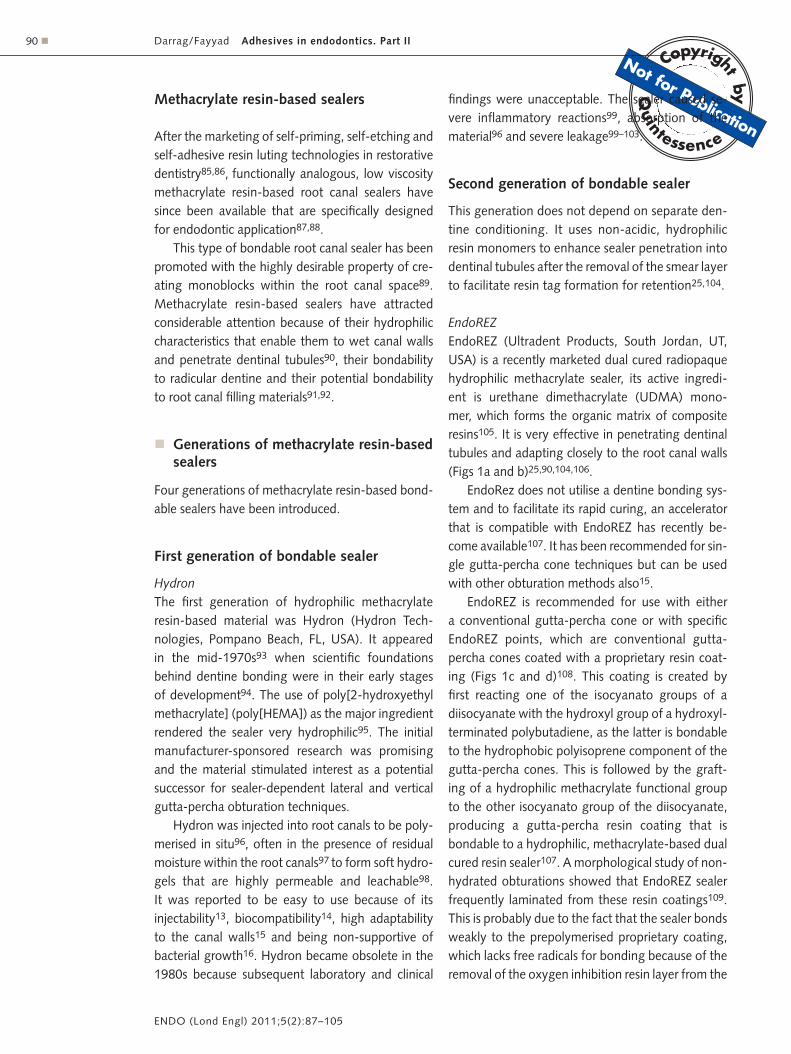
EndoREZEndoREZ (Ultradent Products, South Jordan, UT, USA) is a recently marketed dual cured radiopaque hydrophilic methacrylate sealer, its active ingredi-ent is urethane dimethacrylate (UDMA) mono-mer, which forms the organic matrix of composite resins105. It is very effective in penetrating dentinal tubules and adapting closely to the root canal walls (Figs 1a and b)25,90,104,106.
EndoRez does not utilise a dentine bonding sys-tem and to facilitate its rapid curing, an accelerator that is compatible with EndoREZ has recently be-come available107. It has been recommended for sin-gle gutta-percha cone techniques but can be used with other obturation methods also15.
EndoREZ is recommended for use with either a conventional gutta-percha cone or with specific EndoREZ points, which are conventional gutta-percha cones coated with a proprietary resin coat-ing (Figs 1c and d)108. This coating is created by first reacting one of the isocyanato groups of a diisocyanate with the hydroxyl group of a hydroxyl- terminated polybutadiene, as the latter is bondable to the hydrophobic polyisoprene component of the gutta-percha cones. This is followed by the graft-ing of a hydrophilic methacrylate functional group to the other isocyanato group of the diisocyanate, producing a gutta-percha resin coating that is bondable to a hydrophilic, methacrylate-based dual cured resin sealer107. A morphological study of non-hydrated obturations showed that EndoREZ sealer frequently laminated from these resin coatings109. This is probably due to the fact that the sealer bonds weakly to the prepolymerised proprietary coating, which lacks free radicals for bonding because of the removal of the oxygen inhibition resin layer from the

Copyrig
ht
by
N
otfor
Qu
in
tessence
Not
forPublication
Darrag/Fayyad Adhesives in endodontics. Part II � 91
ENDO (Lond Engl) 2011;5(2):87–105
surface of the resin coating for packing and storage purposes110.
A clinical study with EndoREZ reported a 91.3% success rate after 14 to 24 months111 with low bond strengths to dentine112. However, shear and push-out bond strengths of UDMA-based sealers to radicular dentine have been variously reported to be higher113 or lower20,55,77,91,114–118 than epoxy-based sealers.
Leakage and morphologic studies showed that the seal of the EndoRez system is mediocre, although long resin tags could be identified within the dentinal tu-bules25,104,119. This may be attributed to the polymer-isation shrinkage of the methacrylate-based sealer104. It performed poorly in leakage studies when com-pared to AH Plus sealer87,120,121, with less surface adaptation to dentine119. Moreover, even with the adjunctive use of an adhesive, it is unrealistic to ex-pect the establishment of a mechanically homoge-
nous unit with the root canal wall when using the EndoRez system, as the bulk of the material inside the root canal still consists of thermoplastic gutta-percha, an elastomeric polymer that flows when stressed122.
Several investigators have revealed that EndoREZ sealer is biocompatible with periapical tissues in subhuman primates123 and with rat cells and bone tissue124, while others utilising a different test re-ported it to be somewhat cytotoxic120.
Third generation of bondable sealer
The third generation is technologically analogous to those resin-based luting materials that use a separate self-etching primer85,86 before the application of dual cured resin sealer to the primed dentine. The use of self-etching primer reintroduces the concept of incor-porating smear layer created by root canal instruments
Fig 1 (a) Scanning electron microscopic (SEM) image (150x) taken from the coronal third of a NaOCl/EDTA-irrigated root canal. The latter was filled under moist conditions43 with the EndoREZ sealer. The radicular dentine was demineralised and deproteinised to expose the extent of resin infiltration into the dentinal tubules. (b) High magnification SEM (2000x) of the resin tags shown in the previous image. (c) Environmental SEM (ESEM) image (350x) taken from a non-dehydrated root section that was filled with EndoREZ sealer (S) and resin-coated gutta-percha (GP). Only a fraction of the circumference of the gutta-percha cone was surrounded by the resin coating (open arrow) (RD, radicular dentine). (d) High magnification ESEM image (1000x) of the previous image showing the presence of a thin hybrid layer (H) along the surface of the radicu-lar dentine (RD) that was created after the use of 17% EDTA as the final irrigant. Despite the presence of profuse resin tag formation, a gap (pointer) could be seen between the hybrid layer and the sealer (S). Likewise, a larger gap (asterisk) could be identified between the sealer and the resin coating (RC) of the gutta-percha (GP) cone122.
a b
c d

Copyrig
ht
by
N
otfor
Qu
in
tessence
Not
forPublication
Darrag/Fayyad Adhesives in endodontics. Part II92 �
ENDO (Lond Engl) 2011;5(2):87–105
in the sealer-dentine interface125. Provided that they are aggressive enough to etch through a thick smear layer, the technique sensitivity of bonding to root canals may be reduced when the smear layer is inad-vertently retained in the apical third of instrumented canal walls126.
FibreFill FibreFill root canal sealant (Pentron Clinical Tech-nologies, Wallingford, CT, USA) is a radiopaque dual cure third generation methacrylate resin sealer based on UDMA, used in combination with a self-cured, self-etching primer system (Fibrefill Primer A and B). Bonding between adhesive systems and dentine de-pends on the penetration of monomers into the con-ditioned dentine surface, to create micromechanical interlocking between the dentine collagen and resin, forming a hybrid layer. This system was designed for filling canals with fibre-reinforced obturators that are attached to a thermoplastic root filling material tip. The apical 5 to 8 mm of the obturator is gutta-percha and the coronal two-thirds consist of a resin and glass fibre post that is adhesively bonded within the tooth during the obturation, sealing the coronal portion and providing retention for the core27.
FibreFill root canal sealant is reported to have a good sealing ability27,88,127 and adhesive properties to radicular dentine75. In 2007, Fayyad and Darrag128
stated that the interfacial bond strength achieved with FibreFill root canal obturator to radicu lar den-tine is greatly affected by the type of irrigant solution used during instrumentation. They recommended using MTAD irrigant as a final rinse after sodium hypochlorite (NaOCl) irrigation to ensure the com-plete removal of organic and inorganic components of the smear layer and enhance the bond strength of FibreFill root canal obturation material.
Resilon/EpiphanyRecently, a new dual-curing third generation resin-based sealer commercially named Epiphany (Pentron Clinical Technologies) has been introduced. This system uses a self-etching primer and comprises a Resilon cone, which is a thermoplastic synthetic material (polycapro-lactone) that contains bioactive glass, bismuth oxy-chloride, and barium sulphate to replace gutta-percha and conventional sealers26,129,130. The sealer matrix consists of bisphenol-A-glycidyldimethacrylate (Bis-
GMA), ethoxylated Bis-GMA, UDMA, and hydrophilic methacrylate with calcium hydroxide, barium sulphate, barium glass, bismuth oxychloride and silica26.
A resinous solvent (Epiphany Thinning Resin) also comprises this filling system. The manufacturer rec-ommends the use of 1 to 2 drops of the solvent to adjust the sealer viscosity, when necessary. This prod-uct is an aqueous solution and consists of ethoxylated bisphenol-A-dimethacrylate (EBPADMA) resins with photo-initiators, amines, stabilisers and pigments131. The resinous solvent of the Epiphany system increased the bond strength of Epiphany sealer to dentine walls when followed by photoactivation132. The self-etch-ing primer of Epiphany has been further reduced from a two-bottle to a single-bottle system133.
Controversy has recently emerged concerning the use of this material. One research group demon-strated that the Epiphany/Resilon system chemically interacts with dentine and forms a resin monoblock between the root canal dentine and the respec-tive filling material26,30,129,134, resulting in a lower incidence of apical leakage, strengthening of the tooth16,26,135 and good resistance to bacterial infil-tration26,136. However, a different research group re-ported the potential of the Resilon/Epiphany system for reinforcing the residual tooth structure77 to be lower than using AH Plus and gutta-percha137.
In addition, previous studies verified the adhesive-ness of the Epiphany/Resilon system to radicular den-tine; it was not superior when compared with other resinous sealers77,91 and inferior to the epoxy resin-based sealers associated with gutta-percha cones77, 87,109,119,138. The adhesive quality to root dentine promoted by Epiphany sealer is compromised even when teeth with simple anatomic features were obt-urated under well-monitored laboratory conditions. Tay et al25 found gaps in root canals obturated with Resilon/Epiphany between Epiphany and the dentine wall, with bond strengths of only 4 to 6 MPa77,110,114. This is thought to be due to the inability of the sealer bond to resist shrinkage stresses generated during polymerisation of the root canal sealer139,140. Other findings reported gaps between the core material and the sealer114,141, with bond strength less than 2 MPa77,110,114. This is not surprising, because un-polymerised resin must be available in both materials to achieve co-polymerisation and in Resilon there is no unpolymerised resin110. This led to a conclusion

Copyrig
ht
by
N
otfor
Qu
in
tessence
Not
forPublication
Darrag/Fayyad Adhesives in endodontics. Part II � 93
ENDO (Lond Engl) 2011;5(2):87–105
that the chemical coupling of the methacrylate-based sealer to Resilon is very weak142. However, this chem-ical bond between the sealer and the Resilon points26 might be the reason for significantly better adaptation to the points than EndoREZ and Guttaflow (Coltène Whaledent, Langenau, Germany)143.
Versiani et al129 reported that Epiphany sealer was outside the acceptable range for solubility and dimensional stability on the basis of the American National Standards Institute/American Dental As-sociation (ANSI/ADA) Specification No. 57144, but was within the acceptable range for setting time, flow and thickness. Melker et al145 reported that Resilon was found to exhibit no antimicrobial activ-ity, despite the fact that bioactive glass is one of its components and is considered to have antimicrobial properties. Several studies have tested the cytotoxic-ity of this material, with highly variable results. Sousa et al80 and Garcia et al146 observed that Epiphany/Resilon root canal filling system presented satisfac-tory tissue reaction and only minor inflammatory reactions (Fig 2). Its biocompatibility may be related to a high calcium release from Epiphany and this feature causes a more alkaline pH of the tissue site, which might increase tissue repair. Other authors, however, revealed that the multi-methacrylate resin-based (Epiphany) root canal sealer was significantly more toxic than the silicone-based Roeko-Seal (Roeko, Langenau, Germany), the single methacr-ylate-based EndoREZ root canal sealers147 and more cytotoxic than other conventional sealers129,148.
RealSealRealSeal (SybronEndo, Orange, CA, USA) has been approved for endodontic use for about 5 years and was introduced as an alternative to traditional root canal filling materials. The system includes primer, sealer and core material. The sealer contains UDMA, polyethylene glycol dimethacrylate (PEGDMA), ethoxylated bisphenol A dimethacrylate and 2,2- bis[p-(2-hydroxy-3-methacryloxypropoxy) phenyl] propane (Bis-GMA) resins, silane treated barium borosilicate glass, barium sulphate, silica, calcium hydroxide, bismuth oxychloride with amines, perox-ide, photoinitiator, and pigments. RealSeal core mate-rial contains 57% polyester polymer polycaprolactone, and 42% bioactive glass and radiopaque fillers149.
RealSeal had a significantly higher micro-shear bond strength than EndoREZ, but it is lower than epoxy resin sealer (AH Plus)5. The type, size and shape of filler particles may influence the bond strength of the different sealer types. The large plate-like structure of RealSeal filler particles appeared to align in layers that were parallel to each other and the dentine surface, possibly creating cleavage planes that readily fail in shear mode (Fig 3)118,150.
RealSeal core material is a biocompatible mater-ial151 and tends to be nontoxic152,153. The biocom-patibility of the RealSeal system depends largely on the sealer, which has been reported as cyto-toxic154–166. This cytotoxicity has been proven on human gingival fibroblasts and mouse skin fibro-blasts157,158.
Fig 2 Histologic image after 42 days: the subcutaneous connective tissue of rats represented slight to absent chronic inflammatory reaction to Resilon/Epiphany root filling sys-tem with little residual dispersed sealer146.
Fig 3 SEM image of RealSeal in a thin film showing the gritty irregular resin layer covering the plate-like filler parti-cles over the dentine surface. Notice the filler particles are aligned parallel to the dentine surface, predisposing to shear failure (2000x magnification)5.
Copyrig
ht
by
N
otfor
Qu
in
tessence
Not
forPublication
Darrag/Fayyad Adhesives in endodontics. Part II94 �
ENDO (Lond Engl) 2011;5(2):87–105
Fourth generation of bondable sealer (self-adhesive sealers)
The fourth generation of methacrylate resin-based sealers is comparable to self-adhesive resin luting materials in that both were designed with the in-tention of combining a self-etching primer and a moderately filled flowable composite into a single product. The acidic resin monomers that are origi-nally found in dentine adhesive primers are now incorporated into the resin-based sealer to render them self- adhesive to dentine substrates159.
The combination of an etchant, a primer and a sealer into an all-in-one self-etching, self-adhesive product is advantageous in that it reduces the ap-plication time as well as errors that might occur dur-ing each bonding step160. In theory, the bonding mechanism of self-adhesive sealers is similar to self-adhesive resin materials. They are designed to inte-grate canal wall smear layer into the sealer-dentine interface159.
Thus, it is imperative to evaluate the true etch-ing potential of self-etching and self-adhesive seal-ers. Recently, there have been doubts regarding the bonding efficacy of self-adhesive resin sealers to smear layer-covered radicular dentine with an absence of the adjunctive use of calcium-chelat-ing irrigants67. The manufacturers’ instructions for self-adhesive sealers recommend the use of NaOCl irriga tion followed by EDTA for 1 minute to com-pletely remove the smear layer. So, the retention mechanisms are likely to be contributed by the com-bined dentine demineralisation effects of EDTA and the sealer system161. It was further speculated that the dentine demineralisation effect of EDTA could have surmounted the true self-etching capacity of these self-adhesive sealers, reducing its leakage and improving the seal of the filled canals67,161,162.
The fourth generation self-adhesive root canal sealers are still relatively new, and detailed informa-tion on their adhesive properties to root canal filling materials is limited or lacking. However, they have limited aggressiveness in creating micromechani-cal retention via dentine hybridisation163, and the chemical coupling between contemporary meth-acrylate resin-based sealers to root canal filling ma-terials is generally weak or insufficiently optimised148 with poorer marginal integrity when compared with
conventional resin cements that use etch-and-rinse or self-etch adhesives for bonding164–166.
MetaSealMetaSeal (Parkell, Edgewood, NY, USA) has recently been developed as a hydrophilic and self-adhesive sealer, which was reported to seal the root canal with a 4-META hybrid layer167 and is recommended for use exclusively with cold compaction or single-cone techniques118. A recent report identified a hybrid layer-like structure along the gutta-percha –sealer interface168. However, no data are currently avail-able on the adhesive strength of MetaSeal to gutta-percha via this hybrid layer-like interface. According to the manufacturers, MetaSeal has low cytotoxicity and is thus biocompatible both at the cellular and at the tissue levels; however a recent study revealed that MetaSeal was severely cytotoxic148.
RealSeal SE and RealSeal 1 RealSeal SE (SybronEndo, Orange, CA, USA) is the simplified dual-cured version of RealSeal sealer and uses a polymerisable methacrylate carboxylic acid anhydride (4-META) as the acidic resin mono-mer67,76,169. It might be used with Resilon cones or pellets by using cold lateral or warm vertical compaction techniques or with the more recently introduced RealSeal 1 carrier-based Resilon obtu-rator system. In the RealSeal 1 bonded obturation system, the carrier is a polysulphone-containing polymer with radiopaque filler, and the surrounding Resilon-based filling contains polycaprolactone and polyolefin polymers loaded with fillers. This product combines adhesive bonding technology with a car-rier product and aims to provide the benefits of an efficient obturation technique combined with opti-mal leakage resistance. It appeared to resist bacte-rial penetration more effectively than Thermafil170. MetaSeal 67,169 and RealSeal SE67,76 are unable to etch beyond a thick smear layer created by rotary nickel-titanium instruments into the underlying in-tact radicular dentine in the absence of the adjunc-tive use of EDTA.
Babb et al 67 and Mai et al169 examined the ad-hesive strength of two self-adhesive resin sealers MetaSeal and RealSeal SE. They found that MetaSeal had the highest push-out bond strength and pro-vides a more impermeable seal to coronal leakage.

Copyrig
ht
by
N
otfor
Qu
in
tessence
Not
forPublication
Darrag/Fayyad Adhesives in endodontics. Part II � 95
ENDO (Lond Engl) 2011;5(2):87–105
Both micro-shear bond testing114 and push-out tests 77,91,115,116,150 showed that the bonding of RealSeal SE sealer to root dentine is not superior to other sealer systems.
Contrary to the manufacturers’ claims, neither the second nor the fourth generation sealers are likely to bond well to radicular dentine if EDTA is not used to remove the smear layer and smear plugs, or when EDTA does not reach the apical third of the root canal walls. Inadequate dentine hybridisa-tion might also occur in the calcospherite-containing noninstrumented dentine for those clinicians who elect to use NaOCl as the only active root canal ir-rigant67,76,169.
� Problems associated with resin-based sealer application and curing in root canals
An in vitro study by Bouillaguet et al23 reported lower bond strengths achieved in bonding to root canal wall dentine than to flat prepared samples of radicular dentine. These results are not surprising because effective bonding within a deep, narrow root canal is a challenge mainly as a result of the unfavourable geometry of the root canal system. With resin-based sealers, sealer pene tration into tu-bules is very extensive171,172. The resin component penetrates tubules, whereas filler particles, which are mostly too large to enter tubules, remain at the inter-face. This depletion of resin from the interfacial layer was proposed as a reason for the low bond strength as measured by the push-out test116, although it has been shown that resin tags do not contribute to dentine adhesion in self-etching adhesives173. A cer-tain degree of micromechanical retention is required to achieve an adequate seal, because considerable shrinkage strain might be expected during polymer-isation of low viscosity sealers174 inside long narrow canals with high C-factors175.
Proper and uniform application of a primer or adhesive is essential but it is difficult for effective bonding in the apical third of the canal. Once the primer is applied, the volatile carrier must be evap-orated. This can be problematic in the apical third, as it is difficult and probably a bad idea to blow air into the apical part of the canal. Also, application and drying of the primer with paper points is probably
not very effective for either task. If the acetone or alcohol carrier is not completely removed, the bond is adversely affected175.
In addition, penetration with a curing light is limited in the root canal system, so dual-cured or self-cured resin adhesives must be used. Dual-cured resins contain components that provide rapid light polymerisation in those areas where the curing light penetrates effectively and a slower chemical poly-merisation in those areas where the light is not effect ive. Adhesives and sealers that contain a self-cure component are a mixed blessing, however. On the plus side, the slower chemical polymerisa-tion process and less conversion of monomer to polymer than light-cured resins allows the mate-rial to flow in the pre-gel stage, which provides some stress relief from polymerisation contraction at the resin–dentine interface176,177. Also, air bub-bles incorporated into the resin during the mixing process provide a stress relief mechanism178. On the negative side, with the prolonged time of poly-merisation the hydrophilic primer acts as permeable membranes, permitting diffusion of moisture from the dentine through it and creates water blisters along the interface. This moisture contamination reduces bond strength and facilitates leaching of water-soluble components from the resin, which may further contribute to the breakdown of the bond56,65,179–181. Also, unpolymerised resin and air bubbles incorporated during mixing have negative effects on the mechanical properties and chemical stability of the resin. This phenomenon only occurs within the root canal, it is not a problem in areas that are light polymerised181.
� Retreatment in root canals obturated with methacrylate resin-based sealer
There is a general consensus that methacrylate resin-based sealers used with Resilon or gutta-percha were more effectively removed, with fewer remnants of the filling material than conventional sealer/gutta-percha combinations106,182–187, especially in the apical part of the root canal183. Easier removal and less remnant materials would imply that methacryl-ate resin-based sealers do not bond well to sclerotic dentine that is present in the apical part of the canal walls. Although Resilon is soluble in chloroform and
Copyrig
ht
by
N
otfor
Qu
in
tessence
Not
forPublication
Darrag/Fayyad Adhesives in endodontics. Part II96 �
ENDO (Lond Engl) 2011;5(2):87–105
other solvents182–184, Epiphany is insoluble in the solvents commonly used in dentistry. Thus, removal of resin sealers from fins, accessory canals or canal isthmi remains a challenge15.
� Glass ionomer-based sealer
Glass-ionomer cement (GIC) has been modified for use as a root canal sealer188 to fulfil the criteria for an ideal root canal sealer189. It is the only self-adhesive material currently available that eliminates the need for a separate adhesive system and its associated problems. GIC adheres to dentine through phys-ical and chemical interaction; physical interaction is through a micromechanical interlocking of the ma-terial to tooth surface irregularities, while chemical interaction occurs when polyacrylate ions of GIC displace existing phosphate ions in the hydroxy-apatite crystal and become irreversibly attached to the dentine190. Although theoretically glass ionomer cement may bond to gutta-percha by chelation with the zinc component of gutta-percha191, scanning electron microscopy revealed no evidence of chemi-cal or physical bonding192. In vitro evaluations of the sealing ability of GIC sealers have been equivocal; demonstrating both advantages193 and disadvan-tages194 over other sealer types.
Ketac Endo
Ketac Endo (3M ESPE, St. Paul, MN, USA) is a tradi-tional glass-ionomer cement that was developed as an endodontic sealer to reinforce the root, presum-ably because of its adhesion to dentine72. Its clinical efficacy has, however, been confirmed but never gained wide acceptance. It offered little benefit in leakage studies and was generally considered dif-ficult to re-treat21,31,193,195.
Lee et al30 demonstrated that Ketac Endo can bond to both dentine and gutta-percha although it bonded to dentine (0.74–1.19 MPa) more strongly than to gutta-percha (0.14 MPa). Recently, two other experimental GIC-based sealers have been introduced. KT-308 (GC Corporation, Tokyo, Ja-pan) is a GIC sealer with extended working time and radiopacity. This sealer resisted coronal ingress of bacteria into the root canal system better than a zinc oxide-eugenol-based sealer196. On the other
hand, ZUT (University of Toronto, Ontario, Can-ada) is a combination of GIC and an antimicrobial silver-containing zeolite. ZUT has been shown in vitro to effectively suppress adherent Enterococ-cus faecalis over a 3-month period197. This en-hanced antimicrobial efficacy may be an advantage in treating teeth of persistent apical periodontitis infected with E. faecalis198. Lalh et al199 com-pared the bond strengths of Ketac Endo, KT-308 and ZUT to bovine dentine conditioned with the common irrigants used in endodontic therapy and found that Ketac Endo demonstrated significantly lower (P < 0.05) shear bond strength to dentine than KT-308 or ZUT.
Glass ionomers lack desirable properties for an obturation material and adhesiveness to gutta-percha, which led to the introduction of a new root canal filling system (Active GP) that is claimed to consistently deliver a true monoblock obturation200.
ActiV GP
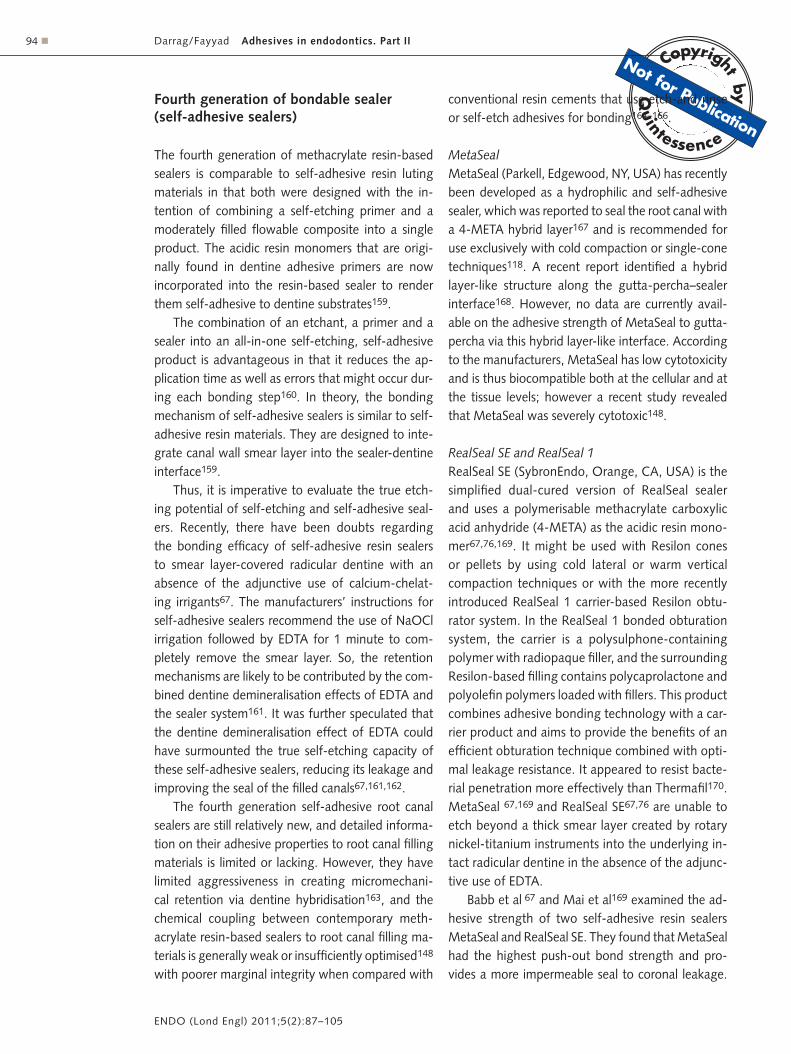
ActiV GP (Brasseler USA, Savannah, GA, USA) is a new glass-ionomer root canal-filling system that has been marketed to create a single-cone monoblock obturation. The system comprises glass ionomer im-pregnated and coated gutta-percha cones that are bondable to a sealing agent composed of barium aluminosilicate glass powder and polyacrylic acid (Fig 4)201. This was claimed to offer adhesive bond-ing of the core material to intraradicular dentine via the glass-ionomer sealer200.
The chemical bonding capacity of the ActiV GP (a diffusion-based adhesion) would have developed by ion exchange between the glass-ionomer and the tooth surface. The polyalkenoic acid chains penetrate the surface of dentine and displace phosphate ions, releasing them into the cement. Each phosphate ion takes with it a calcium ion to maintain electrolytic balance202. By doing so, a stiffer gutta-percha cone is achieved that transforms it into a gutta-percha core/cone, enabling the latter to function as both the tapered filling cone and as its own carrier core, thus avoiding the need for a separate interior carrier of plastic or metal200.
Bond strength of ActiV GP can be improved by final irrigation of the root canal with 17% EDTA20. Root canals obturated with ActiV GP had signifi-

Copyrig
ht
by
N
otfor
Qu
in
tessence
Not
forPublication
Darrag/Fayyad Adhesives in endodontics. Part II � 97
ENDO (Lond Engl) 2011;5(2):87–105
cantly higher bond strengths compared to Resilon and EndoREZ obturation systems203. The system produced an apical seal to fluid filtration that is com-parable to that of gutta-percha and AH Plus sealer. However, being a single-cone technique, coronal leakage of the ActiV GP system to fluid filtration was worse than that achieved with gutta-percha and AH Plus, probably because of the increase in the volume of the glass-ionomer cement sealer204. Kazandag et al205 demonstrated that ActiV GP combined with its glass-ionomer sealer is not superior to the other systems in terms of root reinforcement.
� Bioceramic-based sealers
A problem historically with the early generations of glass ionomer and resin sealers was that neither method was able to develop a true monoblock. Resin-based sealers were shown to have a good seal between sealer and core material, but their seal to the canal wall was questionable and has many limitations. These limitations may be related to the root canal system environment or to the material properties it-self 36. In contrast to resins, glass ionomer cements displayed an excellent seal to the canal wall but their seal to the gutta-percha was less than ideal. So, the search contiunued for a technique that could consist-ently deliver a true monoblock obturation200.
Fig 4 Surface coating of conventional gutta-percha cones with glass-ionomer fillers (ActiV GP, Brasseler USA, Savannah, GA, USA) represents an example of a part of the components of a tertiary endodontic monoblock, in which these filler-coated gutta-percha cones are bonded to intraradicular dentine with the use of a glass-ionomer root canal sealer. (a) A low magnification scanning electron micrograph of a cryofractured ActiV GP gutta-percha cone depicting the representative locations from which the higher magnification micrographs were derived. (b) A high magnification interfacial view show-ing the surface of the fractured gutta-percha cone (between asterisks) with the glass-ionomer fillers (arrow) on top of the surface and the filler-dense gutta-percha cone below. (c) A high magnification surface view showing a region that is heavily coated with glass-ionomer fillers. The dimensions of these angular fillers ranged from submicrometre to 2 μm in diameter. (d) Incomplete or uneven coating of the gutta-percha cone surface could often be observed along different regions of the same coated gutta-percha cone. In this micrograph, the glass-ionomer fillers were sparse (open arrowhead) and numerous dimpled, filler-free areas (pointer) could be identified89.
a b
c d

Copyrig
ht
by
N
otfor
Qu
in
tessence
Not
forPublication
Darrag/Fayyad Adhesives in endodontics. Part II98 �
ENDO (Lond Engl) 2011;5(2):87–105
More recently the introduction of bioceramic technology is considered a game changer in root canal obturation. Bioceramics are exceedingly bio-compatible, non-toxic, do not shrink and are chem-ically stable within the biological environment206.
iRoot SP
iRoot SP (Innovative Bioceramix, Vancouver, Canada; also known as EndoSequence BC sealer, Brasseler, Savannah, GA, USA) is an insoluble, radio paque sealer, and was equivalent to AH Plus sealer in apical sealing ability207. It is an aluminium-free material based on a calcium silicate composition, which requires the presence of water to set and harden. It is composed of biocompatible and nontoxic materials that include calcium phos-phate, calcium silicates, zirconium oxide and cal-cium hydroxide. One of its advantages is its ability during the setting process to form hydroxyapatite and ultimately a bond between dentine and filling materials208. Moreover, the glass components in the bioceramic sealer bond to the ActiV GP glass ionomer coated cones206.
� Monoblocks
The term monoblock, literally meaning a single unit, has been employed in dentistry since the turn of the century89. With increasing success and predictability
of contemporary adhesive strategies used for intra-coronal adhesive seals, likewise, potential improve-ments in apical and coronal seals26 and strengthen-ing of root canal treated teeth16 may be anticipated by establishing monoblocks between the intrara-dicular dentine and adhesive root canal fillings89. A monoblock obturation system is the unit in which the core material, sealing agent and the root canal dentine form a single cohesive unit20. Two prerequi-sites are simultaneously required for a monoblock to function successfully as a mechanically homogenous unit. First, the materials that constitute a monoblock should have the ability to bond strongly and mu-tually to one another, as well as to the substrate that the monoblock is intended to reinforce. Second, these materials should have a modulus of elasticity that is similar to that of the substrate89.
Tay and Pashley89 classified monoblocks created in the root canal space as primary, secondary or ter-tiary depending on the number of interfaces present between the bonding substrate and the bulk material core (Fig 5).
� Primary monoblocks
A primary monoblock has only one interface that extends circumferentially between the material and the root canal wall.
Hydron
Hydron was known as one of the primary monoblocks employed in root canals. This HEMA- containing root canal filling material was marketed commercially for the paste filling of root canals. It was not stiff enough to strengthen roots even if it could have bonded to root canal surfaces. The modulus of elasticity of por-ous poly-HEMA hydrogels such as Hydron ranges from 180 to 250 MPa95. To reinforce roots, the modulus of elasticity of a root canal filling material would need to achieve approximately that of den-tine (14,000–18,600 MPa according to location and orientation of the dentinal tubules)149.
Mineral trioxide aggregate (MTA)
Orthograde obturation with mineral trioxide aggre-gate (MTA) (Dentsply Tulsa Dental, Tulsa, OK, USA)
Fig 5 Classification of endodontic monoblocks89.
Primary Secondary Tertiary
Root dentine
Cementum Root filling material
Resin cement or root canal sealer
Fibre post or root filling material
Bondable coating on
fibre post or root filling
material

Copyrig
ht
by
N
otfor
Qu
in
tessence
Not
forPublication
Darrag/Fayyad Adhesives in endodontics. Part II � 99
ENDO (Lond Engl) 2011;5(2):87–105
as an apical plug material represents a contempor-ary version of the primary monoblock in attempts to strengthen immature tooth roots. Although MTA does not bond to dentine, interaction of the re-leased calcium and hydroxyl ions of MTA with a phosphate-containing synthetic body fluid results in the formation of apatite-like interfacial depos-its209,210. These deposits fill any gaps induced dur-ing the material shrinkage phase and improve the frictional resistance of MTA to the root canal walls. Unlike Hydron, MTA should theoretically be able to strengthen roots.
iRoot SP
iRoot SP root canal sealer has recently been intro-duced to the market. According to the manufacturer’s description, iRoot SP is a convenient, premixed, in-jectable sealer that can be used for permanent filling of root canals without gutta-percha points (primary monoblock). It has a similar composition to white MTA material and has both excellent physical prop-erties and biocompatibility206.
� Secondary monoblocks
The combined use of a core material and a sealer/cement in contemporary endodontic obturations and fibre post adhesion introduces additional inter-faces into a monoblock. Secondary monoblocks are those that have two circumferential interfaces, one between the sealer and dentine and the other be-tween the sealer and the core material. A secondary monoblock is the type of monoblock that is clas-sically perceived in the restorative and endodontic literature117.
However, as conventional root canal sealers do not bond strongly to dentine and gutta-percha30, they do not behave as mechanically homogenous units with the root dentine. Although glass-ionomer cements and resin-modified glass-ionomer cements bond to root dentine and have been marketed as root canal sealers211,212, they do not bond to gutta-percha. Even if they do, the modulus of elasticity of gutta-percha points (80 MPa)149 is 175 to 230 times lower than that of dentine149,213,214, making them too plastic (not stiff enough) to reinforce roots after root canal treatment. Thus, it is dubious that a glass-
ionomer-based sealer can be used to prevent root fracture in gutta-percha-filled root canals215.
Resilon
Interest in utilising the classic monoblock concept for sealing and reinforcing the root canal space was rekindled in 2004 with the advent of bondable root canal filling materials that were advocated as alter-natives to conventional gutta-percha. Resilon is the first true bondable root canal filling material. As Resi-lon is applied using a methacrylate-based sealer to self-etching primer-treated root dentine, it contains two interfaces, one between the sealer and primed dentine and the other between the sealer and Resi-lon, and hence may be classified as a type of second-ary monoblock89.
Resilon, together with the Epiphany primer and sealer system was subsequently referred to as the Resilon monoblock system (RMS)18, which produces ideal root canal obturation in terms of coronal sealing and fracture resistance216. In root canals, however, C-factors can be over 1000. Any polymerising root canal sealer will be subjected to large polymerisa-tion stresses during setting that may cause debond-ing and gap formation along the periphery of the root canal filling. The extremely high C-factor in root canals has been cited as a possibility for not achieving perfect seals in Resilon-filled root canals25. An experimental strategy has also been developed by using a zinc oxide-filled thermoplastic poly-urethane composite root canal filling material and a light-cured urethane-acrylate/tripropylene glycol diacryl ate root canal sealer150,217. Another experi-mental system uses ethylene vinyl acetate (EVA) as the major thermo plastic component of an alterna-tive root canal filling material186 together with a dimethacrylate- conjugated fluorene monomer218 for potential bonding to methacrylate resin-based sealers.
iRoot SP
iRoot SP was a suitable cement paste for use in a single-cone filling technique207. It can be used with conventional gutta-percha (secondary monoblock) or it is recommended to be used with ActiV GP glass ionomer coated cones206 (tertiary monoblock).

Copyrig
ht
by
N
otfor
Qu
in
tessence
Not
forPublication
Darrag/Fayyad Adhesives in endodontics. Part II100 �
ENDO (Lond Engl) 2011;5(2):87–105
� Tertiary monoblocks
The entrepreneurial concept of creating a root canal monoblock to achieve a total bond and hence a total seal of the canal space has been hampered by the lack of chemical union between the polyisoprene component of gutta-percha and resin-based or glass ionomer-based sealers. To circumvent this problem, several strategies have been used in which a third circumferential interface is introduced between the bonding substrate and the abutment material by coating the non-bondable gutta-percha points with materials that render them bondable to the root canal sealers, creating tertiary monoblocks.
EndoREZ
EndoREZ is the first commercialised strategy that was introduced by coating gutta-percha cones with a polybutadiene-diisocyanate-methacrylate adhe-sive219. This proprietary adhesive resin includes a hydrophobic portion that is chemically compatible with the hydrophobic polyisoprene substrate and a hydrophilic portion that is chemically compatible with a hydrophilic methacrylate resin. With the use of this adhesive resin coating, a strong chemical union is achieved between the gutta-percha and the methacrylate resin-based sealer. This thermoplastic resin-coated gutta-percha cone is recommended for use with the EndoREZ system107.
In view of the extremely high C-factor encoun-tered in long, narrow root canals25, it is doubtful whether the core material –sealer bond is capable of resisting the polymerisation shrinkage stresses that develop during the setting of the resin sealer to permit the realisation of the goal of creating a monoblock in the root canal system. It also has to be taken into consideration that the interface be-tween the gutta-percha resin coating and the resin sealer is the only truly bondable interface in this sys-tem. This interface is a weak link that failed during polymerisation shrinkage of the sealer. Removal of
the oxygen inhibition layer from the surface of resin-coated gutta-percha cones during packaging has been hypothesised for their weak adhesion to the methacrylate resin-based root canal sealer, resulting in their frequent delamination from the sealer after root canal obturation160.
ActiV GP
This second commercialised tertiary monoblock strategy uses conventional gutta-percha cones that are surface coated with glass ionomer fillers using a proprietary technique200. Both systems (EndoREZ and ActiV GP) are designed to be used with either a single-cone technique or a technique that involves the passive placement of accessory cones without lateral compaction (harpooning technique), to avoid disruption of these external coatings.
� Conclusions
Although adhesive obturation materials are in the early stages of development and, on the basis of the in vitro and in vivo data available to date, have greater potential than traditional materials, there ap-pears to be no adhesive root canal filling material that can perfectly obturate the canal space with a gap-free solid mass that consists of different mater-ials and interfaces, with the apparent advantages of simultaneously improving the seal and fracture resistance of the root canal-filled tooth. Even in adhesive resin root canal filling materials, despite the hybridisation, a perfect seal of the root canal is difficult to achieve, which may be a result of the complexity of the substrate and the high C-factor. The concept of creating mechanically homogenous units with root dentine is excellent in theory, but accomplishing these ‘ideal monoblocks’ in the root canal space is easier said than done. However, con-tinued research and development is likely to result in improvements and in new more effective materials.

Copyrig
ht
by
N
otfor
Qu
in
tessence
Not
forPublication
Darrag/Fayyad Adhesives in endodontics. Part II � 101
ENDO (Lond Engl) 2011;5(2):87–105
� References
1. Leonardo MR, Barnett F, Debelian GJ, et al. Root canal adhesive filling in dogs’ teeth with or without coronal restoration: a histopathological evaluation. J Endod 2007; 33:1299–303.
2. Lin LM, Skribner JE, Gaengler P. Factors associated with endodontic treatment failures. J Endod 1992;18:625–627.
3. Siqueira JF Jr. Aetiology of root canal treatment failure and why well-treated teeth can fail. Int Endod J 2001;34:1–10.
4. Siqueira JF Jr, Rocas IN, Favieri A, et al. Bacterial leakage in coronally unsealed root canals obturated with 3 different techniques. Oral Surg Oral Med Oral Pathol Oral Radiol Endod 2000;90:647–650.
5. Rahimi M, Jainaen A, Parashos P, Messer HH. Bonding of res-in-based sealers to root dentine. J Endod 2009;35:121–124.
6. Johnson WT, Gutmann JL. Obturation of the cleaned and shaped root canal system. In: Cohen S, Hargreaves KM (eds). Pathways of the pulp, ed 9. St Louis, MO: Mosby, 2006:358–399.
7. White RR, Goldman M, Lin PS. The influence of the smeared layer upon dentinal tubule penetration by endo-dontic filling materials. Part II. J Endod 1987;13:369–374.
8. Heling I, Chandler NP. The antimicrobial effect within dentinal tubules of four root canal sealers. J Endod 1996;22:257–259.
9. Weis MV, Parashos P, Messer HH. Effect of obturation technique on sealer cement thickness and dentinal tubule penetration. Int Endod J 2004;37:653–663.
10. Branstetter J, von Fraunhofer JA. The physical properties and sealing action of endodontic sealer cements: a review of the literature. J Endod 1982;8:312–316.
11. Bouillaguet S, Bertossa B, Krejci I, et al. Alternative adhesive strategies to optimize bonding to radicular dentine. J Endod 2007;33:1227–1230.
12. Grande NM, Plotino G, Lavorgna L, et al. Influence of dif-ferent root canal-filling materials on the mechanical proper-ties of root canal dentine. J Endod 2007;33:859–863.
13. Salehrabi R, Rotstein I. Endodontic treatment outcomes in a large patient population in the USA: an epidemiological study. J Endod 2004;30:846–850.
14. Tilashalski KR, Gilbert GH, Boykin MJ, Shelton BJ. Root canal treatment in a population-based adult sample: status of teeth after endodontic treatment. J Endod 2004;30:577–581.
15. Schwartz RS. Adhesive dentistry and endodontics: part 2: bonding in the root canal system: the promise and the problems—a review. J Endod 2006;32:1125–1134.
16. Teixeira FB, Teixeira EC, Thompson J, et al. Dentinal bond-ing reaches the root canal system. J Esthet Restor Dent 2004;16:348–354.
17. Najar AL, Saquy PC, Vansan LP, Sousa-Neto MD. Adhesion of a glass-ionomer root canal sealer to human dentine. Aust Endod J 2003;29:20–22.
18. Teixeira FB, Teixeira EC, Thompson JY, Trope M. Fracture resistance of roots endodontically treated with a new resin filling material. J Am Dent Assoc 2004;135:646–652.
19. Zmener O, Pameijer CH. Clinical and radiographical evalu-ation of a resin-base root canal sealer: a 5-year follow-up. J Endod 2007;33:676–679.
20. Fisher MA, Berzins DW, Bahcall JK. An in vitro comparison of bond strength of various obturation materials to root canal dentine using a push-out test design. J Endod 2007;33:856–858.
21. Leonard JE, Gutmann JL, Guo IY. Apical and coronal seal of roots obturated with a dentine bonding agent and resin. Int Endod J 1996;29:76–83.
22. Ferrari M, Vichi A, Grandini S, Goracci C. Efficacy of a self-curing adhesive-resin cement system on luting glass-fiber posts into root canals: an SEM investigation. Int J Prostho-dont 2001;14:543–549.
23. Bouillaguet S, Troesch S, Wataha JC, et al. Microtensile bond strength between adhesive cements and root canal dentine. Dent Mater 2003;19:199–205.
24. Gogos C, Stavrianos C, Koloukoris I, et al. Shear bond strength of AH-26 root canal sealer to dentine using three dentine bonding agents. J Dent 2003;31:321–326.
25. Tay FR, Loushine RJ, Monticelli F, et al. Effectiveness of resin-coated gutta-percha cones and a dual-cured, hy-drophilic methacrylate resin-based sealer in obturating root canals. J Endod 2005;31:659–664.
26. Shipper G, Ørstavik D, Teixeira FB, Trope M. An evaluation of microbial leakage in roots filled with a thermoplastic synthetic polymer-based root canal filling material (Resilon). J Endod 2004;30:342–347.
27. Shipper G, Trope M. In vitro microbial leakage of endo-dontically treated teeth using new and standard obturation techniques. J Endod 2004;30:154–158.
28. Yamauchi S, Shipper G, Buttke T, et al. Effect of orifice plugs on periapical inflammation in dogs. J Endod 2006;32:524–526.
29. Britto LR, Borer RE, Vertucci FJ, et al. Comparison of the apical seal obtained by a dual-cure resin based cement or an epoxy resin sealer with or without the use of an acidic primer. J Endod 2002;28:721–723.
30. Lee KW, Williams MC, Camps JJ, Pashley DH. Adhesion of endodontic sealers to dentine and gutta-percha. J Endod 2002;28:684–688.
31. Pommel L, About I, Pashley D, Camps J. Apical leakage of four endodontic sealers. J Endod 2003;29:208–210.
32. Paque F, Sirtes G. Apical sealing ability of Resilon/Epiphany versus gutta-percha/AH Plus: immediate and 16-months leakage. Int Endod J 2007;40:722–729.
33. Cotton TP, Schindler WG, Schwartz SA, et al. A retrospec-tive study comparing clinical outcomes after obturation with Resilon/Epiphany or gutta-percha/Kerr sealer. J Endod 2008;34:789–797.
34. De-Deus G, Namen F, Galan J Jr. Reduced long-term sealing ability of adhesive root fillings after water-storage stress. J Endod 2008;34:322–325.
35. Gogos C, Theodorou V, Economides N, et al. Shear bond strength of AH-26 and Epiphany to composite resin and Resilon. J Endod 2008;34:1385–1387.
36. Fayyad DM, Darrag AM. Adhesives in endodontics. Part I: limitations and effect of different materials. ENDO (Lond Engl) 2009;3:185–204.
37. Erickson RL. Surface interactions of dental adhesive mater-ials. Oper Dent 1995;5:81–94.
38. Nakabayashi N, Pashley D. Hybridization of dental hard tissues. Tokyo: Quintessence Publishing Co, 1998:130.
39. Eick JD, Gwinnett AJ, Pashley DH, et al. Current concepts on adhesion to dentine. Crit Rev Oral Biol Med 1997;8:306–335.
40. Pashley DH, Pashley EL, Carvalho RM, et al. The effect of dentine permeability on restorative dentistry. Dent Clin North Am 2002;46:211–245.
41. Foxton R. Nakajima M, Tagami J, Miura H. Adhesion to root canal dentine using one and two-step adhesives with dual-cure composite core materials. J Oral Rehabil 2005;32:97–104.
42. Inoue S, Vargas MA, Abe Y, et al. Microtensile bond strengths of eleven contemporary adhesives to dentine. J Adhes Dent 2001;3:237–245.
43. Schwartz RS, Fransman R. Adhesive dentistry and endodon-tics: materials, clinical strategies and procedures for restoration of access cavities: A review. J Endod 2005;31: 151–165.
44. Van Meerbeek B, Yoshida Y, Lambrechts P, et al. A TEM study of two water-based adhesive system bonded to dry and wet dentine. J Dent Res 1998;77:50–59.
45. Perdigao J, Van Meerbeek B, Lopes MM, Mbrose WW. The effect of a re-wetting agent on dentine bonding. Dent Mater 1999;15:282–295.

Copyrig
ht
by
N
otfor
Qu
in
tessence
Not
forPublication
Darrag/Fayyad Adhesives in endodontics. Part II102 �
ENDO (Lond Engl) 2011;5(2):87–105
46. Ferrari M, Tay FR. Technique sensitivity in bonding to vital, acid etched dentine. Oper Dent 2003;28:3–8.
47. Crim GA. Prepolymerization of Gluma 4 sealer: effect on bonding. Am J Dent 1990;3:25–27.
48. Erickson RL. Surface interactions of dentine adhesive mater ials. Oper Dent 1992;(Suppl 5):81–83.
49. Van Meerbeek B, De Munck J, Yoshida Y, et al. Buonocore memorial lecture. Adhesion to enamel and dentine: current status and future challenges. Oper Dent 2003;28:215–235.
50. Tagami J, Tao L, Pashley DH. Correlation among dentine depth, permeability, and bond strength of adhesive resin. Dent Mater 1990;6:45–50.
51. Pereira PN, Okuda M, Sano H, et al. Effect of intrinsic wetness and regional difference on dentine bond strength. Dent Mater 1999;15:46–53.
52. Li H, Burrow MF, Tyas M. The effect of load cycling on the nanoleakage of dentine bonding systems. Dent Mater 2002;18:111–119.
53. Bouillaguet S, Gysi P, Wataha JC, et al. Bond strength of composite to dentine using conventional one step and self-etching adhesive systems. J Dent 2001;29:55–61.
54. Armstrong SR, Vargas MA, Fang Q, Laffoon JE. Microten-sile bond strength of a total etch 3-step, total-etch 2-step, self-etch 2-step and a self-etch 1-step dentine bonding system through 15-month water storage. J Adhes Dent 2003;5:47–56.
55. De Munck J, Van Meerbeek B, Yoshida Y, et al. Four-year water degradation of total etch adhesives bonded to den-tine. J Dent Res 2003;82:136–140.
56. Fabianelli A, Goracci C, Ferrari M. Sealing ability of pack-able resin composites in class II restorations. J Adhes Dent 2003;5:217–223.
57. Tay FR, Frankenberger R, Krejci I, et al. Single-bottle adhe-sives behave as permeable membranes after polymeriza-tion. I. In vivo evidence. J Dent 2004;32:611–621.
58. Cobankara FK, Orucoglu H, Sengun A, Belli S. The quanti-tative evaluation of apical sealing of four endodontic seal-ers. J Endod 2006;32:66–68.
59. Williamson AE, Sandor AJ, Justman BC. A comparison of three nickel titanium rotary systems, EndoSequence, Pro-Taper universal, and ProFile GT, for canal-cleaning ability. J Endod 2009;35:107–109.
60. Paqué F, Laib A, Gautschi H, Zehnder M. Hard-tissue debris accumulation analysis by high-resolution computed tomog-raphy scans. J Endod 2009;35:1044–1047.
61. Verissimo DM, do Vale MS, Monteiro AJ. Comparison of apical leakage between canals filled with gutta-percha/AH-Plus and the Resilon/Epiphany System, when submitted to two filling techniques. J Endod 2007;33:291–294.
62. Orucoglu H, Sengun A, Yilmaz N. Apical leakage of resin based root canal sealers with a new computerized fluid filtration meter. J Endod 2005;31:886–890.
63. Onay EO, Ungor M, Orucoglu H. An in vitro evaluation of the apical sealing ability of a new resin-based root canal obturation system. J Endod 2006;32:976–978.
64. Yoshiyama M, Tay FR, Torii Y, et al. Resin adhesion to cari-ous dentine. Am J Dent 2003;16:47–52.
65. Tay FR, Pashley DH. Resin bonding to cervical sclerotic dentine: a review. J Dent 2004;32:173–196.
66. Ørstavik D, Eriksen HM, Beyer-Olsen EM. Adhesive proper-ties and leakage of root canal sealers in vitro. Int Endod J 1983;16:59–63.
67. Babb BR, Loushine RJ, Bryan TE, et al. Bonding of self- adhesive (self-etching) root canal sealers to radicular den-tine. J Endod 2009;35:578–582.
68. Gutmann JL. Biologic perspectives to support clinical choices in root canal treatment. Aust Endod J 2005;31:9–13.
69. Macchi RL, Capurro MA, Herrera CL, et al. Influence of endodontic materials on the bonding of composite resin to dentine. Endod Dent Traumatol 1992;8:26–29.
70. Ngoh EC, Pashley DH, Loushine RJ, et al. Effects of eugenol on resin bond strengths to root canal dentine. J Endod 2001;27:411–414.
71. Zemner O, Spielberg C, Lamberghini F. Sealing properties of a new epoxy resin based root-canal sealer. Int Endod J 1997;30:332–334.
72. Tagger M, Tagger E, Tjan AH, Bakland LK. Measurement of adhesion of endodontic sealers to dentine. J Endod 2002;28:351–354.
73. Nunes V, Silva RG, Alfredo E, et al. Adhesion of Epiphany and AH Plus sealers to human root dentine treated with different solutions. Braz Dent J 2008;19:46–50.
74. Wennberg A, Ørstavik D. Adhesion of root canal sealers to bovine dentine and gutta-percha. Int Endod J 1990;23:13–19.
75. Gogos C, Economides N, Stavrianos C, et al. Adhesion of a new methacrylate resin-based sealer to human dentine. J Endod 2004;30:238–240.
76. Kim YK, Mai S, Haycock JR, et al. The self-etching potential of Realseal vs RealSeal SE. J Endod 2009;35:1264–1269.
77. Gesi A, Raffaelli O, Goracci C, et al. Interfacial strength of resilon and gutta-percha to intraradicular dentine. J Endod 2005;31:809–813.
78. De-Deus G, Di Giorgi K, Fidel S, et al. Push-out bond strength of Resilon/Epiphany and Resilon/Epiphany self-etch to root dentine. J Endod 2009;35:1048–1050.
79. Tai KW, Huang FM, Chang YC. Cytotoxic evaluation of root canal filling materials on primary human oral fibro-blast cultures and a permanent hamster cell line. J Endod 2001;27:571–573.
80. Chang MC, Lin LD, Chen YJ, et al. Comparative cytotoxic-ity of five root canal sealers on cultured human periodontal ligament fibroblasts. Int Endod J 2010;43:251–257.
81. Sousa CJ, Montes CR, Pascon EA, et al. Comparison of the intraosseous biocompatibility of AH Plus, EndoREZ, and Epiphany root canal sealers. J Endod 2006;32:656–662.
82. Xu P, Liang J, Dong G, Zheng L, Ye L. Cytotoxicity of RealSeal on human osteoblast-like MG63 cells. J Endod 2010;36:40–44.
83. Miletic I, Anic I, Karlovic Z, et al. Cytotoxic effect of four root filling materials. Endod Dent Traumatol 2000;16:287–290.
84. Cohen BI, Pagnillo MK, Musikant BL, Deutsch AS. For-maldehyde from endodontic materials. Oral Health 1998; 88:37–39.
85. Salz U, Zimmermann J, Salzer T. Self-curing, self-etching adhesive cement systems. J Adhes Dent 2005;7:7–17.
86. Al-Assaf K, Chakmakchi M, Palaghias G. Interfacial char-acteristics of adhesive luting resins and composites with dentine. Dent Mater 2007;23:829–839.
87. Kardon BP, Kuttler S, Hardigan P. An in vitro evaluation of the sealing ability of a new root-canal-obturation system. J Endod 2003;29:658–661.
88. Economides N, Kokorikos I, Kolokouris I, et al. Compara-tive study of apical sealing ability of a new resin-based root canal sealer. J Endod 2004;30:403–405.
89. Tay FR, Pashley DH. Monoblocks in root canals: a hypo-thetical or a tangible goal. J Endod 2007;33:391–398.
90. Zmener O, Pameijer CH, Serrano SA. Significance of moist root canal dentine with the use of methacrylate-based endodontic sealers: an in vitro coronal dye leakage study. J Endod 2008;34:76–79.
91. Sly MM, Moore BK, Platt JA, Brown CE. Push-out bond strength of a new endodontic obturation system (Resilon/Epiphany). J Endod 2007;33:160–162.
92. De-Deus G, Namen F, Galan J Jr., Zehnder M. Soft chelat-ing irrigation protocol optimizes bonding quality of Resilon/Epiphany root fillings. J Endod 2008; 34:703–705.
93. Benkel BH, Rising DW, Goldman LB, et al. Use of a hydro philic plastic as a root canal filling material. J Endod 1976;2:196–202.
Copyrig
ht
by
N
otfor
Qu
in
tessence
Not
forPublication
Darrag/Fayyad Adhesives in endodontics. Part II � 103
ENDO (Lond Engl) 2011;5(2):87–105
94. Vaidyanathan TK, Vaidyanathan J. Recent advances in the theory and mechanism of adhesive resin bonding to den-tine: a critical review. J Biomed Mater Res B Appl Biomater 2008;88:558–578.
95. Liu Q, Hedberg EL, Liu Z, Bahulekar R, Meszlenyi RK, Mikos AG. Preparation of macroporous poly (2-hydroxyethyl methacrylate) hydrogels by enhanced phase separation. Biomaterials 2000;21:2163–2169.
96. Yesilsoy C. Radiographic evidence of absorption of Hydron from an obturated root canal. J Endod 1984;10:321–323.
97. Hosoya N, Nomura M, Yoshikubo A, Arai T, Nakamura J, Cox CF. Effect of canal drying methods on the apical seal. J Endod 2000;26:292–294.
98. Chirila TV, Chen YC, Griffin BJ, et al. Hydrophilic sponges based on 2 hydroxyethyl methacrylate. I. Effect of mono-mer mixture composition on the pore size. Polym Int 1993; 32:221–232.
99. Langeland K, Olsson B, Pascon EA. Biological evaluation of Hydron. J Endod 1981;7:196–204.
100. Rhome BH, Solomon EA, Rabinowitz JL. Isotopic evaluation of the sealing properties of lateral condensation, vertical condensation, and Hydron. J Endod 1981;7:458–461.
101. Tanzilli JP, Nevins AJ, Borden BG. A histologic study com-paring Hydron and gutta-percha as root canal filling mater-ials in monkeys. J Endod 1981;7:396–401.
102. Osins BA, Carter JM, Shih-Levine M. Microleakage of four root canal sealer cements as determined by an electro-chemical technique. Oral Surg Oral Med Oral Pathol 1983; 56:80–88.
103. Murrin JR, Reader A, Foreman DW, Beck M, Meyers WJ. Hydron versus gutta-percha and sealer: a study of endo-dontic leakage using the scanning electron microscope and energy-dispersive analysis. J Endod 1985;11:101–109.
104. Bergmans L, Moisiadis P, De Munck J, Van Meerbeek B, Lambrechts P. Effect of polymerization shrinkage on the sealing capacity of resin fillers for endodontic use. J Adhes Dent 2005;7:321–329.
105. Adanir N, Cobankara FK, Belli S. Sealing properties of dif-ferent resin-based root canal sealers. J Biomed Mater Res Part B: Appl Biomater 2006;77:1–4.
106. Hammad M, Qualtrough A, Silikas N. Extended set-ting shrinkage behavior of endodontic sealers. J Endod 2008;34:90–93.
107. Jensen SD, Fischer DJ. Method for filling and sealing a root canal. United States Patent & Trademark Office. Patent Number 6,811,400, November 2, 2004.
108. Haschke E. Adhesive endodontic cones and related meth-ods. United States Patent Application 20040202986. US Patent & Trademark Office, October 14, 2004.
109. Tay FR, Hirashi N, Pashley DH, et al. Bondability of Resi-lon to a methacrylate-based root canal sealer. J Endod 2006;32:133–137.
110. Hiraishi N, Loushine RJ, Vano M, et al. Is an oxygen in-hibited layer required for bonding of resin-coated gutta-percha to a methacrylate-based root canal sealer? J Endod 2006;32:429–233.
111. Zmener O, Pameijer CH. Clinical and radiographic evalua-tion of a resin-based root canal sealer. Am J Dent 2004;17: 19–22.
112. Eldeniz AU, Erdemir A, Belli S. Shear bond strength of three resin based sealers to dentine with and without the smear layer. J Endod 2005; 31: 293–296.
113. Skidmore LJ, Berzins DW, Bahcall JK. An in vitro comparison of the intraradicular dentine bond strength of Resilon and gutta-percha. J Endod 2006;32:963–966.
114. Hiraishi N, Papacchini F, Loushine RJ, et al. Shear bond strength of Resilon to a methacrylate-based root canal sealer. Int Endod J 2005;38:753–763.
115. Ungor M, Onay EO, Orucoglu H. Push-out bond strengths: the Epiphany-Resilon endodontic obturation system com-pared with different pairings of Epiphany, Resilon, AH Plus and gutta-percha. Int Endod J 2006;39:643–647.
116. Jainaen A, Palamara JE, Messer HH. Push-out bond strengths of the dentine-sealer interface with and without a main cone. Int Endod J 2007;40:882–890.
117. Ureyen Kaya B, Kececi AD, Orhan H, Belli S. Micropush-out bond strengths of gutta-percha versus thermoplastic synthetic polymer-based systems — an ex vivo study. Int Endod J 2008;41:211–218.
118. Lawson MS, Loushine B, Mai S, et al. Resistance of a 4-META-containing, methacrylate-based sealer to disloca-tion in root canals. J Endod 2008;34:833–837.
119. Sevimay S, Kalayci A. Evaluation of apical sealing ability and adaptation to dentine of two resin-based sealers. J Oral Rehabil 2005;32:105–110.
120. Bouillaguet S, Wataha JC, Lockwood PE, et al. Cytotoxicity and sealing properties of four classes of endodontic sealers evaluated by succinic dehydrogenase activity and confocal laser scanning microscopy. Eur J Oral Sci 2004;112:182–187.
121. Sevimay S, Oztan MD, Dalat D. Effects of calcium hydrox-ide paste medication on coronal leakage. J Oral Rehabil 2004;31:240–244.
122. Kim YK, Grandini S, Ames JM, Gu L, Kim SK, Pashley DH, Gutmann JL, Tay FR. Critical review on methacrylate resin-based root canal sealers. J Endod 2010;36:383–399.
123. Louw NP, Pameijer CH, Norval G. Histopathological evalua-tion of a root canal sealer in subhuman primates (Abstract). J Dent Res 2001;80:654.
124. Zmener O, Banegas G, Pameijer CH. Bone tissue response to a methacrylate-based endodontic sealer: a histological and histometric study. J Endod 2005;31:457–459.
125. Watanabe I, Nakabayashi N, Pashley DH. Bonding to ground dentine by a phenyl-P self-etching primer. J Dent Res 1994;73:1212–1220.
126. Tay FR, Pashley DH. Aggressiveness of contemporary self-etching systems. I: depth of penetration beyond dentine smear layers. Dent Mater 2001;17:296–308.
127. Kurtzman GM, Norby CR, von Fraunhofer JA. The leak-age resistance of endodontic fiber obturators. Gen Dent 2007;55:36–38.
128. Fayyad DM, Darrag AM. Effect of different irrigating solu-tions on push out bond strength of resin obturation system. Cairo Dent J 2007;23:149–157.
129. Versiani MA, Carvalho-Junior JR, Padilha MI, et al. A com-parative study of physicochemical properties of AH Plus and Epiphany root canal sealants. Int Endod J 2006;39:464–671.
130. Onay EO, Ungor M, Ozdemir BH. In vivo evaluation of the biocompatibility of a new resin-based obturation sys-tem. Oral Surg Oral Med Oral Pathol Oral Radiol Endod 2007;104:60–66.
131. Skrtic D, Antonucci JM. Dental composites based on amor-phous calcium phosphate - resin composition/physicochemi-cal properties study. J Biomater Appl 2007;21:375–393.
132. Abi Rached-Junior FJ, Souza-Gabriel AE, Alfredo E, et al. Bond strength of Epiphany sealer prepared with resinous solvent. J Endod 2009;35:251–255.
133. Jia ET. Self-etching primer adhesive and method of use thereof. United States Patent & Trademark Office. Patent number 7,226,900, June 5, 2007.
134. Tanomaru-Filho M, Gouveia EG, Tanomaru JMG, Gonçalves M. Radiopacity evaluation of new root canal filling mater-ials by digitalization of images. J Endod 2007;33:249–251.
135. Hammad M, Qualtrough A, Silikas N. Effect of new ob-turating materials on vertical root fracture resistance of endodontically treated teeth. J Endod 2007;33:732–736.
136. Biggs SG, Knowles KI, Ibarrola JL, Pashley DH. An in vitro assessment of the sealing ability of resilon/epiphany using fluid filtration. J Endod 2006;32:759–761.

Copyrig
ht
by
N
otfor
Qu
in
tessence
Not
forPublication
Darrag/Fayyad Adhesives in endodontics. Part II104 �
ENDO (Lond Engl) 2011;5(2):87–105
137. Ulusoy OI, Genç O, Arslan S, et al. Fracture resistance of roots obturated with three different materials. Oral Surg Oral Med Oral Pathol Oral Radiol Endod 2007;104: 705–708.
138. Nunes V, Silva RG, Alfredo E, Sousa-Neto MD, Silva-Sousa YT. Adhesion of Epiphany and AH Plus sealers to human root dentine treated with different solutions. Braz Dent J 2008;19:46–50.
139. Davidson CL, van Zeghbroeck L, Feilzer AJ. Destruc-tive stresses in adhesive luting cements. J Dent Res 1991;70:880–882.
140. Alster D, Feilzer AJ, de Gee AJ, Davidson CL. Polymeriza-tion contraction stress in thin resin composite layers as a function of layer thickness. Dent Mater 1997;13:146–150.
141. Perdigão J, Lope MMs, Gomes G. Interfacial adapta-tion of adhesive materials to root canal dentine. J Endod 2007;33:259–263.
142. Caliskan S, Karihaloo BL. Effect of surface roughness, type and size of model aggregates on the bond strength of ag-gregate/mortar interface. Interface Sci 2004;12:361–374.
143. Herbert J, Bruder M, DDS, Braunsteiner J, et al. Apical quality and adaptation of Resilon, Endorez, and Guttaflow root canal fillings in combination with a non-compaction technique. J Endod 2009;35:261–264.
144. ANSI/ADA. Specification no.57: endodontic sealing mate-rial. Chicago: ANSI/ADA, 2000.
145. Melker KB, Vertucci FJ, Rojas MF, et al. Antimicrobial ef-ficacy of medicated root canal filling materials. J Endod 2006;32:148–151.
146. Garcia L, Marques A, Roselino L, et al. Biocompatibility evaluation of Epiphany/Resilon root canal filling system in subcutaneous tissue of rats. J Endod 2010;36:110–114.
147. Lodiene G, Morisbak E, Bruzell E, Ørstavik D. Toxic-ity evaluation of root canal sealers in vitro. Int Endod J 2008;41,72–77.
148. Al-Hiyasat AS, Tayyar M, Darmani H. Cytotoxicity evalua-tion of various resin based root canal sealers. Int Endod J 2010;43:148–153.
149. Williams C, Loushine RJ, Weller RN, et al. A comparison of cohesive strength and stiffness of Resilon and gutta-percha. J Endod 2006;32:553–555.
150. Lee BS, Lai EH, Liao KH, et al. A novel polyurethane-based root canal-obturation material and urethane-acrylate-based root canal sealer-part 2: evaluation of push-out bond strengths. J Endod 2008;34:594–598.
151. Verma D, Katti K, Katti D. Bioactivity in in situ hydroxy-apatite-polycaprolactone composites. J Biomed Mater Res 2006;78:772–780.
152. Bodrumlu E, Muglali M, Sumer M, Guvenc T. The response of subcutaneous connective tissue to a new endodontic filling material. J Biomed Mater Res B Appl Biomater 2008;84:463–467.
153. Scotti R, Tiozzo R, Parisi C, et al. Biocompatibility of vari-ous root canal filling materials ex vivo. Int Endod J 2008; 41:651–657.
154. Bouillaguet S, Wataha J, Tay F, et al. Initial in vitro biologi-cal response to contemporary endodontic sealers. J Endod 2006;32:989–992.
155. Key JE, Rahemtulla FG, Eleazer PD. Cytotoxicity of a new root canal filling material on human gingival fibroblasts. J Endod 2006;32:756–758.
156. Susini G, About I, Tran-Hung L, Camps J. Cytotoxicity of Epiphany and Resilon with a root model. Int Endod J 2006;39:940–944.
157. Eldeniz AU, Mustafa K, Ørstavik D, Dahl JE. Cytotoxicity of new resin-, calcium hydroxide- and silicone-based root canal sealers on fibroblasts derived from human gingiva and L929 cell lines. Int Endod J 2007;40:329–337.
158. Economides N, Koulaouzidou EA, Gogos C, Kolokouris I, Beltes P, Antoniades D. Comparative study of the cyto-toxic effect of Resilon against two cell lines. Braz Dent J 2008;19:291–295.
159. Radovic I, Monticelli F, Goracci C, et al. Self-adhesive resin cements: a literature review. J Adhes Dent 2008;10:251–258.
160. Kim YK, Grandini S, Ames JM, et al. Critical review on methacrylate resin-based root canal sealers. J Endod 2010;36:383–399.
161. Tay FR, Gutmann JL, Pashley DH. Microporous, demineral-ized collagen matrices in intact radicular dentine created by commonly used calcium-depleting endodontic irrigants. J Endod 2007;33:1086–1090.
162. Nakashima K, Terata R. Effect of pH modified EDTA solu-tion to the properties of dentine. J Endod 2005;31:47–49.
163. Monticelli F, Osorio R, Mazzitelli C, et al. Limited decalci-fication/diffusion of self-adhesive cements into dentine. J Dent Res 2008;87:974–979.
164. Peumans M, Hikita K, De Munck J, et al. Bond durability of composite luting agents to ceramic when exposed to long-term thermocycling. Oper Dent 2007;32:372–379.
165. Frankenberger R, Lohbauer U, Schaible RB, et al. Luting of ceramic inlays in vitro: marginal quality of self-etch and etch-and-rinse adhesives versus self-etch cements. Dent Mater 2008;24:185–191.
166. Mazzitelli C, Monticelli F, Osorio R, et al. Effect of simulated pulpal pressure on self adhesive cements bonding to den-tine. Dent Mater 2008;24:1156–1163.
167. Pinna L, Brackett MG, Lockwood PE, et al. In vitro cytotox-icity evaluation of a self-adhesive, methacrylate resin-based root canal sealer. J Endod 2008;34:1085–1088.
168. Ori T, Otsuki H, Wakamatsu S, et al. Chemical surface analyses of a 4-meta-containg methacrylate resin-based sealer. J Dent Res 2009;88A (Spec Iss): Abstr 2335.
169. Mai S, Kim YK, Hiraishi N, et al. Evaluation of the true self-etching potential of a fourth generation self-adhesive methacrylate resin-based sealer. J Endod 2009;35:870–874.
170. Duggan D, Arnold RR, Teixeira FB, Caplan DJ, Tawil P. Peri-apical inflammation and bacterial penetration after coronal inoculation of dog roots filled with RealSeal 1 or Thermafil. J Endod 2009;35:852–857.
171. Mamootil K, Messer HH. Penetration of dentinal tubules by endodontic sealer cements in extracted teeth and in vivo. Int Endod J 2007;40:873–881.
172. Patel DV, Sherriff M, Ford TR, Watson TF, Mannocci F. The penetration of RealSeal primer and Tubliseal into root canal dentinal tubules: a confocal microscopic study. Int Endod J 2007;40:67–71.
173. Lohbauer U, Nikolaenko SA, Petschelt A, Frankenberger R. Resin tags do not contribute to dentine adhesion in self-etching adhesives. J Adhes Dent 2008;10:97–103.
174. Baroudi K, Saleh AM, Silikas N, Watts DC. Shrinkage be-haviour of flowable resin composites related to conversion and filler-fraction. J Dent 2007;35:651–655.
175. Tay FR, Loushine RJ, Lambrechts P, Weller RN, Pashley DH. Geometric factors affecting dentine bonding in root canals: a theoretical modeling approach. J Endod 2005;31: 584–589.
176. Braga RR, Ferracane JL, Condon JR. Polymerization con-traction stress in dual-cure cements and its effect on inter-facial integrity of bonded inlays. J Dent 2002;30:333–340.
177. Braga RR, Ferracane JL. Alternatives in polymerization contraction stress management. Crit Rev Oral Biol Med 2004;15:176–184.
178. Alster D, Feilzer AJ, de Gee AJ, Mol A, Davidson CL. The dependence of shrinkage stress reduction on poros-ity concentration in thin resin layers. J Dent Res 1992;71: 1619–1622.

Copyrig
ht
by
N
otfor
Qu
in
tessence
Not
forPublication
Darrag/Fayyad Adhesives in endodontics. Part II � 105
ENDO (Lond Engl) 2011;5(2):87–105
179. Tay FR, Pashley DH, Yiu CK, Sanares AM, Wei SH. Factors contributing to the incompatibility between simplified-step adhesives and chemically-cured or dual-cured composites: part I. Single-step self-etching adhesive. J Adhes Dent 2003;5:27–40.
180. Chersoni S, Suppa P, Grandini S, et al. In vivo and in vitro permeability of one-step self-etch adhesives. J Dent Res 2004;83:459–564.
181. Chersoni S, Acquaviva GL, Prati C, et al. In vivo fluid move-ment through dentine adhesives in endodontically treated teeth. J Dent Res 2005;84:223–227.
182. de Oliveira DP, Barbizam JV, Trope M, Teixeira FB. Compari-son between gutta-percha and resilon removal using two different techniques in endodontic retreatment. J Endod 2006;32:362–364.
183. Ezzie E, Fleury A, Solomon E, Spears R, He J. Efficacy of retreatment techniques for a resin-based root canal obtura-tion material. J Endod 2006;32:341–344.
184. Schirrmeister JF, Meyer KM, Hermanns P, et al. Effectiveness of hand and rotary instrumentation for removing a new syn-thetic polymer-based root canal obturation material (Epipha-ny) during retreatment. Int Endod J 2006;39:150–156.
185. Cunha RS, De Martin AS, Barros PP, et al. In vitro evalu-ation of the cleansing working time and analysis of the amount of gutta-percha or Resilon remnants in the root canal walls after instrumentation for endodontic retreat-ment. J Endod 2007;33:1426–1428.
186. Lin ZM, Jhugroo A, Ling JQ. An evaluation of the sealing ability of a polycaprolactone-based root canal filling mater-ial (Resilon) after retreatment. Oral Surg Oral Med Oral Pathol Oral Radiol Endod 2007;104:846–851.
187. Somma F, Cammarota G, Plotino G, et al. The effectiveness of manual and mechanical instrumentation for the retreat-ment of three different root canal filling materials. J Endod 2008;34:466–469.
188. Ray H, Seltzer S. A new glass ionomer root canal sealer. J Endod 1991;17:598–603.
189. Grossman L. Endodontic practice, ed 10. Philadelphia: Lea & Febiger, 1981:297.
190. Chung HA, Titley K, Torneck CD, et al. Adhesion of glass-ionomer cement sealers to bovine dentine conditioned with intracanal medications. J Endod 2001;27:85–88.
191. McComb D, Smith DC. Comparison of physical properties of polycarboxylate-based and conventional root canal seal-ers. J Endod 1976;2:228–235.
192. Lee C, Harandi L, Cobb C. Evaluation of glass ionomer as an endodontic sealant: an in vitro study. J Endod 1997;23:209–212.
193. Timpawat S, Sripanaratanakul S. Apical sealing ability of glass ionomer sealer with and without smear layer. J Endod 1998;24:343–345.
194. Smith M, Steiman H. An in vitro evaluation of microleakage of two new and two old root canal sealers. J Endod 1994;20:18–21.
195. Cobankara FK, Ungor M, Belli S. The effect of two differ-ent root canal sealers and smear layer on resistance to root fracture. J Endod 2002;28:606–609.
196. Friedman S, Komorowski R, Maillet W, et al. In vivo resistance of coronally induced bacterial ingress by an experimental glass ionomer cement root canal sealer. J Endod 2000;26:1–5.
197. Patel V, Santerre J, Friedman S. Suppression of bacte-rial adherence by experimental root canal sealers. J Endod 2000;26:20–24.
198. Molander A, Reit C, Dahlén G, Kvist T. Microbiological sta-tus of root-filled teeth with apical periodontitis. Int Endod J 1998;31:1–7.
199. Lalh M, Titley K, Torneck C, Friedman S. The shear bond strength of glass ionomer cement sealers to bovine dentine conditioned with common endodontic irrigants. Int Endod J 1999;32:430–435.
200. Koch K, Brave D. A new endodontic obturation technique. Dentistry Today 2006;25:102–107.
201. Donadio M, Jiang J, Safavi KE, et al. Cytotoxicity evaluation of Activ GP and Resilon cones in vitro. Oral Surg Oral Med Oral Pathol Oral Radiol Endod 2008;106:76–79.
202. Ngo H, Mount GJ, Peters MC. A study of glass-ionomer cement and its interface with enamel and dentine using a low-temperature, high-resolution scanning electron micro-scope technique. Quintessence Int 1997;28:63–69.
203. Hashem AA, Ghoneim AG, Lutfy RA, Fouda MY. The effect of different irrigating solutions on bond strength of two root canal-filling systems. J Endod 2009;35:537–540.
204. Monticelli F, Sword J, Martin RL. Sealing properties of two contemporary single-cone obturation systems. Int Endod J 2007;40:374–385.
205. Kazandag MK, Sunay H, Tanalp J. Bayirli G. Fracture resist-ance of roots using different canal filling systems. Int Endod J 2009;42:705–710.
206. Koch K, Brave D. The increased use of bioceramics in endo-dontics. dentaltown, April 2009;39–43.
207. Zhang W, Li Z, Bin Peng B. Assessment of a new root canal sealer’s apical sealing ability. Oral Surg Oral Med Oral Pathol Oral Radiol Endod 2009;107:e79–e82.
208. Göhring KS, Lehnert B, Zehnder M. Indications for use of MTA, a review. Part 1: chemical, physical and bio-logical properties of MTA. Schweiz Monatsschr Zahnmed 2004;114:143–153.
209. Sarkar NK, Caicedo R, Ritwik P, et al. Physicochemical basis of the biologic properties of mineral trioxide aggregate. J Endod 2005;31:97–100.
210. Bozeman TB, Lemon RR, Eleazer PD. Elemental analysis of crystal precipitate from gray and white MTA. J Endod 2006;32:425–428.
211. Saunders WP, Saunders EM, Herd D, Stephens E. The use of glass ionomer as a root canal sealer: a pilot study. Int Endod J 1992;25:238–244.
212. Koch K, Min PS, Stewart GG. Comparison of apical leakage between Ketac Endo sealer and Grossman sealer. Oral Surg Oral Med Oral Pathol 1994;78:784–787.
213. Watanabe T, Miyazaki M, Inage H, Kurokawa H. Determi-nation of elastic modulus of the components at dentine-resin interface using the ultrasonic device. Dent Mater J 2004;23:361–367.
214. Li LL, Wang ZY, Bai ZC, et al. Three-dimensional finite ele-ment analysis of weakened roots restored with different ce-ments in combination with titanium alloy posts. Chin Med J (Engl) 2006;119:305–311.
215. Lertchirakarn V, Timyam A, Messer HH. Effects of root canal sealers on vertical root fracture resistance of endo-dontically treated teeth. J Endod 2002;28:217–219.
216. Teixeira FB. Ideal obturation using synthetic root-filling sys-tems: coronal sealing and fracture resistance. Prac Proced Aesthet Dent 2006;18:S7–S11.
217. Hsieh KH, Liao KH, Lai EH, Lee BS, Lee CY, Lin CP. A novel polyurethane-based root canal-obturation material and urethane acrylate-based root canal sealer: part I — synthe-sis and evaluation of mechanical and thermal properties. J Endod 2008;34:303–305.
218. Liu F, He JW, Lin ZM, Ling JQ, Jia DM. Synthesis and characterization of dimethacrylate monomer with high molecular weight for root canal filling materials. Molecules 2006;11:953–958.
219. Haschke E. Methods of filling a root canal with adhesive endodontic cones and polymerizable filling and sealing materials. United States Patent & Trademark Office. Patent number 7,261,563, April 28, 2007.