Embed Size (px)
Citation preview

Developed by: Administered by:
The American Board of Dental Examiners
The Commission on Dental Competency Assessments
Please read all pertinent manuals in detail prior to attending the examination
Copyright © 2020 American Board of Dental Examiners Copyright © 2020 The Commission on Dental Competency Assessments
2021 Exam Cycle
ADEX DENTAL EXAM SERIES: Fixed Prosthodontics
and Endodontics
2021 CANDIDATE MANUAL

Table of Contents
Examination and Manual Overview 2 I. Examination Overview
A. Manikin Exam Available Formats 4 B. Manikin Exam Parts 4 C. Endodontic and Prosthodontic Typodonts and Instruments 5 D. Examination Schedule Guidelines 6
1. Dates & Sites 6 2. Timely Arrival 6
E. General Manikin-Based Exam Administration Flow 7
1. Before the Exam: Candidate Orientation 7 2. Exam Day: Sample Schedule 7 3. Exam Day: Candidate Flow 8
F. Scoring Overview and Scoring Content 11 1. Section II. Endodontics Content 12 2. Section III. Fixed Prosthodontics Content 12
G. Penalties 13 II. Standards of Conduct and Infection Control
A. Standards of Conduct 15 B. Infection Control Requirements 16
III. Examination Content and CriteriaA. Endodontics Examination Procedures 19 B. Prosthodontics Examination Procedures 20 C. Endodontics Criteria
1. Anterior Endodontics Criteria 23 2. Posterior Endodontics Criteria 25
D. Prosthodontics Criteria
1. PFM Crown Preparation 27 2. Cast Metal Crown Preparation 29 3. Ceramic Crown Preparation 31
IV. Examination Forms
A. Progress Form 34
See the Registration and DSE OSCE Manual for:
• Candidate profile creation and registration
• Online exam application process
• DSE OSCE registration process and examination information / Prometric scheduling processes
• ADEX Dental Examination Rules, Scoring, and Re-test processes
1

2
The CDCA administers the ADEX dental licensure examination. This manual has been designed to assist candidates with the manikin-based examination procedures and other related administrative guidelines. The examination is based on specific performance criteria as developed by the ADEX for the purpose of evaluating the candidate’s clinical competency. Currently there are two testing agencies that administer the ADEX examination series. Although the content, scoring systems, and basic exam flow are uniform, each agency may have some unique administrative elements. Therefore, candidates should obtain and thoroughly read the manual published by the agency administering the examination on the date and at the site the candidate plans to attend. This manual is published by The Commission on Dental Competency Assessments (CDCA) and is specific to its administration of the ADEX examination. For information about available examination dates, examination sites, and fees, visit the CDCA website at www.cdcaexams.org.
Failing to review and master the guidelines provided by the CDCA, to the point that such failure has significant adverse impact upon a candidate’s ability to efficiently and effectively take the ADEX dental examination, may result in dismissal from and subsequent failure of the examination.
Every effort has been made to ensure that this manual is accurate, comprehensive, clear, and current. In the rare instances when examination related instructions need to be updated or clarified during the examination year those changes will be communicated to the candidates either via the website, manual updates, or email. There may also be other test related material sent to candidates which will be made available through their online candidate profiles and/or at registration on the day of the exam.
All candidates who take any parts of the ADEX dental examination through the CDCA between January 1, 2021 and December 31, 2021 are responsible for reading and understanding the 2021 examination manual(s) published by the CDCA, any documented changes to the 2021 manual(s), and for reviewing and understanding all other material provided by the CDCA regarding the exams administered between January 1, 2021 and December 31, 2021. If, while reviewing any exam related materials, questions regarding administrative procedures arise, it is the candidate’s responsibility to resolve those questions by contacting the CDCA office (see contact information below). Questions MUST be submitted in writing.
Prior to taking an examination through the CDCA, each candidate must review the manuals published by the CDCA as well as other material provided by the CDCA.
Please see the Registration and DSE OSCE Manual for step-by-step instructions on how to register for the ADEX clinical dental exam through the CDCA as well as relevant information regarding the Diagnostic Skills Examination OSCE (DSE OSCE), the computer-based portion of the ADEX Dental Examination Series.
1304 Concourse Drive Suite 100
Linthicum, MD 21090 www.cdcaexams.org
contact us: https://www.cdcaexams.org/contact
EXAMINATION AND MANUAL OVERVIEW

3
The ADEX Dental Examination Series:
Manikin Procedures
I. Examination Overview
▪ Manikin Exam Available Formats
▪ Manikin Exam Parts
▪ Endodontics and Prosthodontics Typodonts andInstruments
▪ Examination Schedule Guidelines
▪ General Manikin-based Exam AdministrationFlow
▪ Scoring Overview and Scoring Content
▪ Penalties

4
A. Manikin Exam Available Formats
There are three basic exam formats: The Curriculum Integrated Format (CIF) is the pre- graduation format of the ADEX Dental Examination Series for dental students of record. The Curriculum Integrated Format, the Patient-Centered Curriculum Integrated Format (PC-CIF), and the Traditional Format examinations are identical in content, criteria, and scoring. The major difference between the two formats is in the sequencing of examination sections.
a. Curriculum Integrated Format (CIF): examination parts are administered over the courseof an eligible dental student’s D3 or D4 (or final) year. Typically, the manikin proceduresare administered separately, usually months or weeks apart from the patient-basedprocedures.
b. Patient-Centered CIF (PC-CIF): Similar to the CIF format described above, but the PC- CIF format is more individually tailored to each student’s readiness and is integratedwithin the framework of a student’s faculty-approved, treatment-planned school cliniccaseload. In this format, patients leave with a definitive restoration provided by or underthe supervision of the faculty, if treatment is not completed during the examination.Candidates participating in the PC-CIF format challenge all manikin and patientprocedures in their home school clinic. Candidates register for all exam parts at the sametime prior to challenging the manikin procedures.
c. Traditional Format: the manikin-based and patient-based examination sections areadministered in their entirety at each site over the course of two consecutive days. TheTraditional Format is available several times each year. D4 (or final year) dental studentsas well as candidates who have already graduated from dental school are eligible for theTraditional Format.
B. Manikin Exam Parts
The Endodontics and Prosthodontics parts are performed on a manikin with a typodont in a patient treatment clinic or simulation laboratory, and they are offered on the same day, Endodontics procedures first, followed by the Prosthodontics procedures. Initially, candidates challenge both parts together, but individual parts may be re-challenged as needed.
Endodontics (administered first): Candidates have three hours total to complete both of the following:
• Anterior tooth: access, canal preparation, and obturation
• Posterior tooth: access preparation and canal identification
Prosthodontics (administered second): Candidates have four hours total to complete all of the following:
• Ceramic Crown: preparation of a maxillary incisor for an all ceramic crown
• Cast Metal Crown: preparation of a molar for a cast metal bridge abutment crown
• Porcelain-Fused-to-Metal Crown: preparation of a premolar for a porcelain-fused- to-metal bridge abutment crown
5
THE CDCA PROVIDES TYPODONTS FOR THE EXAM BUT YOU MAY PURCHASE ONE FROM THE MANUFACTURER
FOR PRACTICE PRIOR TO YOUR EXAM
Specified testing sites require candidates to use the Acadental typodont model. Setup and mounting
procedures of Acadental typodonts will be covered on site during registration. To order Acadental typodonts,
visit http://acadental.com/magento/licensure-
candidates/cdca
Only pink or colored gutta-percha may be used for this examination. The use of white gutta-percha is prohibited.
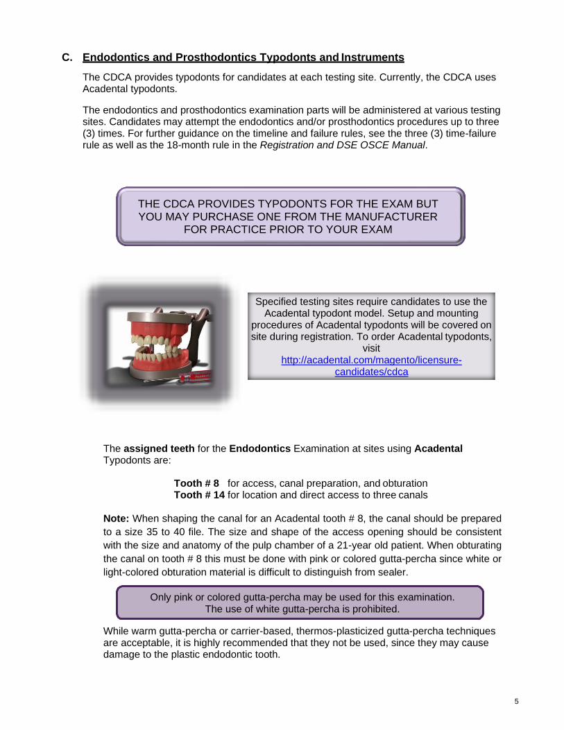
C. Endodontics and Prosthodontics Typodonts and Instruments
The CDCA provides typodonts for candidates at each testing site. Currently, the CDCA usesAcadental typodonts.
The endodontics and prosthodontics examination parts will be administered at various testing sites. Candidates may attempt the endodontics and/or prosthodontics procedures up to three (3) times. For further guidance on the timeline and failure rules, see the three (3) time-failurerule as well as the 18-month rule in the Registration and DSE OSCE Manual.
The assigned teeth for the Endodontics Examination at sites using Acadental Typodonts are:
Tooth # 8 for access, canal preparation, and obturation Tooth # 14 for location and direct access to three canals
Note: When shaping the canal for an Acadental tooth # 8, the canal should be prepared
to a size 35 to 40 file. The size and shape of the access opening should be consistent
with the size and anatomy of the pulp chamber of a 21-year old patient. When obturating
the canal on tooth # 8 this must be done with pink or colored gutta-percha since white or
light-colored obturation material is difficult to distinguish from sealer.
While warm gutta-percha or carrier-based, thermos-plasticized gutta-percha techniques are acceptable, it is highly recommended that they not be used, since they may cause damage to the plastic endodontic tooth.

6
Parallel Preparations and Line of Draw: The two bridge abutment preparations must be parallel and allow a line of draw.
The assigned teeth for the Prosthodontics Examination at sites using the Acadental Typodonts are:
Tooth # 9 for preparation for an all-ceramic crown Tooth # 3 for preparation for a cast metal bridge abutment crown Tooth # 5 for preparation for a porcelain fused to metal bridge abutment
crown
D. Examination Schedule Guidelines
1. Dates and Sites
Specific examination dates for a participating dental school can be found on the CDCA website. Please refer to the Registration and General Administration Supplement manual for the CDCA’s specific policies and administrative guidelines.
The CDCA administers the endodontic and prosthodontic manikin-based examination parts at various dental schools on specified dates as determined by the dean or other official representative of the dental school and agreed upon by the CDCA.
In the event there are extenuating circumstances such as weather, acts of God, or other unforeseen circumstances which may impact or alter the schedule and administration of the examination(s), CDCA will make every attempt to contact candidates with updated information.
2. Timely Arrival
Candidates are responsible for determining their travel and time schedules to ensure they can meet all of the CDCA’s time requirements. All candidates are expected to arrive at the examination site at their designated time. Failure to follow this guideline may result in failure of the examination.
Candidates will be informed in their online candidate profiles as to the date on which they are to challenge each part of the examination. Candidates should note that the manikin- based examination procedures have specific time restraints, and all procedures for each examination must be completed within the allotted time for that part. Examination schedules are not finalized until after the examination application deadline.
7
In order to be granted entrance to the candidate orientation session, you must bring the following:
a. Two forms of identification: one ID must be a photo ID, and both IDs must have thecandidate’s signature. Acceptable forms include such documents as current, valid driver’slicense, passport, military ID, official school ID. A voter registration card (signed) or a creditcard (signed) may be used as a second ID.
b. Proof of your candidate sequential number by bringing with you your registrationconfirmation (available in your online candidate profile). The photo candidate ID badgeyou receive at the candidate orientation session is your admission badge to theexamination day. The candidate ID badge must be worn at all times on your outermostgarment during the course of the examination.
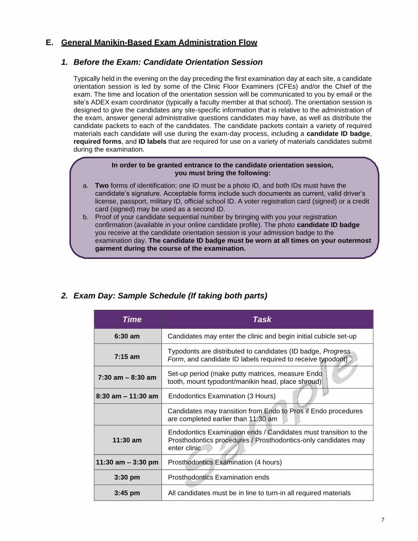
E. General Manikin-Based Exam Administration Flow
1. Before the Exam: Candidate Orientation Session
Typically held in the evening on the day preceding the first examination day at each site, a candidateorientation session is led by some of the Clinic Floor Examiners (CFEs) and/or the Chief of theexam. The time and location of the orientation session will be communicated to you by email or thesite’s ADEX exam coordinator (typically a faculty member at that school). The orientation session isdesigned to give the candidates any site-specific information that is relative to the administration ofthe exam, answer general administrative questions candidates may have, as well as distribute thecandidate packets to each of the candidates. The candidate packets contain a variety of requiredmaterials each candidate will use during the exam-day process, including a candidate ID badge,required forms, and ID labels that are required for use on a variety of materials candidates submitduring the examination.
2. Exam Day: Sample Schedule (If taking both parts)
Time Task
6:30 am Candidates may enter the clinic and begin initial cubicle set-up
7:15 am Typodonts are distributed to candidates (ID badge, Progress Form, and candidate ID labels required to receive typodont)
7:30 am – 8:30 am Set-up period (make putty matrices, measure Endo tooth, mount typodont/manikin head, place shroud)
8:30 am – 11:30 am Endodontics Examination (3 Hours)
Candidates may transition from Endo to Pros if Endo procedures are completed earlier than 11:30 am
11:30 am Endodontics Examination ends / Candidates must transition to the Prosthodontics procedures / Prosthodontics-only candidates may enter clinic
11:30 am – 3:30 pm Prosthodontics Examination (4 hours)
3:30 pm Prosthodontics Examination ends
3:45 pm All candidates must be in line to turn-in all required materials

8
Auxiliary personnel and/or laboratory technicians are not permitted to assist a candidate during the Endodontics or Prosthodontics clinical examination procedures. Violation may result in failure of these examination parts.
An examiner will check each mounting to verify that it is correct. Should an examiner encounter an issue with fashioning a proper occlusal scheme, he/she should contact the Chief immediately.
3. Exam Day: Candidate Flow
Prior to candidate arrival, Clinic Floor Examiners (CFEs) will arrive to the simulation laboratory no later than 6:00 am on the day of the endodontic procedure to assist in setting up. Candidates may enter the clinic or simulation lab used for the examination at 6:30am. Cubicle/work station assignments will be posted in a prominent location in the clinic or simulation lab being used for the exam. Upon arrival at 6:30am, candidates should locate their assigned cubicle/work station and proceed to begin set-up:
a. Arrival to Cubicle/Workstation
Remove the following materials from the white envelope you received at orientation:
• Cubicle card: you will only need ONE card for this examination. Either separate them orfold the cubicle card sheet in half. Write your cubicle number in the appropriate space onthe cubicle card, and then tape it in a prominent place in your cubicle to identify your location
• Candidate ID badge and plastic badge holder: Place the ID badge on your clinic gownor the outermost garment you are wearing for the examination. Candidate ID badgesMUST be clearly visible at all times during the course of the examination
• Candidate ID labels: full sheet of candidate ID labels
• Progress Form: This form will be used for both the Endodontic and Prosthodontic parts.CFEs will use it to track and document your progress. CFEs must verify at specific steps inthe exam process that you received their permission to proceed to the next step. You willuse the Progress Form to record the length of your anterior endodontic tooth, and you mayalso use the “comments” section of the Progress Form to communicate any information tothe examiners that you believe may affect the evaluation of your performance. For example,if your typodont has pre-existing defects on teeth or simulated gingival tissues, you shoulddocument the nature, size, and location of the defect(s) in the “comments” section andrequest that a CFE verify that you made the comments as well as the time at which youmade the comments (Note: Examiners will only consider candidate comments that havebeen verified by a CFE on the Progress Form). Complete the following initial tasks in order:
i. Fill in the cubicle number box on the upper right side ofthe form with YOUR cubicle number (also write on yourcubicle card)
ii. Fill in the typodont number box on the upper right sideof the form with YOUR typodont number
iii. Place one candidate ID label in the space provided (topright of the form)
b. Mounting Manikins
Instructions will be provided regarding any necessary adjustments and mechanisms specific tothe test site to affect those adjustments to create a best fit occlusion.
9
A CFE must be notified immediately in case of equipment failure; he/she will contact school maintenance personnel. The malfunction must be corrected, or
the candidate relocated
Candidates may NOT remove the typodont from the manikin head, loosen it, or re-tighten it after 8:30 am without a CFE present
Isolation dam clamps on the tooth undergoing treatment may loosen or damage the tooth. Clamp adjacent teeth, or use alternate methods—such as ligation—to
secure the isolation dam
Only assigned teeth may be treated. If a candidate begins a procedure on the wrong tooth, he/she must notify a CFE immediately and the Endodontics exam
will be stopped. Please note that the Endodontics and Prosthodontics procedures are scored independently.
c. Set-up Period
• CFEs will circulate to loosen Endo tooth #8 retention screw (Candidates may NOT loosenscrew)
• Candidates measure length of Endo tooth (incisal edge to apex) with CFE present and thenrecord that measurement on the Progress Form
• CFE re-tightens tooth in typodont and then annotates the Progress Form
• Candidates make putty matrices/reduction guides (Candidates are NOT authorized tobring pre- made reduction guides, pre-made putty matrices, pre-made impressions,overlays, clear plastic shells, models, or pre-preparations; use of gloves is not required)
d. 8:30 am: Endodontics Treatment of Simulated Patient Begins
The Chief or a CFE will announce the beginning of the examination at 8:30am, and the candidate may perform the endodontic procedures in any order (separate isolation dam required for each procedure). CFEs will monitor the progress of all candidates and should confirm that all candidates have their manikin heads mounted, properly articulated, shrouds are properly placed, and the manikin heads are ready to undergo treatment. Once the examination has begun, the manikin will-from that point forward-be considered a “live” patient and candidates must observe and maintain all infection control guidelines and barrier controls. Failure to do so is a violation of examination protocol and will result in a penalty being assessed to the candidate. Repeated failure to observe appropriate infection/barrier control protocols will result in failure of the examination.
If you have completed both Endodontics procedures to your satisfaction prior to the end time (11:30 am), you may request a CFE to help you either transition to the Prosthodontics procedures or grant you permission to clean up your cubicle/workstation and turn in the required materials and paperwork prior to exiting. Once you transition to the Prosthodontics procedures, you may not return to any endodontic procedure. The CFE will record your finish time for the Prosthodontics procedures on your Progress Form, which will be four (4) hours from the time you have officially started the Prosthodontics exam. The CFEs also keep a log of all finish times during the course of the exam.

10
CFEs will be checking and collecting all required materials at a central location on the clinic floor. Candidates must bring the following to the check-out station:
• White envelope (received at Candidate Orientation)
• Remaining Candidate ID labels
• Two (2) cubicle cards
• Progress Form
• Any models, impressions, or reduction guides made during theexamination
• Typodont and Typodont box containing: saved wing nut and bolt; tan- colored tag; bubble wrap pouch
e. 11:30 am: All candidates must transition to Prosthodontics Procedures
• The Chief or CFE will announce the end of the Endodontics treatment, and all candidateswho have not yet transitioned to the Prosthodontics exam must stop immediately, request aCFE to help them, and ask the CFE to record the appropriate entries on their Progress Form.Once the CFE has granted the candidate permission to proceed, the candidate may beginthe Prosthodontics procedures in any order, as well as alternate between procedureswithout permission
• Impressions: Candidates may make impressions and pour models for the purpose ofverifying preparations (ie: parallelism for the bridge preps or for an undercut on any prep).Models and impressions MUST be submitted to a CFE at the completion of the exam. Allmodel pouring must be performed in the designated location. Candidates may not removeany examination materials from the Clinic Floor, except impressions to be poured in thelaboratory, and they do not need permission to go to the laboratory
f. 3:30 pm: End of Prosthodontics Treatment Examination
• At 3:30pm, all candidates must stop working immediately and step away from their manikins
• Candidates should request a CFE to grant them permission to dismount the typodont (nomore alterations of teeth may be performed)
• Once dismounted, typodonts should be cleaned with water, soap, and a brush/cotton. Rinseall soap off and then dry the typodont thoroughly with towels and an air syringe.
• Candidates must be in line to turn in all required materials (see list below) no later than 3:45pm.
g. Exam Check-out
If you wish to take a break after the Endodontics section and prior to beginning the Prosthodontics procedures, do so before requesting a CFE to officially start the
Prosthodontic procedures. Otherwise, you will lose valuable time.
11
Adheres to Criteria: The treatment is of acceptable quality, demonstrating competence in clinical judgment, knowledge and skill.
Marginally Substandard: The treatment is of marginal quality, demonstrating less than expected clinical judgment, knowledge or skill.
Critically Deficient: The treatment is of unacceptable quality, demonstrating critical areas of incompetence in clinical judgment, knowledge or skill.
*3-SUB rule: If examiners confirm 3 marginally substandard over-preparation
criteria on the same procedure, then the procedure will be determined to be
critically deficient and the candidate will fail that procedure. SUB criteria that are
part of this rule have been highlighted in yellow on the criteria sheets beginning
on page 23.
To pass the ADEX Dental Examination, you must score 75 or higher on all
procedures. State statutes have set 75 as the minimum passing score and
the CDCA is not permitted to round up or accept any score less than 75.
1) The CFE at the check-out station will inspect the typodont and ask you for your sheet ofcandidate ID labels
2) Acadental typodonts:
a. CFE will attach a candidate ID label to the tan-colored tag, and then attach it tothe typodont
b. CFE will attach wing nut and bolt as well, to prevent damage during shipment
c. CFE will place the typodont in the bubble wrap pouch and put it in the typodontbox along with the Progress Form.
3) Candidate will clean cubicle/workstation and exit the clinic.
F. Scoring Overview and Scoring Content
Dental Boards throughout the U.S. have worked together through ADEX to draft and refine the performance criteria for each procedure in this examination. For the majority of those criteria, gradations of competence are described across a 3-level rating scale. Those criteria appear in the manual and are the basis for the evaluation system. The three rating levels as follows:

12
1. Section II: Endodontics Content
Endodontics Examination – 100 points
CONTENT FORMAT
1. Access opening on a first molar Performed on a manikin
2. Access opening, canal instrumentation, Time:
obturation (tooth #8) 3 hours
Endodontics Examination Content – 100 Points
The Endodontics Examination is a manikin-based examination consisting of three procedures:
1. Access opening and identification of canals on a posterior typodont tooth (6 criteria)
2. Access opening canal instrumentation, and obturation of an anterior typodont tooth
(10 criteria)
2. Section III: Fixed Prosthodontics Content
Fixed Prosthodontics Examination – 100 points
CONTENT FORMAT
1. Preparation – PFM crown asone 3-unit bridge abutment
Performed on a manikin
on a bicuspid 2. Preparation – Full cast metal Time:
crown on a first molar as 4 hours the other abutment for the same 3-unit bridge
3. Preparation – Ceramic crown
(tooth #9)
Fixed Prosthodontics Content – 100 Points
The Prosthodontics Examination is a manikin-based examination consisting of three procedures completed on artificial teeth:
1. Cast metal crown preparation as a posterior abutment for a 3-unit bridge (12 criteria)
2. Porcelain-fused-to-metal crown preparation as an anterior abutment for the same3-unit bridge, plus an evaluation of the line of draw for the bridge abutmentpreparations (12 criteria)
3. All-ceramic crown preparation on an anterior central incisor (12 criteria)

13
G. Penalties
The integrity of the examination process depends on fairness, accuracy, andconsistency. Penalties are assessed proportionally for violations of examinationstandards for certain procedural errors as described below:
• Any of the following may result in a deduction of points from the score of the entireexamination procedure or dismissal from the examination:
o Improper/incomplete record keeping
o Improper treatment selection
o Improper/inadequate isolation
o Improper retraction of simulated facial tissue
o Removing or dismantling the teeth, typodont, or manikin without authorization from aCFE
o Violation of universal precautions, infection control or disease barrier technique orfailure to dispose of potentially infectious materials and clean the operatory afterindividual examination sections
o Poor patient management and/or disregard for simulated patient welfare or comfort
o Improper operator/patient/manikin position (The manikin must be mounted andmaintained in a physiologically acceptable operating position while performing theFixed Endodontics and Prosthodontics Clinical Examination procedures. The facialshroud must be maintained in the same position as the normal facial tissue)
o Unprofessional demeanor: unkempt, unclean or unprofessional appearance;inconsiderate or uncooperative behavior with other candidates, examiners, or anymember of the exam team
• The following will result in the loss of all points for an individual examination:
o Performing treatment procedures other than those assigned
o Performing procedures outside the authorized examination clinic area
o Failure to complete a finished preparation.
o Violation of examination standards, rules or guidelines (treatment may not be initiatedprior to the established starting time, and treatment must be completed within theallotted time for each procedure)
o Use of prohibited electronic devices in the designated examination area during theexam (ie: cell phones, pagers, computers, cameras, smart watches, recordingdevices, etc.)
o Treatment of teeth other than those approved or assigned by examiners (once aprocedure has been started, the procedure must be carried to completion on theassigned tooth/teeth with no substitutions permitted; if a candidate discovers that thewrong tooth has been prepared, he/she must immediately contact the CFE, andwhichever procedure is in progress will be stopped)
o Critical lack of clinical judgement
o Non-compliance with anonymity requirements
This listing is not exhaustive, and penalties may be applied for errors not specifically listed, since some procedures will be classified as unsatisfactory for other reasons or for a combination of several deficiencies.

14
The ADEX Dental Examination Series:
Manikin Procedures
II. Standards of Conductand Infection Control
▪ Standards of Conduct
▪ Infection Control

15
A. Standards of Conduct
All candidates are required to adhere to the rules, regulations and standards of conduct for the ADEX Dental Examination Series. Only the candidate manual, or portions thereof, are permitted in the clinic area. No other reference material is allowed. Candidate notes written in the manual are acceptable. Penalties may be assessed for violation of examination standards and/or for certain procedural errors, as defined and further described below:
1. Unethical personal/professional conduct: Falsification or intentional misrepresentationof registration requirements, dishonesty, collusion, receiving unauthorized assistance,misappropriation of equipment (theft), alteration of examination records, or a candidate’sfailure to follow the instructions of the chief examiner or CFEs will automatically result infailure of all five examination sections. All candidates are expected to behave in anethical and proper manner. Manikins (acting as substitute patients) shall be treated withproper concern for their safety and comfort. Improper behavior is cause for dismissalfrom the examination at the discretion of the chief examiner and will result in failure ofthe examination. Additionally, the candidate shall be denied re-examination through anytesting agency who administers the ADEX dental licensure exam for one full year fromthe time of the infraction.
2. Termination of the examinations: The CDCA reserves the right to delay or terminatethe exam at any time if the candidate or examiners are threatened in any manner orother interfering events occur that are beyond the CDCA’s control.
3. Completion of the examinations: Examination procedures performed outside theassigned time will be considered incomplete, and the candidate will fail theexamination part.
4. Misappropriation (theft) and/or damage of equipment: No equipment, instruments,or materials shall be removed from the examination site without written permission of theowner. Willful or careless damage of dental equipment, typodonts, manikins or shroudsmay result in failure. All resulting repair or replacement costs will be charged to thecandidate and must be paid to the host site before the candidate’s examination resultswill be released.
5. Assigned procedures: Only the treatment and/or procedures assigned may beperformed. Performing other treatment or procedures may result in failure of theexamination.
6. Electronic equipment: The use of any electronic equipment is prohibited on the clinicfloor by candidates, auxiliaries, or patients during the examination. Any such use willbe considered unprofessional conduct and may result in dismissal from theexamination. The use of electronic recording devices or cameras by the candidate, anauxiliary, or a patient during any part of the examination is a violation of examinationguidelines and may result in failure of the entire ADEX Dental Examination Series.However, intra-oral photographs may be taken by authorized examiners or schoolpersonnel during the course of the examination for the purpose of future examinerstandardization and calibration.
The ADEX examination strives to evaluate the candidate’s clinical judgment and skills in a fair manner. In addition, conduct, decorum, and professional demeanor are evaluated. Professional misconduct is a most serious violation of examination guidelines. Substantiated evidence of professional misconduct during the course of the examination will result in automatic failure of the entire examination series. In addition, there will be no refund of examination fees and the candidate may not be allowed to re-apply for the ADEX exam for a period of one year from the time of the infraction.

16
B. Infection Control Requirements
Although this is a simulated patient examination, all candidates must comply with and follow the current recommended infection control procedures as published by the Centers for Disease Control and Prevention once the examination treatment time officially begins (8:30 am). Infection control procedure compliance begins with the initial set-up of the unit, continues throughout the Endodontics and Fixed Prosthodontics clinical examination procedures, and includes the final clean-up of the operatory. It is the candidate’s responsibility to fully comply with these procedures, as failure to do so will result in a loss of points, and any violation that could lead to direct patient harm will result in failure of the examination.
As much as is possible, dental professionals must help prevent the spread of infectious diseases. Because many infectious patients are asymptomatic, all patients must be treated as if they are, in fact, contagious. The use of barrier techniques, disposables whenever possible, and proper disinfection and sterilization procedures are essential. Candidates must adhere to the following infection control guidelines:
1. Barrier protection
• Gloves must be worn while setting up or performing any intra-oral procedures and whencleaning up after any treatment; if rips or tears occur, don new gloves; do not wear glovesoutside the operatory
• Wash and dry hands between procedures and whenever gloves are changed; do not wearhand jewelry that can tear or puncture gloves
• Wear clean, long-sleeved, closed neck uniforms, gowns, or laboratory coats, and changethem if they become visibly soiled; remove gowns or laboratory coats before leaving theclinic area at any point; wear facemasks and protective eyewear during all procedures inwhich splashing of any body fluids could occur during actual patient care; discard masksafter each patient (or sooner if the masks become damp or soiled)
• Do not wear sandals or open-toed shoes
• Cover surfaces that may become contaminated with impervious-backed paper, aluminumfoil or plastic wrap; remove these coverings (while gloved), discard them, and replace thembetween procedures (after removing gloves)
2. Sterilization and Disinfection
• Instruments that become contaminated must be placed in an appropriate receptacleand identified as contaminated.
• Instruments do not need to be sterilized for this simulated examination but could beprovided in an equivalent sterilization bag. However, once the examination has begun, allCDC infection control guidelines must be followed.
• If not barrier wrapped, surfaces and counter tops must be pre-cleaned and disinfected witha site-approved tuberculocidal hospital-level disinfectant
ONE EXCEPTION: candidates are not required to maintain protective eyewear on the manikin during manikin procedures. Infection control will be monitored by the CFEs. The Endodontics and Prosthodontics procedures are considered to be on the same patient, so it is not necessary to sanitize the operatory or sterilize instruments between these procedures.

17
• Handpieces, prophy angles, and air/water syringes must be sterilized before and after useor properly disposed of after use
• Used sharps are to be placed in a spill-proof, puncture- resistant container; needles are tobe recapped with a one- handed method or with special devices designed to preventneedle-stick injuries and disposed of properly
• Candidates must follow any additional infection control requirements established by school orstate authorities during the examination.
• All waste and disposable items must be considered potentially infectious and shall bedisposed of in accordance with federal, state, and local regulations

18
The ADEX Dental Examination Series:
Manikin Procedures
III. Examination Content▪ Endodontics Examination Procedures
▪ Prosthodontics Examination Procedures
▪ Anterior Endodontic Preparation
▪ Posterior Endodontic Preparation
▪ Porcelain Fused to Metal Crown Preparation(PFM Crown)
▪ Cast Metal Crown
▪ Ceramic Crown

19
A. Endodontics Examination Procedures
During the Endodontics Examination, each candidate will perform:
• An access opening on a posterior tooth (#14). Candidates must achieve direct access to allthree canals.
• An access opening, canal instrumentation and obturation on an anterior tooth (#8). Tooth #8
is considered to have a normal size pulp chamber for a 21-year old. The size, shape, and
extent of the prepared access opening should reflect such anatomy and will be graded
accordingly. Canal instrumentation to a minimum size equivalent with a 35-40 file on tooth
#8 will be required prior to obturation.
1. Important Notes About the Endodontics Examination
Radiographs: Since the tooth length is directly measuredprior to the procedure, no radiographs are utilized beforeor after treatment.
Isolation dam: A separate isolation dam is required for each endodonticprocedure, and the dam must be placed prior to beginning accesspreparation. Using separate isolation dams most closely simulates theapproved clinical protocols in that it would reduce the potential of crosscontamination between the two teeth and ease the radiographs takingprocess on an actual patient. An isolation dam clamp should not beplaced on the teeth on which the endodontic procedures areperformed, as doing so may cause the crown to separate from the root. Clamping ofadjacent teeth or ligation is acceptable. All treatment must be done with the dam in place.
Instruments: Other than the instruments and materials provided by the testing site, thecandidates are responsible for providing the instruments, files, and materials of their choice.Rotary instruments are acceptable.
Crown Fractures: If the anterior endodontic tooth crown fractures during filling, contact a CFEimmediately.
Reference point: The cemento-enamel junction (CEJ) on the facial surface should be usedas the reference point to determine the fill depth in the pulp chamber.
Filling material: No temporary filling material, cotton pellet or restorative material should beplaced in the pulp chamber.
Marking Teeth: Teeth may be marked on the typodonts prior to treatment as a precautionagainst preparing the wrong tooth. However, they may only be marked after the typodont hasbeen mounted, then approved by a CFE, and the treatment-time portion of the examination hasbegun. The marking must be done intra-orally and must follow CDC guidelines (barrier-wrappedwriting instrument). Teeth may be marked prior to placing the isolation dam.
Gutta-Percha: Only pink or colored gutta-percha should be used for the examination. Theuse of white gutta-percha is strongly discouraged. While warm gutta-percha or carrier- based, thermo-plasticized gutta-percha techniques are acceptable, it is highlyrecommended they not be used since they may cause damage to the plastic endodontictooth.

20
2. Summary of Endodontics Start:
Between 7:30 – 8:30 am, CFEs will oversee the setup of typodonts and manikinheads. By 8:30 am candidates must have in their possession all necessaryinstruments and materials to begin the Endodontics examination. Candidates shouldensure that:
a. The tooth for the endodontic fill has been measured and secured in the typodontb. The manikin head is properly assembled; andc. Any defective equipment or materials have been identified and corrected or
replaced. Candidates may NOT begin the endodontics examination until instructedby the CFEs.
Upon completing the endodontics procedures, candidates should request a Clinic Floor Examiner (CFE) who will confirm the above by entering the appropriate information on the Progress Form and, if you are ready to begin, will provide a start time for the Prosthodontic Examination.
B. Prosthodontics Examination Procedures
During the Prosthodontics Examination, each candidate will perform:
• Preparation for a PFM crown as one 3-unit bridge abutment (#5)
• Preparation for a full cast crown (#3) as the other abutment for the same 3-unit bridge – bothpreps must be parallel
• Preparation for a ceramic crown (#9)
1. Important Notes about the Prosthodontics Examination
Air/Water spray: Water spray is not recommended. However, if water spray is utilized, amechanism to collect and remove the water must be in place.
Parallel preparation and line of draw: The two bridge abutment preparations must beparallel and allow a line of draw.
Patient simulation: The correct patient/operator position must be maintained whileoperating. Throughout the manikin procedures, the treatment process will be observed byCFEs and evaluated as if the manikin were a patient. Manikins are not required to wearprotective eyewear but are subject to the same treatment standards as a patient. Thefacial shroud may not be displaced other than with those retracting methods that wouldbe reasonable for a patient’s facial tissue.
Security requirements: No written materials may be in the operating area other than acopy of the candidate manual or parts thereof, notes written on these copies, and pertinentexamination forms.

21
Prohibited materials: Impressions, registration, overlays, pre-made putty matrices, clear plastic shells, models, extra teeth, or pre-preparations are not permitted to be brought to the examination site. Failure to follow these requirements will result in confiscation of the materials as well as dismissal from and failure of the examination.
Isolation dam: No isolation dam is required for the crown preparations.
Fractures: If the crown fractures any time before treatment is complete, contact a CFE immediately.
Margins: If the simulated gingival margin is recessed below the CEJ, prepare the margins to within 0.5 mm of the supra-gingival CEJ. The lingual margin for the porcelain-fused-to- metal crown should be prepared for a metal margin, 0.5 mm.
Occlusal reduction: The tooth for the PFM crown should be prepared for a porcelain occlusal surface with an optimal occlusal reduction of 2 mm. For the full cast metal crown preparation, the occlusal reduction is optimally 1.5 mm.
Equilibration prohibited: No equilibration is permitted on the typodont.
Crown Preparation Reduction Guide (putty matrix): Putty matrices or reduction guides must be fabricated during the setup time or (using full infection control procedures) once the Prosthodontic Exam has begun. Two putty matrices are to be fabricated for the ceramic crown and two for the combination of the cast metal crown and PFM preps. One of each of the sets of putty matrices is to be sectioned mesio-distally and one facio-lingually over each tooth to be prepared. This may be done without the use of gloves prior to typodont mounting. Other impressions may be taken during the exam using the CDC infection control procedures. The reduction guides or putty matrices must be placed into the typodont box with the typodont at the end of the examination. All other impressions, casts, or models must also be turned in.
Taper: To taper is defined as to gradually become narrower in one direction. For the purposes of this examination the requirements for tapering are illustrated below:
Adheres to Criteria Marginally Substandard
Taper greater than 16 Degrees is considered Critically Deficient
The lingual margin on the porcelain-fused-to-metal crown preparation should be prepared to receive a metal margin. The transition from the facial shoulder to the lingual margin should begin to occur at the interproximal-buccal line angles.

22
2. Prosthodontics exam flow
Before the start of the examination, candidates must have in their possession allnecessary instruments and materials to begin the prosthodontics examination.
At 11:30 am, the fixed-prosthodontics treatment begins for candidates who arechallenging both the Endodontics and Prosthodontics parts during the exam day. Thereis no extension of time due to starting treatment after 11:30 am for these candidates andthey MUST complete the prosthodontics examination by 3:30 pm. Candidates taking onlythe Prosthodontic examination may start as early as 8:30 am but will still have just 4hours to complete the examination.
When the candidate has finished the prosthodontic portion of the examination, thecandidate must first obtain permission from the Clinic Floor Examiner (CFE) to dismantlethe typodont. A CFE will come to the candidate’s clinic area, oversee the dismantling ofthe typodont, and assist the candidate in submitting the typodonts at the designatedcheck-out station.
When the candidate has finished all procedures, the candidate should request a ClinicFloor Examiner (CFE) who will oversee the dismantling of the typodont and assist thecandidate in submitting the carrier trays and typodonts at the check-out station.

ANTERIOR ENDODONTIC PROCEDURE
CRITICAL ERRORSWrong tooth/surface treated
The crown of the tooth has been reducedYesNo YesNo
Any part of the tooth is fractured during instrumenta on or obtura on YesNo Any part of the tooth is perforated YesNo
Procedure not challenged YesNo
ADEX 2021
21
ACCESS OPENINGPlacement
Placement of the access opening is on the lingual surface directly over the pulp chamber and allows for pulp horns to be fully removed and complete debridement of the pulp chamber and straight-line access to the root canal system.ACC
Placement of the access opening is NOT over the pulp chamber and/or does NOT allow complete debridement of the pulp chamber or access to the root canal system.DEF
ACC = Adheres to Criteria SUB = Marginally Substandard DEF = Cri cal Defi ciency
A. The incisal aspect of the access opening is ≥ 2.0 mm from the incisal edge which provides for a fully supported incisal edge.B. The cervical aspect of the access opening is ≥ 3.0 mm from the lingual CEJ which provides for a fully supported cingulum.C. The widest por on of the prepara on mesio-distally is ≤ ⁄ of the lingual surface which provides for fully supported marginal
ridges (approximately 1.0 mm).D. Size of the access opening allows for complete removal of pulp horns.
ACC
A. The incisal aspect of the access opening is < 2.0 mm from the incisal edge which compromises the incisal edge.B. The cervical aspect of the opening is < 3.0 mm from the lingual CEJ which compromises the cingulum.C. The prepara on compromises the mesial and/or distal marginal ridge(s) (≤ 1.0 mm).D. The size of the access opening does NOT allow for removal of pulp horns.
DEF
Internal Form
DEF Internal form exhibits excessive gouges, which compromise the integrity of the tooth.From the lingual surface to the cervical por on, the internal form tapers to the canal opening with slight ledges.ACC
CANAL INSTRUMENTATIONCervical Por on
DEF Cervical por on of the canal is grossly over-prepared aff ec ng the integrity of the tooth structure.
Canal is shaped to a con nuous taper to allow adequate debridement and obtura on and the cervical por on of the canal is of appropriate loca on and size to allow access to the apical root canal system.ACC
Mid-Root Por on
DEF Mid-root por on of the canal has signifi cant instrumenta on irregulari es that will compromise obtura on.Mid-root por on of the canal blends smoothly with the cervical por on without ledges or shoulders.ACC
Size
Apical Por onApical por on of the canal is prepared to the anatomical apex of the tooth or ≤ 3.0 mm short of the anatomical apex.ACC A. Apical por on of the canal is over-prepared beyond the anatomical apex.B. Apical por on of the canal is transported to the extent that the apical por on of the canal is not instrumented.C. Apical por on is under-prepared > 3.0 mm short of the anatomical apex.
DEF
23
Anterior Endodon c Procedure - con nued
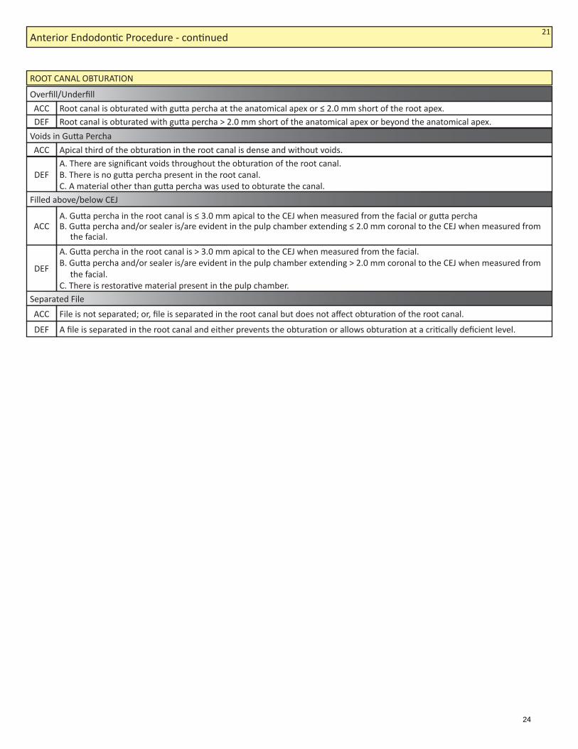
ROOT CANAL OBTURATION
Overfi ll/Underfi ll
Root canal is obturated with gu a percha > 2.0 mm short of the anatomical apex or beyond the anatomical apex.DEFRoot canal is obturated with gu a percha at the anatomical apex or ≤ 2.0 mm short of the root apex.ACC
Voids in Gu a Percha Apical third of the obtura on in the root canal is dense and without voids.ACC A. There are signifi cant voids throughout the obtura on of the root canal.B. There is no gu a percha present in the root canal.C. A material other than gu a percha was used to obturate the canal.
DEF
Filled above/below CEJ
A. Gu a percha in the root canal is > 3.0 mm apical to the CEJ when measured from the facial.B. Gu a percha and/or sealer is/are evident in the pulp chamber extending > 2.0 mm coronal to the CEJ when measured from
the facial.C. There is restora ve material present in the pulp chamber.
DEF
A. Gu a percha in the root canal is ≤ 3.0 mm apical to the CEJ when measured from the facial or gu a perchaB. Gu a percha and/or sealer is/are evident in the pulp chamber extending ≤ 2.0 mm coronal to the CEJ when measured from
the facial.ACC
Separated File
DEF A fi le is separated in the root canal and either prevents the obtura on or allows obtura on at a cri cally defi cient level.File is not separated; or, fi le is separated in the root canal but does not aff ect obtura on of the root canal.ACC
21
24

ACCESS OPENINGPlacement
Placement of the access opening is over the pulp chamber allowing for debridement of the pulp chamber and straight-line access to the three root canals located in the tooth.ACC
Placement of the access opening is not over the pulp chamber and/or does not allow complete debridement of the pulp chamber or access to the 3 root canals. DEF
POSTERIOR ENDODONTIC PROCEDURE
ACC = Adheres to Criteria SUB = Marginally Substandard DEF = Cri cal Defi ciency
CRITICAL ERRORS
Size
Access opening is in the mesial triangular pit and central fossa of the tooth and the following are true: A. The mesial extent of the access prepara on is ≥ 2.0 mm from the mesial marginal ridge.B. The buccal extent of the access prepara on is ≥ 1.0 mm from the line bisec ng the mesio-buccal and disto-buccal cusp ps.C. The distal extent of the access opening is ≥ 1.0 mm mesial to the distal oblique groove.D. The palatal extent of the access prepara on is ≥ 1.0 mm from the mesiolingual cusp p.
ACC
Wrong Tooth/surface treatedThe crown of the tooth has been reduced
YesNo YesNo
Any part of the tooth is fractured during instrumenta on or obtura on YesNo Any part of the tooth is perforated YesNo
Procedure not challenged YesNo
DepthThe depth of the access prepara on removes the en re roof of the pulp chamber.ACC
A. The pulpal fl oor at the center of the fl oor is > 10.0 mm deep when measured from the buccal cavosurface margin of theaccess prepara on.
B. The depth of the access prepara on does not remove the roof of the pulp chamber to the extent that all pulp ssue can beremoved.
DEF
ADEX 2021
A. The mesial extent of the access prepara on is < 2.0 mm distal to the mesial marginal ridge.B. The buccal extent of the access prepara on is < 1.0 mm from the line bisec ng the mesio-buccal and disto-buccal cusp ps.C. The distal extent of the access prepara on is < 1.0 mm mesial to the distal oblique groove.D. The palatal extent of the access prepara on is < 1.0 mm from the mesio-lingual cusp p access opening.E. The access size is too small: <2.5 mm at its widest mesio-distally and/or < 2.5 mm at its widest bucco-lingually.
DEF
Internal Form
DEF The internal form of the access prepara on leaves < 1.0 mm of lateral supported tooth structure at any point of the prepara on and/or tapers to the canal orifi ces with gross ledges that will inhibit access to the root canal orifi ces.
The internal form of the access prepara on leaves ≥ 1.0 mm of supported lateral tooth structure at any point of the prepara on and tapers to the canal orifi ces with no or slight gouges. ACC
21
25

TREATMENT MANAGEMENTCondi on of Adjacent Tooth/Teeth
DEF There is gross damage to adjacent tooth/teeth, requiring a restora on.
Any damage to adjacent tooth/teeth can be removed with polishing without adversely altering the shape of the contour and/or contact.ACC
Condi on of Surrounding Tissue
DEF There is gross iatrogenic damage to the simulated gingiva and/or typodont inconsistent with the procedure.There is slight damage to simulated gingiva and/or typodont consistent with the procedure.ACC
Posterior Endodon c Procedure - con nued 21
26

CERVICAL MARGIN AND DRAWMargin/Extension
The cervical margin is ≤ 0.5 mm below to ≤ 1.5 mm above the simulated free gingival margin.
A. The cervical margin is over-extended > 0.5 mm below the simulated free gingival margin.
ACC
SUB
A. The cervical margin is over-extended by > 0.5 mm below the simulated free gingival margin, causing visual damage to thetypodont.
B. The cervical margin is under-extended by > 1.5 mm above the simulated free gingival margin.DEF
Margin/Defi ni on
PFM CROWN PREPARATION
ACC = Adheres to Criteria SUB = Marginally Substandard DEF = Cri cal Defi ciency
CRITICAL ERRORSWrong tooth/surface treated
Procedure not challengedYesNo
YesNo
Line of Draw
The cervical margin is con nuous but may be slightly rough and may lack some defi ni on. The cervical bevel, when used, is ≤ 1.5 mm, and/or may lack some defi ni on.A. The cervical bevel, when used, is > 1.5 mm but ≤ 2.0 mm.
ACC
SUB A. The cervical bevel, when used, is > 2.0 mm.B. The cervical margin has no con nuity or defi ni on.C. The cervical margin is cupped or J-shaped.
DEF
DEF The path of inser on/line of draw deviates 20° to < 30° from the long axis of the tooth. The path of inser on/line of draw deviates ≥ 30° from the long axis of the tooth.
SUB The path of inser on/line of draw deviates < 20° from the long axis of the tooth.ACC
ADEX 2021
Margin/Facial Width
DEF
A. The facial shoulder is reduced > 2.0 mm but ≤ 2.5 mm.A. The facial shoulder is > 2.5 mm in width.B. The facial shoulder is < 0.5 mm in width.
SUB The facial shoulder is > 0.5 mm but ≤ 2.0 mm in width.ACC
Margin/Lingual Width
DEF
A. The lingual margin is > 1.0 mm but ≤ 2.0 mm.A. The lingual margin is > 2.0 mm.B. The lingual margin is feathered and/or is not explorer detectable.
SUB The margin width varies slightly from visually & explorer detectable to ≤ 1.0 mm.ACC
21
Note: those SUBs that are highlighted are part of the 3-SUB rule
27

PFM CROWN PREPARATION (CONTINUED)
Taper
DEF There is excessive taper that is > 12° and ≤ 16° per wall.Taper is grossly over-reduced > 16° per wall.
SUB Taper is present, from nearly parallel to ≤ 12° per wall.ACC
TREATMENT MANAGEMENTCondi on of Adjacent/Opposing Teeth
Any damage to adjacent tooth/teeth can be removed with polishing without adversely altering the shape of the contour and/or contact.ACC
A. Damage to adjacent tooth/teeth requires recontouring that changes the shape and/or posi on of the contact.B. Opposing hard ssue shows minimal evidence of damage and/or altera on inconsistent with the procedure.SUB
A. There is gross damage to adjacent tooth/teeth requiring a restora on.B. There is evidence of gross damage and/or altera on to opposing hard ssue inconsistent with the procedure.DEF
Condi on of Surrounding Tissue
DEF There is iatrogenic damage to the simulated gingiva and/or typodont inconsistent with the procedure.There is gross iatrogenic damage to the simulated gingiva and/or typodont inconsistent with the procedure.
SUB There may be slight damage to simulated gingiva and/or typodont consistent with the procedure.ACC
Occlusal Reduc on
A. Occlusal reduc on is > 2.5 mm but ≤ 3.0 mm.SUB Occlusal reduc on is ≥ 1.0 mm but ≤ 2.5 mm.ACC
A. Occlusal reduc on is > 3.0 mm.B. Occlusal reduc on is < 1.0 mm.DEF
Internal Line Angles
DEF The internal line angles or cusp p areas are excessively sharp with no evidence of rounding.Internal line angles and cusp p areas may not be completely rounded and may show a slight tendency of being sharp.ACC
WALLS, TAPER, AND SHOULDERAxial Tissue Removal
DEF
A. The axial ssue removal is > 2.0 mm but ≤ 2.5 mm.A. The axial ssue removal is > 2.5 mm.B. The axial ssue removal is < 0.5 mm.
SUB The axial ssue removal is ≥ 0.5 mm but ≤ 2.0 mm.ACC
Axial Walls Smoothness/Undercut
DEF There is an undercut, which, when blocked out, would compromise margin width criteria and/or is > 0.5 mm deep.The walls may be slightly rough and may lack some defi ni on.ACC
21
28

CERVICAL MARGIN AND DRAWMargin/Extension
The cervical margin is at the level of or ≤ 1.5 mm occlusal to the simulated free gingival margin.
A. The cervical margin is over-extended > 0.5 mm below crest of the simulated free gingival margin.
ACC
SUB
A. The cervical margin is over-extended > 0.5 mm below the crest of the simulated free gingival margin and causes visual dam- age to the typodont.B. The cervical margin is under-extended > 1.5 mm above the simulated free gingival margin.
DEF
Margin/Defi ni on/Bevel
CAST METAL CROWN PREPARATION
ACC = Adheres to Criteria SUB = Marginally Substandard DEF = Cri cal Defi ciency
CRITICAL ERRORSWrong tooth/surface treated
Procedure not challengedYesNo YesNo
Line of Draw
WALLS, TAPER, AND MARGINAxial Tissue Removal
The cervical margin is con nuous but may be slightly rough and/or may lack some defi ni on. The cervical bevel, when used, is ≤ 1.5 mm and/or may lack some defi ni on.A. The cervical bevel, when used, is > 1.5 mm but ≤ 2.0 mm.B. The cervical bevel, when used, has very poor defi ni on.
ACC
SUB
A. The cervical bevel, when used, is > 2.0 mm in length.B. The cervical margin has no con nuity and/or defi ni on.C. The cervical margin is cupped or J-shaped.
DEF
DEF The path of inser on/line of draw deviates 20° to < 30° from the long axis of the tooth.The path of inser on/line of draw deviates ≥ 30° from the long axis of the tooth.
SUB The path of inser on/line of draw deviates < 20° from the long axis of the tooth.ACC
DEF
A. The axial ssue removal is > 2.0 mm but ≤ 2.5 mm.A. The axial ssue removal is > 2.5 mm.B. The axial ssue removal is < 0.5 mm.
SUB The axial ssue removal is > 0.5 mm but ≤ 2.0 mm.ACC
ADEX 2021
Margin/Width
DEF
The margin width is > 1.0 mm but ≤ 2.0 mm.A. The margin width is > 2.0 mm.B. The margin is not detectable and/or is feathered.
SUB The margin varies slightly in width from visually and explorer detectable to ≤ 1.0 mm.ACC
21
Axial Walls Smoothness/Undercut
DEF There is an undercut, which, when blocked out, would compromise margin width criteria and/or is > 0.5 mm deep.The walls may be slightly rough and may lack some defi ni on.ACC
Note: those SUBs that are highlighted are part of the 3-SUB rule
29

CAST METAL CROWN PREPARATION (CONTINUED)
The taper is grossly over-reduced > 16° per wall.There is excessive taper that is > 12° or ≤ 16°.
Taper
DEF SUB
Taper is present, from nearly parallel to ≤ 12°.ACC
TREATMENT MANAGEMENTCondi on of Adjacent/Opposing Teeth
Any damage to adjacent tooth/teeth can be removed with polishing without adversely altering the shape of the contour and/or contact.ACC
A. Damage to adjacent tooth/teeth requires recontouring that changes the shape and/or posi on of the contact.B. Opposing hard ssue shows minimal evidence of damage and/or altera on inconsistent with the procedure.SUB
A. There is gross damage to adjacent tooth/teeth, requiring a restora on.B. There is evidence of gross damage and/or altera on to opposing hard ssue inconsistent with the procedure.DEF
Condi on of Surrounding Tissue
DEF There is iatrogenic damage to the simulated gingiva and/or typodont inconsistent with the procedure.There is gross iatrogenic damage to the simulated gingiva and/or typodont inconsistent with the procedure.
SUB There may be slight damage to simulated gingiva and/or typodont consistent with the procedure.ACC
Occlusal Reduc on
A. Occlusal reduc on is > 2.0 mm but ≤ 2.5 mm.SUB Occlusal reduc on is ≥ 1.0 mm but ≤ 2.0 mm.ACC
A. Occlusal reduc on is > 2.5 mm.B. Occlusal reduc on is < 1.0 mm.DEF
Internal Line Angles
DEF Internal line angles or cusp p areas are excessively sharp with no evidence of rounding.
Internal line angles and cusp p areas may not be completely rounded and may show a slight tendency of being sharp.ACC
BRIDGE FACTORPath of Inser on/Line of Draw
The line of draw or path of inser on is direct or may require altering the path of inser on from a direct ver cal axis to allow full sea ng.ACC
No line of draw or path of inser on exists through any plane of rota on without the removal of addi onal tooth structure in the apical ⁄ of either/both of the prepara ons.DEF
21
30
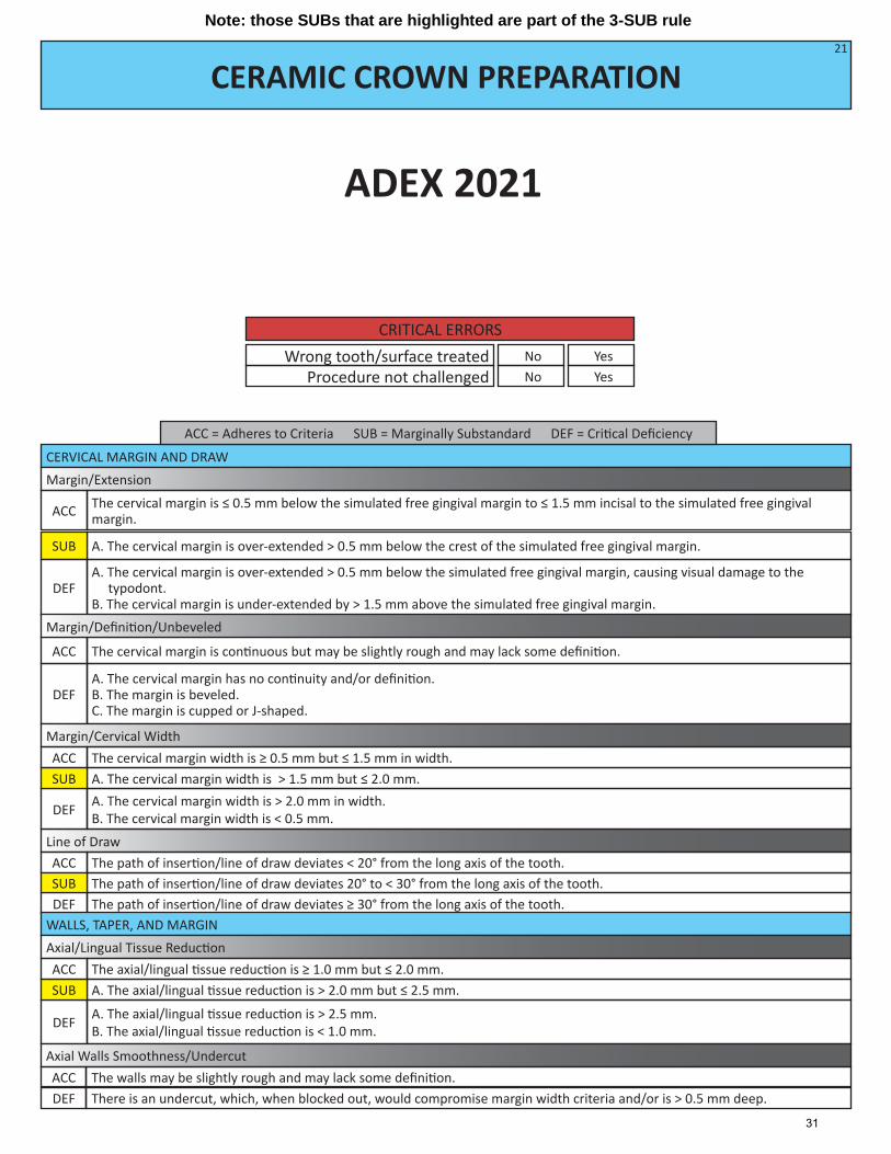
CERVICAL MARGIN AND DRAWMargin/Extension
The cervical margin is ≤ 0.5 mm below the simulated free gingival margin to ≤ 1.5 mm incisal to the simulated free gingival margin.
A. The cervical margin is over-extended > 0.5 mm below the crest of the simulated free gingival margin.
ACC
SUB
The cervical margin is con nuous but may be slightly rough and may lack some defi ni on.ACC
A. The cervical margin is over-extended > 0.5 mm below the simulated free gingival margin, causing visual damage to thetypodont.
B. The cervical margin is under-extended by > 1.5 mm above the simulated free gingival margin.DEF
Margin/Defi ni on/Unbeveled
CERAMIC CROWN PREPARATION
ACC = Adheres to Criteria SUB = Marginally Substandard DEF = Cri cal Defi ciency
CRITICAL ERRORSWrong tooth/surface treated
Procedure not challengedYesNo YesNo
DEF A. The cervical margin has no con nuity and/or defi ni on.B. The margin is beveled.C. The margin is cupped or J-shaped.
Line of DrawThe path of inser on/line of draw deviates < 20° from the long axis of the tooth.ACC
DEF The path of inser on/line of draw deviates 20° to < 30° from the long axis of the tooth.The path of inser on/line of draw deviates ≥ 30° from the long axis of the tooth.
SUB
WALLS, TAPER, AND MARGINAxial/Lingual Tissue Reduc on
DEF
A. The axial/lingual ssue reduc on is > 2.0 mm but ≤ 2.5 mm.A. The axial/lingual ssue reduc on is > 2.5 mm.B. The axial/lingual ssue reduc on is < 1.0 mm.
SUB The axial/lingual ssue reduc on is ≥ 1.0 mm but ≤ 2.0 mm.ACC
Margin/Cervical Width
DEF
A. The cervical margin width is > 1.5 mm but ≤ 2.0 mm.A. The cervical margin width is > 2.0 mm in width.B. The cervical margin width is < 0.5 mm.
SUB The cervical margin width is ≥ 0.5 mm but ≤ 1.5 mm in width.ACC
21
ADEX 2021
Axial Walls Smoothness/Undercut
DEF There is an undercut, which, when blocked out, would compromise margin width criteria and/or is > 0.5 mm deep.The walls may be slightly rough and may lack some defi ni on.ACC
Note: those SUBs that are highlighted are part of the 3-SUB rule
31

CERAMIC CROWN PREPARATION ( CONTINUED)
Taper
DEF There is excessive taper that is > 12° but ≤ 16° per wall.The taper is grossly over-reduced > 16° per wall.
SUB Taper is present, from nearly parallel to ≤ 12° per wall.ACC
Incisal Reduc on
DEF
The incisal reduc on is > 3.0 mm but ≤ 3.5 mm.A. The incisal reduc on is > 3.5 mm.B. The incisal reduc on is < 1.0 mm.
SUB The incisal reduc on is ≥ 1.0 mm but ≤ 3.0 mm.ACC
External/Internal Line Angles
DEF The external and/or internal line angles are excessively sharp with no evidence of rounding.External and/or internal line angles may be rounded but irregular.ACC
Lingual Wall Height
DEF The lingual wall height is < 1.0 mm.The lingual wall height is ≥ 1.0 mm.ACC
TREATMENT MANAGEMENTCondi on of Adjacent/Opposing Teeth
Any damage to adjacent tooth/teeth can be removed with polishing without adversely altering the shape of the contour and/or contact.ACC
A. Damage to adjacent tooth/teeth requires recontouring that changes the shape and/or posi on of the contact.B. Opposing hard ssue shows minimal evidence of damage and/or altera on inconsistent with the procedure.SUB
A. There is gross damage to adjacent tooth/teeth, requiring a restora on.B. There is evidence of gross damage and/or altera on to opposing hard ssue inconsistent with the procedure.DEF
Condi on of Surrounding Tissue
DEF There is iatrogenic damage to the simulated gingiva and/or typodont inconsistent with the procedure.There is gross iatrogenic damage to the simulated gingiva and/or typodont inconsistent with the procedure.
SUB There may be slight damage to the simulated gingiva and/or typodont consistent with the procedure.ACC
21
32
33
The ADEX Dental Examination Series:
Manikin Procedures
IV. Examination Forms
▪ Progress Form

34
A. Progress Form
Progress Forms are utilized to track the candidate’s progress through each procedure, document treatment provided, collect examiner signatures for all completed portions of the examination, and provide appropriate progress notes from the candidate to examiners during the course of treatment.
Candidates will be provided with identification labels to place on each procedure’s Progress Form, as indicated on the form.
The Endodontic and Prosthodontic Examination Progress Form will be collected by the Clinic Floor Examiners once the candidate has completed all procedures for which he/she was registered.
General exam registration forms and registration procedures may be found in the Registration and DSE OSCE Manual.


![Pontics [Fixed Prosthodontics Seminar @AmCoFam]](https://img.pdfslide.us/doc/110x75/5571fe2a49795991699ac64b/pontics-fixed-prosthodontics-seminar-amcofam.jpg)