Embed Size (px)
Citation preview

8
Additive Manufactured Models of Fetuses Built from 3D Ultrasound, Magnetic Resonance Imaging and
Computed Tomography Scan Data
Jorge Lopes Santos1, Heron Werner2, Ricardo Fontes3 and Simone Belmonte4
1Laboratório de Modelos Tridimensionais, Instituto Nacional de Tecnologia (INT), Laboratório de Processamento de Imagens Digitais, Museu Nacional (UFRJ),
Laboratório de Modelagem e Simulação 3D - Departamento de Artes e Design, (PUC-Rio), Rio de Janeiro
2Radiologia, Clinica de Diagnóstico por Imagem (CDPI), Universidade Federal do Rio de Janeiro (UFRJ), Rio de Janeiro
3Laboratório de Modelos Tridimensionais, Instituto Nacional de Tecnologia (INT), Rio de Janeiro
4Laboratório de Modelos Tridimensionais, Instituto Nacional de Tecnologia (INT), Laboratório de Processamento de Imagens Digitais,
Museu Nacional (UFRJ), Rio de Janeiro, Brazil
1. Introduction
The importance of additive manufacturing (AM) in the biomedical sector has been
increasing steadily during the past decade. Different uses of AM models have been reported
widely in the medical scientific literature. This study introduces the use of digital generated
didactic models into fetal medicine, an area where little work has so far been done in
relation to digital 3D modeling. The project proposes a new form of interaction with the idea
of the unborn child during the pregnancy, through physically recreating the interior of the
womb during gestation: the physical appearance, the actual size, and sometimes also the
unexpected event of malformation.
Nowadays, scientific and technological developments in the field of medical imaging
scanning and automated interpretation of imagery are having an increasingly significant
role in healthcare in order to achieve earlier diagnosis through advanced non-invasive
procedures. This integration between medical non invasive imaging systems and additive
manufacturing technologies is currently a very promising area covering a wide range of
medical imaging modalities, and several researches are being carried out, from the high
advances in tissue engineering, to the production of customized implants. Since different
uses have been reported widely in the medical literature but little has been published on its
www.intechopen.com

Rapid Prototyping Technology – Principles and Functional Requirements
180
application to the gravid uterus, so we applied AM technology to fetal images obtained by
3DUS, MRI and CT (Armillotta et al., 2007; Willis et al., 2007; Robiony et al., 2007; Werner et
al., 2010).
Additive manufacturing is the automatic, layer-by-layer construction of physical models
using solid freeform fabrication. The first AM techniques were used in the late 1980s to
produce models and prototypes and classified by different authors into three main
categories, according to the physical state of the materials to be transformed: liquid-based
systems (associated with photosensitive resins), powder- based systems (associated with
sintering or agglutination of grain particles) and solid- based systems (associated with non-
powder formats, such as sheets or thermoplastic extruded filaments).
There are currently several different systems of additive manufacturing technologies on the
market that, although using dissimilar material procedures, are based on the same principle
of manufacturing by layer deposition. One of the most important features of additive
technology is the possibility of manufacturing parts with significant geometrical complexity,
a process which in conventional technologies is more expensive and lengthy, affecting both
the time taken to launch the product commercially and the total costs of production.
Additive systems can typically produce models in a few hours, although this time varies
widely depending on the type of machine being used, the size of the model to be built, the
spatial positioning, the level of accuracy and the number of parts being produced
simultaneously. Nowadays, interesting trends relating to additive equipment are the so-called ‘3D printers’, a market for which is growing rapidly. Being smaller in terms of overall size than the regular equipment, and consequently having a reduced area of construction envelope, these 3D printers also have the potential to become more ‘office friendly’ and also less expensive, in order to be accepted in the same way as the laser and inkjet paper printers in common use in offices.
2. Imaging scanning technologies carried out in pregnancy
Image-scanning technologies have led to vast improvements in medicine, especially in the
diagnosis of fetal anomalies (Steiner et al., 1995; Campbell, 2006). In general, three main
technologies are used to obtain images within the uterus during pregnancy i.e. 3DUS, MRI
and CT. The development of ultrasound scanning during the 1960’s opened a new window
into the study of the fetus. It is currently the primary method of fetal assessment during
pregnancy because it is patient friendly, useful, cost effective and considered to be safe
(Campbell, 2006).
Although there is a history of more than fifty years of Ultrasonography scanning to assist
the diagnostic visualization by two-dimensional methods, enhanced in the late 1980s with
the advent of the 3D visualization through the use of USG 3D1 equipment, the
representation in a physical context presented in this work is a new approach, since it is
focused on three-dimensional physical didactic models of fetuses from the first, second and
third trimesters, created from data files obtained from Ultrasonography 3D, Magnetic
Resonance Imaging and even Computed Tomography scans, together with Additive
manufacturing systems.
1 Von Ramm, O; Smith, S. Three-dimensional imaging system (U.S.Patent 4,694,434. Duke University, 1987)
www.intechopen.com

Additive Manufactured Models of Fetuses Built from 3D Ultrasound, Magnetic Resonance Imaging and Computed Tomography Scan Data
181
Many centers are exploring 3D US because of the life-like images if the fetus it provides
(Blaas et al., 2006; Peralta et al., 2006). MRI is a non-invasive method that has been used in
obstetrics since the 1980s (Smith et al., 1983). It offers high-resolution fetal images with
excellent contrast that allow visualization of internal tissues (Brugger et al., 2006; Jani et al.,
2007; Daltro et al., 2008). When ultrasound yields equivocal results, MRI is generally used,
because it provides additional information about fetal abnormalities and conditions in
situations where ultrasound cannot provide high-quality images (Frates et al., 2004; Prayer
et al., 2006). CT is used only in specific cases of suspected fetal malformation, particularly
those related to the skeleton, because of potential risks associated with radiation exposure to
the fetus. Its use during pregnancy must be adequately justified and its application is
limited to specific pathologies such bone dysplasia, which can, in some cases, be difficult to
diagnose by ultrasound especially in the absence of family history of the disease (Cassart et
al., 2007; Werner et al., 2008).
Fig. A. 34 weeks Physical model of fetus built from CT scan through SLA process (3D systems – VIPER Si).
Fig. B. 34 weeks, 3D virtual and physical models of the skeleton of the fetus built on SLA (3D systems - Viper Si) from CT scan.
www.intechopen.com

Rapid Prototyping Technology – Principles and Functional Requirements
182
Fig. C. Virtual 3D model of skeleton including support structures for the Stereolitography process.
Fig. D. 34 weeks Physical model of fetus built from CT scan through SLA process (3D systems – VIPER Si).
3. Brief history of the use of didactic models in medicine
The early registered attempts to represent the fetus and the interior of the womb during the
gestation, dates from around 1500, and examples of artistic drawings are spread around
museums and private collections throughout the world. Among the artists who achieved a
refined quality in terms of visual representation of fetuses, Leonardo Da Vinci, is
undoubtedly the one who demonstrated through various detailed anatomic studies, the
growing process of the fetus in the womb environment.
The use of didactic physical models in medicine began in Italy from the Renaissance
onwards with the appearance of the colored wax teaching models representing different
parts of the human body with visual realism, including the alterations of the woman body
during the pregnancy period. The important cities regarding artistic and cultural aspects on
that stage were Bologna and Florence, and the diffusion of anatomic models made of wax,
created in order to teach anatomy without having to directly observe a real corpse, was
emphasized through the famous “Florentine school”, an art introduced in Florence by
Ludovico Cigoli (1559-1613).
Among the recognized artists, Clemente Susini (1754-1814) who made the most important
pieces of the collection on the laboratorial facilities of the Museum and the Sicilian sculptor
Gaetano Zumbo (1656-1701), were the skilled wax modelers who contributed to the
development of the medical sciences by carrying out an intensive research on the physical
reproduction of anatomy by the use of wax models.
Many of these didactical models were constructed during the period between
approximately 1550 and 1800 reaching its maximum period of technical and scientific
splendour during the 18th century. Diverse models still can be appreciated in the permanent
exhibition at the Museum of Zoology and Natural History "La Specola" in Florence, which
was officially inaugurated in 1775 and until the early years of the l9th century was the only
scientific museum specifically created for the public. Nowadays the Museum is unique on
its collection of anatomic wax models.
Another interesting initiative happened in France, more specifically on the villages of
Royaume, when around 1778, a very interesting set of hand made didactical models called
www.intechopen.com

Additive Manufactured Models of Fetuses Built from 3D Ultrasound, Magnetic Resonance Imaging and Computed Tomography Scan Data
183
“La machine", made of different types of fabric, were created and produced by Madame Du
Coudray, being its use largely adopted around different villages in order to teach and
disseminate information about the birth process through the stylized models of fetuses and
the mother.
The fetus as a real image of human form appeared in the pioneering work of the
photographer Lennart Nilsson, who started to take pictures of dead fetuses, having his
works published on the “Life magazine” in 1965. Within the years and the technological
advance of fiber optics, Nilsson developed more advanced photographic techniques, being
possible to see very clear bi dimensional images of live fetuses when inside the womb.
Nowadays, it is possible to find several companies that dedicate its production to the
construction of didactical physical models representing all the gestation phases, from the
surge of the embryo to the new born babies, including examples of the normal and healthy
expectance and as well malformations occurred during the pregnancy, possible diseases and
others. These models are intended to be used in medicine schools and related areas as a
visual and tactile aiding information. In some cases, their appearance can have very
impressive characteristics, exhibiting details such as veins, blood vessels and others. The
fabrication processes of these models begin in general with the production of a prototype,
hand made by a specialized modelmaker, which after the approval, will be replicated
through the use of moulds.
Fig. G, H and I. Physical models built on Z Corp 510 developed from Ultrasound 3D scan files obtained in different initial pregnancy periods: twin embryos with 9 weeks in the womb, twin fetuses with 11 weeks in the womb and 13 weeks fetus in the womb.
www.intechopen.com

Rapid Prototyping Technology – Principles and Functional Requirements
184
Fig. J. Virtual 3D model of 35 weeks conjoined twins generated during the CT scanning process
4. Methods and technologies applied on the project
AM was performed after 3DUS, MRI or CT of fetuses at different gestational ages. The
indications for MRI were central nervous system, thoracic, gastrointestinal or genitourinary
malformations, and for CT, skeletal malformations after the period of 30 weeks. The ethical
issues associated with this work were carefully considered. All cases were examined first by
ultrasound imaging. 3DUS scans were performed transvaginally and/or transabdominally
using a high-resolution ultrasound probe with harmonic imaging for all examinations (4-8
MHz transducer, Voluson 730 Pro/Expert system, General Electric, Kretztechnik, Zipf,
Austria). MRI examinations were performed using a 1.5-T scanner (Siemens, Erlangen,
Germany). The protocol consisted of: T2-weighted sequence in the three planes of the fetal
body (HASTE; repetition time, shortest; echo time, 140 ms; field of view, 300–200 mm; 256 ×
256 matrix; slice thickness, 4 mm; acquisition time, 17 s; 40 slices) and (TRUFI; repetition
time, 3.16 ms; echo time, 1.4 ms; field of view, 340 mm; slice thickness, 1,5 mm; acquisition
time, 18s; 96 slices). The entire examination time did not exceed 40 min. CT was performed
using a multislice 64 scanner (Philips, Solingen, Germany) with the following parameters: 40
mA, 120 kV, 64 slices per rotation, pitch 0.75 and slice thickness 0.75 mm. This corresponds
to a mean radiation dose to the fetus of 3.12 mGy (CT dose index weighted). The acquisition
lasted around 20 s and was performed during maternal apnea.
In order to construct physical models from the medical examinations (3DUS, MRI and/or CT), for several researches, the first step was the production of three-dimensional (3D) virtual models. All 3DUS, MRI and CT images were exported to a workstation in Digital
www.intechopen.com

Additive Manufactured Models of Fetuses Built from 3D Ultrasound, Magnetic Resonance Imaging and Computed Tomography Scan Data
185
Imaging and Communications in Medicine (DICOM) format for segmentation done by a 3D modeling technician and supervised by the physician responsible. The 3D structure of the fetus was reconstructed by generating skinning surfaces that joined the resulting profiles. Software that converts medical images into numerical models (Mimics v. 12, Materialize, Leuven, Belgium) was used for 3D virtual model reconstruction, and the model was exported into a Standard triangular language (STL) format and converted into an "OBJ" extension for adjustment using 3D modeling polygonal software (Autodesk Mudbox, San Francisco, USA). Using this software, the volumetric surface was smoothed, to be later compared and analyzed as a topographic construction. After this procedure, the 3D model was again converted and exported as an STL extension. The model file was again opened in Mimics software for correlating the contours of the 3DUS, MR or CT images with the generated 3D surface. The physical modeling process was done through using four different AM technologies (SLA Viper, Objet Connex 350, ZCorp 510 and FDM Vantage). Essentially, the technological processes of medical imaging acquisition and AM systems are very similar in terms of their logical procedure: the idea of acquiring images from medical scans is based on “slicing” the physical body being scanned. Through the capture of several slices (from Ultrasonography, Magnetic Resonance and Computed Tomography) the later construction of a virtual 3D CAD model can be achieved, through the superimposition of those same layers. The AM process begins with the virtual 3D CAD model which is “sliced” in layers in order to later deposit various materials, layer on layer, resulting in a physical 3D model.
Fig. O. Physical model of 34 weeks fetus built on Z Corp 510 from Magnetic Resonance scan.
www.intechopen.com

Rapid Prototyping Technology – Principles and Functional Requirements
186
Fig. P. 11 weeks virtual and physical model of fetus built on Z Corp 510 from Ultrasonography 3D files.
Fig. L. 35 weeks conjoined twins physical models of the skeleton built in SLA (3D systems - Viper Si)
www.intechopen.com

Additive Manufactured Models of Fetuses Built from 3D Ultrasound, Magnetic Resonance Imaging and Computed Tomography Scan Data
187
Fig. N. 32 weeks physical model of fetus built in Objet Connex 350 showing the skeleton and the structure of the uterus (transparent).
Fig. E. 32 weeks fetus built on Z Corp 510 from MRI files – on the left including the uterus and the umbilical cord and on the right only the body of the fetus.
www.intechopen.com

Rapid Prototyping Technology – Principles and Functional Requirements
188
Fig. F. 26 weeks physical model of fetus built in Z Corp 510 from Ultrasonography 3D files.
www.intechopen.com

Additive Manufactured Models of Fetuses Built from 3D Ultrasound, Magnetic Resonance Imaging and Computed Tomography Scan Data
189
5. Results
In this study the main outcomes presented were the possibility to create 3D virtual and physical models from 3DUS, MRI or CT both separately and also in various combinations. AM systems allow the conversion of a 3D virtual model to a physical model in a fast, easy and dimensionally accurate process. The construction process transfers a 3D data file that specifies surfaces and solid internal structures to AM equipment that builds physical models through the superimposition of thin layers of raw materials. The models were remarkably similar to the postnatal appearance of the aborted fetus or newborn baby, especially in cases with pathology.
Fig. K. Slice showing the laser beam during the hardening process of the photo sensible resin on SLA VIPER Si
www.intechopen.com
Rapid Prototyping Technology – Principles and Functional Requirements
190
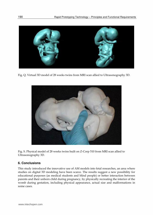
Fig. Q. Virtual 3D model of 28 weeks twins from MRI scan allied to Ultrasonography 3D.
Fig. S. Physical model of 28 weeks twins built on Z-Corp 510 from MRI scan allied to Ultrasonography 3D.
6. Conclusions
This study introduced the innovative use of AM models into fetal researches, an area where studies on digital 3D modeling have been scarce. The results suggest a new possibility for educational purposes (as medical students and blind people) or better interaction between parents and their unborn child during pregnancy, by physically recreating the interior of the womb during gestation, including physical appearance, actual size and malformations in some cases.
www.intechopen.com

Additive Manufactured Models of Fetuses Built from 3D Ultrasound, Magnetic Resonance Imaging and Computed Tomography Scan Data
191
7. References
Armillotta A, Bonhoeffer P, Dubini G, Ferragina S, Migliavacca F, Sala G, Schievano S. Use
of rapid prototyping models in the planning of percutaneous pulmonary valve
stent implantation. Proc Inst Mech Eng H 2007; 221: 407–416.
Blaas HG, Taipale P, Torp H, Eik-Nes SH. Three-dimensional ultrasound volume
calculations of human embryos and young fetuses: a study of the volumetry of
compound structures and its reproducibility. Ultrasound Obstet Gynecol 2006; 27:
640–646.
Brugger PC, Stuhr F, Lindner C, Prayer D. Methods of fetal MR: beyond T2-weighted
imaging. Eur J Radiol 2006; 57: 172–181.
Campbell S. 4D and prenatal bonding: still more questions than answers. Ultrasound Obstet
Gynecol 2006; 27: 243–244.
Cassart M, Massez A, Cos T, Tecco L, Thomas D, Van Regemorter N, Avni F. Contribution
of three-dimensional computed tomography in the assessment of fetal skeletal
dysplasia. Ultrasound Obstet Gynecol 2007; 29: 537–543.
Daltro P, Werner H. Fetal MRI of the Chest. In Pediatric Chest Imaging, Lucaya J, Strife JL
(eds). Springer-Verlag: Berlin and Heidelberg, 2008; 397–416.
Frates M, Kumar AJ, Benson CB, Ward VL, Tempany CM. Fetal anomalies: comparison of
MR imaging and US for diagnosis. Radiology 2004; 232: 398–404.
Jani J, Cannie M, Done E, Van Mieghem T, Van Schoubroeck D, Gucciardo L, Dymarkowski
S, Deprest JA. Relationship between lung area at ultrasound examination and lung
volume assessment with magnetic resonance imaging in isolated congenital
diaphragmatic hernia. Ultrasound Obstet Gynecol 2007; 30: 855–860.
Nelson TR, Bailey MJ. Solid object visualization of 3D ultrasound data. Med Imaging 2000;
3982: 26–34.
Peralta CF, Cavoretto P, Csapo B, Falcon O, Nicolaides KH. Lung and heart volumes by
three-dimensional ultrasound in normal fetuses at 12–32 weeks’ gestation.
Ultrasound Obstet Gynecol 2006; 27: 128–133.
Prayer D, Brugger PC, Kasprian G, Witzani L, Helmer H, Dietrich W, Eppel W, Langer M.
MRI of fetal acquired brain lesions. Eur J Radiol 2006; 57: 233–249.
Robiony M, Salvo I, Costa F, Zerman N, Bazzocchi M, Toso F, Bandera C, Filippi S, Felice M,
Politi M. Virtual reality surgical planning for maxillofacial distraction osteogenesis:
the role of reverse engineering rapid prototyping and cooperative work. J Oral
Maxillofac Surg 2007; 65: 1198–1208.
Smith FW, Adam AH, Phillips WD. NMR imaging in pregnancy. Lancet 1983; 1: 61–62.
Steiner H, Spitzer D, Weiss-Wichert PH, Graf AH, Staudack A. Three-dimensional
ultrasound in prenatal diagnosis of skeletal dysplasia. Prenat Diagn 1995; 15: 373–
377.
Werner H, dos Santos JR, Fontes R, Gasparetto EL, Daltro PA, Kuroki Y, Domingues RC.
The use of rapid prototyping didactic models in the study of fetal malformations.
Ultrasound Obstet Gynecol 2008; 32: 955–956.
Werner H, Dos Santos JRL; Fontes R, Daltro P, Gasparetto E, Marchiori E, Campbell S.
Additive manufacturing models of fetuses built from three-dimensional
www.intechopen.com

Rapid Prototyping Technology – Principles and Functional Requirements
192
ultrasound, magnetic resonance imaging and computed tomography scan data.
Ultrasound Obstet Gynecol 2010; 36: 355–361.
Willis A, Speicher J, Cooper DB. Rapid prototyping 3D objects from scanned measurement
data. Image Vision Comput 2007; 25: 1174–1184.
www.intechopen.com

Rapid Prototyping Technology - Principles and FunctionalRequirementsEdited by Dr. M. Hoque
ISBN 978-953-307-970-7Hard cover, 392 pagesPublisher InTechPublished online 26, September, 2011Published in print edition September, 2011
InTech EuropeUniversity Campus STeP Ri Slavka Krautzeka 83/A 51000 Rijeka, Croatia Phone: +385 (51) 770 447 Fax: +385 (51) 686 166www.intechopen.com
InTech ChinaUnit 405, Office Block, Hotel Equatorial Shanghai No.65, Yan An Road (West), Shanghai, 200040, China
Phone: +86-21-62489820 Fax: +86-21-62489821
Modern engineering often deals with customized design that requires easy, low-cost and rapid fabrication.Rapid prototyping (RP) is a popular technology that enables quick and easy fabrication of customizedforms/objects directly from computer aided design (CAD) model. The needs for quick product development,decreased time to market, and highly customized and low quantity parts are driving the demand for RPtechnology. Today, RP technology also known as solid freeform fabrication (SFF) or desktop manufacturing(DM) or layer manufacturing (LM) is regarded as an efficient tool to bring the product concept into the productrealization rapidly. Though all the RP technologies are additive they are still different from each other in theway of building layers and/or nature of building materials. This book delivers up-to-date information about RPtechnology focusing on the overview of the principles, functional requirements, design constraints etc. ofspecific technology.
How to referenceIn order to correctly reference this scholarly work, feel free to copy and paste the following:
Jorge Lopes Dos Santos, Heron Werner, Ricardo Fontes and Simone Belmonte (2011). Additive ManufacturedModels of Fetuses Built from 3D Ultrasound, Magnetic Resonance Imaging and Computed Tomography ScanData, Rapid Prototyping Technology - Principles and Functional Requirements, Dr. M. Hoque (Ed.), ISBN: 978-953-307-970-7, InTech, Available from: http://www.intechopen.com/books/rapid-prototyping-technology-principles-and-functional-requirements/additive-manufactured-models-of-fetuses-built-from-3d-ultrasound-magnetic-resonance-imaging-and-comp

© 2011 The Author(s). Licensee IntechOpen. This chapter is distributedunder the terms of the Creative Commons Attribution-NonCommercial-ShareAlike-3.0 License, which permits use, distribution and reproduction fornon-commercial purposes, provided the original is properly cited andderivative works building on this content are distributed under the samelicense.


![“CHANGING TRENDS IN ANTENATAL & POST NATAL CRANIAL … · 2016-06-20 · spinal ]Anomalies in fetuses by Antenatal Ultrasound Examination is 1.10 % [23/2089] : ... Only twelve [neonatal](https://img.pdfslide.us/doc/110x75/5f03fc4b7e708231d40bbfd1/aoechanging-trends-in-antenatal-post-natal-cranial-2016-06-20-spinal-anomalies.jpg)


![Minimally invasive implantable fetal micropacemaker ... · ing using ultrasound or fetoscopy [23]. Fetuses with hydrops develop fluid collections in the pleural and per-icardial spaces,](https://img.pdfslide.us/doc/110x75/5f2d761c9172f467120f31f8/minimally-invasive-implantable-fetal-micropacemaker-ing-using-ultrasound-or.jpg)