Embed Size (px)
Citation preview
Additions to Galapagos Fungi
G. W. MARTINI
IN CONNECTION with the investigations ontropical deterioration conducted by the Quartermaster Corps of the United States Army, opportunity was afforded for a brief visit to SouthSeymour Island, in the Galapagos group, inearly September, 1945, in company with Dr. E.S. Barghoorn and Mr. R. T. Darby.
Traveling by plane from the Canal Zone, wealso made short stops at Salinas, Ecuador, andTalara, Peru. In all three areas samples of textiles, chiefly tentage, paulins, sandbags, andcamouflage cloth, which had been exposed in thecourse of service, were collected. All of theseregions are extremely arid, and it seemed worthwhile to attempt to learn what fungi had beenable to attack fabrics under such conditions.That deterioration had occurred was abundantlyevident from the state of the material sampledand, while the relative importance of biologicalagencies as compared with chemical and physical factors in causing such deterioration is difficult to evaluate, the suggestion is very strongthat in fabrics in contact with or near the soil,the bulk of the deterioration is due to fungi.
Since cultures were to be made from all samples, a supply of previously sterilized test tubes,bottles, and heavy paper folders was carried, andall samples were placed in such sterilized containers at the time of collection. In each locality, a few hours were available for miscellaneouscollections, and these also were placed, whenever it was suspected that cultures might profitably be made, in such sterile packets. The following account treats only of those samplestaken in the Galapagos.
1 Professor of Botany, State University of Iowa, IowaCity, Iowa. Manuscript received May 13, 1947.
South Seymour Island is small, roughly triangular in shape, about 5 miles long and 3Y2 mileswide in the southern portion, separated fromthe much larger Indefatigable (Santa Cruz)Island to the south by a narrow strait scarcelyY2 mile wide. It is relatively low, although inthe southeast it fronts the sea with precipitouscliffs arising abruptly for 200 feet. The surfaceis extremely irregular, with volcanic boulders ofevery size making progress difficult, except onthe excellent roads.
Svenson (1946) has recently published anextensive account of the vegetation of all threeareas visited, and more than a casual mentionof particular features connected with the fungiwould be superfluous. The average annual rainfall on South Seymour Island is less than 4.5inches, virtually all of it falling in the first 4months of the year. Yet, despite this and thenumerous goats roaming the island, vegetationwas surprisingly abundant in early September.The two most conspicuous plants are Burseragraveolens (HBK) Triana & Planch., a small,pale-barked tree, and a columnar-trunked Opun
tia, presumably O. insularis Stewart, but thereare numerous other woody species, including thedark-green Scsti« spicata (Willd.) Weberb.,looking like a juniper or yew at a short distance, and several legumes, one of which was inbloom at the time, its bright yellow flowers attracting numerous bees. Everywhere there isevidence of what must be a rather abundantgrowth of grass in the rainy season.
The only extensive account of Galapagosfungi appears to be that of Bonar (1939), whocites the scanty earlier reports (four speciesunder five names) and reports 59 species and
[71 ]

72
varieties represented in the material he studied,one of which is duplicated in an earlier report,making a total of 62 species or varieties fromthe archipelago. None of the species reportedby Bonar was recognized in the collections herenoted.
All Myxomycetes and a majority of the otherfungi were developed in moist chambers in IowaCity. The collections were removed from theirsterile containers or wrappings, put into sterilePetri dishes with flamed forceps, wet with sterile carbon water, and incubated at room temperature. In several dishes Myxomycetes fruited in3 to 5 days after wetting. Molds usually appeared a little later. On the other hand, severalspecies of Myxomycetes were slow in appearingbut, once started, continued to develop over aconsiderable period.
In the listing which follows, species markedwith an asterisk are those which developed inmoist chambers. The numbers given are my owncollection numbers. All specimens are depositedin the herbarium of the State University ofIowa. Where material permits, portions will bedistributed to other institutions. A number ofspecies not listed here are in the hands of various specialists for study.
Acknowledgments: I am indebted to Dr. H.K. Svenson for determination of host species, toDr. G. R. Bisby for determining the H ysterographium and for comments on other specimensexamined by him, to Dr. 1. E. Wehmeyer fordescribing and illustrating the new Pbaeopeltosphaeria, and to Dr. D. P. Rogers for determining the Sebacina .
MYXOMYCETES
*Arcyria cinerea (Bull .) Pers.
On dead wood of Bursera, 6314, 6322 2; onthorns of Scuti« spicata, 6318. This is the small,slender, long-stalked phase of this common species which is often encountered in the tropics.
• Two numbers listed as occurring on the same substratum indicate two different collections.
PACIFIC SCIENCE, Vol. II, April, 1948
*Badhamia affinis Rost.
On wood of Bursera, 6329. The early fruitings, which began to appear the third day afterwetting, were typical. Later fruitings tended tobe smaller, with smaller spores, relatively longerstalks, and a somewhat physaroid capillitium,but the manner of appearance was such as tosuggest that all arose from the same plasmodium, although the plasmodium itself was notobserved.
*Badhamia gracilis Macbr.
On dead stems of Opuntia, 6326. Cacti andyuccas are favorite substrata for this species.
*Clastoderma Debaryanum Blytt
On wood of Bursera, 6311.
*Comatr icha elegans (Racib. ) Lister
On wood of Bursera, 6315, 6324.
*Cribraria languescens Rex
On wood of Bursera, 6310. Originally wet onOctober 17, 1945, the wood on which this grewproduced this species later in the same month.Still later, it bore six additional species of Myxomycetes (6311 to 6316) but no more C. languescens until it was allowed to become completely dry in January, 1947. It was again wetwith sterile carbon water about March 1, andby March 6 a typical fruiting had matured.
*Cribraria violacea Rex
On wood of Bursera, 6312.
*Echinostelium minutum deBary
On wood of Bursera, 6313.
*Perichaena corticalis (Batsch ) Rost.On wood of Bursera, 6316, 6323. This spe
cies appeared shortly after the wood was wetand continued to develop singly or in smallclusters for a period of about 3 months. Themajority of the sporangia are characterized bya prominent circumscissile ridge marking theline of dehiscence, and this often joins with acoarse and prominent reticulation on the uppersurface. The spores were at first bright ochra-

Galapagos Fungi-MARTIN
ceous in mass, but have tended to become dullerWIth age. They are uniformly warted and 1011fL in diameter. The plasmodium was dingyon emergence, becoming dull rose just beforetransformation. Numerous mounts have revealed no trace of capillitium. P. corticalis var.liceoides G. Lister (1911: 251) was erected forforms with scanty or no capillitium and withfew granular deposits in the wall of the peridium. Examination of a large series of collections from numerous localities shows that thesetwo characters vary independently and suggeststhat the varietal name is superfluous. This is,of course, even more true of the specific nameswhich the varietal name was intended to supersede.
*Perichaena depressa Libert
On thorns of Scutia, 6317. Typical, exceptthat the majority of the fructifications are solitary and very small, correlated with the smallthorns on which they developed. All are stronglyflattened, with the circumscissile dehiscencecharacteristic of the species, and there are several small clusters. This material was wet onNovember 28, 1945. The Perlcbaena began toappear about a month later, and a few sporangiawere still developing as late as April, 1947. Thisperiod of over 16 months is, in my experience,by far the longest time during which any collection has produced myxomycete fructifications.Also on goat dung, 6309,. larger and more clustered.
*Perichaena vermicularis (Schw.) Rosr,
On wood of Bursera, 6327.
*Stemonitis pallida Wingate
On thorns of Scutia, 6319. On wood of Bursera, 6328.
ASCOMYCETES
*Ascophanus argenteus (Curr.) Boud.
On goat dung, 6282.
73
*Ascophanus carneus (Fries ) Boud.
On goat dung, 6331.
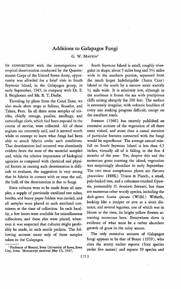
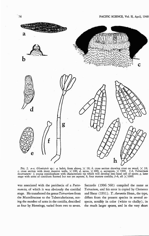
Gloniopsis sp. (Fig. 2a-e)
On the dead wood of Bursera there were numerous elongate black bodies suggesting hysterothecia, These were extremely abundant, occurring on perhaps a majority of the dead branchesseen. It was not until they were examined microscopically that it was recognized that threespecies were involved. Two were the Hysterographium and the Phaeopeltosphaeria listed below; the third was a Gloniopsis. The hysterothecia (Fig. 2a) are black, fusoid, and striate,and most of them appear to be raised well abovethe general surface of the wood. A cross section(Fig. 2b, c) shows that the base is composed ofscarcely altered wood flanked on either side bya black stromatic layer representing a continuation of the walls of the hysterothecium. Thesubhymenial layer is distinctly thinner than thehymenium. The latter is composed of denselycompacted, gelatinous, apparently unbranchedparaphyses penetrated by scattered asci in various stages of development, only a few at a timebearing mature spores. The asci (Fig. 2d) areshort-cylindrical and for the most part 4-6spored. The ascospores (Fig. 2e ) are oval, hyaline, muriform or somewhat irregular in theirseptation, and extremely variable in size, thegreat majority ranging from 25-31fL in lengthby 11-18fL in width. One ascus was seen containing but two ascospores, one of which measured 58X20fL. A number of species of Gloniopsis with large spores are listed in Saccardo. Ofthese G. somala Baccarini (see Saccardo, 1928:1119), from Italian Somaliland, could representthis species, and the specimens are provisionallyfiled under Baccarini's name. On dead limbs andbranches of Bzasera, 6245, 6254.
Hysterographium mori (Schw.) Rehm
On dead wood of Bursera, 6252, 6255. Determined by G. R. Bisby. Dr. Bisby notes thatNo. 6252 approaches H. guaranicum Speg., as

74
described, but does not believe that it is sufficiently distinct to be worthy of recognition.
Phaeopeltosphaeria irregularis Wehmeyer,sp. nov. (Fig. 1)
In superficie caulis maculas dense dispersas,ellipticas, 1-1.5 mm. longas, 0.5 mm. crassas,turnidas, nigricantes formans; ostiolo cenrrali papilliformi vix erumpenti; perithecia 300- 550/Adiametro, 200-350,... alta, singula in lignum submaculis clypeiformibus immersa; pariete crasso,prosenchymatoso, ab ligno adjacenti separato;asci late cylindrici, 90-95,... longi, 12.5,... crassi,saepe 6-7-spori; paraphyses numerosae, filiformes, persistentes, 1,... diametro; sporae uniseriatae, subglobosae vel ellipsoidales, 1O.5-18p.longae, 7-9,... crassae, olivaceae, varie septatae,l-cellulae vel muriformes, cum 1-3 septis transversalibus, ad septa constricrae, cellulis aliquibus verticaliter L-septatis.
Appearing on the surface as thickly scattered,elliptic, raised, blackened spots, 1-1.5 XO.5 mm.,with a central, barely erumpent, papillateostiole; perithecia 300-550 X200-350p., immersed singly in the wood, beneath a clypeuslike blackening of the surface tissues; wall10-20,... thick, prosenchymatous , free from thesurrounding wood. tissue which is somewhatblackened; asci stout-cylindric, 90-95 X 12.5p.,with a claw-like base, and often with only 6 to7 spores; paraphyses numerous, persistent, filiform, Ip. in diameter; spores uniseriate, subglobose to ellipsoid, 10.5-18X 7-9p., olive-brown,variously septate, one-celled to muriform withone to three transverse septa and one or more
FIG. 1. Pbaeopeltospbaeria irregularis: a, radialsection of a perithecium showing the clypeate blackening of the surface; b, ascospores, illustratin g variationin septation; c, ascus with ascospores.
PACIFIC SCIENCE, Vol. II, April, 1948
cells with vertical septa, somewhat constrictedat the septa.
GALAPAGOS: South Seymour Island. Ondead, decorticated wood of Bursera graveolens,September 6, 1945, 6251, type.
The genus Phaeopeltosphaeria Berl. and Peg!.(1892 :139) was based upon P. caudata, onwoody stems, which might be considered as aPelto sph aeria with brown spores or a Pleosporawith a clypeate blackening about the perithecium. It has fusoid spores which are muchlarger than those of P. irregularis. Ph aeopelt osphaeria panam ensis Stev, and King. (Stevens,1927: 50) seems to be the only subsequentlydescribed species. Its spores are describedmerely as "muriforrn, fusiform; olivaceous orstraw-colored; 16X 5,...," but the figures (81-83)given show them to be more irregularly .3septate than in this species and with taperedrather than rounded ends. It is also found onleaves of Chaetochloa, and the authors state thatit resembles the spots of Phyllachora Chaeto chloae Stev. on this host. On the basis of thislatter statement, Petrak (1929:387) claims thatP. panamensis is a Pleospora [Pleospora panamensis (S. & K.) Petr.} and probably parasiticin the Phyllachora stroma.
The collection from the Galapagos Islands isquite distinct from either of these describedspecies. It is true that there is a variable degreeof blackening of the tissues above the perithecia of certain species of Pleospors, but if thegenus Pbaeopeltospbaeria is to be recognized atall, this collection is a typical species.
BASIDIOMYCETES
Sebacina petiolata Rogers
On dead wood of Bursera, 6246. Recentlydescribed (Rogers, 1947: 99) fro m Cuba,Hawaii, and the Marshall Islands. The Galapagos collection, determined by D. P. Rogers,was growing at the base of an old dead trunk ofBursera beneath the soil level and was revealedonly when the trunk was pulled over. On the

'Galapagos Fungi-MARTIN
.basis of examination with a hand lens it wasrecognized in the field as probably a Sebacina'and so entered. Early in October it was soaked'and put in a moist chamber and a scanty but .adequate spore-print was secured. Rogersdescribes the spores as "evenly oblong to ellipsoid-oblong, 9-11 X 6-7.5 p., or ellipsoid-subglobose, 7-9X6-8p.." This description is inclose agreement with that of spores found inmounts from the dried specimen. The sporesfrom the spore-print are almost all globose,1O-11p. in diameter.
The only other Basidiomycetes collected area unique, rough-spored Coprinus isolated fromgoat dung and a small Pleurotus which appearedon Bursera wood. Both were secured in pureculture. The Coprinus fruits readily in culture,and has been referred to Dr. A. H. Smith fordetailed study. The Pleurotus has thus far failedto form fructifications.
FUNGI IMPERFECT!
*Helicosporium guianensis Linder
Referred to this species on the basis of theyellow color of the conidia in mass; the sle';derconidiophores, 4.5p. in diameter below, with atendency to slightly swollen, rounded tips; thebranching, bladder-like projections on which thespores are borne; and the size of the spores.Differing from the species as described (Linder, 1929: 280) in the branching of the conidiophores, which is much like that of H. aureum
(Cda.) Linder, from which species it differs,however, in the more slender conidiophores andthe character of the spore-bearing branches.Further study may reveal that such forms mergeby imperceptible degrees into H. aureum, butfor the present it seems permissible to maintainthe distinction. On thorns of Scu#a spicata.
*Memnoniella echinata (Riv.) Galloway
This widespread species occurred in severalcultures and was particularly abundant on deadOpuntia sterns.
75
*T etracri um incarnatum sp. nov. (Fig. 2f-h)
Sporodochiis pulvinatis, pallide cinnamomeisvel incarnatis, 0.4-0.8 mm. diam.; conidiophoriselongatis, tenuatis, basibus 5p. diam., apicibus 2p.diam., prorrudentibus usque ad 80p.; conidiis 38-digitatis, ramis radiatio-cylindraceis, pluriseptatis, 45-50p. longis, 3.5-4.5p. latis.
.Sporodochia pulvinate to subglobose, at firstwhite, becoming pale cinnamon, pallid ochraceous or flesh-colored . (close to pale ochraceousbuff of Ridgway) , 0.4-0.8 mm. in diameter;conidiophores slender, protruding from body ofsporodochium 60-80p., 5p. in diameter at base,tapering to 2,t at apex just below constrictionmarking junction of spore; conidia digitate, of3-8 multiseptate, subparallel, cylindrical arms,45-50p. in total length, the arms 3.5-4.5p. indiameter.
GALAPAGOS: South SeymourIsland. On deadstem of Opuntia sp. collected September 5, 1945,moistened October 25, 1945, developed January,1947, 6333, type.
After the appearance of the Myxomycetesalready noted, the material in the moist chambers became covered with various molds whichsoon disappeared and were replaced by a densegrowth of the Memnoniella, which appeared tocover the substratum completely. It was notuntil January, 1947, that the sporodochia of theT etracrium were noted, although they may haveappeared earlier., They tended to form at thetips of spines or other projections. The conspicuously protruding conidiophores made thesporodochia appear, under the binocular, asthough covered with glandular hairs. Furtherexamination showed that the Memnoniella hadbeen almost completely replaced by a Curvularia.
The genus T etracrium was established byHennings (1902: 116) for a fungus from Brazil occurring on orange leaves covered with insect larvae. Hennings believed the fungus attacked the larvae first and then spread to the[eaves and twigs. Although a few mites werepresent in the chambers, there was no evidenceof any connection between them and the fungushere described. Hennings assigned his genus tothe Mucedinaceae. Hohnel (1911 : 405) reexamined Hennings' material and found that it
76
~a
b
PACIFIC SCIENCE, Vol. II, April, 1948
g
FIG. 2. a-e, Gloniopsis sp.: a, habit, from above, X 18; b.. cross section showing crust on wood, X 18;c, cross secrion with more massive walls, X 100; d, ascus, X 400; e, ascospore, X 1000. I-h, Tetracriumincarnatu1n: I, young conidiophore with characteristic tip which will develop into basal cell of spore; g, laterstage with arms of conidium formed but not yet septate; b, four mature conidia; I-h, all X 1000.
was associated with the perithecia of a Puttemansia, of which it was obviously the conidialstage. He transferred the genus T etracrium fromthe Mucedinaceae to the Tuberculariaceae, noting the number of arms in the conidia, describedas four by Hennings, varied from two to seven.
Saccardo (1906: 560 ) compiled the name asTetracium, and his error is copied by Clementsand Shear (19 31). T . A urantia Henn., the type,differs from the present species in several respects, notably in color (white to chalky), inthe much larger spores, and in the very short

Galapagos Fungi-MARTIN
conidiophores. Hohnel established a second species, T. coccicola, based on the conidial stage ofOphionectria coccicola (Ell. & Ev.) BerI. &
Vogl. as described and illustrated by Zimmermann (1901:874). In this species the threearms are very long, up to 240p. according toZimmermann, and at right angles to each other.Seaver (1909: 198) describes the conidia of thesame species, using the name Scoleconectria coccicola (Ell. & Ev.) Seaver, as having three tofive arms, each up to 150p. long. It is certainlydistinct from the Galapagos fungus.
The species was readily secured in pure culture. It grows rather slowly and fruits sparselyon most of the ordinary culture media, butforms good sporodochia on weak malt-extractagar and on agars prepared from soil-grass decoction and dung decoction.
REFERENCES
BERLESE, A. N., and V. PEGLION. 1892. Micromiceti Toscani. Contribuzione alla flora micologia della Toscana. Nuovo Gior. Bot. Ital,24: 97-172.
BONAR, LEE. 1939. Fungi from the Galapagosand other Pacific coastal islands. Calif. Acad.ss; Proc, (IV), 22: 195-206.
CLEMENTS, F. E., and C. 1. SHEAR. 1931. Thegenera of fungi. 496 p., 58 pI. Wilson, NewYork. .
HENNINGS, P. 1902. Fungi S. Paulenses I. a d.Puttemans collecti. Hedwigia 41 : 104-118.
77
HOEHNEL, FRANZ VON. 1911. Fragmente zurMykologie XIII. Akad. der Wiss. Wien,Math .-Nat. Kl. Sitzber., Abt. 1, 120: 379484.
LINDER, DAVID H. 1929. A monograph of thehelicosporous Fungi Imperfecti. Mo. Bot.Gard., Ann. 16 (3) : 227-388.
LISTER, ARTHUR. 1911. A monograph of theMycetozoa. (ed. 2, rev. G. Lister). 302 p.,200 pI. Trustees of the British Museum,London.
PETRAK, F. 1929. Mykologische Notizen. X.Ann. Mycol. 27 : 324-410.
ROGERS, DONALD P. 1947. Fungi of the Marshall Islands, Central Pacific Ocean. PacificSci. 1: 92-107. .
SACCARDO, PIER ANDREA. 1882-1931. Syllogefungorztm omnium hucusque cognitorum.
1906. vol. 18. Supplementum universale,Part VII.
1928. vol. 24 (sec. II). Supplementumuniversale, Part X.
SEAVER, FRED]. 1909. The Hypocreales ofNorth America-II. Mycologia 1: 177-207.
STEVENS, FRANK LINCOLN. 1927. Fungi fromCosta Rica. Ill. Bioi. M011Og. 11: 1-102.
SVENSON, HENRY K. 1946. Vegetation of thecoast of Ecuador and Peru and its relation tothe Galapagos Islands. Amer. Jour. Bot. 33:394-498.
ZIMMERMANN, A. 1901. Einige javanische, aufCoccidenparasitierende Ascomyceten. Centbl.f. Bakt., Abr, 2, 7: 872-876.