Embed Size (px)
Citation preview
Additional file
Overexpression of a truncated CTF7 construct leads to pleiotropic defects in reproduction
and vegetative growth in Arabidopsis
Desheng Liu and Christopher A. Makaroff*
Department of Chemistry and Biochemistry, Miami University, Oxford, OH 45056, United
States of America
*Author for correspondence
Christopher A. Makaroff,
Tel: phone: +513 529 2813,
Fax :+513 529 5715,
e-mail: [email protected]

Figure S1. Schematic diagrams of wild type AtCTF7 and AtCTF7 constructs used in this
study. Blue arrow represents 35 promoter and red arrow represents AtCTF7 promoter.
Table S1. Transfer efficiency of 35S:NTAP:AtCTF7∆B mutants
Female (♀) X male(♂) Basta Resistant Basta Sensitive Seeds not germinate
WT X Line 11 137 (38.3%) 188 (52.5%) 33 (9.3%)
Line 11 X WT 71 (25%) 124 (43.7%) 89 (31.3%)
Line 11 self-pollinated 354 (30.4%) 49 (4.2%) 763 (65.4%)
Line 11 plants were used in the crosses.

Figure S2. SYN1 distribution pattern is not altered in 35S:NTAP:AtCTF7∆B male
meiocytes. A to C, Meiocytes from wild type plants. D to F, Meiocytes from
35S:NTAP:AtCTF7∆B plants. Interphase/early leptotene (A, D); zygotene (B, E), pachytene (C,
F). Merged images of 4′,6-diamidino-2-phenylindole (DAPI) stained chromosomes (red) and
SYN1 (green) are shown. Scale bar =10 μm.

Figure S3. Female gametophyte development in wild type plants, revealed by differential
interference contrast (DIC) microscopy. A, Pre-meiotic ovule at stage 1-II. Arrow denotes the
megaspore mother cell (MMC). B, Meiotic ovule at stage 2-IV containing two longitudinally
megaspore cells (dyad) after meiosis I. C, Meiotic ovule at stage 2-IV containing a dyad and the
bottom megaspore containing two nuclei. D, Meiotic ovule at stage 2-V containing a tetrad after
meiosis II. E, Meiotic ovule at stage 2-V containing a linear tetrad. F, Early Female Gametophyte
stage 1 (FG1) ovule (stage 3-I). G, FG1 ovule (stage 3-I). FM (arrow) is uni-nucleate and there is
still space at the distal end of the ovule. H, FG2 ovule containing two nuclei (arrows) (stage 3-
II). I, FG3 ovule containing a two-nucleate embryo sac with a central vacuole (stage 3-III). J,
FG4 ovule containing a four-nucleate embryo sac (stage 3-IV). K, FG5 ovule after cell

differentiation and cellularization (stage 3-V). L, FG7 ovule (stage 3-VI). A diploid central cell
(CC) is formed and the antipodal cells (ANs) are degenerated. CC, central cell; EC, egg cell;
MMC, megaspore mother cell; SN, synergid nucleus; V, vacuole. Scale bar=10 μm. Ovule stages
were determined according to Schneitz et al. [32].

Figure S4. Female gametophyte development in wild type plants, revealed by confocal laser
scanning microscopy (CLSM). A, FG0 ovule. The MMC is surrounded by the nucellar
epidermis. The outer and inner integuments just start to initiate. B, Early FG1 ovule containing
FM and DM. The nucellus is not surrounded by the integuments. C, FG1 ovule. The nucellus is
surrounded by the outer integuments but not the inner integuments. The space is not filled by the
inner integuments (arrow). D, FG3 ovule containing a two-nucleate embryo sac. The two nuclei
are separated by a central vacuole and the nucellus is enclosed by the inner integument. E, Early
FG4 ovule containing a four-nucleate embryo sac. The division planes of the two pair nuclei are
orthogonal to each other. F, Late FG4 ovule. A vacuole appears at the chalazal end of the
embryo sac. G, FG5 ovule. The embryo sac contains eight nuclei in a 4n+4n configuration. Not
all the nuclei are visible on this plane. H, FG6 ovule after the cellularization. Three ANs are
degenerating (arrow) and CC does not form. I, FG7 ovule containing a four-nuclei embryo sac.
CC forms and ANs have degenerated. J, FG8 ovule prior to fertilization. The embryo sac
consists of EC and CC. AN, antipodal nucleus; CC, central cell; DM, degenerated megaspore;
EC, egg cell; FM, functional megaspore; MMC, megaspore mother cell; SN, synergid nucleus; V,

vacuole. Bar scale=10 μm. Ovule developmental stages were defined according to Christensen
et al. [34] [35].
Table S2. Female gametophyte development in wild type (Col) plants
Pistil # FG1 FG2 FG3 FG4 FG5 FG6 FG7 FG8
1 30
2 1 2 26
3 4 23
4 18 13
5 8 26 1
6 1 7 38
7 1 7 16 15
8 1 42
Developmental stages are defined according to Christensen et al. (1997, 1998)[34][35].

Table S3. Female gametophyte development in 35S:NTAP:AtCTF7∆B plants
Pistil # FG0 FG1 FG2 FG-A FG3 FG4 FG5 FG6 FG7 FG-B FG-C
1 25 1
2 22 6 0 1
3 15 10 0 2
4 8 18 0 3
5 1 25 0 1
6 20 2 6
7 4 15 2 6
8 2 4 20
9 1 0 16 1 2 0 0 0 3
10 1 0 17 2 0 0 0 0 8
11 3 0 10 0 0 3 0 0 13
12 12 3 0 0 0 0 1 0 18
13 8 0 1 0 0 0 0 2 20
14 4 0 0 0 0 0 0 12 15
A 4th
generation Line 11 plant was used.
FG0, Ovules before FG1 (Fig. 4F; Supplemental Fig. S5, A, B, E and F); FG-A, Small ovules
with no observable nuclei (Supplemental Fig. S5D); FG-B, Terminal ovules without embryo sac
(Supplemental Fig. S5M); FG-C, Terminal ovules with nuclei (Supplemental Fig. S5, N-P).
Female gametophyte stages are defined according to Christensen et al. (1997, 1998) [34],[35.]

Figure S5. 35S:NTAP:AtCTF7∆B ovules exhibit various defects during meiosis and mitosis.
A, Ovule containing two degrading megaspores. B, Ovule containing a degenerated megaspore
(DM) and anucleate megaspore (arrow), indicating the nucleus is completely degraded. C, Ovule
containing a mis-shaped functional megaspore-like (FML) cell with a degrading nucleus (arrow).
D, Ovule containing no visible megaspore(s), indicating the megaspore(s) are completely
degraded. E, Two elongated cells with no defined nuclei but diffuse signal throughout the cells
(arrows). F&F’, DM at the distal position (arrow) and a chalazal end cell (star) containing
multiple nuclei. Nuclei are not separated by a cell wall. G, Two elongated cells (arrows). H&H’,,
Cell at the chalazal end contains two nuclei (star). I to K, DIC showing degenerating
megaspores/embryo sacs during mitosis. I, FG1 ovule containing a FML-cell (arrow). J&J’,
Embryo sac containing one degenerating nucleus (arrow) and one surviving nucleus. Non-

degenerated L1 layer is marked with a star. K&K’, Ovule containing two degrading nuclei
(arrows) on the side of the vacuole. Other nuclei are not visible. Ovule resembles FG5 stage. L,
Wild type ovule at FG7. CC and EC are marked. M to P, Terminal 35S:NTAP:AtCTF7∆B
ovules containing various defective embryo sacs by DIC. M, Ovule containing the debris of an
embryo sac (arrow). N, Embryo sac containing two nuclei (arrows), morphologically similar to
EC. No vacuole is present. O, Embryo sac containing EC (black arrow) and CC (white arrow).
EC and CC are not separated. No vacuole is present. P, Embryo sac containing EC (black arrow)
and CC (white arrow). EC and CC are not separated. A large vacuole is present at the basal end.
FMLs and DMs are identified according to Barrell and Grossniklaus [37]. Bar scale=10 μm.
Ovule stages are determined according to Schneitz et al in DIC [32] and to Christensen in CLSM
[34],[35].

Figure S6. Female gametophytes from Lines 13 and Line 15 resemble those from Line 11. A,
Female gametophytes from Line 13 develop slowly. Ai, Ovule containing clustered megaspores.
Aii to Aiii, Ovules containing FMLs (arrows) and clustered megaspores (arrows). A megaspore
separates from the clustered megaspores and becomes a FML at chalazal end. Aiv, Ovule
containing a FML and two DMs. B, Two FMLs co-exist in ovules of Line 13. Bi, Ovule
containing megaspore(s) with bright fluorescence signal (arrow). Bii, Two cells with strong
fluorescence. Biii,iii’, Two cells with highly stained nuclei at the chalazal end, resembling FMLs.

They are clearly separated and the right has enlarged nucleus. iii’, Magnified view of iii. C,
Female gametophytes from Line 15 develop slowly. Ci, Ovule containing a FML (arrow) and
clustered megaspores. Cii to Ciii, FG3 ovules. The separated megaspore at chalazal-end becomes
a FML. FML completes one round of mitosis to produce two nuclei and a vacuole forms between
them. D, Middle megaspores become FMLs in ovules of Line 15. Di, Ovule containing only a
FML (arrow). Its nucleus is brightly stained but no associated DM(s) is identified. Dii to Diii,
Middle megaspores (arrows) become FMLs while the associated megaspores (stars) degrade.
DM, degenerated megaspore; FML, functional megaspore like. Bar scale=10 μm. FMLs are
identified as distinctly bright autofluorescence in the nuclei and DMs containing a diffuse signal
throughout the cells but no clearly defined nucleus, as defined according to Barrell and
Grossniklaus [37]. Ovule stages are determined according to Schneitz et al. in DIC [32] and
Christensen et al. in CLSM [34],[35].
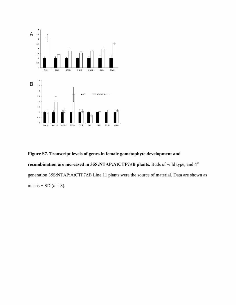
Figure S7. Transcript levels of genes in female gametophyte development and
recombination are increased in 35S:NTAP:AtCTF7∆B plants. Buds of wild type, and 4th
generation 35S:NTAP:AtCTF7∆B Line 11 plants were the source of material. Data are shown as
means ± SD (n = 3).

Figure S8. Morphological alterations are associated with 35S:AtCTF7∆B and atctf7-1
plants transformed with CTF7pro:AtCTF7∆B. A, Wild type plant. B, 35S:AtCTF7∆B plants
from Line 21. Dwarf plants appear at a high frequency with varying severity. C, 35S:AtCTF7∆B
plants from Line 24. “Normal” reduced fertile plants and plants without an inflorescence are
present in the same progeny. D, 35S:AtCTF7∆B plant from Line 29 containing multiple
inflorescences with downward-pointing siliques. E, Homozygous atctf7-1 plants containing
CTF7pro:AtCTF7∆B construct. All plants are dwarf. Some plants (E1) lack stems and some
plants (E3) produce reduced numbers of rosette leaves. F, Wild type silique with full seed set. G,
Silique from atctf7-1:CTF7pro:AtCTF7∆B plant containing about 40 aborted ovules. All plants

are 30-days old and grown under the same environmental conditions. All 35S:AtCTF7∆B plants
are 3rd generation. Scale bar= 5 cm in A-E. Scale bar=0.5 cm in F/G.
Table S4. Primers used
Primers Sequence 5’-3’ Purpose
Genotyping
SALK059500LP ATGAATAGAAATGAGCGATAGGC Genomic forward
SALK059500RP GAAGCTATGAGCTTAAATGCTTCC Genomic reverse
SALK LBb1.3 ATTTTGCCGATTTCGGAAC Left border forward for
SALK lines
Cloning
AtCTF7∆B
gateway F
CACCATGCAAGCCAAAATCAATTC Forward primer for entry
clone of
35S:NTAP:CTF7∆B
AtCTF7∆B
gateway R
GCTAGTTATTGCTCAGCGG Reverse primer for entry
clone of
35S:NTAP:CTF7∆B
AtCTF7 ∆B F GGAGCCATGGATATGCAAGCCAAAA
TC
Forward primer for 35S
CTF7∆B
AtCTF7 ∆B R GAGACTAGTTTAATTCTAGTTATTGC Reverse primer for 35S

TCAGCGG
AtCTF7 gateway F CACCATGCAAGCCAAAATCAATTC Forward primer for entry
clone of 35S:NTAP:CTF7
AtCTF7 gateway R TTAAGAAAAGTGAGTATCAATT Reverse primer for entry
clone of 35S:NTAP:CTF7
CTF7pro F CCGGAATTCCACATCCT
GGAAATATCTTTGCAA
Forward primer for
CTF7promoter
CTF7pro R GGAGCCATGGCGTCTAGAG
AGAGCTCGAATCCTTGTT
Reverse primer for
CTF7promoter
qRT-PCR To quantify expression
Tubulin F TGGATCATGAGTGAGTGAAAAGA Expression control
Tubulin R AAAACCACAATGGACAATTTC
CTF7 native+O/E F GTTGGGTGAGGATTGGAT TC
CTF7 native+O/E R GCGAGGATGAGCTCTCTTTT
CTF7 native F GTGGGATTAGAGCGATTTGG
CTF7 native R TTCCTATGGAGCTTGGTTGTG
WUS F TGGATCTATGGAACAAGACTGTT
WUS R GGCTTTGCTCTATCGAAGAAGT
CLV3 F CGAAGGGTTTAGGACTACATGAAG
CLV3 R GTGGGTTCACATGATGGTGCAA
SYN3-1 F AAAGAAATTTGGGGCTTTCA
SYN3-1 R TGGTGTTCCTACTGGGGAAT
SYN3-2 F CGAGTTCGACTTGGAAGATG

SYN3-2 R AAGGATCAATGCCAGTAGGAA
DMC1 F GTTCATATCAGACCCAAAAAAGCC
DMC1 R AGATTCGGAGCATCGTAGACTTTG
OSD1 F GAATCTCCGGTGAATCCAGA
OSD1R AGAAGGCAACAAACCACCAC
MMD1 F TATCCGCGGTATGACTGTGT
MMD1 R GCAATAGGGTTCCGATGAAT
RBR F GGTGGAGGAGAAACTTGTGC
RBR R GTGGTTGCTTCCGGTAGTTG
MU F TAATTTGGCTGACGGAATCAC
MU R ATTTGGGGGAAAACAAATGAG
COPIA28 F AGTCCTTTTGGTTGCTGAACA
COPIA28 R CCGGATGTAGCAACATTCACT
SoloLTR F AACTAACGTCATTACATACACATCTTG
SoloLTR R AATTAGGATCTTGTTTGCCAGCTA
HDA19 F GACTGTGATTACAACACACCGT
HDA19 R AATTGCCGCCAGTATCCAT
AGO1 F TGGACCACCGCAGAGACAAT
AGO1 R CATCATACGCTGGAAGACGACT
AGO4 F CACTCGCTCTCCTATGTGTACCAAAG
AGO4 R CATGGCTTGATGATGTCTCAGACTGATC
RDR2 F TGGCGAGAGATAACCGGAGGTATG
RDR2 R CTTCTCATCGCGATGGTTTGGATTG

DCL3 F GCCTACTTTCGATACCTCGGAAGA
DCL3 R GCATACATCACAGCCTCACGATTG
NRPD1A F GACTTGTGAAGATGGTTCTGCAGTTG
NRPD1A R GTCTTCGAATGTCCCGTCTATTCTTAC
MIR 156 F CTCTCCCTCCCTCTCTTTGATTC
MIR 156 R AGGCCAAAGAGATCAGCACCGG
MIR 172 F TTTCTCAAGCTTTAGGTATTTGTAG
MIR 172R TCGGCGGATCCATGGAAGAAAGCTC
MET1 F GTGATTCTTAGGGCTATAATGG
MET1 R CATTGATGAAGTCCACTTGAC
DMT7 F CCCACCTGAGTTTGTGGACT
DMT7 R CATTCTGGCCACCATCTCTT
RDM4 F ATGGATGGGGTGGGTGAAAG
RDM4 R TAGCACCTTCTTCGGTTTCAC