Embed Size (px)
Citation preview

FOR RESEARCH USE ONLYwww.genovis.com
ADC kit 2 mg
STORE CONTENT AT DIFFERENT
TEMPERATURES(See page 7)

2
INSTRUCTIONS FOR PRODUCTS
GlyCLICK® ADC kit PNU 2 mg (L1-T01-200) Conjugation of up to 2 mg of IgG
GlyCLICK® ADC kit MMAE 2 mg (L1-T02-200) Conjugation of up to 2 mg of IgG
Last revised December 2020
ADC kit
Immobilized GlycINATOR
Click Reaction
1 2 3
Azide-Activation
DeglycosylationGalT + UDP-GalNAz DBCO-toxin

3
WORKFLOW
Overview of the protocol for antibody conjugation using GlyCLICK
For conjugation of up to 2 mg of IgG .
1 Day 1
• Modification of the antibody Fc N-glycan using Immobilized GlycINATOR® in a spin column. 120 min incubation, 3 centrifugation steps, approx. 2.5 h.
• Azide attachment. The deglycosylated Ab is mixed with UDP-GalNAz, GalT, Buffer additive and TBS. Incubation at 30 °C overnight.
2 Day 2
• Removal of excess UDP-GalNAz using a 2 ml 40 K desalting column. Approx. 1 h.
• Toxin conjugation. Conjugation of chosen DBCO-toxin. The azide-activated Ab is mixed with DBCO-modified toxin. Incubation at 25°C overnight.
3 Day 3
• Removal of excess toxin reagent using affinity purification. Approx. 1 h.

4
Immobilized GlycINATOR
Click Reaction
1 2 3
Azide-Activation
DeglycosylationGalT + UDP-GalNAz DBCO-toxin
PRODUCT DESCRIPTION
GlyCLICK ADC is for toxin conjugation of up to 2 mg of antibody. The conserved N-linked glyco-sylation site on the CH2 domain of each heavy chain of the Fc region is used by GlyCLICK for site-specific conjugation.
Immobilized GlycINATOR removes all Fc N-glycans, including high-mannose, hybrid-type and bisected glycans to the inner GlcNAc. The subsequent azide activation at the GlcNAc is followed by a click reaction for attachment of a selected dibenzo-cyclooctyne (DBCO)-functionalized toxin molecule. The conjugation of the desired molecule occurs at the azide activated sites on the Fc region for incorporation of two toxin molecules per antibody (DAR=2), see Figure 1.
The conjugation procedure is performed by combining enzymatic steps and copper-free click chemistry to covalently link the toxin to the Fc domain of the IgG. All steps are performed under physiological conditions, thus maintaining the quality of the antibody. The site-specific conjugation on the Fc domain preserves the affinity of the antigen-binding sites.

5
Immobilized GlycINATOR
Click Reaction
1 2 3
Azide-Activation
DeglycosylationGalT + UDP-GalNAz DBCO-toxin
GlyCLICK ADC is a reliable tool for conjugation of toxins to generate antibody-drug conjugates from any IgG.
The conjugation is performed in four steps:
1. Deglycosylation: Immobilized GlycINATOR hydrolyzes the N-glycans on the Fc-part of the IgG to the inner GlcNAc.
2. Azide Activation: Azide attachment on the GlcNAc using GalT(Y289L)* and UDP-GalNAz*.
3. Click reaction: The azide activated antibody reacts with a DBCO-toxin in a strain-promoted, copper-free click reaction to form a stable and homogenous antibody drug conjugate.
4. Purification: Excess toxin reagent is removed from the antibody-drug conjugate by affinity chromatography.
* GalT(Y289L) and UDP-GalNAz are components of SiteClick™ and are provided under an intellectual property license from Life Technologies Corporation. The trademark SiteClick™ is the property of Life Technologies Corporation.
Figure 1. Schematic overview of the GlyCLICK technology for ADC generation.
ADC kit
6
PRODUCT DESCRIPTION
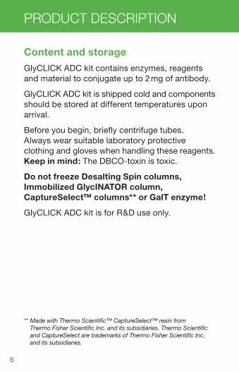
Content and storage
GlyCLICK ADC kit contains enzymes, reagents and material to conjugate up to 2 mg of antibody.
GlyCLICK ADC kit is shipped cold and components should be stored at different temperatures upon arrival.
Before you begin, briefly centrifuge tubes. Always wear suitable laboratory protective clothing and gloves when handling these reagents. Keep in mind: The DBCO-toxin is toxic.
Do not freeze Desalting Spin columns, Immobilized GlycINATOR column, CaptureSelect™ columns** or GalT enzyme!
GlyCLICK ADC kit is for R&D use only.
** Made with Thermo Scientific™ CaptureSelect™ resin from Thermo Fisher Scientific Inc. and its subsidiaries. Thermo Scientific and CaptureSelect are trademarks of Thermo Fisher Scientific Inc. and its subsidiaries.

7
Table 1. Content and storage temperatures of
GlyCLICK components.
Name Amount Store at
Desalting Spin column, 0.5 ml, 40 K
1 piece 4 °C to 8 °C
Antibody concentrator (incl 2 collection tubes), 0.5 ml, 50 K
1 piece 4 °C to 25 °C
Desalting Spin column, 2 ml, 40 K
1 piece 4 °C to 8 °C
Immobilized GlycINATOR, microspin column
1 piece 4 °C to 8 °C
UDP-GalNAz 1 vial solid4 °C to 8 °C
Protect from light
20× TBS pH 7.4 (0.5 M) 3 × 2 ml 4 °C to 8 °C
Buffer additive 1 × 50 µl4 °C to 8 °C
Protect from light
β-1,4-galactosyltransferase (Y289L) (GalT)
1 × 40 µl4 °C to 8 °C
Protect from light
DBCO-modified toxin:
DBCO-Val-Ser(GlcA)- EDA-PNU (L1-T01-200) or DBCO-Val-Ser(GlcA)- PAB-MMAE (L1-T02-200)
1 vial solid-25 °C to -5°C
Protect from light
CaptureSelect Fc, Microspin 2 pieces 4 °C to 8 °C
ADC kit
8
DETAILED PROTOCOL
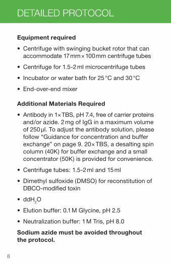
Equipment required
• Centrifuge with swinging bucket rotor that can accommodate 17 mm × 100 mm centrifuge tubes
• Centrifuge for 1.5-2 ml microcentrifuge tubes
• Incubator or water bath for 25 °C and 30 °C
• End-over-end mixer
Additional Materials Required
• Antibody in 1× TBS, pH 7.4, free of carrier proteins and/or azide. 2 mg of IgG in a maximum volume of 250 µl. To adjust the antibody solution, please follow “Guidance for concentration and buffer exchange” on page 9. 20× TBS, a desalting spin column (40K) for buffer exchange and a small concentrator (50K) is provided for convenience.
• Centrifuge tubes: 1.5-2 ml and 15 ml
• Dimethyl sulfoxide (DMSO) for reconstitution of DBCO-modified toxin
• ddH2O
• Elution buffer: 0.1 M Glycine, pH 2.5
• Neutralization buffer: 1 M Tris, pH 8.0
Sodium azide must be avoided throughout the protocol.

9
Guidance for concentration and buffer exchange
It is advisable to start with more antibody than 2 mg if concentration or buffer exchange of the sample is needed prior to “Deglycosylation: Modification of the N-glycan on the Antibody Fc domain” on page 13.
Concentration step
This step is required if:
• The volume of the antibody is more than 250 µl.
If the sample volume is 250 µl but needs a buffer exchange (if it contains phosphate or azide), con centrate the sample to <200 µl and then follow the steps in section “Buffer exchange with Desalting Spin column, 0.5 ml”.
1. Add 500 µl of ddH2O to the small antibody concentrator and cap the device as shown in Figure 2.
2. Centrifuge at 5000 × g for 6 min. Make sure that the cap strap and one membrane panel of the concentrator face the center of the rotor (Fig. 2).
3. Discard the flow-through.
ADC kit

10
4. Add the antibody solution to the small antibody concentrator.
5. Centrifuge at 5000 × g for 2-6 min. Make sure that the cap strap and one membrane panel of the concentrator face the center of the rotor (Fig. 2).
Note: If the antibody volume in the concentrator is more than 200 µl and the sample needs a buffer exchange, centrifuge for an additional 2 min at 5000 × g, or until the appropriate volume is achieved.
6. Invert the small antibody concentrator into the collection tube as shown in Figure 2.
7. Centrifuge at 1000 × g for 3 min to collect the concentrated antibody. After collection, the volume of concentrated Ab should be approximately 150-200 µl in the collection tube.
Figure 2. Antibody concentration step.
DETAILED PROTOCOL

11
Buffer exchange with Desalting Spin column, 0.5 ml
This step is required if:
• The antibody is in a phosphate-based buffer (e.g. PBS), and/or
• The antibody is in a buffer containing azide
1. Prepare 10 ml of 1× TBS buffer by adding 500 µl of 20× TBS to 9.5 ml of ddH2O in a 15 ml tube. Vortex briefly to mix.
2. Break off the bottom closure of the Desalting Spin column. Loosen the lid (do not remove the lid).
3. Place the column in a collection tube (1.5-2 ml) and centrifuge at 1500 × g for 1 min to remove the storage solution.
ADC kit

12
DETAILED PROTOCOL
4. Discard the flow-through and place the column in the collection tube.
5. Add 300 µl of 1× TBS buffer on top of the resin. Centrifuge the column at 1500 × g for 1 min and discard the flow-through.
6. Repeat TBS wash in step 5 two more times. Last spin for 2 min.
7. Blot the bottom of the column to remove excess liquid. Place the column in a new collection tube (1.5-2 ml).
8. Apply the antibody solution on top of the resin (100-200 µl).
9. Centrifuge at 1500 × g for 2 min and collect the flow-through containing the antibody in 1× TBS buffer.

13
Protocol for conjugation of 2 mg of antibody
1 Deglycosylation: Modification of the N-glycan on the Antibody Fc domain.
The antibody solution should be in 1× TBS buffer pH 7.4, with no azide. Max 2 mg in 250 µl.
Time Required: 15 min hands-on, 120 min hands-off.
Materials from kit:
• 1× TBS buffer (prepared from 20× TBS)
• Spin column with Immobilized GlycINATOR
• Let the Immobilized GlycINATOR column equilibrate to room temperature before use
• The lid and the cap of the spin column are used during the incubation
• Before the centrifugations, remove the bottom cap and slightly open the lid
1.1 Break off the bottom plastic cap of the GlycINATOR column (save the cap) and slightly open the lid. Place the column in a microcentrifuge collection tube.
ADC kit

14
DETAILED PROTOCOL
1.2 Centrifuge the column at 200 × g for 1 min to remove the storage solution.
1.3 Discard the flow-through.
1.4 Place the column in the collection tube.
1.5 Add 300 µl of 1× TBS buffer on top of the resin. Centrifuge the column at 200 × g for 1 min and discard the flow-through.
1.6 Repeat the steps in 1.5 two more times.
1.7 Re-insert the bottom cap at the bottom of the spin column.
1.8 Adjust the antibody sample volume (containing 2 mg of antibody) to 250 µl using 1× TBS and immediately add the antibody solution to the column.
1.9 Seal the column with the lid.
1.10 Fully suspend the resin manually and make sure there is a flow in the column.
1.11 Incubate the column by end-over-end mixing at room temperature for 120 min.
1.12 Remove the bottom cap and place the column in a clean microcentrifuge tube. Loosen the top lid.

15
1.13 Centrifuge the column at 1000 × g for 1 min to collect the deglycosylated antibody sample.
1.14 Attach the bottom cap. Add 100 µl of 1× TBS and seal the column with the lid.
1.15 Invert the column a couple of times.
1.16 Remove the bottom cap and place the column in a clean microcentrifuge tube. Loosen the lid.
1.17 Centrifuge at 1000 × g for 1 min to collect the deglycosylated antibody sample.
1.18 Repeat steps 1.14 to 1.17 one more time.
1.19 Pool the collected deglycosylated antibody material and adjust the sample volume to 550 µl with 1× TBS buffer.
ADC kit
16
DETAILED PROTOCOL
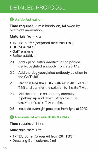
3 Removal of excess UDP-GalNAz
Time required: 1 hour
Materials from kit:
• 1× TBS buffer (prepared from 20× TBS) • Desalting Spin column, 2 ml
2 Azide Activation
Time required: 5 min hands-on, followed by overnight incubation.
Materials from kit:
• 1× TBS buffer (prepared from 20× TBS) • UDP-GalNAz • GalT enzyme • Buffer additive
2.1 Add 7 µl of Buffer additive to the pooled deglycosylated antibody from step 1.19.
2.2 Add the deglycosylated antibody solution to the GalT vial.
2.3 Reconstitute the UDP-GalNAz in 40 µl of 1× TBS and transfer the solution to the GalT vial.
2.4 Mix the sample solution by carefully pipetting up and down. Wrap the tube cap with Parafilm® or similar.
2.5 Incubate overnight protected from light, at 30 °C.

17
3.1 Break off the bottom plastic cap of the column and slightly open the lid. Place the column in a 15 ml collection tube.
3.2 Centrifuge the column at 1000 × g for 2 min to remove the storage solution. Discard the flow-through.
3.3 Place the column in the collection tube.
3.4 Add 1 ml of 1× TBS buffer on top of the resin. Centrifuge the column at 1000 × g for 2 min and discard the flow-through.
3.5 Repeat the steps in 3.4 two more times. The last centrifugation should be 3 min.
3.6 Place the column in a new 15 ml collection tube.
3.7 Apply the azide activated antibody sample (from step 2.5) on top of the resin.
3.8 Centrifuge the column at 1000 × g for 3 min and collect the flow-through that contains the azide activated antibody.
3.9 At this stage, the azide activated antibody can be stored at 2-8 °C protected from light for conjugation at a later time.
ADC kit

18
DETAILED PROTOCOL
4 Conjugation with DBCO-modified toxin
Time required: 10 min hands-on, followed by overnight incubation.
Materials from kit:
• DBCO-modified toxin
4.1 Reconstitute the DBCO-modified toxin in 26 µl of DMSO.
4.2 Transfer the azide activated antibody in 1× TBS (from step 3.9) to a 1.5 ml centrifuge tube and add all of the DBCO-modified toxin from step 4.1. Mix by carefully pipetting up and down.
4.3 Seal the tube with Parafilm® or similar.
4.4 Incubate overnight protected from light, at 25 °C.
4.5 After the incubation, the antibody conjugate can be stored at +4-8 °C, protected from light, until purification.
5 Removal of excess toxin reagent
Time required: 1 h.
Materials from kit:
• Two CapureSelect™ Fc Microspin columns, 0.5 ml.

19
1× TBS or PBS may be used for the purification of the conjugated antibody. 20× TBS is provided in the kit for convenience.
Additional materials:
• Elution buffer: 0.1 M Glycine, pH 2.5
• Neutralization buffer: 1 M Tris, pH 8.0
Equilibration
5.1 Break off the bottom seals of the CaptureSelect Fc columns (save the caps) and place the columns in collection tubes. Loosen the lids.
5.2 Centrifuge for 1 min at 200 × g to remove the storage solution.
5.3 Equilibrate the columns by the following steps:
• seal the columns with the bottom caps
• add 400 µl 1× TBS to each column
• seal the columns with the top lids
• fully suspend the resin, mix it by inversion
• remove the bottom caps and loosen the lid
• centrifuge for 1 min at 200 × g
5.4 Repeat the equilibration steps twice.
5.5 Seal the spin columns with the bottom caps.
ADC kit
20
DETAILED PROTOCOL
Binding of the antibody conjugate
5.6 Equally divide the pooled fractions from step 4.5 and add them to the CaptureSelect Fc col-umns and seal the columns with the top lids.
5.7 Fully suspend the resin, mix it by inversion and make sure there is a flow in the columns.
5.8 Incubate the columns with end-over-end mixing at room temperature for 30 min.
Wash
5.9 Remove the bottom caps and place the col-umns in collection tubes. Loosen the top lids.
5.10 Centrifuge the columns for 1 min at 200 × g.
5.11 Wash the columns by the following steps:
• seal the columns with the bottom caps
• add 400 µl 1× TBS to each column
• seal the columns with the top lids
• fully suspend the resin, mix it by inversion
• remove the bottom caps and loosen the lids
• centrifuge for 1 min at 200 × g
5.12 Repeat the wash steps three more times.
21
ADC kit
Elution of purified, conjugated antibody
5.13 Prepare two collection tubes with 5 µl of 1 M Tris, pH 8.0.
5.14 Seal the columns with the bottom caps.
5.15 Add 50 µl of 0.1 M Glycinea), pH 2.5 to each of the CaptureSelect Fc column and seal the columns.
5.16 Fully suspend the resin by inverting the col-umns a couple of times.
5.17 Remove the bottom caps and place the col-umns in the collection tubes containing Tris. Loosen the top lids.
5.18 Centrifuge the columns 1 min at 200 × g to elute the conjugated antibody.
5.19 Repeat elution (steps 5.13-5.18) three additional times with centrifugation 1 min at 1000 × g.
5.20 Pool the eluted fractions.
5.21 The antibody conjugate can now be stored protected from light at +4-8 °C.
a) Addition of 0.02% Polysorbate 20 is possible to increase the recovery.

22
Related Products
Immobilized GlycINATOR Deglycosylation of IgG Fc domain
GlyCLICK Azide Activation kit Azide activation of IgG
GlyCLICK Labeling kits Labeling of IgG with options for Fluorophores, DFO and Biotin
ADC kit
References1. Sjögren, J. et al., 2013. EndoS2 is a unique and conserved enzyme
of serotype M49 group A Streptococcus that hydrolyses N-linked glycans on IgG and α1-acid glycoprotein. The Biochemical Journal, 455(1), pp.107-118.
2. Ramakrishnan, B. & Qasba, P.K., 2002. Structure-based design of beta 1,4-galactosyltransferase I (beta 4Gal-T1) with equally efficient N-acetylgalactosaminyltransferase activity: point mutation broadens beta 4Gal-T1 donor specificity. J Biol Chem, 277(23), pp.20833-20839.

23
GlyCLICK® Legal and Disclaimers
All rights reserved. Genovis products are covered by one or more patents, trademarks and/or copyrights owned or controlled by Genovis AB. For more information about commercial rights, please contact Genovis team at [email protected]. Genovis products are intended for research use only. They are not intended to be used for therapeutic or diagnostic purposes in humans or animals. All goods and services are sold subject to Genovis’ General Terms and Conditions of Sale.
SiteClick™ Included in GlyCLICK®
This product is provided under an intellectual property license from Life Technologies Corporation. The transfer of this product is conditioned on the buyer using the purchased product solely in research conducted by the buyer, excluding contract research or any fee for service research, and the buyer must not (1) use this product or its components for (a) diagnostic, therapeutic or prophylactic purposes; (b) testing, analysis or screening services, or infor-mation in return for compensation on a per-test basis; or (c) manufacturing or quality assurance or quality control, and/or (2) sell or transfer this product or its components for resale, whether or not resold for use in research. For information on purchasing a license to this product for purposes other than as described above, contact Life Technologies Corporation, 5791 Van Allen Way, Carlsbad, CA 92008 USA or [email protected].
The trademark SiteClick™ is the property of Life Technologies Corporation.
Linker-toxin Payloads Included in GlyCLICK®
This product is provided under an intellectual property license from Glykos Finland ltd. The transfer of this product is conditioned on the buyer using the purchased product solely in research conducted by the buyer.
The buyer must not (1) use this product or its components for (a) diagnostic, therapeutic or prophylactic purposes; or (b) manufacturing or quality assurance or quality control, and/or (2) sell or transfer this product or its components for resale, whether or not resold for use in research.
CaptureSelect™ Included in GlyCLICK®
Thermo Scientific™ CaptureSelect™ resin from Thermo Fisher Scientific Inc. and its subsidiaries. Thermo Scientific and CaptureSelect are trademarks of Thermo Scientific Inc. and its subsidiaries.
©2020 Genovis AB.

24
USA & Canada
Genovis Inc. 245 First Street, Suite 1800 Cambridge, MA 02142 USA
Customer service: 1-617-444-8421 Order phone (toll free): 1-855-782-0084 Order fax: 1-858-524-3006 Email: [email protected]
EMEA & Asia
Genovis AB Box 790 SE-220 07 Lund Sweden
Customer service: 0046 (0)46 10 12 30 Order phone: 0046 (0)46 10 12 30 Order fax: 0046 (0)46 12 80 20 Email: [email protected]