Embed Size (px)
Citation preview
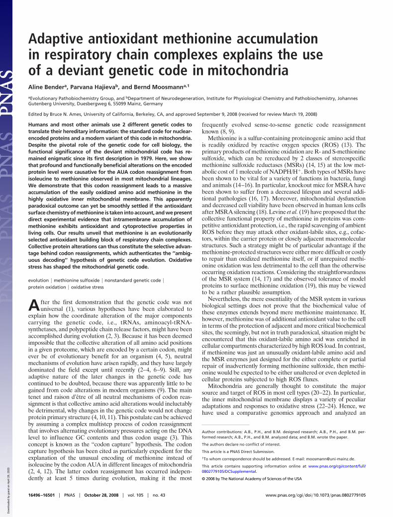
Adaptive antioxidant methionine accumulationin respiratory chain complexes explains the useof a deviant genetic code in mitochondriaAline Bendera, Parvana Hajievab, and Bernd Moosmanna,1
aEvolutionary Pathobiochemistry Group, and bDepartment of Neurodegeneration, Institute for Physiological Chemistry and Pathobiochemistry, JohannesGutenberg University, Duesbergweg 6, 55099 Mainz, Germany
Edited by Bruce N. Ames, University of California, Berkeley, CA, and approved September 9, 2008 (received for review March 19, 2008)
Humans and most other animals use 2 different genetic codes totranslate their hereditary information: the standard code for nuclear-encoded proteins and a modern variant of this code in mitochondria.Despite the pivotal role of the genetic code for cell biology, thefunctional significance of the deviant mitochondrial code has re-mained enigmatic since its first description in 1979. Here, we showthat profound and functionally beneficial alterations on the encodedprotein level were causative for the AUA codon reassignment fromisoleucine to methionine observed in most mitochondrial lineages.We demonstrate that this codon reassignment leads to a massiveaccumulation of the easily oxidized amino acid methionine in thehighly oxidative inner mitochondrial membrane. This apparentlyparadoxical outcome can yet be smoothly settled if the antioxidantsurface chemistry of methionine is taken into account, and we presentdirect experimental evidence that intramembrane accumulation ofmethionine exhibits antioxidant and cytoprotective properties inliving cells. Our results unveil that methionine is an evolutionarilyselected antioxidant building block of respiratory chain complexes.Collective protein alterations can thus constitute the selective advan-tage behind codon reassignments, which authenticates the ‘‘ambig-uous decoding’’ hypothesis of genetic code evolution. Oxidativestress has shaped the mitochondrial genetic code.
evolution � methionine sulfoxide � nonstandard genetic code �protein oxidation � oxidative stress
A fter the first demonstration that the genetic code was notuniversal (1), various hypotheses have been elaborated to
explain how the coordinate alteration of the major componentscarrying the genetic code, i.e., tRNAs, aminoacyl-tRNA-synthetases, and polypeptide chain release factors, might have beenaccomplished during evolution (2, 3). Because it has been deemedimpossible that the collective alteration of all amino acid positionsin a given proteome, which are encoded by a certain codon, mightever be of evolutionary benefit for an organism (4, 5), neutralmechanisms of evolution have arisen rapidly, and they have largelydominated the field except until recently (2–4, 6–9). Still, anyadaptive nature of the later changes in the genetic code hascontinued to be doubted, because there was apparently little to begained from code alterations in modern organisms (9). The maintenet and raison d’etre of all neutral mechanisms of codon reas-signment is that collective amino acid alterations would ineluctablybe detrimental, why changes in the genetic code would not changeprotein primary structure (4, 10, 11). This postulate can be achievedby assuming a complex multistep process of codon reassignmentthat involves alternating evolutionary pressures acting on the DNAlevel to influence GC contents and thus codon usage (3). Thisconcept is known as the ‘‘codon capture’’ hypothesis. The codoncapture hypothesis has been cited as particularly expedient for theexplanation of the unusual encoding of methionine instead ofisoleucine by the codon AUA in different lineages of mitochondria(2, 4, 12). The latter codon reassignment has occurred indepen-dently at least 5 times during evolution, making it the most
frequently evolved sense-to-sense genetic code reassignmentknown (8, 9).
Methionine is a sulfur-containing proteinogenic amino acid thatis readily oxidized by reactive oxygen species (ROS) (13). Theprimary products of methionine oxidation are R- and S-methioninesulfoxide, which can be rereduced by 2 classes of stereospecificmethionine sulfoxide reductases (MSRs) (14, 15) at the low met-abolic cost of 1 molecule of NADPH/H�. Both types of MSRs havebeen shown to be vital for a variety of functions in bacteria, fungiand animals (14–16). In particular, knockout mice for MSRA havebeen shown to suffer from a decreased lifespan and several addi-tional pathologies (16, 17). Moreover, mitochondrial dysfunctionand decreased cell viability have been observed in human lens cellsafter MSRA silencing (18). Levine et al. (19) have proposed that thecollective functional property of methionine in proteins was com-petitive antioxidant protection, i.e., the rapid scavenging of ambientROS before they may attack other oxidant-labile sites, e.g., cofac-tors, within the carrier protein or closely adjacent macromolecularstructures. Such a strategy might be of particular advantage if themethionine-protected structures were either more difficult or costlyto repair than oxidized methionine itself, or if unrepaired methi-onine oxidation was less detrimental to the cell than the otherwiseoccurring oxidation reactions. Considering the straightforwardnessof the MSR system (14, 17) and the observed tolerance of modelproteins to surface methionine oxidation (19), this may be viewedto be a rather plausible assumption.
Nevertheless, the mere essentiality of the MSR system in variousbiological settings does not prove that the biochemical value ofthese enzymes extends beyond mere methionine maintenance. If,however, methionine was of additional antioxidant value to the cellin terms of the protection of adjacent and more critical biochemicalsites, the seemingly, but not in truth paradoxical, situation might beencountered that this oxidant-labile amino acid was enriched incellular compartments characterized by high ROS load. In contrast,if methionine was just an unusually oxidant-labile amino acid andthe MSR enzymes just designed for the either complete or partialrepair of inadvertently forming methionine sulfoxide, then methi-onine would be expected to be either unaltered or even depleted incellular proteins subjected to high ROS fluxes.
Mitochondria are generally thought to constitute the majorsource and target of ROS in most cell types (20–22). In particular,the inner mitochondrial membrane displays a variety of peculiaradaptations and responses to oxidative stress (22–24). Hence, wehave used a comparative genomics approach and analyzed an
Author contributions: A.B., P.H., and B.M. designed research; A.B., P.H., and B.M. per-formed research; A.B., P.H., and B.M. analyzed data; and B.M. wrote the paper.
The authors declare no conflict of interest.
This article is a PNAS Direct Submission.
1To whom correspondence should be addressed. E-mail: [email protected].
This article contains supporting information online at www.pnas.org/cgi/content/full/0802779105/DCSupplemental.
© 2008 by The National Academy of Sciences of the USA
16496–16501 � PNAS � October 28, 2008 � vol. 105 � no. 43 www.pnas.org�cgi�doi�10.1073�pnas.0802779105
Dow
nloa
ded
by g
uest
on
Apr
il 28
, 202
0

extensive set of mitochondrial and nuclear genomes for the encodedmethionine contents. Thereby, we have (i) investigated our hypoth-esis that methionine usage in proteins exposed to particularly highROS fluxes may show characteristic idiosyncrasies and (ii) testedthe main tenet of the codon capture hypothesis, i.e., that encodedprotein structures must not be altered by the use of a nonstandardgenetic code. We have found that methionine is strikingly enrichedin many mitochondrially encoded proteomes, especially in animalswith high aerobic metabolic rate, and that the enrichment dependedalmost exclusively on the use of a second codon (AUA) in thesemitochondria to encode the amino acid methionine.
Results and DiscussionWe have determined methionine content in the mitochondriallyencoded respiratory chain proteins of 361 neutrally selectedanimals and various other eukaryotes, and we have comparedthe obtained methionine contents in mitochondria with baselinemethionine usage in nuclear-encoded proteomes of a variety ofgenomically sequenced species. The results in Fig. 1 and sup-porting information (SI) Table S1 demonstrate that methionineuse in nuclear-encoded proteins is very uniform in all eukaryotes(animals: 2.32% � 0.14%; fungi: 2.15% � 0.07%; other eu-karyotes: 2.06% � 0.32%, Arabidopsis thaliana: 2.45%). Mito-chondrially encoded methionine contents in animals using thestandard coding for this amino acid, i.e., echinoderms, platyhel-minthes, cnidarians, and sponges, were quite similar to thesevalues (overall average: 2.74% � 0.48%). Moreover, also fungi,plants, and a collection of other eukaryotes all sharing the useof a single codon (AUG) for mitochondrial methionine did notdiffer from the rule that proteomic methionine usage generallyamounts to 2–3% (fungi: 2.53% � 0.64%; plants: 2,67% �0.63%; other eukaryotes: 2.42% � 0.46%). In contrast, animalsthat also used a second codon for methionine in mitochondriaconsiderably accumulated this amino acid in the encoded respi-ratory chain proteins (overall average: 6.17% � 1.33%), withseveral insects reaching contents of 10% and more (Fig. 1 A).Using 3 different approaches, the latter association was found tobe statistically significant: regarding animal species as indepen-dent entities, the level of significance was P � 2.1�10�41 (n �361); regarding only phyla as independent entities, significancewas P � 5.7�10�5 (n � 10); phylogenetically independentcontrast analysis on clades sharing the same AUA codon assign-ment returned a significance level of P � 0.003 (n � 5).
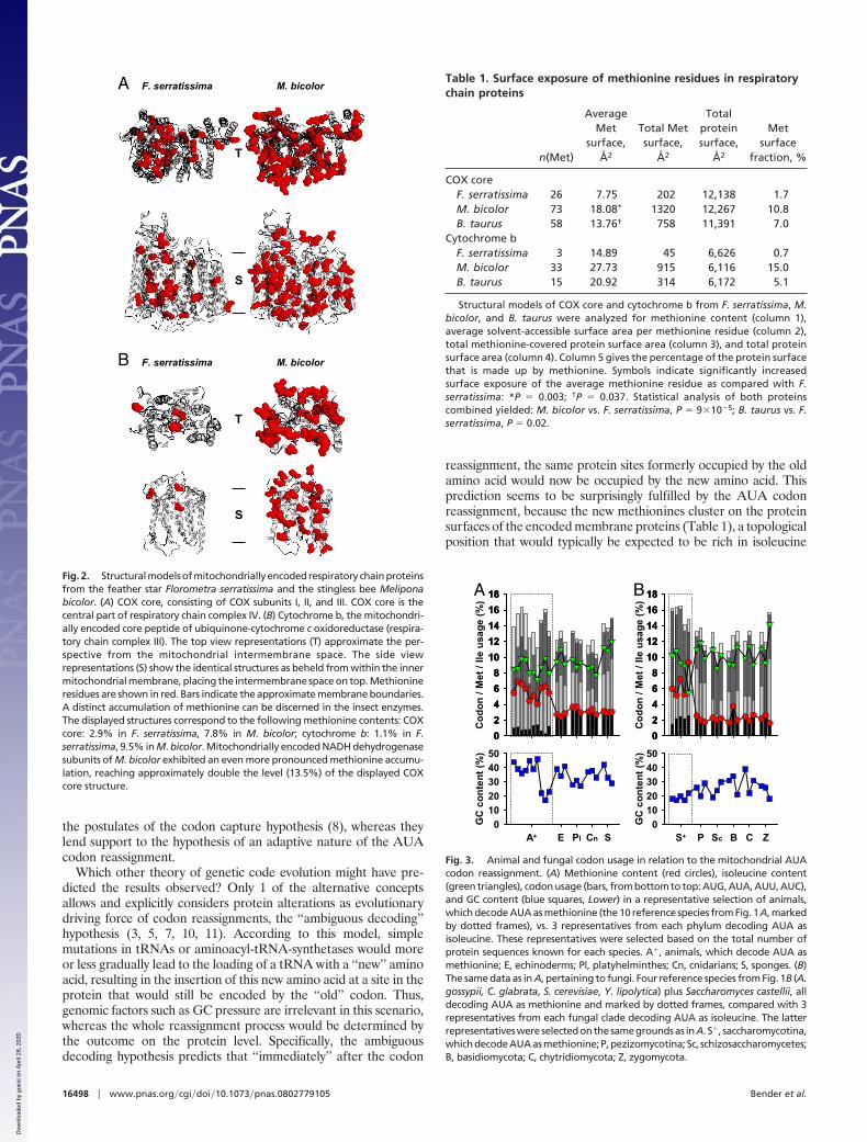
We have investigated the protein structural consequences of theresulting methionine accumulation for 2 of the encoded respiratorychain complexes, the core subunits of cytochrome c oxidase (COX),and cytochrome b, the central subunit of ubiquinone-cytochrome coxidoreductase (Fig. 2 and Table 1). A comparison of the modeledstructures of Florometra serratissima, an echinoderm using thestandard code (2.0% methionine), and Melipona bicolor, an insectusing the nonstandard code (11.2% methionine), indicates thatmethionine accumulation in the insect is massive, particularlypronounced in transmembrane domains, and primarily affectsprotein surfaces. A quantification of these observations is given inTable 1, for which the absolute surface exposure of all methionineresidues was analyzed. Respecting COX, the average M. bicolormethionine is �2.5-fold more surface-exposed than the average F.serratissima methionine, which adds to the effect that the M. bicolorenzyme contains �3 times more methionine than its echinodermcounterpart. Ultimately, methionine builds 10.8% of the insectenzyme’s surface, as opposed to 1.7% in the echinoderm.
In summary, the recent evolutionary change of the genetic codein different lineages of mitochondria has resulted in an extraordi-nary increase in the use of methionine. Consequently, the codoncapture hypothesis is inappropriate to account for the basic out-come of the AUA codon reassignment in mitochondria, namelymethionine accumulation in the encoded proteins. To investigatewhether this conclusion was also borne by the underlying coding
characteristics, we have performed codon usage analyses on avariety of animals and fungi (Fig. 3). Although methionine wasclearly increased in AUA-reassigning species, isoleucine was onlymildly decreased due to an increased use of isoleucine codons(AUU and AUC) and an increased overall usage of the AUX familybox. GC content was unrelated to the codon reassignment. Signif-icantly, the AUA codon is not used less frequently in species thathave undergone the reassignment than in species that have not;rather, animals make less use of the AUG codon if they can decodemethionine by AUA. These observations diametrically contradict
Met
(%)
0
2
4
6
8
10
12
P M B R A F E I C Ar N MoPl Br
An Cn
S
Met
(%)
0
2
4
6
8
10
12
0
2
4
6
8
10
12
0
2
4
6
8
10
12
A
B C D
Met
(%)
Met
(%)
P S+ S-
Sc
BC
ZH
A M R S al. SC
Fig. 1. Mitochondrially encoded methionine contents in 361 animal species, 39fungi, 34 unicellular eukaryotes, and 16 plants, compared with a referenceselection of nuclear-encoded, proteomic methionine contents. (A) Methioninecontents of mitochondrially encoded proteomes of 361 animal species (blackcircles). P, primates; M, other mammals; B, birds; R, reptiles; A, amphibians; F, fish;E, echinoderms; I, insects; C, crustaceans; Ar, arachnids; N, nematodes; Pl, platy-helminthes; Mo, molluscs; An, annelids; Br, brachiopods; Cn, cnidarians; S,sponges. Genomically encoded methionine contents of 10 reference species areshown for comparison (red squares). Adjustment of these nuclear-encoded pro-teomes to the higher transmembrane domain contents of the belonging mito-chondrially encoded proteomes led to only marginally higher reference values(green triangles). The 10 reference species were (from left to right): Homosapiens, Bos taurus, Mus musculus, Rattus norvegicus, Gallus gallus, Danio rerio,Tetraodon nigroviridis, Anopheles gambiae, Drosophila melanogaster, Caeno-rhabditis elegans. Clades, which use 2 codons (AUG and AUA) to encode mito-chondrial methionine are marked by dotted frames. (B) Mitochondrially encodedmethionine contents in 39 fungi. Symbols and frames are used as in A. P, pezizo-mycotina; S�, saccharomycotina, which decode AUA as methionine; S�, saccha-romycotina, which decode AUA as isoleucine; Sc, schizosaccharomycetes; B, ba-sidiomycota; C, chytridiomycota; Z, zygomycota; H, Hyaloraphidium curvatum.The 7 reference species were: Ashbya gossypii, Candida glabrata, Saccharomycescerevisiae, Yarrowia lipolytica, Kluyveromyces lactis, Schizosaccharomycespombe, Cryptococcus neoformans. (C) Mitochondrially encoded methionine con-tents in 34 unicellular eukaryotes. Symbols are used as in A. A, alveolata; M,mycetozoa; R, rhodophyta; S, stramenopiles; al., other eukaryotes. The 3 refer-ence species were: Paramecium tetraurelia, Plasmodium falciparum, Dictyosteli-um discoideum. (D) Mitochondrially encoded methionine contents in 16 plants.Symbols are used as in A. C, chlorophyta; S, streptophyta. The reference specieswas Arabidopsis thaliana. All species names and calculated numeric values per-taining to this figure are given in Table S1.
Bender et al. PNAS � October 28, 2008 � vol. 105 � no. 43 � 16497
BIO
CHEM
ISTR
Y
Dow
nloa
ded
by g
uest
on
Apr
il 28
, 202
0
the postulates of the codon capture hypothesis (8), whereas theylend support to the hypothesis of an adaptive nature of the AUAcodon reassignment.
Which other theory of genetic code evolution might have pre-dicted the results observed? Only 1 of the alternative conceptsallows and explicitly considers protein alterations as evolutionarydriving force of codon reassignments, the ‘‘ambiguous decoding’’hypothesis (3, 5, 7, 10, 11). According to this model, simplemutations in tRNAs or aminoacyl-tRNA-synthetases would moreor less gradually lead to the loading of a tRNA with a ‘‘new’’ aminoacid, resulting in the insertion of this new amino acid at a site in theprotein that would still be encoded by the ‘‘old’’ codon. Thus,genomic factors such as GC pressure are irrelevant in this scenario,whereas the whole reassignment process would be determined bythe outcome on the protein level. Specifically, the ambiguousdecoding hypothesis predicts that ‘‘immediately’’ after the codon
reassignment, the same protein sites formerly occupied by the oldamino acid would now be occupied by the new amino acid. Thisprediction seems to be surprisingly fulfilled by the AUA codonreassignment, because the new methionines cluster on the proteinsurfaces of the encoded membrane proteins (Table 1), a topologicalposition that would typically be expected to be rich in isoleucine
M. bicolorF. serratissima
T
A
S
T
S
M. bicolorF. serratissimaB
Fig. 2. Structural models of mitochondrially encoded respiratory chain proteinsfrom the feather star Florometra serratissima and the stingless bee Meliponabicolor. (A) COX core, consisting of COX subunits I, II, and III. COX core is thecentral part of respiratory chain complex IV. (B) Cytochrome b, the mitochondri-ally encoded core peptide of ubiquinone-cytochrome c oxidoreductase (respira-tory chain complex III). The top view representations (T) approximate the per-spective from the mitochondrial intermembrane space. The side viewrepresentations (S) show the identical structures as beheld from within the innermitochondrial membrane, placing the intermembrane space on top. Methionineresidues are shown in red. Bars indicate the approximate membrane boundaries.A distinct accumulation of methionine can be discerned in the insect enzymes.The displayed structures correspond to the following methionine contents: COXcore: 2.9% in F. serratissima, 7.8% in M. bicolor; cytochrome b: 1.1% in F.serratissima, 9.5% in M. bicolor. Mitochondrially encoded NADH dehydrogenasesubunits of M. bicolor exhibited an even more pronounced methionine accumu-lation, reaching approximately double the level (13.5%) of the displayed COXcore structure.
Table 1. Surface exposure of methionine residues in respiratorychain proteins
n(Met)
AverageMet
surface,Å2
Total Metsurface,
Å2
Totalproteinsurface,
Å2
Metsurface
fraction, %
COX coreF. serratissima 26 7.75 202 12,138 1.7M. bicolor 73 18.08* 1320 12,267 10.8B. taurus 58 13.76† 758 11,391 7.0
Cytochrome bF. serratissima 3 14.89 45 6,626 0.7M. bicolor 33 27.73 915 6,116 15.0B. taurus 15 20.92 314 6,172 5.1
Structural models of COX core and cytochrome b from F. serratissima, M.bicolor, and B. taurus were analyzed for methionine content (column 1),average solvent-accessible surface area per methionine residue (column 2),total methionine-covered protein surface area (column 3), and total proteinsurface area (column 4). Column 5 gives the percentage of the protein surfacethat is made up by methionine. Symbols indicate significantly increasedsurface exposure of the average methionine residue as compared with F.serratissima: *P � 0.003; †P � 0.037. Statistical analysis of both proteinscombined yielded: M. bicolor vs. F. serratissima, P � 9�10�5; B. taurus vs. F.serratissima, P � 0.02.
Co
do
n/
Met
/Il
eu
sag
e(%
)
0
2
4
6
8
10
12
14
16
18
0
2
4
6
8
10
12
14
16
18
GC
co
nte
nt
(%)
0
10
20
30
40
50
0
2
4
6
8
10
12
14
16
18A
Co
do
n/
Met
/Il
eu
sag
e(%
)
0
2
4
6
8
10
12
14
16
18
0
2
4
6
8
10
12
14
16
18
0
2
4
6
8
10
12
14
16
18
GC
co
nte
nt
(%)
0
10
20
30
40
50
B
PS+ Sc B C ZA+ E Pl Cn S
Fig. 3. Animal and fungal codon usage in relation to the mitochondrial AUAcodon reassignment. (A) Methionine content (red circles), isoleucine content(green triangles), codon usage (bars, from bottom to top: AUG, AUA, AUU, AUC),and GC content (blue squares, Lower) in a representative selection of animals,which decode AUA as methionine (the 10 reference species from Fig. 1A, markedby dotted frames), vs. 3 representatives from each phylum decoding AUA asisoleucine. These representatives were selected based on the total number ofprotein sequences known for each species. A�, animals, which decode AUA asmethionine; E, echinoderms; Pl, platyhelminthes; Cn, cnidarians; S, sponges. (B)The same data as in A, pertaining to fungi. Four reference species from Fig. 1B (A.gossypii, C. glabrata, S. cerevisiae, Y. lipolytica) plus Saccharomyces castellii, alldecoding AUA as methionine and marked by dotted frames, compared with 3representatives from each fungal clade decoding AUA as isoleucine. The latterrepresentativeswereselectedonthesamegroundsas inA. S�, saccharomycotina,whichdecodeAUAasmethionine;P,pezizomycotina; Sc, schizosaccharomycetes;B, basidiomycota; C, chytridiomycota; Z, zygomycota.
16498 � www.pnas.org�cgi�doi�10.1073�pnas.0802779105 Bender et al.
Dow
nloa
ded
by g
uest
on
Apr
il 28
, 202
0

(encoded by any codon), but not methionine. In animals, the oldAUG-encoded internal methionines would even be partially lost(Fig. 3), whereas pressures from the fewer isoleucine-encodingcodons (2 instead of 3) would be counterbalanced by an increasedfrequency of the remaining codons and the complete family box.
What might have been the selective advantage of the observedaccumulation of methionine in inner mitochondrial membranerespiratory chain complexes? Two biochemically fundamental pre-mises point toward a specific solution: first, methionine is a veryeasily oxidized amino acid, and it may be the most readily oxidizedresidue in certain biochemical settings (13). Despite efficient re-duction enzymes, steady-state levels of methionine sulfoxideamount to 5–10% of all protein methionine residues in the majorityof tissues (25). Second, the mitochondrial compartment, particu-larly the inner mitochondrial membrane, is characterized by excep-tionally oxidative conditions (20, 22). Thus, pronounced methio-nine oxidation in mitochondria is inevitable, such that the observedmethionine accumulation would be clearly counterproductive for atleast 2 reasons if there were no compensatory beneficial mecha-nisms of exceeding selective advantage: first, the inescapable struc-tural alterations imposed by the mere exchange of amino acids (4,5), even if those alterations may be uncommonly mild in the presentexample (Table 2), and second, the need to permanently repair orreplace the newly introduced unstable methionine residues, whichare henceforth used instead of stable isoleucine residues.
The most parsimonious explanation for this conundrum wouldpostulate that the redox properties of methionine themselvesconstituted the exceeding selective advantage, because most otherphysicochemical properties of isoleucine and methionine are notgreatly dissimilar (Table 2). In fact, several biochemical studies havedescribed protein methionine as an intrinsic antioxidant shield ofproteins against the oxidative destruction of other, potentially morecritical biochemical sites within the same or in neighboring proteins(15, 17, 19). Thus, targeted antioxidant activities of methioninemight provide a plausible origin of significant vantages of methio-nine accumulation as effected by the AUA codon reassignment.This concept is supported by the following observations: first, thestructural models of Fig. 2 evidence that methionine is selectivelyenriched on the protein surface or the lipid bilayer interface. Atthese sites, methionine residues are especially reactive (17, 19),whereas their oxidation to sulfoxides does not entail major struc-tural distortion of the carrier protein (19). Second, methionine canbe economically repaired by MSRs, which are known to be ex-pressed in mitochondria and whose silencing has been shown tocompromise mitochondrial membrane potential in cultivated cells(18). We have examined all genomically sequenced, methionine-accumulating animals and fungi for sequences homologous toEscherichia coli MSR-A and -B, and we have found correspondingsequences in all cases (data not shown), indicating that this poten-
tially mandatory condition for the extensive use of methionine asantioxidant was fulfilled. In contrast, various unicellular organismsreading AUA for isoleucine, particularly anaerobes, did not revealMSR-homologous sequences. It remains to be determined, though,how far MSRs are capable of repairing intramembrane sulfoxides,or whether those methionine residues are rather installed for‘‘single use.’’ Third, mitochondrial methionine in animals appar-ently runs parallel to aerobic metabolic capacity. The anaerobicplatyhelminthes of Fig. 1A and slowly respiring sponges and echi-noderms do not accumulate methionine and have refrained fromaltering their genetic code, whereas insects, arachnids, and mam-mals synthesize particularly methionine-rich respiratory chain com-plexes (Fig. 1).
We sought to experimentally confirm our biochemical andevolutionary reasoning. All proteins affected by the AUA codonreassignment are integral membrane proteins, whose overall trans-membrane domain content amounts to some exceptional 55%. Inaddition, they seem to expose the majority of their newly gainedmethionines to the lipid bilayer (Fig. 2; Table 1). Thus, we havesynthesized lipophilic methionine derivatives (Fig. 4A) to mimic thebiochemical result of the evolutionary AUA codon reassignmentfrom isoleucine to methionine and have investigated these com-pounds for their potential to decrease oxidative stress in intacteukaryotic cells and isolated mitochondria. The results in Fig. 4show that intramembrane methionine enrichment in fact sup-presses the measurable flux of oxidants in respiring tumor cells (Fig.4B). A similar result was found after the exogenous addition of thephysiological peroxide H2O2, which can emerge from numerousmetabolic processes and mediates signaling and toxic responses (26,27). A comparable reduction of cellular ROS was not observed withisoleucine enrichment of the cells (Fig. 4C). In addition, thereduction of ROS by methionine derivatives depended on theintramembrane localization of the applied thioether moiety (ofmethionine), because more hydrophilic but cell-permeable com-pounds, such as acetyl methionine methyl ester, were hardly effec-tive (Fig. 4D).
The cell penetration and intramembrane accumulation of bothmodel compounds of Fig. 4A were verified by means of anHPLC-based analytical protocol. The results in Fig. 4E indicatethat, under the authentic conditions of the antioxidative activityassay in Fig. 4C, both compounds achieved approximately the samecell penetration (NDo-Met-OMe: 44%; NDo-Ile-OMe: 40%). Inaddition, both compounds elicited considerable accumulation inmembranes as expected: the membrane-to-cytosol distributionfactor was 13.4 for NDo-Met-OMe and 13.5 for NDo-Ile-OMe.Tracking the organelle distribution of the applied compounds bysubcellular fractionation (Fig. 4F), it was found that both com-pounds attained the highest concentrations in the mitochondrialfraction (P2), followed by the nuclear fraction (P1). Hence, bothcompounds reached their assumed site of action to a similar degree,which rules out differential distribution as an explanation for theirdisparate antioxidant activities.
In primary mesencephalic neurons, which were used for theirhigh sensitivity to oxidants of mitochondrial origin (28), intramem-brane methionine enrichment resulted in increased survival underthe physiologically hyperoxic conditions of the incubator (20% O2)(Fig. 4G). Moreover, treatment with 100 �M NDo-Met-OMe wasable to partially prevent cell death from enhanced mitochondrialoxidant production (Fig. 4 H and I) as induced by the mitochondrialtoxin rotenone (29). Rotenone is a well-established inhibitor ofmitochondrial complex I that has been demonstrated to inducemitochondrial oxidative stress in vitro, resulting in a syndromeresembling Parkinsons’s disease in vivo (29).
Finally, we aspired to obtain a quantitative comparison betweenthe degree of methionine accumulation effected by the mitochon-drial codon reassignment and the pharmacological application ofour model compounds. Rat liver mitochondria arguably constitutethe best-characterized mitochondrial species. Using literature val-
Table 2. Chemical structures and physicochemical properties ofisoleucine, methionine, and methionine sulfoxide
NH2
α
β S
O
HO
NH2
α
β S
O
HO
NH2
α
β
O
HO
O
LogP 2.38 1.63 �0.50p �D� 0.01 1.56 3.79A �Å2� 117.7 122.0 125.0Ap �Å2� 0.0 0.0 22.3
The following properties were calculated for the displayed amino acid sidechains depicted in bold: LogP, octanol-water partition coefficient; p, dipolemoment; A, Connolly molecular surface area; Ap, polar surface area. Transi-tions from isoleucine to methionine are accompanied by a loss in stericaldemand in the � position, whereas the overall elongated and hydrophobiccharacter of the side chain is largely maintained.
Bender et al. PNAS � October 28, 2008 � vol. 105 � no. 43 � 16499
BIO
CHEM
ISTR
Y
Dow
nloa
ded
by g
uest
on
Apr
il 28
, 202
0

ues on the frequency of each respiratory chain complex permitochondrion (30) and data from the National Center for Bio-technology Information (NCBI) Genome Database, we were ableto calculate the approximate increase in protein methionine ef-fected by the codon reassignment (Table S2). In rat liver mitochon-dria, a ‘‘sudden loss’’ of the codon reassignment would decrease thetotal protein-bound methionine concentration by �8 mM. Simi-larly, a difference of 11.5 mM methionine can be calculated for M.bicolor vs. F. serratissima mitochondria, if similar respiratory chaincomplex copy numbers as in rat liver mitochondria are assumed(Table S2). In return, freshly prepared rat liver mitochondria weresignificantly protected from iron/hydrogen peroxide toxicity by 100�M NDo-Met-OMe (Fig. 4J). Under the conditions of the exper-iment, 42% of the applied substance entered the mitochondria(data not shown). Using published values (30) for rat liver mito-chondrial volume (2.7�10�16 L) and protein content (1.1�10�13 g),these figures translate into a mitochondrial NDo-Met-OMe con-centration of �17 mM. Hence, the mitochondrial genetic codereassignment and the pharmacological application of NDo-Met-OMe appear to match on a topological, qualitative, and evenquantitative level. Thus, intramembrane methionine enrichmentseems to provide significant antioxidant protection to eukaryoticcells, illustrating why mitochondrial inner membranes as primetargets of ROS have been selected by evolution to acquire andsustain unrivalled contents of methionine.
In summary, mitochondrially encoded proteomes of most ani-mals contain conspicuously higher contents of the easily oxidizedamino acid methionine than nuclear-encoded proteomes. Thisincrease is due to the use of a second codon for methionine as partof a nonstandard genetic code, which could be installed againststrong evolutionary pressures of conservation, because isoleucineand methionine were of similar size and shape, minimizing proteinstructural distortion that may have been caused by their exchange,whereas methionine provided an immediate selective advantage asantioxidant on the formerly isoleucine-occupied hydrophobic sur-faces. Our results offer a specific and functional explanation for themodern evolution of a nonstandard genetic code as a uniqueadaptation of mitochondria to oxidative stress.
Materials and MethodsData Sources and Calculations. Sequences for mitochondrial genome and pro-teomeanalysiswereobtainedat the indicatedtimepoints fromtheNCBIgenomedatabase (www.ncbi.nlm.nih.gov/genomes/static/euk�o.html). Nuclear-encodedproteomes were from the European Bioinformatics Institute (EBI) Integr8 data-base(www.ebi.ac.uk/integr8).Proteomicaminoacidcontentswerequantifiedby
SH-SY5Y cells were treated with both compounds (100 �M) as in C, after whichthe different compartments were separated by centrifugation. (S) superna-tant (incubation medium); M, membrane fraction; C, cytosolic fraction. Dataare presented as relative recovery of each applied compound. (F) Subcellulardistribution of NDo-Met-OMe (black bars) and NDo-Ile-OMe (gray bars) inSH-SY5Y cells. Organelle-enriched fractions were prepared after 1-h incuba-tion (100 �M concentration) as in C. P1, nuclear fraction; P2, mitochondrialfraction; P3, Golgi fraction; P4, endoplasmic reticulum fraction; S4, cytosolicfraction. (G) Primary mesencephalic cell culture survival under hyperoxicculture conditions (20% oxygen). Metabolic MTT reduction was measured inmature cultures after a differential incubation with NDo-Met-OMe vs. NDo-Ile-OMe for 3 d. (H) Micrographs of primary mesencephalic cells incubatedwith rotenone (50 nM) and the indicated amino acid derivatives (100 �M) for3 d. (I) The same experiment as in H, quantified by microscopic evaluation ofneuronal survival. (J) Lipid peroxidation in rat liver mitochondria (1 mg/mlprotein), induced by 10-min incubation with 200 �M Fe2�/100 �M H2O2.Mitochondria were preincubated with the compounds to be tested for 30 min.TBARS formation was measured by fluorimetric assay. Black bars representNDo-Met-OMe, gray bars represent NDo-Ile-OMe. Numeric results are given asmean � standard deviation. Asterisks denote significantly decreased oxidantflux (C and D), significantly increased cellular survival (G and I), or significantlydecreased lipid peroxidation (J) as compared with the control (n � 3; P 0.01by ANOVA with Student–Newman–Keul’s test).
NH
O
O
O
S
NH
O
O
O
NDo-Met-OMe
NDo-Ile-OMe
Time (h)0 2 4 6 8
Oxi
da
nt
flu
x(a
.u.)
0
10
20
30
40
50
60
Concentration (µM)0 25 50 100
Oxi
dan
tfl
ux
(%)
020406080
100120140160
* *
Ac Do Pa
Oxi
da
nt
flu
x( %
)
0
20
40
60
80
100
120
**
***
*
ROT + NDo-Ile-OMeConcentration (µM)0 25 50 100
Cel
lvia
bili
ty(%
)
020406080
100120140160
*
ROT + NDo-Met-OMe
CTRL ROT
S M C
Rec
ove
ry(%
)
0
20
40
60
80
100
P1 P2 P3 P4 S4
Co
mp
ou
nd
/Pro
tein
0,0
0,5
1,0
1,5
2,0
C R R+M R+I
Ne
uro
nal
su
rvi v
al(%
)
0
20
40
60
80
100
120
*
Concentration (µM)0 25 50 100
Per
oxi
dat
ion
(%)
0
20
40
60
80
100
120
*
A B
C D
G H
E F
I J
Fig. 4. Antioxidant and cytoprotective effects of intramembrane methionylaccumulation. (A) Chemical structures of 2 model compounds used to mimicthe cellular effect of the AUA codon reassignment in mitochondria. The 2structures are N-dodecanoyl methionine methyl ester (NDo-Met-OMe) andN-dodecanoyl isoleucine methyl ester (NDo-Ile-OMe). (B) Metabolic oxidantflux in respiring SH-SY5Y neuroblastoma cells as measured by the temporalincrease in cellular DCF fluorescence. Treatments were vehicle (circles), 50 �MNDo-Ile-OMe (squares), and 50 �M NDo-Met-OMe (triangles). (C) Inducedoxidant flux in SH-SY5Y cells after addition of hydrogen peroxide (H2O2; 500�M). Shown are normalized increases in cellular DCFA fluorescence 1 h afterapplication of the peroxide. Black bars represent NDo-Met-OMe, gray barsrepresent NDo-Ile-OMe. (D) Comparison of N-acylated methionine derivativesas oxidant quenchers in SH-SY5Y cells. Experiments were done as in C; mea-surements were done after 3 h of incubation with H2O2. Ac is N-acetylmethionine methyl ester, Do is N-dodecanoyl methionine methyl ester, Pa isN-palmitoyl methionine. Each stack of bars represents concentrations of 0, 25,50, 100 �M of the corresponding compound from left to right. (E) Cellpermeability and membrane accumulation of NDo-Met-OMe (black bars) andNDo-Ile-OMe (gray bars), analyzed by HPLC with fluorescence detection.
16500 � www.pnas.org�cgi�doi�10.1073�pnas.0802779105 Bender et al.
Dow
nloa
ded
by g
uest
on
Apr
il 28
, 202
0
using customized Perl scripts. Protein transmembrane domains were identifiedby means of a hidden Markov model algorithm (TMHMM) (31).
Inclusion Criteria for the Analyzed Species. Allmetazoanphylawith3ormorefullysequenced mitochondrial genomes were considered for analysis. Phyla with 50sequenced representatives were analyzed in full (sponges, cnidarians, brachiopods,and annelids were processed in April 2006; mollusks, platyhelminthes, nematodes,and echinoderms were processed in November 2005). Phyla with 50 sequencedmitochondrial genomes were partially considered: of the arthropods, all insects,crustaceans, and arachnids were analyzed in November 2005; of the chordates, allspecies listed in an arbitrarily chosen reference book (32) were sampled in August2005. Pertaining to fungi, plants, and other eukaryotes, all totally sequenced mito-chondria were analyzed in April 2006. All nuclear proteomes available from the EBIIntegr8 database were analyzed in November 2005.
Molecular Modeling. 3D models of respiratory chain proteins were generated byalignment with the experimental crystal structures of corresponding bovinesequences [COX: Protein Data Bank (PDB) 1v54; cytochrome b: PDB 1bgy], whichwere obtained from the PDB (www.rcsb.org). Structural calculations were per-formed on the Geno3D server of the Pole Bioinformatique Lyonnais (33) (http://geno3d-pbil.ibcp.fr). Surface exposure data were calculated on the WHAT IFserver (34) (http://swift.cmbi.ru.nl/whatif).
Biochemical and Cell Culture Experiments. All experimental procedures wereconducted according to established protocols published elsewhere; customizedchemicalsweresynthesizedasdescribed(35)fromcommerciallyavailableacylhalides(Sigma–Aldrich) and amino acid esters (Bachem) and were analyzed by TLC, massspectrometry, and 1H-NMR. Clonal and primary cell culture were essentially done asdescribed (36, 37); rat primary mesencephalic cells were cultivated in neurobasalmedium (Invitrogen) supplemented with B27 without antioxidants. Primary cellswere used for experiments after 9 days of differentiation in vitro.
Cellular oxidative flux was quantified on stringently washed SH-SY5Y neuro-blastoma cells in suspension (106 cells per ml) loaded with the redox-sensitivefluorescentdye2�,7�-dichlorofluorescindiacetate(DCFA) (38) (5�M).Cellviabilityanalyses were either done by the metabolic 3-(4,5-dimethylthiazol-2-yl-)-2,5-diphenyltetrazolium bromide (MTT) reduction method or by microscopic exam-
ination for morphologically intact neurons and counting of the resulting cellnumbers (38).
Subcellular fractionation of neuroblastoma cells was done by stepwise differ-ential centrifugation (39). Cell membranes were prepared by sonication of thecells in authentic homogenization buffer (39), followed by ultracentrifugation at90,000 �g for 40 min.
NDo-Met-OMe and NDo-Ile-OMe were analyzed by means of a fluorescencederivatization RP-HPLC routine. In brief, cellular fractions were supplementedwith 50 nmol of the internal standards (N-dodecanoyl valine ethyl ester, NDo-Val-OEt, and heptadecanoic acid), after which they were extracted with chloro-form-methanol as described (40). The resulting lipids were dried and hydrolyzedin 0.5 M methanolic KOH (80 °C, 1 h). After acidification with HCl, the hydrolysatewas extracted with hexane, dried, redissolved in acetonitrile, and derivatizedwith the carboxylic acid-reactive fluorescent label 4-bromomethyl-7-methoxy-coumarin as published (41). The obtained fluorescently labeled fatty acids anddodecanoyl amino acids were quantified by reversed-phase HPLC at 325-nmexcitation/398-nm emission, using a 35-min acetonitrile-water gradient (60–100% acetonitrile).
Rat liver mitochondria were prepared following standard protocols (30, 42).Thiobarbituric acid reactive substances (TBARS) were measured after 10-mininduction of lipid peroxidation with 200 �M Fe2�/100 �M H2O2 as free radicalinitiation system (42).
Statistical Analyses. Experimentally generated data were tested for statisticalsignificance by 1-way ANOVA followed by Student–Newman–Keul’s multiplecomparisons test. Mitochondrially encoded methionine contents and surfaceexposures were analyzed with the same algorithm. Evolutionary trees and phy-logenetically independent contrasts were generated as described (24). The ob-tained independent contrasts were evaluated by correlation analysis.
ACKNOWLEDGMENTS. We thank P. Dehghanpoor for developing softwaretools, A. Hohberger for assisting with the analysis of mitochondrial genomes, A.Krogh for providing transmembrane domain prediction software (TMHMM), T.Garland for contributing phylogenetic analysis software (PDAP), J. Mocko forsharing primary mesencephalic cultures, and C. Behl for stimulating discussions.This work was supported by the Hans-Gottschalk-Stiftung.
1. Barrell BG, Bankier AT, Drouin J (1979) A different genetic code in human mitochondria.Nature 282:189–194.
2. Knight RD, Freeland SJ, Landweber LF (2001) Rewiring the keyboard: Evolvability of thegenetic code. Nat Rev Genet 2:49–58.
3. Santos MA, Moura G, Massey SE, Tuite MF (2004) Driving change: The evolution ofalternative genetic codes. Trends Genet 20:95–102.
4. Osawa S, Jukes TH, Watanabe K, Muto A (1992) Recent evidence for evolution of thegenetic code. Microbiol Rev 56:229–264.
5. Crick FH (1968) The origin of the genetic code. J Mol Biol 38:367–379.6. Pezo V, et al. (2004) Artificially ambiguous genetic code confers growth yield advantage.
Proc Natl Acad Sci USA 101:8593–9597.7. Schultz DW, Yarus M (1996) On malleability in the genetic code. J Mol Evol 42:597–601.8. Swire J, Judson OP, Burt A (2005) Mitochondrial genetic codes evolve to match amino acid
requirements of proteins. J Mol Evol 60:128–139.9. Sengupta S, Yang X, Higgs PG (2007) The mechanisms of codon reassignments in mito-
chondrial genetic codes. J Mol Evol 64:662–688.10. Jukes TH, Osawa S (1997) Further comments on codon reassignment. J Mol Evol 45:1–3.11. Yarus M, Schultz DW (1997) Further comments on codon reassignment. Response. J Mol
Evol 45:3–6.12. Castresana J, Feldmaier-Fuchs G, Paabo S (1998) Codon reassignment and amino acid
composition in hemichordate mitochondria. Proc Natl Acad Sci USA 95:3703–3707.13. Vogt W (1995) Oxidation of methionyl residues in proteins: Tools, targets, and reversal.
Free Radic Biol Med 18:93–105.14. Weissbach H, et al. (2002) Peptide methionine sulfoxide reductase: Structure, mechanism
of action, and biological function. Arch Biochem Biophys 397:172–178.15. Moskovitz J (2005) Methionine sulfoxide reductases: Ubiquitous enzymes involved in
antioxidant defense, protein regulation, and prevention of aging-associated diseases.Biochim Biophys Acta 1703:213–219.
16. Moskovitz J, et al. (2001) Methionine sulfoxide reductase (MsrA) is a regulator of antiox-idant defense and lifespan in mammals. Proc Natl Acad Sci USA 98:12920–12925.
17. Stadtman ER, Moskovitz J, Berlett BS, Levine RL (2002) Cyclic oxidation and reduction ofprotein methionine residues is an important antioxidant mechanism. Mol Cell Biochem234- 235:3–9.
18. Marchetti MA, et al. (2006) Silencing of the methionine sulfoxide reductase A gene resultsin loss of mitochondrial membrane potential and increased ROS production in human lenscells. Exp Eye Res 83:1281–1286.
19. Levine RL, Mosoni L, Berlett BS, Stadtman ER (1996) Methionine residues as endogenousantioxidants in proteins. Proc Natl Acad Sci USA 93:15036–15040.
20. Cadenas E, Davies KJ (2000) Mitochondrial free radical generation, oxidative stress, andaging. Free Radic Biol Med 29:222–230.
21. Shigenaga MK, Hagen TM, Ames BN (1994) Oxidative damage and mitochondrial decay inaging. Proc Natl Acad Sci USA 91:10771–10778.
22. Kowaltowski AJ, Vercesi AE (1999) Mitochondrial damage induced by conditions ofoxidative stress. Free Radic Biol Med 26:463–471.
23. Kowaltowski AJ, Castilho RF, Vercesi AE (2001) Mitochondrial permeability transition andoxidative stress. FEBS Lett 495:12–15.
24. Moosmann B, Behl C (2008) Mitochondrially encoded cysteine predicts animal lifespan.Aging Cell 7:32–46.
25. Stadtman ER, Van Remmen H, Richardson A, Wehr NB, Levine RL (2005) Methionineoxidation and aging. Biochim Biophys Acta 1703:135–140.
26. Behl C, Davis JB, Lesley R, Schubert D (1994) Hydrogen peroxide mediates amyloid betaprotein toxicity. Cell 77:817–827.
27. Giorgio M, Trinei M, Migliaccio E, Pelicci PG (2007) Hydrogen peroxide: A metabolicby-product or a common mediator of ageing signals? Nat Rev Mol Cell Biol 8:722–728.
28. Moon Y, Lee KH, Park JH, Geum D, Kim K (2005) Mitochondrial membrane depolarizationand the selective death of dopaminergic neurons by rotenone: Protective effect ofcoenzyme Q10. J Neurochem 93:1199–1208.
29. Sherer TB, et al. (2003) Mechanism of toxicity in rotenone models of Parkinson’s disease.J Neurosci 23:10756–10764.
30. Schwerzmann K, Cruz-Orive LM, Eggman R, Sanger A, Weibel ER (1986) Molecular archi-tecture of the inner membrane of mitochondria from rat liver: A combined biochemicaland stereological study. J Cell Biol 102:97–103.
31. Sonnhammer EL, von Heijne G, Krogh A (1998) A hidden Markov model for predictingtransmembrane helices in protein sequences. Proc Int Conf Intell Syst Mol Biol 6:175–182.
32. Carey JR, Judge DS (2000) Life Spans of Mammals, Birds, Amphibians, Reptiles, and Fish.Monographs on Population Aging (Odense Univ Press, Odense, Denmark), No. 8.
33. Combet C, Jambon M, Deleage G, Geourjon C (2002) Geno3D: Automatic comparativemolecular modelling of protein. Bioinformatics 18:213–214.
34. Vriend G (1990) WHAT IF: A molecular modeling and drug design program. J Mol Graphics8:52–56.
35. Fincher TK, Yoo SD, Player MR, Sowell JW, Michniak BB (1996) In vitro evaluation of a seriesof N-dodecanoyl-L-amino acid methyl esters as dermal penetration enhancers. J Pharma-col Sci 85:920–923.
36. Moosmann B, Behl C (1999) The antioxidant neuroprotective effects of estrogens andphenolic compounds are independent from their estrogenic properties. Proc Natl Acad SciUSA 96:8867–8872.
37. Bayatti N, Zschocke J, Behl C (2003) Brain region-specific neuroprotective action andsignaling of corticotropin-releasing hormone in primary neurons. Endocrinology144:4051–4060.
38. Moosmann B, Skutella T, Beyer K, Behl C (2001) Protective activity of aromatic amines andimines against oxidative nerve cell death. Biol Chem 382:1601–1612.
39. PetitN,etal. (2003)SelenoproteinN:Anendoplasmic reticulumglycoproteinwithanearlydevelopmental expression pattern. Hum Mol Genet 12:1045–1053.
40. Bligh EG, Dyer WJ (1959) A rapid method of total lipid extraction and purification. Can JBiochem Physiol 37:911–917.
41. Wolf JH, Korf J (1988) Automated solid-phase catalyzed pre-column derivatization of fattyacids for reversed-phase high-performance liquid chromatographic analysis with fluores-cence detection. J Chromatogr 436:437–445.
42. Nagababu E, Lakshmaiah N (1992) Inhibitory effect of eugenol on non-enzymatic lipidperoxidation in rat liver mitochondria. Biochem Pharmacol 43:2393–2400.
Bender et al. PNAS � October 28, 2008 � vol. 105 � no. 43 � 16501
BIO
CHEM
ISTR
Y
Dow
nloa
ded
by g
uest
on
Apr
il 28
, 202
0


![Dietary supplementation with free methionine or methionine … · 2019. 6. 27. · with MHA or DL-methionine in heat stress-exposed broilers [23, 24]. In this study, we hypothesize](https://img.pdfslide.us/doc/110x75/60e337666b3f9a31a45a96d1/dietary-supplementation-with-free-methionine-or-methionine-2019-6-27-with-mha.jpg)