Embed Size (px)
Citation preview
1
Transfusion (Original research)
Revised version
ADAMTS13 unbound to larger von Willebrand factor multimers in cryosupernatant: Implications for selection of plasma preparations for
TTP treatment
Short title:
ADAMTS13 unbound to larger VWF multimers in cryosupernatant
Yuji Hori,1 Masaki Hayakawa,1 Ayami Isonishi,1
Kenji Soejima,2Masanori Matsumoto,1 Yoshihiro Fujimura1
1Department of Blood Transfusion Medicine, Nara Medical University, Kashihara, Nara
Japan. 2The Chemo-Sero-Therapeutic Research Institute, Kikuchi, Kumamoto, Japan
Address reprint requests to: Yoshihiro Fujimura, MD.
Department of Blood Transfusion Medicine, Nara Medical University
Shijyo-cho 840, Kashihara, Nara 634-8522, Japan
Tel:+81-744-22-3051 (ext. 3289)
Fax:+81-744-29-0771
e-mail: [email protected]
Word counts: text 4598, abstract 248
The number of Figure :6, Table: 1
Conflict of Interest YH, MH, AI and KS: None
MM is a member of clinical advisory boards for Alexion Pharma.
YF is a member of clinical advisory boards for Baxter BioScience and Alexion Pharma.
2
Abstract
BACKGROUND: Thrombotic thrombocytopenic purpura (TTP) is characterized by
deficient ADAMTS13 activity. Treatment involves plasma exchange (PE). Both fresh
frozen plasma (FFP) and cryosupernatant (CSP) are used, but it remains to be
determined which is more effective.
STUDY DESIGN AND METHODS: To analyze the interaction between von
Willebrand factor (VWF) and ADAMTS13, we used large-pore isoelectric focusing
(IEF) analysis followed by detection with anti-ADAMTS13 monoclonal antibody. FFP,
CSP, cryoprecipitate (CP), and purified ADAMTS13 were analyzed for their effects on
high shear stress–induced platelet aggregation (H-SIPA).
RESULTS: IEF analysis of normal plasma revealed three groups of ADAMTS13 bands
with pI of 4.9–5.6, 5.8–6.7, and 7.0/7.5. Two band groups (pI 4.9–5.6 and 5.8–6.7) were
found in plasma of a patient with type 3 von Willebrand disease, in which VWF is
absent, whereas no bands were found in plasma of a patient with congenital
ADAMTS13 deficiency. Mixing these plasmas generated the bands at pI 7.0/7.5,
representing the VWF-ADAMTS13 complex; these bands were absent in CSP. FFP and
purified ADAMTS13 down-regulated H-SIPA in a dose-dependent manner. However,
CP did not inhibit H-SIPA in the initial phase, and the degree of inhibition at the
endpoint was almost indistinguishable from those of the other two plasma products.
CONCLUSION: Both plasma products (FFP, and CSP) were effective for PE in TTP
patients. However, CSP may be more favorable, because it has lower levels of VWF,
and almost normal ADAMTS13 activity, but lower levels of ADAMTS13 in complex
with larger VWF multimers.
Key words: IEF, VWF-ADAMTS13 complex, VWF, ADAMTS13
ABBREVIATIONS: ADAMTS13=a disintegrin-like and metalloprotease with
thrombospondin type-1 motifs 13, VWF=von Willebrand factor,
UL-VWFMs=unusually large VWF multimers, CP=cryoprecipitate,
CSP=cryosupernatant, IEF: isoelectric focusing, H-SIPA= high shear stress induced
platelet aggregation
3
von Willebrand factor (VWF), a multimeric hemostatic glycoprotein, is secreted from
vascular endothelial cells into circulation as unusually large VWF multimers
(UL-VWFMs).1 The UL-VWFM is the most biologically active form with regard to
platelet adhesion properties.2 Under conditions of high shear stress, UL-VWFMs cause
enhanced platelet aggregation and gives rise to VWF-rich thrombi in the
microvasculature. ADAMTS13 (a disintegrin-like and metalloprotease with
thrombospondin type-1 motifs 13) down-regulates the function of UL-VWFMs by
cleaving the VWF A2 domain at the Tyr1605–Met1606 bond, yielding a series of
smaller molecular forms.3-5 The proteolytic activity of ADAMTS13 is located in its
amino-terminal metalloprotease domain, but optimal enzyme activity requires
cooperative interactions with other domains of the ADAMTS13 molecule. 6
Deficiency of ADAMTS13 activity causes a life-threatening generalized disease,
thrombotic thrombocytopenic purpura (TTP), which can be caused either by mutation of
the ADAMTS13 gene (Upshaw-Schulman syndrome, USS) or by acquisition of
autoantibodies against the ADAMTS13 enzyme.7 USS is often treated by prophylactic
infusions of fresh frozen plasma (FFP) as a source of ADAMTS13, but cryoprecipitate
(CP) has also been effective.8,9 On the other hand, for patients with acquired TTP,
plasma exchange (PE) is the first-line treatment.10 For PE treatment, cryosupernatant
(CSP) is preferentially used in Canada, but FFP is used in many other countries,
including Japan. However, it has not been firmly established which material is more
favorable for PE.11 In this context, immunoprecipitation using anti-VWF antibodies
revealed that approximately ~3% of ADAMTS13 in plasma is bound to VWF, with a

4
stoichiometry of one ADAMTS13 molecule to 250 VWF monomeric subunits.12
However, the characteristics of the VWF-ADAMTS13 complex in the plasma milieu
remain unclear, as does the physiological relevance of functional differences, if any,
between the bound and unbound forms of ADAMTS13.
To address these issues and analyze the VWF-ADAMTS13 complex in the plasma
milieu, we employed isoelectric focusing (IEF) analysis using a large-pore
agarose-acrylamide composite gel. Using this method, we were able to visualize the
VWF-ADAMTS13 complex. We found that in the plasma milieu, ADAMTS13 forms a
complex with larger VWFMs, but is less likely to do so with smaller VWFMs (dimers
and tetramers); the complex can be separated from its unbound counterpart by
cryoprecipitation. Based on these observations, we hypothesize that the bound and
unbound forms of ADAMTS13 possess functional differences with respect to the
microvascular condition of the patient. Furthermore, we evaluated the functional
differences between ADAMTS13 in CSP and CP, by testing their inhibitory effects on
high shear stress–induced platelet aggregation (H-SIPA).
MATERIALS and METHODS
Plasma samples
Anti-coagulated blood containing one-tenth volume of 3.8% sodium citrate was
collected from normal individuals and from patient with either type 3 von Willebrand
5
disease (T3-VWD) or Upshaw-Schulman syndrome (USS: patient USS-EE4). The
citrated plasmas were then separated by centrifugation and stored at −80°C until use.
T3-VWD plasma had less than 3% of the normal control levels of both VWF antigen
and ristocetin cofactor. The USS-EE4 patient had plasma levels of both ADAMTS13
activity and antigen less than 0.5% and 0.1% of the normal control, respectively; the
ADAMTS13 gene mutation was identified as 2259delA/2259delA.8 Informed consent
was obtained from all subjects.
Preparation of cryosupernatant (CSP) and cryoprecipitate (CP)
FFP was prepared at Nara Red Cross Blood Center and stored in inventory at −30°C.
However, FFP preparations beyond one year for the inventory were kindly provided to
us. These FFP preparations were then kept frozen at −80°C in our institution. Outdated
FFP was then thawed overnight at 4°C, followed by centrifugation at 7,000×g for 30
min at 4°C. After centrifugation, the CSP was separated and kept frozen in aliquots at
−80°C. For analysis of ADAMTS13 activity and VWF antigen, the CP was dissolved in
one-fifth volume of 20 mM Tris-buffered saline (TBS, pH 7.4) without rinsing.
For H-SIPA, the CP was rinsed with cold TBS containing 0.38% Na3-citrate, 2 mM
benzamidine-HCl, 20 mM 6-amino-n-caproic acid, and 0.02% NaN3, and then
centrifuged at 4°C. This procedure was repeated twice. Ultimately, the CP was
dissolved in one-tenth volume of TBS containing 0.38% Na3-citrate, and then stored in
aliquots at −80°C.
6
Purified plasma VWF and ADAMTS13
Purification of plasma VWF was performed essentially as previously described13:
cryoprecipitation of outdated pooled FFP collected from normal volunteers, removal of
fibronectin by gelatin-agarose affinity chromatography, precipitation with 40%
saturated (NH4)2SO4, and finally purification by size-exclusion chromatography in
Sepharose 4B gel. Fractions eluted in the anterior half of the void volume of the
Sepharose 4B column was pooled; the resulting protein consisted of higher VWF
multimers and migrated as a single 250 kD band on a SDS-5% polyacrylamide gel
under reducing conditions.13 After dialysis against TBS, the purified plasma-derived
(pd)-VWF was kept frozen in aliquots at −80°C until use.
Purification of plasma ADAMTS13 was achieved using anti-ADAMTS13
monoclonal antibody A10 (IgG2b-k)–coupled beads as recently described.14 The
epitope of A10 resides on the disintegrin-like domain of ADAMTS13.15 Briefly, the
CSP was prepared essentially as described above, from outdated FFP in the presence of
two protease inhibitors (2 mM benzamidine-HCl and 20 mM 6-amino-n-caproic acid)
and 0.02% NaN3. The CSP was then applied to an A10-coupled column at 4°C and
washed extensively. The ADAMTS13 bound to the column was eluted in two steps, first
with 10% dimethylsulfoxide (DMSO), and then with 40% DMSO. The ADAMTS13
eluted with 40% DMSO was pooled and concentrated, and then purified by size
exclusion chromatography on a Superdex HR10 column. The purified pd-ADAMTS13
migrated on a SDS-5% polyacrylamide gel as a single 170 kD band before reduction,
and a single 190 kD band after reduction; specific activity was 300 unit/mg.14 One unit
7
of ADAMTS13 activity was defined as the amount contained in 1 mL of pooled normal
plasma.
Assays for ADAMTS13 and VWF
The ADAMTS13 activity and antigen were measured with a chromogenic
ADAMTS13-act-ELISA,16 and an in-house sandwich ELISA using two monoclonal
antibodies, respectively.17 The VWF antigen was determined with a sandwich ELISA
using a rabbit polyclonal anti-human VWF antibody (Dako Cytomation, Kyoto,
Japan).18 A value of one hundred percent of the ADAMTS13 activity and antigen were
defined as the amount in the pooled normal human plasmas, which were prepared from
a total of 40 normal volunteers, consisted of each 10 individuals with different ABO
blood groups.
Preparation of ADAMTS13-depleted plasma
The ADAMTS13-depleted (dp) plasma was prepared from the whole FFP using an
A10-agarose column equilibrated with TBS at room temperature. Flowthrough fractions
were monitored with ADAMTS13 activity and antigen, both the values indicated less
than 0.5% and 0.1% of the normal, respectively, and were pooled and stored in aliquots
at -80 ºC.
IEF using an agarose-acrylamide composite gel
IEF gel plate was assembled with two glass plates and 1mm thick plastic spacers.
8
Four grams sucrose and 0.3 g agarose (final 0.75%, Agarose IEF, GE Healthcare
Bio-Science AB, Sweden) were mixed with 34.2 mL distilled water. The mixture was
dissolved by microwave oven and kept at 56 ºC. Then, 1.67 mL 30%
acrylamide-bisacrylamide (final 1.25%), 1.67 mL distilled water, 2.5 mL 40%
Pharmalyte TM 3-10 (GE Healthcare Bio-Science AB, Sweden), 0.27 mL ammonium
peroxodisulfate (22.8 mg/mL) and 0.01 mL N,N,N’,N’-tetramethylethylenediamine
were added to this mixture. The mixture was poured into the IEF gel plate quickly and
left it for more than 1 hour at room temperature followed by 4 °C overnight.
The IEF gel was placed on the Multiphor apparatus (GE Healthcare Bio-Science
AB, Sweden) equilibrated at 10°C. The electrode strips were prepared using 0.5 M
acetic acid at the anode and 0.5 M sodium hydroxide at the cathode. The electrical
conditions used for IEF were the first 30 min at a maximum of 100 V, 5 mA and 15 W:
then 60 min at a maximum of 200 V, 10 mA and 6 W: and finally 90 min at a maximum
of 1500 V, 15 mA and 6 W. After IEF, the isolated proteins were electrophoretically
transferred to nitrocellulose membrane.
Iodoacetamide effect on complex of ADAMTS13 and VWF in plasma milieu
Recent studies have indicated that free thiols exposed in ADAMTS13 play an
important role to regulate thiol-disulfide exchange of VWF under a high shear stress.
Furthermore, blocking these active thiols decreases ADAMTS13 activity in cleaving
UL-VWFM under flow conditions.19 We evaluated the effect of iodoacetamide (IAA),
which blocks the free thiols and prevents the formation of a covalent complex through
9
disulfide bonds. For this experiment, each reagent of ADAMTS13-dp plasma, purified
pd-ADAMTS13, and pd-VWF was separately treated with or without 100 mM IAA
before mixing for 30 min at room temperature. The mixture of these 3 reagents was
exposed to a high shear stress generated by a vortex mixer at 3200 rpm for 5 min. The
final concentration of each reagent in this mixture (a total of 130 µL) was 60 μg/mL
for pd-VWF, 2.3 μg/mL for purified pd-ADAMTS13, and 65 µL for ADAMTS13-dp
plasma.
Two-dimensional gel electrophoresis using either PAGE or agarose
In some experiments, after IEF the two-dimensional gel electrophoresis was
performed using either SDS-5% PAGE under reducing conditions or SDS-0.9% agarose
gel electrophoresis under non-reducing conditions. The former was used for an analysis
of ADAMTS13 antigen and the latter for VWF multimer patterns. In both the instances,
the separated proteins were electrophoretically transferred to PVDF membrane or
nitrocellulose membrane, and then the blot proteins were immuno-reacted with
anti-ADAMTS13 mAb (WH2-11-1, an epitope residing on the 4th thrombospondin
type-1 domain of ADAMTS13)20 or rabbit polyclonal anti-human VWF antibody, and
then were visualized by chemiluminescent detection kits (Perkin-Elmer Life Science,
Inc., Boston, MA).
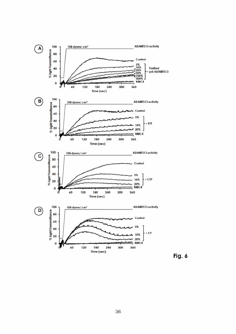
H-SIPA in the absence of ADAMTS13
To reproduce platelet aggregation assumed to be occurring in TTP patients, H-SIPA
10
at a constant shear rate of 108 dynes/cm2 was measured with an argon laser-assisted
cone platelet aggregometer (Toray Medical, Tokyo),21 using a mixture of normal
washed platelets (300x109/L, final), ADAMTS13-dp plasma (29% v/v, final), and the
purified pd-VWF (250 % of the normal plasma, final).
For this assay, normal washed platelets were prepared and suspended in a
Hepes-Tyrode buffer (pH 7.3) containing 1.8 mmol/L CaCl2.22 The mixture with a total
volume of 400 µL was preincubated at 37 ºC for 5 min, and then H-SIPA was measured
for 6 min. The maximum platelet aggregation was seen in the absence of any additives,
and the minimum or nonspecific platelet aggregation was determined in the presence of
anti-VWF monoclonal antibody NMC-4 (10 µg IgG/mL, final), which totally blocks the
VWF binding to platelet GPIb.13
For assessment of the inhibitory effect of various forms of pd-ADAMTS13 to
H-SIPA, they were spiked into the abovementioned assay mixtures and incubated for 5
min at 37 ºC before measurement. H-SIPA was measured at room temperature and
completed within 2.5 h after blood collection. The inhibition rate of H-SIPA was
calculated in the following formula: Inhibition rate (%) = [1- (% light transmittance of
tested sample / % light transmittance of control)] x100. These data were expressed as
the mean±SD. We calculated the inhibition rate in two points at 140 seconds and 340
seconds after the initiation of H-SIPA. Comparison between these 2 points were tested
for statistical significance using paired t-test using StatView (SAS Institute Inc, Cary,
NC, USA). A p-value <0.05 was considered significant.
11
RESULTS
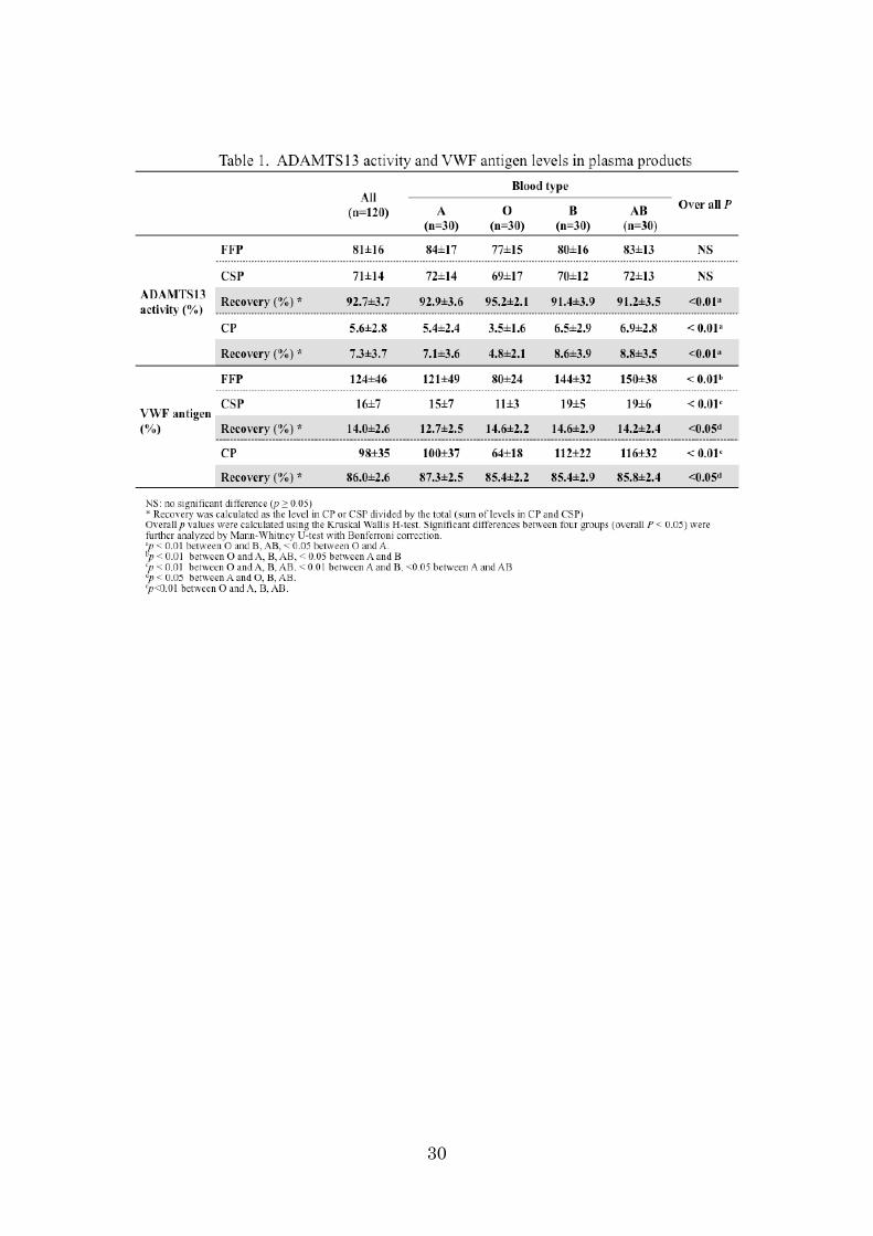
ADAMTS13 and VWF on IEF agarose-acrylamide composite gels
We detected pd-ADAMTS13 (15 ng) as one band at pI 4.9–5.6 (median 5.4) using
anti-ADAMTS13 monoclonal antibody (WH2-11-1) on IEF agarose-acrylamide
composite gels (Fig 1, left panel). Next, we analyzed various amounts (1–10 µL) of
normal citrated plasma, and found that ADAMTS13 antigen in the plasma milieu could
also be detected as a major band at pI 4.9–5.6, as in the case of purified pd-ADAMTS13.
In plasma, however, two additional bands of ADAMTS13 antigen were also detected:
one was composed of a cluster of blurred bands at pI 5.8–6.7, and the other consisted of
two clear bands at pI 7.0/7.5. In T3-VWD plasma, two groups of ADAMTS13 bands, pI
4.9–5.6 and 5.8–6.7, were detected, but the bands at pI 7.0/7.5 were totally absent (Fig.1
right panel). T3-VWD plasma lacks VWF antigen; therefore, the two groups of bands at
pI 4.9–5.6 and 5.8–6.7 appear to exist independently of the presence of plasma VWF.
Conversely, we assumed that the bands at pI 7.0/7.5 represented a complex with VWF
that exists within the plasma milieu. The bands at pI 7.0/7.5 were also detected after
mixing FFP with 1 M NaCl (final), excluding the possibility that the complex is formed
by an ionic linkage (data not shown).
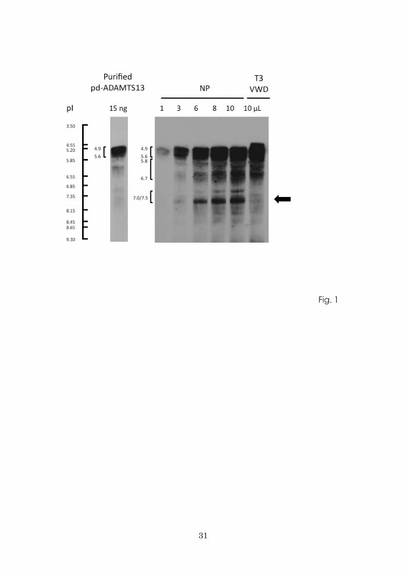
Generation of the pI 7.0/7.5 band of ADAMTS13 complex with VWF
Next, we performed the mixing experiments shown in Fig. 2A. T3-VWD plasma
12
spiked with purified pd-VWF yielded a new band at pI 7.5. USS-EE4 plasma initially
lacked three groups of ADAMTS13 bands (pI 4.9–5.6, 5.8–6.7, and 7.0/7.5), but once
that plasma was spiked with purified pd-ADAMTS13, the band at pI 7.5 clearly
appeared. When T3-VWD and USS-EE4 plasma samples were mixed together, the band
at pI 7.5 also appeared, confirming that it represents a complex of VWF and
ADAMTS13.
ADAMTS13 (pI 7.5) is a non-covalent complex with VWF in the plasma milieu
We next evaluated the effects of IAA, which blocks free thiols and prevents the
formation of disulfide bond–mediated covalent complexes, under high shear stress in a
vortex mixer. As shown in Fig. 2B, the band at pI 7.5, representing the
VWF-ADAMTS13 complex, was generated irrespective of the presence of IAA. When
pd-VWF was spiked into this mixture, the density of the band at pI 7.5 increased. These
results indicate that in our experiments, formation of the VWF-ADAMTS13 complex
does not depend upon disulfide bond bridges.
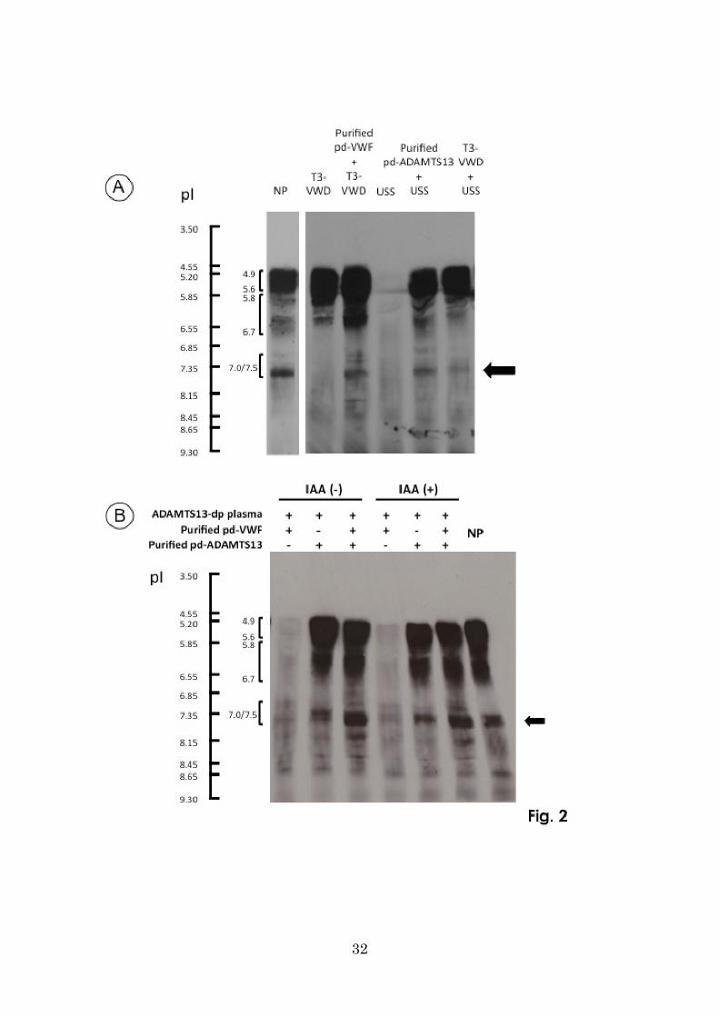
ADAMTS13 is present in plasma in complex with a large VWFM
As shown in Fig. 3, IEF gel analysis of normal plasma revealed ADAMTS13 as three
groups of bands (pI 4.9–5.6, 5.8–6.7, and 7.0/7.5) (also see Fig. 1), and VWF antigen
was largely separated into four series of bands: pI 4.8–5.6 (trace), 6.0–6.7, 7.1–7.8, and
7.9–8.4 (Fig. 3, middle).
Since two ADAMTS13 bands with pI 4.9–5.6 and 5.8–6.7 were seen in T3-VWD
13
plasma (Fig. 1), both bands appeared to be present in plasma irrespective of the
presence of VWF. Further, two-dimensional analysis of normal plasma (IEF gel
followed by SDS-0.9% agarose gel electrophoresis) confirmed that ADAMTS13 forms
a complex with a larger VWFM with pI 7.1-7.8, but less likely with a smaller VWFM
(dimers and tetramers) with pI 4.8-5.6 (Fig. 3 lower panel).
Amounts of ADAMTS13 and VWF in CP and CSP
Gill et al.23 reported that the level of VWF antigen in plasmas from normal
individuals with blood group O is significantly lower than that in plasmas with non-O
blood groups. Further, Feys et al.12 indicated that ADAMTS13 is bound to VWF with a
stoichiometry of one ADAMTS13 molecule to 250 VWF monomeric subunits. Taken
these two reports together, it is conceivable that the amount of a complex of
ADAMTS13 and VWF in CP could be influenced by the ABO blood groups.
We analyzed ADAMTS13 activity and VWF antigen in FFP, CSP, and CP from
120 normal volunteers, with 30 individuals of each ABO blood type (A, B, O, and AB).
The recovery rates of ADAMTS13 activity and VWF antigen in CP or CSP were
expressed as the level in CP or CSP divided by the sum of the levels in both (CP + CSP).
As summarized in Table 1, an average of 7.3% (range, 4.8–8.8%) of plasma
ADAMTS13 was recovered in CP, whereas an average of 92.7% (range, 91.2–95.2%)
remained in CSP. The amounts of ADAMTS13 remaining in CP from A, O, B, and AB
plasmas were 5.4±2.4%, 3.5±1.6%, 6.5±2.9%, and 6.9±2.8%, respectively; the amount
of ADAMTS13 in CP was significantly lower in blood group O than in other blood
14
groups. On the other hand, an average of 86.0% (range, 85.4–87.3%) of plasma VWF
antigen was recovered in CP, whereas an average of 14.0% (range, 12.7–14.6 %)
remained in CSP. The amounts of VWF in FFP, CP, and CSP from blood group O were
significant lower than in samples from other blood groups. The recovery rate of VWF
antigen in CP was significantly higher in blood group A than in other blood groups.
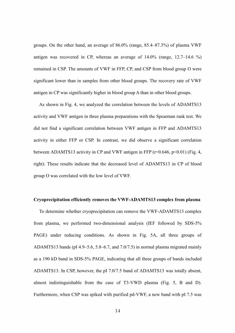
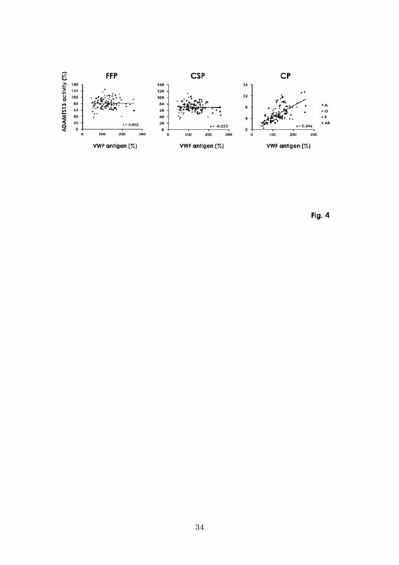
As shown in Fig. 4, we analyzed the correlation between the levels of ADAMTS13
activity and VWF antigen in three plasma preparations with the Spearman rank test. We
did not find a significant correlation between VWF antigen in FFP and ADAMTS13
activity in either FFP or CSP. In contrast, we did observe a significant correlation
between ADAMTS13 activity in CP and VWF antigen in FFP (r=0.646, p<0.01) (Fig. 4,
right). These results indicate that the decreased level of ADAMTS13 in CP of blood
group O was correlated with the low level of VWF.
Cryoprecipitation efficiently removes the VWF-ADAMTS13 complex from plasma
To determine whether cryoprecipitation can remove the VWF-ADAMTS13 complex
from plasma, we performed two-dimensional analysis (IEF followed by SDS-5%
PAGE) under reducing conditions. As shown in Fig. 5A, all three groups of
ADAMTS13 bands (pI 4.9–5.6, 5.8–6.7, and 7.0/7.5) in normal plasma migrated mainly
as a 190 kD band in SDS-5% PAGE, indicating that all three groups of bands included
ADAMTS13. In CSP, however, the pI 7.0/7.5 band of ADAMTS13 was totally absent,
almost indistinguishable from the case of T3-VWD plasma (Fig. 5, B and D).
Furthermore, when CSP was spiked with purified pd-VWF, a new band with pI 7.5 was
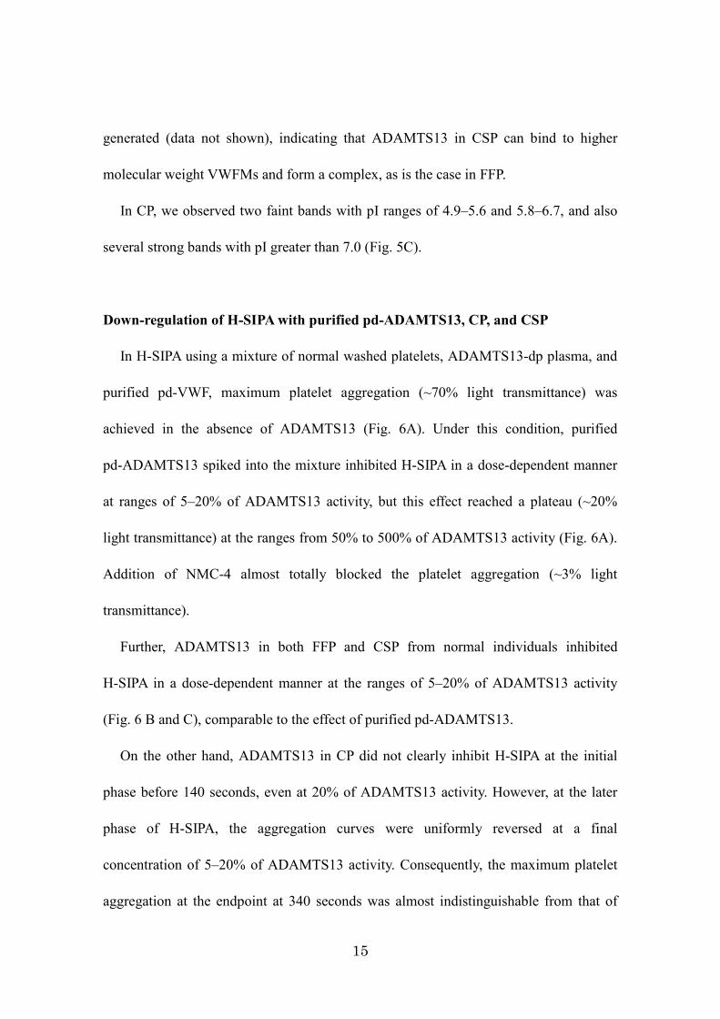
15
generated (data not shown), indicating that ADAMTS13 in CSP can bind to higher
molecular weight VWFMs and form a complex, as is the case in FFP.
In CP, we observed two faint bands with pI ranges of 4.9–5.6 and 5.8–6.7, and also
several strong bands with pI greater than 7.0 (Fig. 5C).
Down-regulation of H-SIPA with purified pd-ADAMTS13, CP, and CSP
In H-SIPA using a mixture of normal washed platelets, ADAMTS13-dp plasma, and
purified pd-VWF, maximum platelet aggregation (~70% light transmittance) was
achieved in the absence of ADAMTS13 (Fig. 6A). Under this condition, purified
pd-ADAMTS13 spiked into the mixture inhibited H-SIPA in a dose-dependent manner
at ranges of 5–20% of ADAMTS13 activity, but this effect reached a plateau (~20%
light transmittance) at the ranges from 50% to 500% of ADAMTS13 activity (Fig. 6A).
Addition of NMC-4 almost totally blocked the platelet aggregation (~3% light
transmittance).
Further, ADAMTS13 in both FFP and CSP from normal individuals inhibited
H-SIPA in a dose-dependent manner at the ranges of 5–20% of ADAMTS13 activity
(Fig. 6 B and C), comparable to the effect of purified pd-ADAMTS13.
On the other hand, ADAMTS13 in CP did not clearly inhibit H-SIPA at the initial
phase before 140 seconds, even at 20% of ADAMTS13 activity. However, at the later
phase of H-SIPA, the aggregation curves were uniformly reversed at a final
concentration of 5–20% of ADAMTS13 activity. Consequently, the maximum platelet
aggregation at the endpoint at 340 seconds was almost indistinguishable from that of
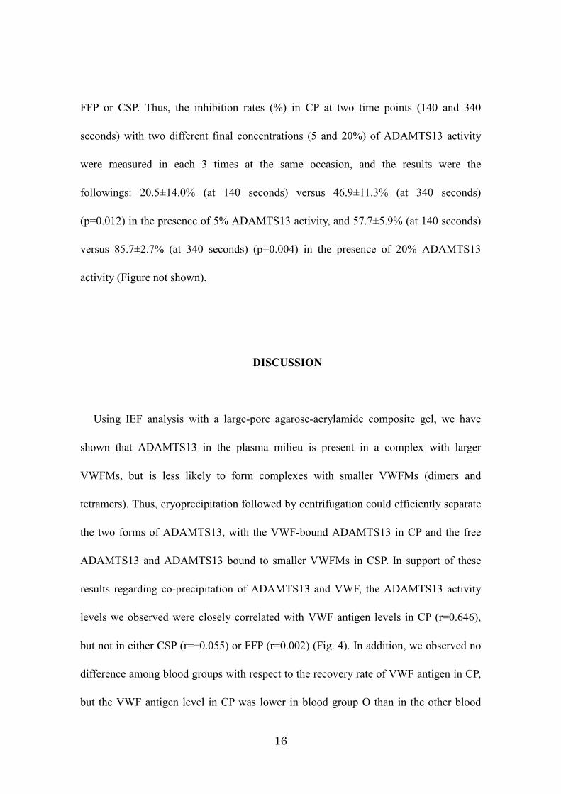
16
FFP or CSP. Thus, the inhibition rates (%) in CP at two time points (140 and 340
seconds) with two different final concentrations (5 and 20%) of ADAMTS13 activity
were measured in each 3 times at the same occasion, and the results were the
followings: 20.5±14.0% (at 140 seconds) versus 46.9±11.3% (at 340 seconds)
(p=0.012) in the presence of 5% ADAMTS13 activity, and 57.7±5.9% (at 140 seconds)
versus 85.7±2.7% (at 340 seconds) (p=0.004) in the presence of 20% ADAMTS13
activity (Figure not shown).
DISCUSSION
Using IEF analysis with a large-pore agarose-acrylamide composite gel, we have
shown that ADAMTS13 in the plasma milieu is present in a complex with larger
VWFMs, but is less likely to form complexes with smaller VWFMs (dimers and
tetramers). Thus, cryoprecipitation followed by centrifugation could efficiently separate
the two forms of ADAMTS13, with the VWF-bound ADAMTS13 in CP and the free
ADAMTS13 and ADAMTS13 bound to smaller VWFMs in CSP. In support of these
results regarding co-precipitation of ADAMTS13 and VWF, the ADAMTS13 activity
levels we observed were closely correlated with VWF antigen levels in CP (r=0.646),
but not in either CSP (r=−0.055) or FFP (r=0.002) (Fig. 4). In addition, we observed no
difference among blood groups with respect to the recovery rate of VWF antigen in CP,
but the VWF antigen level in CP was lower in blood group O than in the other blood
17
groups. As a result, both the ADAMTS13 activity and VWF antigen levels in CP were
significantly lower in blood group O than in non-O blood groups (Table 1). Further, we
determined that ~95% of the original ADAMTS13 activity in FFP is recovered after
cryoprecipitation; ~93% of the recovered ADAMTS13 activity remained in CSP,
whereas 7% was found in CP. This relative distribution of ADAMTS13 in FFP and CSP
was consistent with previous reports.24-26
Evidence that the pI 7.0/7.5 band is a complex of VWF and ADAMTS13 is clearly
provided by the following observations: 1) plasmas from both VWF antigen–defective
T3-VWD and ADAMTS13 antigen–defective USS patients lacked the bands at pI
7.0/7.5; 2) an equal mixture of plasmas from T3-VWD and USS generated the bands at
pI 7.0/7.5; and 3) CSP prepared from normal plasma lacked the bands at pI 7.0/7.5,
whereas CSP spiked with purified VWF regenerated these bands. On the other hand, we
assume that the proteins in the two other band groups (pI 4.9–5.6 and 5.8–6.7) are less
involved in complex formation with VWF, because both band groups are present in
T3-VWD plasma. Furthermore, because pd-ADAMTS13 purified from pooled normal
plasmas has only one band with pI 4.9–5.6,14 the pI 5.8-6.7 band might represent a
complex with proteins other than VWF. This speculation originates from the
observation that ADAMTS13 can bind in vitro to a soluble form of CD3627 and
Lys-plasminogen.28
In a previous study, immunoprecipitation method using anti-VWF antibodies was
used to show that ~3% of the total in plasma ADAMTS13 is bound to VWF.12 By
contrast, in our IEF gel analysis, coupled with densitometry, we observed that the
18
amount of VWF-bound ADAMTS13 in plasma appeared to be much lower than the
amount of unbound ADAMTS13, but greater than the 3% of total ADAMTS13 (Fig.
1).12 This discrepancy might be attributable to differences in the experimental designs
employed in these studies.
The mechanism by which ADAMTS13 binds to VWF in the plasma milieu is a
critical issue that remains to be addressed. Because Fujikawa et al.29 succeeded in
purifying ADAMTS13 from a commercial concentrate of factor VIII/VWF, prepared
from cryoprecipitate, such concentrates might contain the VWF-ADAMTS13 complex
itself. After extensive fractionation, including fibrin-clot formation, ammonium sulfate
precipitation, and sequential chromatography, the purified ADAMTS13 described in
that study was free of VWF. However, in our experience, the VWF-ADAMTS13
complex in CP is not readily dissociated by size-exclusion chromatography in the
presence of either 0.15 M or 1 M NaCl (data not shown). In fact, when we treated CP
with 1 M NaCl for 1 h at room temperature before IEF, the bands at pI 7.0/7.5 persisted,
indicating that no dissociation of VWF-ADAMTS13 complex had taken place under
conditions of high ionic strength. These results may indicate that VWF binding to
ADAMTS13 is independent of ionic strength. In this regard, McKinnon et al.30 reported
that the N-linked glycans of VWF exert a modulatory effect on the interaction with
ADAMTS13, and that removal of the N-linked glycans from VWF increased its affinity
for ADAMTS13 under static conditions. Furthermore, Yeh et al.19 recently reported that
ADAMTS13 possesses a disulfide bond–reducing activity that regulates shear-induced
thiol-disulfide exchange. Therefore, one of the mechanisms underlying formation of
19
VWF-ADAMTS13 complexes might involve disulfide-bond formation between
ADAMTS13 and VWF. To address this issue, we investigated whether IAA, a blocker
of free thiols, might prevent the formation of a disulfide bond–mediated covalent
complex under high shear stress. We observed, however, that VWF-ADAMTS13
complex formation was unaffected by IAA treatment, suggesting that in CP, the amount
of VWF-ADAMTS13 complex formed in a thiol-dependent fashion is marginal. This
finding rules out a major role for disulfide bonds, but otherwise we have not elucidated
the binding mechanism of VWF and ADAMTS13; this issue remains to be addressed in
future studies.
PE is a first-line treatment for acquired TTP. For this purpose, either FFP or CSP is
commonly used,31 but the results regarding CP have been controversial.32 At least one
case of congenital TTP (USS) has been successfully treated with CP.9 Therefore, it is
important to determine whether there is a functional difference between bound and
unbound ADAMTS13, and whether any such difference has physiological relevance.
Here we have clearly shown that CSP contains the unbound or less bound ADAMTS13,
whereas CP contains much more bound ADAMTS13 and lower levels of its unbound
counterpart. An authoritative determination regarding which form of ADAMTS13 more
efficiently down-regulates H-SIPA will be crucial in establishing the optimal treatment
modality for TTP patients.
In most acquired TTP patients, plasma ADAMTS13 activity is less than 5% of
normal. As a consequence, UL-VWFMs are not cleaved after secretion from endothelial
cells, and remain anchored to the cell surface in long strings.33 Circulating platelets
20
adhere to these long strings, resulting in occlusive platelet thrombi. However, smaller
VWFMs do not induce this spontaneous adhesion and aggregation of platelets.
Consequently, increased fluid shear stress is required to induce platelet aggregation in
vitro.34
In order to reproduce the platelet aggregation generated in the microvasculature of
ADAMTS13 activity–deficient TTP patients, here we employed an H-SIPA assay
system that uses a mixture of washed normal platelets and ADAMTS13-dp plasma
spiked with purified pd-VWF to mimic TTP plasmas. In this assay, the purified
pd-ADAMTS13 inhibited H-SIPA in a dose-dependent manner, reaching a plateau of
20% ADAMTS13 activity at final pd-ADAMTS13 concentrations up to 500%. Under
the same experimental conditions, FFP, CSP, and CP inhibited H-SIPA in a
dose-dependent manner to the same extent at the end-points; in CP, however, the
aggregation inhibition curves were different, and in fact no distinct inhibition was
observed at the initial phase of platelet aggregation. This might be simply explained by
the fact that the VWF concentration in the H-SIPA reaction mixtures was much higher
than in CSP or FFP. Alternatively, the binary complex of ADAMTS13 and larger
VWFMs might modulate a different phase of H-SIPA than unbound ADAMTS13,
because CSP spiked with purified VWF readily generates the ADAMTS13–larger
VWFM complex with pI 7.0/7.5. The larger VWFM is required in the earliest phase of
platelet thrombi formation and high shear stress, but in the later phase the
ADAMTS13–larger VWF complex embedded in the thrombi may play a role in
regulating the size of the thrombi in order to prevent microvascular occlusion. Further
21
studies are required to determine the functional differences between ADAMTS13 in
CSP and CP.
In conclusion, our results indicated that both plasma products of FFP and CSP are
effective in treatment of TTP. However, CSP may be more favorable for PE in acquired
TTP patients: relative to FFP, CSP has a lower level of VWF and a comparable
ADAMTS13 activity, but lower amounts of ADAMTS13–larger VWFM complex.
Acknowledgements
Plasma of T3-VWD patient was kindly provided by Dr. Midori Shima, Department
of Pediatrica, Nara Medical University. This study was supported in part by research
grants from the Ministry of Health, Labor, and Welfare of Japan, from the Ministry of
Education, Culture, Sports, Science and Technology of Japan, and from the Takeda
Science Foundation.
Authorship
Contribution: YH performed research, analyzed and interpreted data, and wrote the
manuscript. MH, AI performed research. KS contributed vital reagent. MM analyzed
data and wrote the manuscript. YF designed research, interpreted data, and wrote the
manuscript.
22
REFERENCES
1. Ruggeri ZM. Von Willebrand factor, platelets and endothelial cell interactions. J Thromb Haemost 2003;1: 1335-42.
2. Moake JL, Turner NA, Stathopoulos NA, et al. Involvement of large plasma von Willebrand factor (vWF) multimers and unusually large vWF forms derived from endothelial cells in shear stress-induced platelet aggregation. J Clin Invest 1986;78: 1456-61.
3. Zheng X, Chung D, Takayama TK, et al. Structure of von Willebrand factor-cleaving protease (ADAMTS13), a metalloprotease involved in thrombotic thrombocytopenic purpura. J Biol Chem 2001;276: 41059-63.
4. Levy GG, Nichols WC, Lian EC, et al. Mutations in a member of the ADAMTS gene family cause thrombotic thrombocytopenic purpura. Nature 2001;413: 488-94.
5. Soejima K, Mimura N, Hirashima M, et al. A novel human metalloprotease synthesized in the liver and secreted into the blood: possibly, the von Willebrand factor-cleaving protease? J Biochem 2001;130: 475-80.
6. Crawley JT, de Groot R, Xiang Y, et al. Unraveling the scissile bond: how ADAMTS13 recognizes and cleaves von Willebrand factor. Blood 2011;118: 3212-21.
7. Sadler JE. Von Willebrand factor, ADAMTS13, and thrombotic thrombocytopenic purpura. Blood 2008;112: 11-8.
8. Fujimura Y, Matsumoto M, Isonishi A, et al. Natural history of Upshaw-Schulman syndrome based on ADAMTS13 gene analysis in Japan. J Thromb Haemost 2011;9 Suppl 1: 283-301.
9. Allford SL, Harrison P, Lawrie AS, et al. Von Willebrand factor-cleaving protease activity in congenital thrombotic thrombocytopenic purpura. Br J Haematol 2000;111: 1215-22.
10. Scully M, Hunt BJ, Benjamin S, et al. Guidelines on the diagnosis and management of thrombotic thrombocytopenic purpura and other thrombotic microangiopathies. Br J Haematol 2012;158: 323-35.
11. Rock G, Anderson D, Clark W, et al. Does cryosupernatant plasma
23
improve outcome in thrombotic thrombocytopenic purpura? No answer yet. Br J Haematol 2005;129: 79-86.
12. Feys HB, Anderson PJ, Vanhoorelbeke K, et al. Multi-step binding of ADAMTS-13 to von Willebrand factor. J Thromb Haemost 2009;7: 2088-95.
13. Fujimura Y, Usami Y, Titani K, et al. Studies on anti-von Willebrand factor (vWF) monoclonal antibody NMC-4, which inhibits both ristocetin- and botrocetin-induced vWF binding to platelet glycoprotein Ib. Blood 1991;77: 113-20.
14. Hiura H, Matsui T, Matsumoto M, et al. Proteolytic fragmentation and sugar chains of plasma ADAMTS13 purified by a conformation-dependent monoclonal antibody. J Biochem 2010;148: 403-11.
15. Uemura M, Tatsumi K, Matsumoto M, et al. Localization of ADAMTS13 to the stellate cells of human liver. Blood 2005;106: 922-4.
16. Kato S, Matsumoto M, Matsuyama T, et al. Novel monoclonal antibody-based enzyme immunoassay for determining plasma levels of ADAMTS13 activity. Transfusion 2006;46: 1444-52.
17. Yagi H, Ito S, Kato S, et al. Plasma levels of ADAMTS13 antigen determined with an enzyme immunoassay using a neutralizing monoclonal antibody parallel ADAMTS13 activity levels. Int J Hematol 2007;85: 403-7.
18. Bartlett A, Dormandy KM, Hawkey CM, et al. Factor-VIII-related antigen: measurement by enzyme immunoassay. Br Med J 1976;1: 994-6.
19. Yeh HC, Zhou Z, Choi H, et al. Disulfide bond reduction of von Willebrand factor by ADAMTS-13. J Thromb Haemost 2010;8: 2778-88.
20. Soejima K, Nakamura H, Hirashima M, et al. Analysis on the molecular species and concentration of circulating ADAMTS13 in blood. J Biochem 2006;139: 147-54.
21. Ikeda Y, Handa M, Kawano K, et al. The role of von Willebrand factor and fibrinogen in platelet aggregation under varying shear stress. J
24
Clin Invest 1991;87: 1234-40. 22. Fujimura Y, Ikeda Y, Miura S, et al. Isolation and characterization of
jararaca GPIb-BP, a snake venom antagonist specific to platelet glycoprotein Ib. Thromb Haemost 1995;74: 743-50.
23. Gill JC, Endres-Brooks J, Bauer PJ, et al. The effect of ABO blood group on the diagnosis of von Willebrand disease. Blood 1987;69:1691-5.
24. Yarranton H, Lawrie AS, Purdy G, et al. Comparison of von Willebrand factor antigen, von Willebrand factor-cleaving protease and protein S in blood components used for treatment of thrombotic thrombocytopenic purpura. Transfus Med 2004;14:39-44.
25. Rock G, Yousef H, Lu M. ADAMTS-13 in fresh, stored, and solvent/detergent-treated plasma. Transfusion 2006;46:1261-2.
26. Scott EA, Puca KE, Pietz BC, et al. Comparison and stability of ADAMTS13 activity in therapeutic plasma products. Transfusion 2007;47:120-5.
27. Davis AK, Makar RS, Stowell CP, et al. ADAMTS13 binds to CD36: a potential mechanism for platelet and endothelial localization of ADAMTS13. Transfusion 2009;49: 206-13.
28. Shin Y, Akiyama M, Kokame K, et al. Binding of von Willebrand factor cleaving protease ADAMTS13 to Lys-plasmin(ogen). J Biochem 2012;152: 251-8.
29. Fujikawa K, Suzuki H, McMullen B, Chung D. Purification of human von Willebrand factor-cleaving protease and its identification as a new member of the metalloproteinase family. Blood 2001;98: 1662-6.
30. McKinnon TA, Chion ACK, Millington AJ, et al. N-linked glycosylation of VWF modulates its interaction with ADAMTS13. Blood 2008;111:3042-9.
31. Byrnes JJ, Moake JL, Klug P, Periman P. Effectiveness of the cryosupernatant fraction of plasma in the treatment of refractory thrombotic thrombocytopenic purpura. Am J Hematol 1990;34: 169-74.
32. Zeigler ZR, Shadduck RK, Gryn JF, et al. Cryoprecipitate poor plasma
25
does not improve early response in primary adult thrombotic thrombocytopenic purpura (TTP). J Clin Apher 2001;16: 19-22.
33. Dong J, Moake JL, Nolasco L, et al. ADAMTS-13 rapidly cleaves newly secreted ultralarge von Willebrand factor multimers on the endothelial surface under flowing conditions. Blood 2002;100:4033-9.
34. Moake JL, Turner NA, Stathopoulos NA, et al. Involvement of large plasma von Willebrand factor (vWF) multimers and unusually large vWF forms derived from endothelial cells in shear stress-induced platelet aggregation. J Clin Invest 1986;78: 1456–61.
26
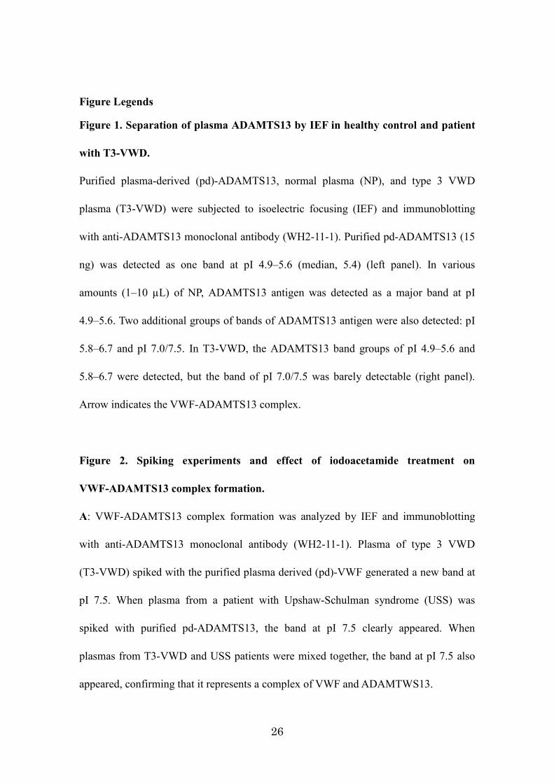
Figure Legends
Figure 1. Separation of plasma ADAMTS13 by IEF in healthy control and patient
with T3-VWD.
Purified plasma-derived (pd)-ADAMTS13, normal plasma (NP), and type 3 VWD
plasma (T3-VWD) were subjected to isoelectric focusing (IEF) and immunoblotting
with anti-ADAMTS13 monoclonal antibody (WH2-11-1). Purified pd-ADAMTS13 (15
ng) was detected as one band at pI 4.9–5.6 (median, 5.4) (left panel). In various
amounts (1–10 µL) of NP, ADAMTS13 antigen was detected as a major band at pI
4.9–5.6. Two additional groups of bands of ADAMTS13 antigen were also detected: pI
5.8–6.7 and pI 7.0/7.5. In T3-VWD, the ADAMTS13 band groups of pI 4.9–5.6 and
5.8–6.7 were detected, but the band of pI 7.0/7.5 was barely detectable (right panel).
Arrow indicates the VWF-ADAMTS13 complex.
Figure 2. Spiking experiments and effect of iodoacetamide treatment on
VWF-ADAMTS13 complex formation.
A: VWF-ADAMTS13 complex formation was analyzed by IEF and immunoblotting
with anti-ADAMTS13 monoclonal antibody (WH2-11-1). Plasma of type 3 VWD
(T3-VWD) spiked with the purified plasma derived (pd)-VWF generated a new band at
pI 7.5. When plasma from a patient with Upshaw-Schulman syndrome (USS) was
spiked with purified pd-ADAMTS13, the band at pI 7.5 clearly appeared. When
plasmas from T3-VWD and USS patients were mixed together, the band at pI 7.5 also
appeared, confirming that it represents a complex of VWF and ADAMTWS13.
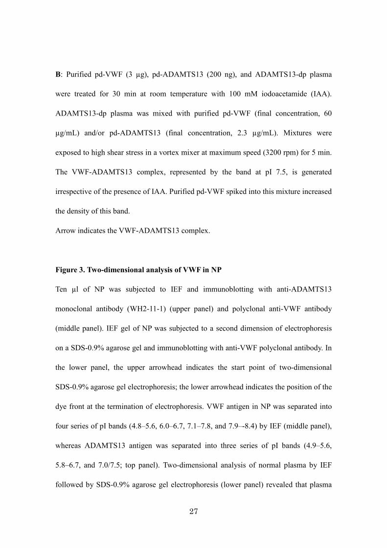
27
B: Purified pd-VWF (3 µg), pd-ADAMTS13 (200 ng), and ADAMTS13-dp plasma
were treated for 30 min at room temperature with 100 mM iodoacetamide (IAA).
ADAMTS13-dp plasma was mixed with purified pd-VWF (final concentration, 60
µg/mL) and/or pd-ADAMTS13 (final concentration, 2.3 µg/mL). Mixtures were
exposed to high shear stress in a vortex mixer at maximum speed (3200 rpm) for 5 min.
The VWF-ADAMTS13 complex, represented by the band at pI 7.5, is generated
irrespective of the presence of IAA. Purified pd-VWF spiked into this mixture increased
the density of this band.
Arrow indicates the VWF-ADAMTS13 complex.
Figure 3. Two-dimensional analysis of VWF in NP
Ten µl of NP was subjected to IEF and immunoblotting with anti-ADAMTS13
monoclonal antibody (WH2-11-1) (upper panel) and polyclonal anti-VWF antibody
(middle panel). IEF gel of NP was subjected to a second dimension of electrophoresis
on a SDS-0.9% agarose gel and immunoblotting with anti-VWF polyclonal antibody. In
the lower panel, the upper arrowhead indicates the start point of two-dimensional
SDS-0.9% agarose gel electrophoresis; the lower arrowhead indicates the position of the
dye front at the termination of electrophoresis. VWF antigen in NP was separated into
four series of pI bands (4.8–5.6, 6.0–6.7, 7.1–7.8, and 7.9–-8.4) by IEF (middle panel),
whereas ADAMTS13 antigen was separated into three series of pI bands (4.9–5.6,
5.8–6.7, and 7.0/7.5; top panel). Two-dimensional analysis of normal plasma by IEF
followed by SDS-0.9% agarose gel electrophoresis (lower panel) revealed that plasma
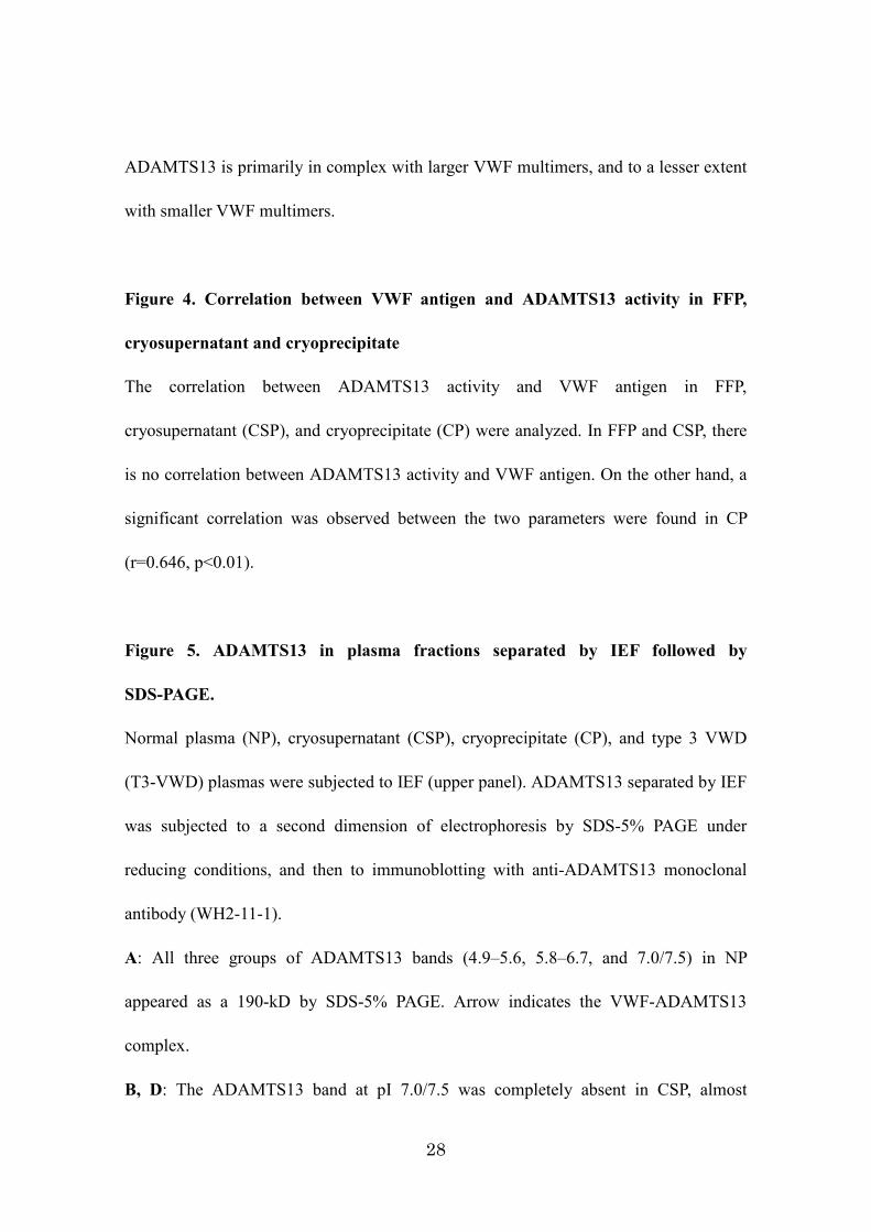
28
ADAMTS13 is primarily in complex with larger VWF multimers, and to a lesser extent
with smaller VWF multimers.
Figure 4. Correlation between VWF antigen and ADAMTS13 activity in FFP,
cryosupernatant and cryoprecipitate
The correlation between ADAMTS13 activity and VWF antigen in FFP,
cryosupernatant (CSP), and cryoprecipitate (CP) were analyzed. In FFP and CSP, there
is no correlation between ADAMTS13 activity and VWF antigen. On the other hand, a
significant correlation was observed between the two parameters were found in CP
(r=0.646, p<0.01).
Figure 5. ADAMTS13 in plasma fractions separated by IEF followed by
SDS-PAGE.
Normal plasma (NP), cryosupernatant (CSP), cryoprecipitate (CP), and type 3 VWD
(T3-VWD) plasmas were subjected to IEF (upper panel). ADAMTS13 separated by IEF
was subjected to a second dimension of electrophoresis by SDS-5% PAGE under
reducing conditions, and then to immunoblotting with anti-ADAMTS13 monoclonal
antibody (WH2-11-1).
A: All three groups of ADAMTS13 bands (4.9–5.6, 5.8–6.7, and 7.0/7.5) in NP
appeared as a 190-kD by SDS-5% PAGE. Arrow indicates the VWF-ADAMTS13
complex.
B, D: The ADAMTS13 band at pI 7.0/7.5 was completely absent in CSP, almost
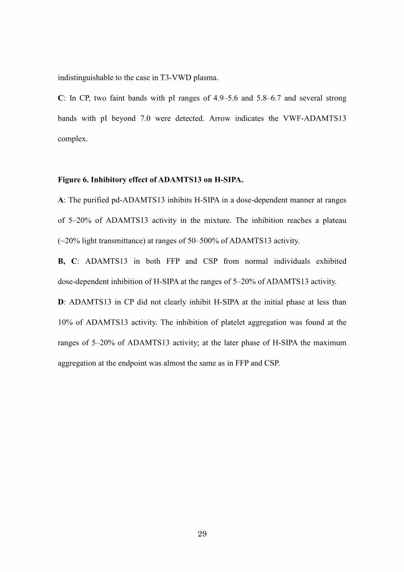
29
indistinguishable to the case in T3-VWD plasma.
C: In CP, two faint bands with pI ranges of 4.9–5.6 and 5.8–6.7 and several strong
bands with pI beyond 7.0 were detected. Arrow indicates the VWF-ADAMTS13
complex.
Figure 6. Inhibitory effect of ADAMTS13 on H-SIPA.
A: The purified pd-ADAMTS13 inhibits H-SIPA in a dose-dependent manner at ranges
of 5–20% of ADAMTS13 activity in the mixture. The inhibition reaches a plateau
(~20% light transmittance) at ranges of 50–500% of ADAMTS13 activity.
B, C: ADAMTS13 in both FFP and CSP from normal individuals exhibited
dose-dependent inhibition of H-SIPA at the ranges of 5–20% of ADAMTS13 activity.
D: ADAMTS13 in CP did not clearly inhibit H-SIPA at the initial phase at less than
10% of ADAMTS13 activity. The inhibition of platelet aggregation was found at the
ranges of 5–20% of ADAMTS13 activity; at the later phase of H-SIPA the maximum
aggregation at the endpoint was almost the same as in FFP and CSP.
30
31
32
33
34

35
36