Embed Size (px)
Citation preview

AD-A257 9941111111 l fm| ii lIII
CHAPTER
2
Long-Term Storage and ) TPreservation of Red Blood Cells C"
Gerald L. Moore 1992 .
CommMnderATf1I: S(RD-ULZ-TEI I Army atutitute of ResearchPmedio of San Pranciao. CA 94129-6800
Humans have experimented with blood transfusions for over 300 years andhave attempted to preserve human blood since the early 1900s. The firstmodern attempts to store blood were stimulated by World War I whenblood was stored in citrate-glucose solutions (Robertson 1918; Rous andTurner 1916). During World War II, the increased need for blood plasma
Sand whole blood resulted in the development of a solution called acid-___ I citrate-dextrose (ACD) for 21-day refrigerated storage of blood. A slight• variation of ACD, called CPD, was introduced in the late 1950s. For CPD,UM 0phosphate was added to the citrate and dextrose, which slightly improved
the viability of stored red cells, although the dating period was held to 21Sdays. Blood preservation solutions remained unchanged until the late 1970s- 1 when adenine was first added to CPD to produce CPDA-1, which extended
the shelf life of blood to 35 days (Peck et al. 1981). CPDA-l appears to be
the industry's final attempt to modify the anticoagulant solution for betterblood preservation. However, the success of U.S. and European blood bankswith CPDA- 1 has encouraged the development of modern additive solutionsfor component-specific preservation.
Red cell preservation research has traditionally centered around threeissues, maximizing viability and function, while minimizing cell lysis, andassuring that the storage systems maintain sterility. Obviously, any pre-
31
S

32 Long-Tom Storag, and Presrvaioon of Red Blood Cell
servatives must be nontoxic. Percent viability is defined as the percentageof the stored red cells remaining in circulation for 24 hours after infusion.For many years the U.S. Food and Drug Administration (FDA) set thismean percentage at 70%, but in 1985 this percentage was raised to 75%.Viability must be measured by in vivo red cell survival, but adenosinetriphosphate (ATP) has been traditionally used in developmental experi-ments as an indicator assay for viability since some correlation exists be-tween ATP level and viability (Peck et al. 1981). This correlation is bestdefined as a threshold, since it is known that ifATP levels drop below about30% of normal, the cells will have low viability, although a high ATP levelwill not necessarily insure good viability. Red cell function, i.e., oxygendelivery, is closely associated with the level of cellular 2,3-diphosphogly-cerate (2,3-DPG). During storage in any commercially available system, the2,3-DPG level falls to near zero in about 2 weeks. This results in a 50%reduction in the cells' ability to deliver oxygen to tissues, all other thingsbeing equal (Moore 1983). Red cell lysis during storage is limited to 1% byFDA policy. Red cells stored with white cells or without any plasma aremost likely to lyse, but the degree varies dramatically among donors.
This chapter covers the use of modern additive solutions for red cellstorage, the use of freezing to preserve red cells, some methods being de-veloped to make freezing of blood more practical, and new, methods ofprocessing stored red cells. The storage of platelets, white cells, and plasmacomponents will not be discussed.
2.1 CURRENT STATE OF LIQUID PRESERVATION AT 4*C
In 1983 the USA and parts of Europe shifted from preserving red cells by VAanticoagulant fortification to using CPD coupled with separate additive so-lutions. Employing this approach, blood was drawn into a basic anticoag-ulant and processed into components. The red cell component was then
TABLE 2-1 Composition of Commrdal Additive Solutions for Red Cells
Component CPD ADSOL AS-3' SAG SAGM Cir/Pk'
Adenine (mg) - 27 17 17 17 7 ForGlucose (g) 1.61 2.2 0.4 0.9 0.9 0.4 1NaH2PO, (mg) 140 1-- 285 - - 285Na Citrate (g) 1.66 - 588 - - 588Citric acid (mg) 206 - 42 - - 42Mannitol (g) - 0.75 - - 0.52 -NaCl (mg) - 900 718 877 877 718Water (ml) 63 100 100 100 100 100'AS-3 and Cr/Pk also use double glucose in their CPD. " -
. U .
2.1 CuWn Stat. of Lkqid Pmn.ftlon at 4C 33
mixed with an isotonic solution containing a nutrient mixture designed for42-day red cell preservation. The four principle solutions are ADSOL (orAS-I), AS-3 (or Nutricel), and in Europe, SAG and SAGM. Table 2-1 givesrecipes for these solutions.
The first additive solution was developed in Sweden in the late 1970sand contained saline, adenine, and glucose (and thus was named SAG).SAG was later modified by the addition of mannitol (SAGM) to retard lysis(Hogman et al. 1978a, 1987b, 1981). Hogman was the first to show thatwhite cell enzymes contaminating red cell suspension will increase red celllysis rates (Hogman et al. 1978b) and that lysis can be reduced to manageablelevels by adding mannitol to the storage solution (Hogman et al. 1981).Buffy-coat-poor red cells can be stored in SAGM for 35 days.
In 1983 Fenwal Laboratories (Deerfield, IL) introduced ADSOL (AS-1) solution for 49-day red cell storage (Heaton et al. 1984). In 1985 a con-troversy over the viability of red cell in this product (Heaton et al. 1985;Page 1985; eteutler 1985; and Valeri 1985) resulted in a reduction of thestorage time in AS-I to 42 days.
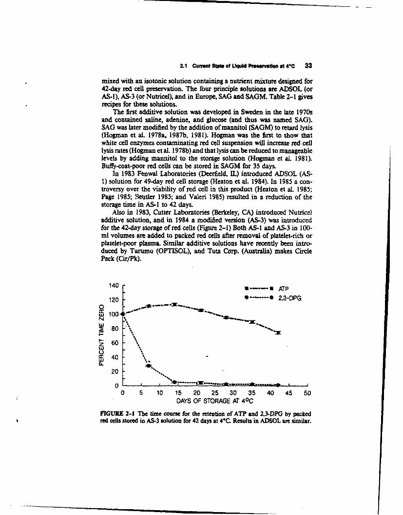
Also in 1983, Cutter Laboratories (Berkeley, CA) introduced Nutriceladditive solution, and in 1984 a modified version (AS-3) was introducedfor the 42-day storage of red cells (Figure 2-1) Both AS-I and AS-3 in 100-ml volumes are added to packed red cells after removal of platelet-rich orplatelet-poor plasma. Similar additive solutions have recently been intro-duced by Turumo (OPTISOL), and Tuta Corp. (Australia) makes CirclePack (Cir/Pk).
140 8-.---.U ATP
120 ........ 0 2,3-DPG0w 10080N '
S80 '
Z 6040a: 40
LU .
20
0 --------- .....- " .
0 5 10 15 20 25 30 35 40 45 50DAYS OF STORAGE AT 40 C
FIGURE 2-1 The time course for the retention of ATP and 2,3-DPG by packedred cells stored in AS-3 solution for 42 days at 4C. Results in ADSOL are similar.

34 Long-Team Storage and Preservatlon of Red Blood Calls
The switch to additive solutions for red cell preservation resulted in anincrease of storage time from 35 days (in CPDA-I) to 42 days. Other ad-vantages of the additive systems include lower viscosity, elimination ofexcessive nutrients in platelets, and better control of optimal ratios of redcells to nutrients. The current 42-day storage limit is felt by many to be allthat is needed in modern blood banking. Further extension in storage timewould require dramatically different approaches since the drop in pH andmembrane changes become critical after 42 days. None of these additivesolutions preserve 2,3-DPG beyond 7-14 days. Expanded reviews of ad-ditive solutions are presented elsewhere (Heaton 1986; Moore 1987).
2.2 CURRENT RESEARCH IN NONFROZEN SYSTEMS
2.2.1 Long-Term Uquid StorageIn 1986, Meryman investigated the use of osmotic swelling to extend redcell storage time in an attempt to retard lysis (Meryman et al. 1986). Heused ammonium salts of low concentration and found that they were ef-fective in maintaining red cell ATP for long periods (50% left after 12-16weeks). In vivo red cell survivals were measured on these cells after storageat 40C for 84-131 days. Percent survival varied from 46-86%. Lysis variedfrom 0.5-7%. The in vitro portion of these studies were repeated in ourlaboratory, with 50% ATP remaining at 8--10. weeks with a rate of lysisunder 1%. The system does have limitations, however, since the 2,3-DPGdrops to near zero by week 2, the pH (at 370C) drops to 6.0 by 7 weeks,and the cells must be washed extensively prior to use.
2.2.2 Maintaining 2,3-DPG for Preservation of FunctionThe concept of maintaining 2,3-DPG during storage has been investigatedfor two decades. Many metabolites have been tested as elevators of 2,3-DPG, including dihydroxyacetone, inosine, ascorbate (active component isoxalate), and methylene blue. To date these compounds have been eitherof only marginal benefit, or toxic (Moore 1983, 1987).
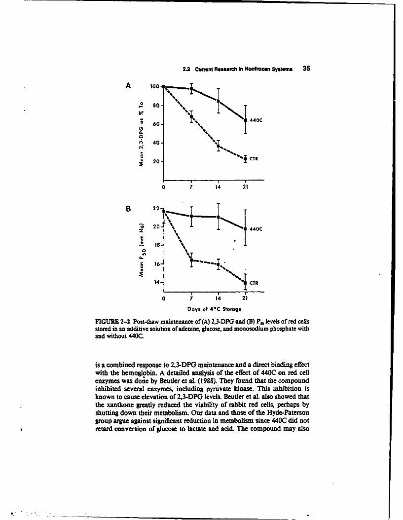
Several studies have been published using a modified xanthone, 2-hy-droxyethoxy-6-(5-tetrazoyl) xanthone, which was named BW A440C by itsdevelopers (Hyde et al. 1984). Our data.(Moore, unpublished observations)supports the findings of Hyde et al. (1984) and Paterson et al. (1988) thatBW A440C (440C) elevates 2,3-DPG and P.,, while not affecting ATP, pH,the use of glucose, or the production of lactose. We tested the 440C bothas a supplement to ADSOL storage for 42 days and as a supplement to anadenine/glucose/mannitol solution used in post-thaw preservation (Figure2-2). We showed that the xanthone could bind to pure AO hemoglobin (thepredominant genetic form) and raise the P30 of hemoglobin in a mannersimilar to the addition of 2,3-DPG (Table 2-2). The P50 effect on red cells
2.2 Curmt Research In Nonfrozen Systems 35
A 100 4I....
S60- 440C
A• 40-
C44
CT~20 aR:• 20-
I I I'
0 7 14 21
B 2
"• 20 . 1 440C
E
14 - Oi
7 14 21Days of 4*C Storage
FIGURE 2-2 Post-thaw maintenance of(A) 2,3-DPG and (B) P. levels of red cellsstored in an additive solution of adenine, glucose, and monosodium phosphate withand without 440C
is a combined response to 2,3-DPG maintenance and a direct binding effect
with the hemoglobin. A detailed anlysis of the effect of 440C on red cellenzymes was done by Beutler et al. (1988). They found that the compoundinhibited several enzymes, including pyruvate kinase. This inhibition isknown to cause elevation of 2,3-DPG levels. Beutler et al. also showed thatthe xanthone greatly reduced the viability of rabbit red cells, perhaps byshutting down their metabolism. Our data and those of the Hyde-Patersongroup argue against significant reduction in metabolism since 440C did notretard conversion of glucose to lactate and acid. The compound may also

36 Long-Term Storage and Preservation of Red tood Cells
TABLE 2-2 Mean P. Values as Measured on Hemoscan at 37 0C and pH 7A
Mean P,Sample (N - 3)
Red cells in buffer 19.0Red cells plus 440C 19.0'Hemoglobin AO in buffer 17.5Hemoglobin plus 440C 27.5Hemoglobin plus 2,3-DPG 32.0Hemoglobin plus 440C and 2,3-DPG 31.5
'440C and 2,3-DPG were added at time of Po assay.
have some hypotensive activity, which could preclude its genural use as ablood additive (unpublished data, Burroughs Wellcome Co.).
Meryman discussed a washing procedure which used nonionic bufferssuch as citrate to remove plasma components from red cells (Meryman1989). Subsequent storage of red cells in this buffer appears effective inmaintaining 2,3-DPG, perhaps by producing hydroxyl ions, which enter thered cells in order to replace the lost chloride ions. Other efforts will un-doubtedly be made to maintain 2,3-DPG during storage, if a nontoxic ad-ditive can be found.
2.2.3 Concern Over Phthalate ToxicityPolyvinylchloride (PVC) bags have been used for 30 years to store blood.PVC is made pliable by addition of up to 40% of di(2-ethylhexyl)phthalate(DEHP). The DEHP is mechanically trapped in the vinyl matrix and isinsoluble in water, but it will leach into hydrophobic materials such asplasma lipoproteins or cell membranes. The acute toxicity of DEHP is verylow, but it does bind to red cells, platelets, and plasma during blood storage,and is converted into the monoester MEHP (Rock et aL 1978). There hasbeen a long-standing concern that DEHP may have toxic effects. An excellentand current review of this subject raises new concern that DEHP may pro-mote, if not induce, cancer in some test animals (Rubin and Ness 1989).This effect cannot be reproduced in human hepatoottes (Turnbull and Rod-ricks 1985). The primary component of concern in vivo is the water solublemonoester MEHP formea by lipase activity on DEHP (Rubin and Ness1989).
The concern over DEHP/MEHP has influenced the search for alternatenonleachable plastics for blood components. Another concern, which willnot be discussed here, is the need for greater oxygen permeability to allow5-day platelet storage. Platelet storage bags of polyolefins or of PVC withthe plasticizer tri-(2-ethylhexyl)trimellitate have been available for severalyears. Shimizu et al. (1989) recently reported good results with platelet

2.2 Current Research In Nonfrozen System 37
storage using a PVC bag containing di-n-decyl phthalate. There has howeverbeen some reluctance to modify the storage bag used with red cells sinceDEHP actually stabilizes red cell membranes during storage (Estep et al.1984). Fenwal Laboratories recently has developed an entirely new bag sys-tem based on a citrate-plasticized PVC. This new bag is made of PVCcontaining butyl-trihexylcitrate and is effective for the storage of both redcells and platelets (Buchholz et al. 1989a, 1989b). Other bag companies willprobably follow Fenwal with a non-DEHP plasticized PVC bag.
2.2.4 Methods of Measuring Red Cell SurvivalThe efficacy of red cell preservation can only be measured by tagging thecells with a radioactive label, reinjecting them into the donor and evaluatingtheir survival. The two common methods for this procedure are defined asthe single and double label chromium methods. With the single-labelmethod, red cells are tagged with 5 Cr, injected, and blood volume is cal-culated by back-extrapolation of the 5-15 rmin dilutions of tagged red cells.In the double-label method, red cells are tagged with 3tCr, but blood volumeis measured by a separate isotope, usually 115I bound to albumin. A stan-dardized method for each of these approaches has-been published (Moroffet al. 1984), with the recognition that each method contains assumptionswhich can reduce its accuracy.
The controversy concerning relative accuracy and problems with singleversus double-label methods has been discussed (Moore 1987). To evaluatethe single label method independently, Beutler proposed an alternate doublelabel method using "Tc and fresh red cells to measure blood volumes(Beutler and West 1984). He showed that if the viability was above 80%the two methods gave identical results, but below 80% viability, the singlelabel technique overestimated blood volumes. However, he showed that theerror in absolute percentage of viable red cells remains small because thelarge (10 to 20) percentage error is applied to a small percentage ofremainingviable red cells, making the largest overestimation of viability only 4% (Beu-tier and West 1984). Marcus et al. (1987) studied red cell survival mea-surements using isotopes of chromium, technetium, and indium, andshowed that chromium produced higher and more accurate viabilities, dueto lower isotope elution rates. Heaton developed a modified technetiumprocedure which ninimized the elution of the technetium label, and con-firmed the 3.4% higher values found with single versus double label methods(Heaton et al. 1989a). AuBuchon and Brightman studied five methods ofindium-labeling red cells, and they found that four of them are effective asmeasures of blood volume (AuBuchon and Brightman 1989). One problemwith the use of indium is its overlap with the energy window with chromium,so that the two cannot be used together in most gamma counters (AuBuchonand Brightman 1989).

38 Long-Term Storage and Preservation of Red Blood Cells
The double label methods, while providing slightly superior viabilitydata, subject the donor to additional radiation and are technically muchmore difficult. To overcome this, a double label method has been developedusing nonradioactive Cr for blood volume measurements and "Cr for storedcell recovery (Heaton et al. 1989b, 1989c). This technique has a high cor-relation with older double-label methods (66 t 5% vs. 69 t 8%) but requiresa Zeeman electrothermal atomic absorption spectrophotometer to measure"2Cr. Another nonradioactive, red cell tagging method is being developedusing rabbit red cells. In this technique, the cells are tagged with biotin byreaction with N-hydroxysuccinimidobiotin (Suzuki and Dale 1987).
2.3 FROZEN RED CELLS
The technology currently employed for freezing red cells was developed inthe 1960s and early 1970s by the American Red Cross Research Lab (Wash-ington, D.C.) and the U.S. Naval Blood Research Lab (Boston, MA). Thismethod, known as the "high glycerol" procedure, has been extensively re-viewed (Valeri 1970, 1976, 1988; Meryman and Hornblower 1972; Meryman1979). The high glycerol method consists of mixing packed red cells with 6M glycerol and freezing in a special polyolefin freezing bag at -80°C. Cellsstored in this manner can be kept for at least 21 years (Valeri 1988; Valeriet al. 1989). Upon demand, the cells are thawed in a 37°C water bath anddeglycerolized by centrifugal washing with 2-3 liters of sterile saline solution(Widmann 1985). While fresh red cells are the usual starting material forthis process, Valeri et al. (1979) and Valeri (1988) have shown that outdatedred cells may be rejuvenated with a solution of pyruvate, inosine, phosphate,and adenine (PIPA) for 1 h at 37°C and then frozen. PIPA restores thelevels of the red cell ATP and 2,3-DPG to fresh blood levels, but must beremoved by washing prior to infusion since inosine promotes hypotension.The deglycerolizing step removes the residual PIPA after freezing and thaw-ing. Several washing machines developed by IBM (now COBE Labs, Denver,CO), and Haemonetics (Braintree, MA) have been approved by the FDAfor washing thawed red cells.
This frozen red cell technology has been available for over two decades,but has not gained popularity, except for very rare blood types, for severalreasons. First and foremt, the procedure is labor intensive and expensive,costing two to three times as much as nonfrozen red cells. Early hopes thatthe washing step might remove viruses from red cells was shown to beunfounded (Haugen 1979). An additional difficulty is the FDA requirementthat a thawed-washed unit be used within 24 hours due to both the possiblecompromise of sterility, and the lack of adequate nutrient support for thecells in the final wash solution. Another problem was identified when at-tempts to ship the frozen units in their special bags resulted in an unac-ceptably high rate (10-15%) of breakage (Valeri 1988).

2.3 Frozen Red Cefl 39
In 1981, the U.S. Naval Blood Research Lab developed a minor, butimportant, change in the freezing/thawing/washing procedure. This changeallowed the red cells to be frozen in the primary bag in which the cells hadbeen drawn (Valeri et al. 1981). The size of the primary bag for this pro-cedure was increased from 600 to 800 ml. This change eliminated the needfor transferring red cells to a freezing bag and reduced the breakage rate ofred cells shipped in these bags from 1S% to I% (Valeri 1988). The oversizedprimary bag containing CPDA-I and attached to the usual two or threesatellite bags can then be used for component preparation and either frozenor nonfrozen red cell storage. If 4°C-stored cells are not used in one week,they may be frozen in the same bag; if not used within 35 days, they maybe rejuvenated with PIPA and frozen in this same bag. In 1989, the U.S.Military Blood Program adopted this bag system, with the intention ofdeveloping a stockpile of several hundred thousand units of frozen type 0red cells for emergency use. The frozen cells will also be used routinely tomaintain turnover and familiarity with frozen blood manipulation. Stock-piled units will be maintained at various depots worldwide and kept for upto 21 years.
The explosive development of the biotechnolog industry in recent yearshas stimulated many improvements in membrane technology designed tofacilitate the separation of cells from supernatant solutions. Thus, it wasnatural that researchers consider using membranes to deglycerolize frozen-thawed red cells. Membrane technology offers several potential advantagesover the current centrifugal washing to remove glycerol. First, the membraneand its integral connecting tubing harness could be sterilized. This wouldremove the FDA objection to the nonclosed nature of the centrifugal bowl,potentially allowing for extended storage of the washed red cells. Otherpotential advantages include smaller, less expensive hardware, faster washtimes, less operator interface, and the ability to control hematocrit in thefinish'd product.
To deglycerolize red cells, the Millipore Corp., Sterimatics Division(Boston, MA), has developed a prototype device which is a modification oftheir plasmapheresis machine. One prototype machine was extensively eval-uated in the Naval. Blood Research Lab and another was examined in ourlab (Moore, unpublished observations). These prototypes are microproces-sor-controlled and use three pumps and pressure transducers to regulate therate of flow ofpline into the wash; the rate of flow across the membrane,and the rate of permeate removal. The membrane is a 10-stack, tangentialflow cartridge, 11.5 X 5.7 cm in size. When red cells are washed with theflow program established by C.R. Valeri, the red cell lysis, potassium leak,morphology, ATP, and 2,3-DPG levels are similar to those obtained withthe centrifugal washer (Moore, unpublished observations). Unfortunately,we found that the time required to wash a red cell unit was 1.2 to 1.5 hours,which is much slower than the 0.6 hours required using the Haemoneticsmodel 115 washer. However, development of a larger, 20-stack membrane

40 Long-Teno Storage and PreawmUon of Red mood Celia
could make the wash times competitive with the centrifugal methods. TheMiflipore Sterimatics washing tube set (with membrane and wash bag) in-cludes sterilizing microfilters on all input lines, and sterile splicing tabs toattach the thawed red cells to the wash bag, thereby assuring sterilitythroughout the process.
Feasibility studies have also been done to test the ability of hollow fibermembrane systems to deglycerolize red cells. Numerous hemofilter and plas-mapheresis devices were tested, and the best were superior to centrifugalwashing, producing washed red cell units in 19±4 min using 1,750±130ml of saline (Radovich 1989). At least eight red cell units per cartridge couldbe run with no decrement in membrane performance. Further developmentof hollow-fiber washing devices appears desirable as a quick, low-cost al-ternative to washing glycerol from red cells.
The issue of sterility of thawed-washed red cells has been one of themain reasons the technique has received limited use in blood banking. TheFDA has limited dating of the final product to 1 day at 4°C, since sterilitymay have been compromised during processing. This could occur with theseveral additions of solutions: glycerol when freezing, then 12% and 0.9%saline when washing. Also, most centrifugal wash systems have a spinningbowl with a stationary center hub which is "closed to air," but not technicallysealed. This step could, in theory, allow the introduction of bacteria. How-ever, after processing "thousands" of units by this method, the Naval BloodResearch reports no problems with bacterial contaminition, even on unitskept at 4°C for up to one week (Valeri 1988).
In November 1987, the FDA approved the Haemonetics platelet prep-aration system for use and agreed that the system was a "technically closed"unit. The platelet preparation unit is a spinning centrifugal bowl similar tothe unit used to wash red cells. Haemonetics is currently gathering data forsubmission to the FDA to show that their red cell washing bowl is also"technically closed," with the hope that the question of compromised ste-rility can be overcome (D. Mareci, personal communication). This may giveanother washing system the potential for use in extended 40C storage ofthawed washed red cells.
2.4 POST-THAW PRESERVATION OF RED CELLS
The 24-hour shelf life of post-thawed red cells is a factor which seriouslylimits their practical use. For example, blood thawed for the elective surgeryof a particular patient will usually be discarded if the surgery is postponedat the last minute, or if excess units are thawed. Such units are not recycledthrough the blood bank inventory. Should these units be autologous or rare,the loss is even greater. Another example of the limitations inherent in a24-hour shelf life is the large fluctuation in combat casualties seen in militarysurgical hospitals during wartime. Maintaining the balance between frozen-

2.4 Post-Thaw Prewvation of Red Cds 41
thawed red cells and casualty admission rates would be very difficult, atbest, with 24-hour dating. The use of post-thaw, 40C, dating period for redcells would eliminate these types of problems and make frozen blood a morepractical product of both homologous and autologous transfusions.
The requirements for extended post-thaw storage of blood are similarto regular blood bank storage: 1) sterility must be maintained; 2) red cellpost-transfusion viability must be adequate (>75% ?); 3) hemolysis mustbe low (< I% ?); and 4) any additive must be nontoxic. A post-thaw storagetime of 2-3 weeks at 4VC appears to be optimal for most needs. The sterilityissues for post-thaw preservation have been discussed above, and will prob-ably resolve themselves as a combination of closed washing systems, sterilesplicing, and in-line filtration of wash solutions. Additive solutions areneeded to maintain viability and to retard lysis.
We have evaluated in vitro several solutions as potential post-thawadditive solutions. ADSOL preserves ATP adequately to infer good viabilityon 2 1-day stored cells; however, ADSOL does not work as well with post-thaw cells as with fresh blood (Moore et al. 1987a). In our studies the redcells were washed on a Haemonetics model 115 centrifugal washer andrestored in ADSOL (Figure 2-3). The lysis produced in this system wasunacceptably high, exceeding 2% of the cells by'day 21 (Figure 2-4). Rosset al. (1989), working with cells that were washed on the IBM washer andrestored in ADSOL, showed I% lysis over 21 days. This study also reportedin vivo red cell survivals of 90 ± 1% after 10 days of storage in ADSOL.The difference in lysis rates in the two studies may reflect the more gentlewashing technique in the IBM cell washer, which resulted in less cell-mem-brane damage.
100 100ý
0 so- #O_9I-
-60 t... 0 60-aA.
40- - 40-C ('4
* Cc20 ; 20-
0 7 14 21 0 7 14 21
Days al 41C
FIGURE 2-3 Post-thaw storage of red cells in ADSOL at 40C. ATP and 2,3-DPGlevels are shown as a percent of time 0, which was equivalent to fresh cells.
0)

42 Long-Term Storage and Preservation of Red Bood Conls
g CTR/1000- 1/
, ADSOL800.
S600.S400
400 /I T
0 7 14 21
Days of 4*C Storage
FIGURE 2-4 Supernatant hemoglobin levels of'post-thaw red cells stored in saline/glucose (CTR), ADSOL, or adenine/glucose/PO./mannitol (AS). In this study 1%lysis was about 300-350 mg/dl Hib.
Since thawed-washed red cells have been stripped of al' plasma, whichprovided them with some buffering capacity and protection from lysis, andhave also been depleted of intracellular metabolites due to the washingprocess, their metabolic needs are somewhat different from those of freshcells. Therefore, we have examined modified additive solutions (AS) forpost-thaw preservation (Moore et al. 1987a, 1987b). A solution containingadenine, glucose, mannitol, and trisodium phosphate showed greater po-tential than ADSOL This AS produced lower lysis (Figure 2-4) and higher2,3-DPG than ADSOL By using D-optimality experimental design tech-niques we were able to optimize the formulation of this mixture to producethe maximal levels of both ATP and 2,3-DPG; however, the lysis of redcells in this mixture still exceeded 1% in some units (Figure 2-5). In aneffort to lower lysis rates, we studied many antilysis/antioxidant reagents,including plasma, as components to the additive system. Mannitol helpsretard lysis, but it alone is not sufficient. The mechanism of action of man-nitol is not known, in spite of extensive studies of its actions (Beutler andKuhl 1988). We have looked at the changes in membrane oxidation due tomannitol, but have seen no differences. Plasma and specifically a low-mo-lecular-weight fraction of plasma is very effective in retard;ng lysis (Figure2-6). Further investigation revealed that the active component was citrate,and substitution of citrate for mannitol in the AS solution was effective inreducing lysis by 50%, while only reducing the 2,3-DPG by 20% (Moore,
II i 1 116

24 Post-Thaw Prwvgtion of Rod CoNs 43
u.5nm ATP --- 2.3-DPG A ........ A LYSIS150
02
Uw 5010 .
0-
0'.
0 2 4 6 8 10 12 14 16 18 20 22 24DAYS OF STORAGE AT 40C
FIGURE 2-5 Post-thaw storage of red cells in an optimized additive solution (AS)containing adenine, glucose, sodium phosphate, and mannitol. Mean values for ATP,2,3-DPG, and cell lysis are shown.
1. .. CONTROL *---**PLASMA
a....:.5- -
FIGURE 0.0 ~2
0 7 14 21
DAYS OF 4C POST-THAW STORAGE
FIGURE 2-6 Lysis during post-thaw preservation of red cells in AS solution con-taining mannitol with or without 50 ml of added autologous plasma.
unpublished observations). Clinical trials on this additive solution are inprogress.
In a preliminary report from Australia, a new method of freezing at- 20OC and re-storing red cells at 4*C was described (Lovric and Klarkowski1989). This procedure is done in a five-bag circle pack and uses manualmixing and centrifugation to wash the thawed red cells. The cells are frozenat -- 20°C in a mixture of glycerol and glucose, and they are viable for 6
44 Long-Term Storage and Preservation of Red Blood Cela
months. Manual washing was done with an attached saline/glucose solution.The final wash was with an additive solution containing adenine, glucose,citrate, and disodium phosphate. The cells could be restored at 4C for upto 35 days with "good in vitro parameters." Red cell survivals on such cellsafter storage at 4VC for 14-21 days were 70-81%. In its present configurationthis system is bulky, slow, and labor intensive. However, it does show apotential for itoring cells frozen in a commercial, -20°C freezer in a closedbag system. If such a system could be automated and optimized, it couldhave the potential of offering low-cost frozen storage. However, the maxi-mum length of frozen storage time must be carefully explored.
2.5 CONCLUSIONS
Research in the 1960s and 1970s provided blood banks with quality red cellproducts having a 42-day, 4VC dating or frozen cells with a 3-year storagetime. However, current medical and social challenges have created the needfor new and different approaches to banked blood. With the advent of AIDSand increased awareness of other blood-borne viral diseases, autologousblood programs are growing at exponential rates. The need for autologous,long-term-stored, red cells are receiving new attention. In addition, the ca-pability of frozen red cells to be stored for 10-20 years, and to'oe post-thaw-preserved for 2-3 weeks adds to the attractiveness, safety, and flexibility offrozen blood programs. The U.S. Department of Defense plan to stockpilefrozen type 0 cells may lead to a reevaluation of stockpiling frozen cellsfor civilian emergencies. An additional impetus for developing new strat-egies for storing and processing red cells comes from the recent advancesin the biotechnology field, where new innovations in membrane filtration,sterile transferring, and product manipulation are constantly appearing.Many of these technical advances will be incorporated into future bloodproducts.
REFERENCES
Aubuchon, J.P., and Brightman, A. (1989) Transfusion 29, 143-147.Beutler, E. (1985) New Eng. 1. Med. 312, 1392.Beutler, E, and Kuhl, W. (1988) Transfusion 28, 353--357.Beutler, E., and West, C. (1984) Transfusion 24, 100-104.Beutler, E., Forman, L, West, C., and Gelbart, T. (1988) Biocher. Pharm. 37, 1057-
1060.Buchholz, D., Aster, R., Menitone, J., et al. (1989a) Transfusion 29, 8s (Abstract).Buchholz, D., Aster, R., Menitone, J., et al. (1989b) Transftusion 29, 5Is (Abstract).Estep, T.N., Pederson, R.A., Miller, TJ., and Stupar, K.R. (1984) Blood 64, 1270-
1274.Haugen, R.K. (1979) New Eng. J. Med. 301, 393-395.

Referenceg 45
Heaton, A., Miripol, J., Aster, R., et al. (1984) Brit. J. Haem. 57, 467-478.Heaton, A., Aster, R.A., and Button, L (1985) New Eng. J. Med 312, 1391.Heaton, W.A. (1986) in New Frontiers in Blood Banking (Wallas, C.H., and Mc-
Carthy, LJ., eds.), pp. 89-125. American Association of Blood Banks, Arlington,VA.
Heaton, W.A., Keegan, T., Holme, S., and Momoda, G. (1989a) Vox Sang. 57, 37-42.
Heaton, W.A.L., Hambury, C.M., Keegan, T.E., Pleban, P., and Holme, S. (1989b)Transfusion 29, 696-702.
Heaton, W.A.L., Keegan, T., Hambury, C.M., Holme, S., and Pleban, P. (1989c)Transfusion 29, 703-710.
Hogman, C.F., Hedland, K, Ackerblom, 0., and Venge, P. (1978a) New Eng. J.Med. 299, 1377-1382.
Hogman, C.F., Hedland, K., Ackerblom, 0., and Venge, P. (1978b) Transfusion 18,233-241.
Hogmnan, C.F., Hedland, K., and Sahlestrom, Y. (1981) Vox Sang. 41, 274-281.Hyde, R.M., Paterson, R.A., Livingstone, D.J., Batchelor, J.F., and King, W.R.
(1984) Lancet 2, 15-16.Lovric, V.A., and Klarkowski, D.B. (1989) Lancet 1, 71-73.Marcus, C.S., Myhrv, B.A., Angulo, M.L., et al. (1987) Transfusion 27, 415-419.Meryman, H.T. (1979) Prog. Hemat. 11, 193-227.Meryman, H.T. (1989) Public Communication, American Assoc. Blood Banks 42nd
Annual Meeting, New Orleans, LA.
Meryman, H.T., and Hornblower, M. (1972) Transfusion 12, 145-156.Meryman, H.T., Hornblower, M., and Syring, R.L. (1986) Transfusion 26, 500-505.Moore, G.L. (1983) Diag. Med 6(Sept.), 33-43.Moore, G.L. (1987) CRC Crit. Revs. in Clin. Lab. Sci. 25, 211-229.Moore, G.L., Ledford, M.E., Mathewson, PJ., and Hankins, D.J. (1987a) Trans-
fusion 27, 496-498.Moore, G.L., Ledford, ME., Mathewson, PJ., Hankins, DJ., and Shah, S.B. (1987b)
Vox Sang. 53, 15-18.Moroflf G., Sohmer, P.R, Button, LN., et al. (1984) Transfusion 24, 109-114.Page, P.U. (1985) New Eng. I. Med 312, 1391-1392.Paterson, R.A., Dawson, J., Hyde, R.M., et al. (1988) Transfusion 28, 34-37.Peck, C.C., Moore, G.L, and Bolin, R.B. (1981) CRC Crit. Revs. in Clin. Lab. Sci.
13, 173-212.Radovich, J.M. (1989) U.S. Army Medical Research and Development Command
Contract DAMD-17.86-E-6142, Final Report, Fort Dietrick, MD.Robertson, O.H. (1918) Br. Med. 1. 1, 691-695.Rock, G., Secours, V.E., Franklin, C.A., Chu, L, and Villeneve, D.C. (1978) Trans-
fusion 18, 553-558.Ross, D.G., Heaton,,W.A.L, and Holmes, S. (1989) Vox Sang. 56, 75-79.Rous, P., and Turner, J.R. (1916) . Expl. Med. 23, 219-237.Rubin, RJ., and Ness, P.M. (1989) Tranfusion 29, 358-361.Shimizu, T., Kouketsu, K., Morishima, Y., et al. (1989) Transfusion 29, 292-297.Suzuki, T., and Dale, G.L. (1987) Blood 70, 791-795.Turnbull, D., and Rodricks, J.V. (1985) J. Am. Coil. Toxicol. 21, 111.Valeri, C.R. (1970) CRC Crit. Revs. in Clin. Lab. Sci. 1, 381-425.Valeri, C.R. (1976) Blood Banking and the Use of Frozen Blood Products, CRC Press,
Boca Raton, FL.

46 Long-Tom Storage and Preservation of Red Blood Cells
Valeri, CR. (1985) New Eng. I. Med, 312, 1392-1393.Valeri, C.R. (1988) Methods in Hemat. 17, 277-304.Valeri, C.R, Valeri, D.A., Dennis, R.C., Vecmhione, J.J., and Emerson, C.P. (1979)
Crit. Care Med 7, 439-447.Valeri, C.R., Valexi, D.A., Anastasi, J., et al. (1981) Transfusion 21, 138-149.Valeri, C.R., Pivacek, LE., and Gray, A.D. (1989) Transfusion 29, 429-437.Widmann, F.K. (1985) in Technical Manual, 9th ed., pp. 59-69, AABB Press, Ar-
lington, VA.
-- 4 ! !! ! |


![IIIl111 1 1111111/67531/metadc621141/m2/1/high_re… · IIIl1111_ 1111111'.---4'.__---6 • :. t'::l:) (] S-69,172 rD _u N m O C A DEVICE FOR THE DETERMINATION OF LOW CONCENTRATIONS](https://img.pdfslide.us/doc/110x75/5ebc7e41ac152c6857261e7b/iiil111-1-1111111-67531metadc621141m21highre-iiil1111-1111111-4-6.jpg)