Embed Size (px)
Citation preview

USAFSAM-TR-90-3
AD-A221 127
PROGRAM REVIEW: THE LIFETIME EFFECTSOF SPACE RADIATION IN RHESUS MONKEYS
Thomas M. Butler, D.V.M, M.S.
Southwest Foundation for Biomedical ResearchP.O. Box 28147San Antonio, TX 78284
This Document ContainsMissing Page/s That AreUnavailable In The
Original Document C t' ,OMarch 1990 °
Final Report for Period March 1989 - July 1989
Approved for public release; distribution is unlimited.
Prepared forUSAF SCHOOL OF AEROSPACE MEDICINEHuman Systems Division (AFSC)Brooks Air Force Base, TX 78235-5301
0 05 0o 114-

NOTICES
This final report was submitted by the Southwest Foundation for
Biomedical Research, P.O. Box 21847, San Antonio, TX 78284, under contractF33615-87-D-0627/0008, job order 7757-04-48, with the USAF School of Aerospace
Medicine, Human Systems Division, AFSC, Brooks Air Force Base, Texas. Dr.David H. Wood (USAFSAM/RZB) was the Laboratory Project Scientist-in-Charge.
When Government drawings, specifications, or other data are used for anypurpose other than in connection with a definitely Government-related
procurement, the United States Government incurs no responsibility nor anyobligation whatsoever. The fact that the Government may have formulated or in
any way supplied the said drawings, specifications, or other data is lot t& be
regarded by implication, or otherwise in any manner construed, as licensing
the holder or any other person or corporation; or as conveying any rights orpermission to manufacture, use, or sell any patented invention that may in anyway be related thereto.
The animals involved in this study were procured, maintained and used in
accordance with the Animal Welfare Act and the "Guide for the Care and Use of
Laboratory Animals" prepared by the Institute of Laboratory Animal Resources -National Research Council.
The Office of Public Affairs has reviewed this report and it is
releasable to the National Technical Information Service (NTIS), where it willbe available to the general public, including foreign nationals.
This report has been reviewed and is approved for publication.
DAVID H. WOOD, D.V.M, Ph.D. STANLEY L. HARTGRA<ES, Lt Col, USAF, BSCProject Scientist Supervisor
EQ Colone, A, , CFS
Commander

UNCLASSIFIEDSECURITY CLASSIFICATION OF THIS PAGE
RP TI P E Form ApprovedREPORT DOCUMENTATION PAGE 7 OMBNo. 0704-0188
la. REPORT .FrIIRITY CLASSIFICATION lb. RESTRICTIVE MARKINGSUnclassified
2a. SECURITY CLASSIFICATION AUTHORITY 3. DISTRIBUTION/AVAILABILITY OF REPORT
Approved for public release; distribution2b. DECLASSIFICATION / DOWNGRADING SCHEDULE is unlimited.
4. PERFORMING ORGANIZATION REPORT NUMBER(S) 5. MONITORING ORGANIZATION REPORT NUMBER(S)
USAFSAM-TR-90-3
6a. NAME OF PERFORMING ORGANIZATION 6b. OFFICE SYMBOL 7a. NAME OF MONITORING ORGANIZATIONSouthwest Foundation for (If applicable) Texas A&M Research FoundationBiomedical Research I
6c. ADDRESS (City, State, and ZIP Code) 7b. ADDRESS(City, State, and ZIP Code)
P.O. Box 28147 Box 3578San Antonio, TX 78284 College Station, TX 77843
8a. NAME OF FUNDING/SPONSORING 8b. OFFICE SYMBOL 9. PROCUREMENT INSTRUMENT IDENTIFICATION NUMBERORGANIZATION USAF School (If applicable)
of Aerospace Medicine USAFSAM/RZB F33615-87-D-0627/00088c ADDRESS (City, State, and ZIP Code) 10. SOURCE OF FUNDING NUMBERS
PROGRAM PROJECT TASK IWORK UNITHuman Systems Division (AFSC) ELEMENT NO. NO. NO. CCESSION NO.Brooks AFB, TX 78235-5301 62202F 7757 04 48
11. TITLE (Include Security Classification)
Program Review: The Lfetime Effects of Space Radiation in Rhesus Monkeys
12. PERSONAL AUTHOR(S)Butler, Thomas M.
13a. TYPE OF REPORT 13b. TIME COVERED 14. DATE OF REPORT (Year, Month, Day) 15. PAGE COUNTFinal FROM 89/3/9 TO 89/7/15 1990, March 27
16. SUPPLEMENTARY NOTATION
17. COSATI CODES 18. SUBJECT TERMS (Continue on reverse if necessary and identify by block number)FIELD GROUP SUB-GROUP Space radiation; Radiobiology; Protons; Radiation- ,0624 076 - saety; R adfition protection ...___
19. ABSTRACT (Continue on reverse if nec.sary and identify by block number)./ This report summarizes findins from a select panel of scientists charged with reviewing the
quality and productivity of the S AFSAM 24-year in-house study of the lifetime effects of
space radiation in the rhesus monk The panel also had the responsibility of assessingthe value of the study in both military. and civilian scientific applications and makingrecommendations for maximizing the yield--of relevant data as the animals near the end oftheir life span. The panelists reviewed results presented by project scientists during a2-day conference in San Antonio, TX, 9-10 March 1989. They were unanimous in theirendorsement of the study as a uniquely important resource of information on the late effectsof space radiation and the interaction of late effects with t e normal aging processes. Inasserting that the study should be continued for at least anoth 5 years, the panel madespecific recommendations for additional work to be carried out in cllaboration withscientists at other laboratories who could provide the breadth of te nical expertiserequired to fully exploit the potential of this project. scienti ic proceedings of theconference will be published as separate journal articles. , -
20. DISTRIBUTION/AVAILABILITY OF ABSTRACT 21. ABSTRACT SECURICY CLASSIFICATIONMUNCLAS IFIEDUNLIMITED 0 SAME AS RPT 0 DTIC USERS Unclassified
22a NAME OF RESPONSIBLE INDIVIDUAL 22b. TELEPHONE (Include Area Code) 22c OFFICE SYMBOLDavid H. Wood, D.V.M., Ph.D. (512) 536-3416 USAFSAM/RZB
DD Form 1473, JUN 86 Previous editions are obsolete. SECURITY CLASSIFICATION OF THIS PAGE
UNCLASSIFIED
CONTENTS
INTRODUCTION ............................................................. 1
TASK DESCRIPTION AND APPROACH ........................ .................... 1
PANEL REPORT ............................................................... 2
Dosimetry ......... .................................................. 4
Mortality,............................................................. 5
Cataracts)...........................................................
Cancer,.. ............................................................. 6
Endometriosis ........................................................... 6
Hematology and Biochemistry ........................................ 7
Skin Effects,.. ....................................................... 7
(-Induced Radiosensitivity .............................................. 8
Immunocompetence . .. . . . . . . .. . . . . .. . . . . .. . . . . . .. .. . . . . . . . .. . . . . . . . . .. . 9
Diabetes ............................................................. 10
Anatomic Pathology... ........................ i
SUMMARY AND RECOMMENDATIONS .............................................. 10
BIBLIOGRAPHY .............................................................. 12
APPENDIX A: LIST OF PANEL MEMBERS ....................................... 19
APPENDIX B: PRESENTATIONS AT THE CONFERENCE 9-10 MARCH 1989 ............. 21
APPENDIX C: LIST OF EXPERTS IN RESEARCH AREAS RELATED TO THE LONG-TERM RHESUS MONKEY STUDY ..................................... 23
Aooession ForNTIS GRA&I
DTIC TAB
Unannounced
Justificatio
By
Distribut on/
Aa1 t 1 Codes 0 e0EAvaiJ and/or
Dist special
iii I

PROGRAM REVIEW: THE LIFETIME EFFECTSOF SPACE RADIATION IN RHESUS MONKEYS
INTRODUCTION
The commitment of the U.S. Government to manned lunar exploration in the1960s was made at a time when the radiation hazards of such a venture werelargely undefined. To help fill this urgent requirement, the U.S. Air ForceSchool of Aerospace Medicine (USAFSAM), Brooks AFB, TX, with the support ofthe National Aeronautics and Space Administration, conducted a series ofexperiments to determine the acute and long-term effects of the types ofradiation that could be encountered in extended manned space flights.Adolescent rhesus monkeys of similar age and of both sexes were exposed tosingle, total body doses of one of several types and energies of radiation,including protons, electrons, and x-rays. A select population of theoriginal group of exposed animals and their age-matched controls was set asidefor long-term observation of possible delayed effects.
The delayed effects study began with 301 irradiated and 57 sham-irradiated animals. The animals have been individually housed under the sameenvironmental conditions since the study began. Though individually caged,the monkeys are in visual, aural, and tactile contact with other monkeys. Noanimals have been euthanized, except for humane reasons, and all animals thathave died received a thorough post-mortem examination. The study is now inits 25th year and is the only existing source of controlled data on thelifetime effects of proton irradiation in a primate species. As a scientificdocument, the study would not be complete without a set of biological datacovering the natural life span of every animal in the experiment.Unfortunately, the original experiments were not specifically designed as thebasis of a lifetime study; therefore, both the biological observations and thestatistical analyses have been hampered by insufficient numbers of subjects insome groups and missing data. Despite these limitations, observation of theseanimals has contributed to the development of improved radiation protectionstandards for military space crews. Recent publication by the USAFSAM of the20-year cancer and mortality data along with new career dose limitrecommendations for astronauts represents a milestone in the study.
In late 1988, 127 of the 358 original colony members were still alive.The Bibliography includes 66 scientific articles based on research datacollected from the colony.
TASK DESCRIPTION AND APPROACH
The USAFSAM decided that, after 25 years, all aspects of the study on thelifetime effects of space radiations in the rhesus monkey (Macaca mulatta)should be reexamined to determine how the best interest of the U.S. Air Forceand the U.S. manned space program might be served. It was also decided thatthe most acceptable approach for this effort would be to have a panel ofindependent experts provide this assessment. Under subcontract from the
1
Texas A&M Research Foundation, the Southwest Foundation for Biomedical
Research (SFBR) selected a panel of scientists, who were not directly workingfor the U.S. Air Force, to assess the potential of the colony to provideadditional relevant data on delayed radiation effects, to critically evaluatedata collection and management procedures, and to identify the most promisingapproaches to the resolution of unanswered questions. The panel members are
listed in Appendix A.
The panel members were selected by the principal investigator for theirexpertise in the scientific disciplines of radiation biology, radiation
physics, pathology, aging, primatology, veterinary medicine, human medicine,genetics, and biostatistics. Each member is nationally recognized by hispeers as an authority in his field and fully qualified to accomplish thedescribed task.
The panel met on 9-10 March 1989 at the Southwest Foundation forBiomedical Research, San Antonio, TX. At that time, USAF scientists,including USAFSAM military and civilians as well as selected contractors,
presented reports of past and current research and considerations for futurework concerning the study of lifetime effects of radiation in rhesus monkeys.The presentations are 1 sted by title in Appendix B. Draft manuscripts onthese topics were prepared by the presenters and distributed to each panel
member in advance of the meeting.
The panel was also charged with reviewing the manuscripts forpublication in the journal, Radiation Research. Critiques of the manuscriptswere provided to the authors separate from this final report.
PANEL REPORT1
The experiment to study the effects of proton radiation on rhesusmonkeys begn in 1964 under the direction of Dalrymple, Lindsay, and theircolleagues. The aim of the project was to provide information aboutpossible effects of radiation in space. It was an ambitious undertaking.Protons predominate in the radiation environment in space and they have a
broad energy spectrum. The experiments included exposures to 5 protonenergies, from 32 MeV to 2300 MeV, in order to obtain information for areasonably representative spectrum of energies. The experiment is unique andhas provided interesting and important information. It is clear that there isconsiderable potential for providing equally important data in the future.
1. A summary of the report of the panel as submitted by the chairman, Dr. R.
J. M. Fry.
2. The panel was pleased to have both Dr. Dalrymple and Air Commodore Lindsayat the meeting to discuss the earlier work.
2
The return on the investment of time, expertise, and money in theoriginal experiments came quickly in the case of acute effects. The resultsof the assays of lethality after acute high doses of radiation were publishedpromptly. Since then, progress and interim reports on the late effects haveappeared as the data accumilated. It will be some years before the finalchapters on tumor induction, life shortening, and nonstochastic effects can bewritten; nevertheless, it is very important that it be done.
The determination of late effects, such as cancer and degenerativediseases, is a lengthy process and to obtain the full return requires unusualpersistence, management skills, and support. The colony has been maintainedin an exemplary manner. The median survival time of the control monkeys is 24years. This is higher than for any other colony of rhesus monkeys in thenation and attests to the remarkable care that these animals have received. Itis clear that there has been a remarkable commitment to the care of theanimals and the management of the project. As a result of the success inmaintaining this colony, there are now two resources. The colony, which nowstands Pt 88 irradiated monkeys and 29 control nonirradiated animals, is aresource not only for further information about the effects of protonradiation but also for gerontological studies. How long it should bemaintained cannot be answered now. It is clear that the ideal would be tofollow out the experiment to the end because each of the old monkeys canproduce useful information. In relatively small sample sizes one or twomonkeys can make a difference, especially in the case of cancer incidence. Werecommend, unequivocally, that the colony be maintained and that the case forfurth-r maintenance should be reviewed in about 5 years.
In all lifetime radiation studies, especially studies on large animals,there is the conflict between minimal intervention, so as not to compromisethe objective of obtaining the data for late effects, and the desire to obtainas much data as possible at each stage of the study, which necessarily entailsintervention. This study is no exception. The panel has made certainrecommendations, which are described herein, but their exposure to the actualwork and the staff was brief. They acknowledged that there may be practicalaspects of which they were unaware and which could have influenced theirsuggestions; some of which will involve more intervention with the animals
than is currently accepted.
The panel felt that the project could benefit from more collaborativestudies and interaction with research workers in other institutions. Adiversity of expertise is required to exploit the potential of this colony ofrhesus monkeys to its fullest. Such collaborative studies should enhance theresearch produced by the team at USAFSAM in both breadth and quality. Some ofthe suggestions for collaborative work would entail extra examinations of theanimals and perhaps more sampling than is currently permitted or planned.Decisions will have to be made about whether increased intervention in theanimals will have an unacceptable impact on carrying out the initial objectiveor compromises the original experimental design. In general, it is ouropinion that with care, there will be a benefit from obtaining moreinformation although it does involve more intervention. The veterinariansintervene, quite correctly, in cases of disease, especially when medical and
3

surgical procedures are carried out. However, such procedures also impact onthe studies of mortality. In order to capitalize on this resource forgerontological and radiobiological studies, more intervention should beconsidered. The philosophy and policies of the control of the experimentaldesign should be reviewed for adequacy, consistency, and practicality. If theinvestigators have to design their experiments around semiannual examinations,there will be severe restrictions on the information that can be obtained.
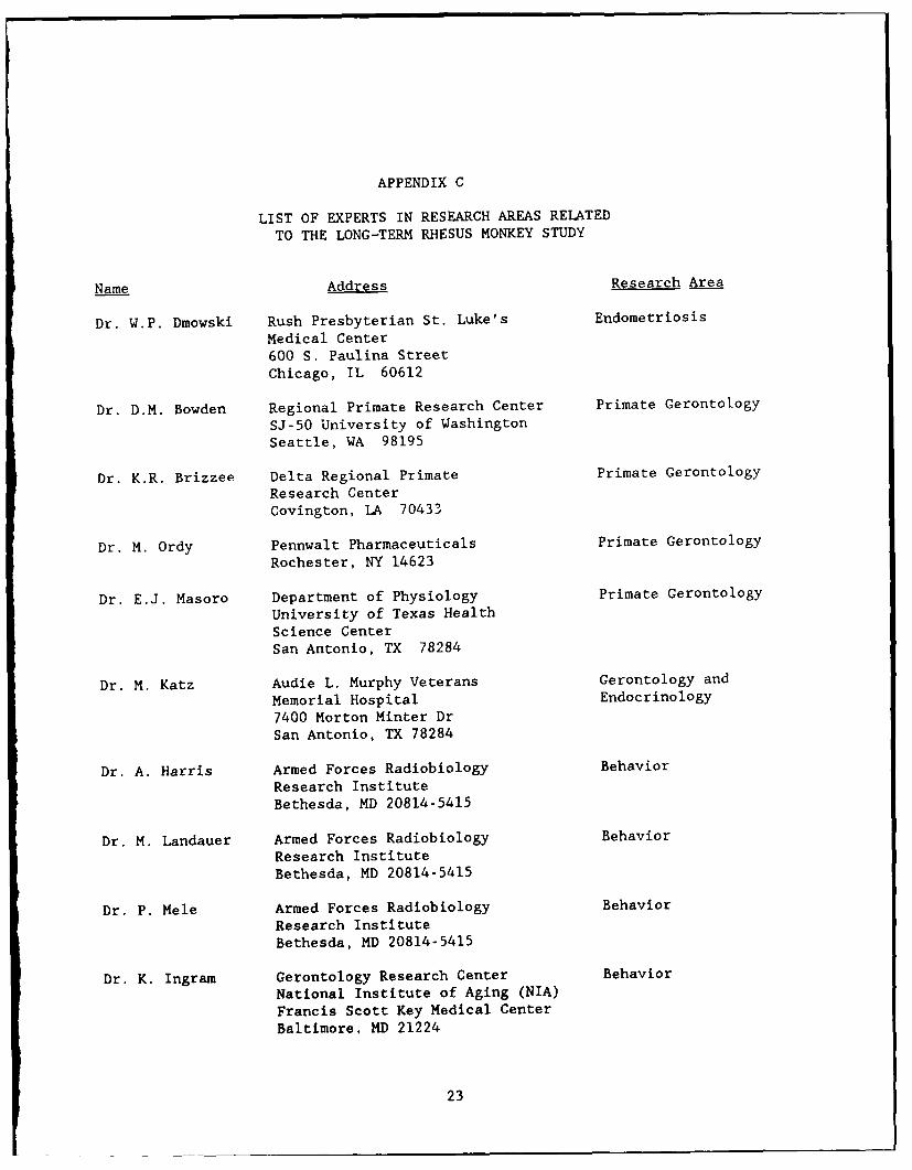
The collaborative studies, on dosimetry, cataracts, and immunology thatare underway, are a good example of what can be done and are to be commended.There are a number of other areas of investigation that have potential andshould be explored. A number of suggestions for interaction and collaborationare mentioned below in the sections on the specific areas of research. Theresearch team at Brooks AFB has obviously considered collaborative studies ina number of areas. The review panel suggests that such areas asneuroscience, behavioral sciences, and molecular biology could be extremelyfertile areas for collaborative research and should be considered. Names ofpotential collaborators or researchers that might interact with the USAF teamare given in Appendix C.
The potential for future investigations, by both the research staff atBrooks AFB and collaborators, will be greatly enhanced if state-of-the-artlaboratory methodology is available for the collection and processing ofvaluable tissues. The panel recommends that the attainment, fixation, andother methods of maintenance of samples be reviewed immediately and advicesought where necessary. Storage of DNA, or tissues from which DNA can beobtained at a later date, would make it possible to use both current andfuture techniques in molecular biology. Immortalization of fibroblasts and
lymphocytes, in order to establish specific cell lines, should be considered.Samples for the sake of samples is neither wise nor economical, but thisresource is unique and every possibility of extracting information should betaken.
In its assessment of the value of the current studies, the panelsummarized its impressions of each area of study as presented by theprincipal investigators.
Dosimetry
Overall, the dosimetry has been carried out well and carefully, and thevaried circumstances have been evaluated dosimetrically; however, severaladditional dosimetry studies should be seriously considered. Further workshould be carried out on the identification and distribution of secondaryradiation which becomes an important consideration at higher proton energies.The head of the juvenile rhesus monkey could be examined in a CAT scan inorder to determine whether the location of hot spots and the site of origin ofthe tumors coincide. Above a critical proton energy level, irradiationeffects in the brain become important. Calculations based on data from CATscans, which take into account the variation in size of the human head, shouldbe made to determine the critical proton energy range. Drs. Wilson and
4
• m mw

Townsend at NASA (Langley) and W. Schimmerling (Lawrence Berkeley Laboratory)are studying heavy ion irradiation of the head and interaction with them wouldbe worthwhile.
Tissue depth dosi:,etry should be carried out for each of the fatalcancers. The circumstances of each monkey should be evaluated so that theorgan dose for the cancer concerned is specified in detail. For uniformirradiation this should be straightforward, while for nonuniform exposurespecific work will have to be done.
Biological dosimetry, while not normally in the domain of thephysicists,is worth considering. These animals represent a good test (dose upto 8 Gy given 20 years ago) of the persistence of chromosome aberrations. Bothfibroblasts and lymphocytes could be assayed. If the cytogenetic study, whichshould include the current techniques, cannot be done at Brooks AFB, one of anumber of cytogeneticists could be asked to collaborate (see Appendix C).
Mortality
All of the control monkeys should be maintained for their life span, butthere is the question of whether some of the irradiated monkeys, perhaps inthe lower proton energy group, might be dedicated to other experimental work.It is important that the causes of death in both the control and irradiatedgroups be established precisely. Determination of the contribution of thedifferent causes of death to radiation-induced excess risk is particularlyimportant.
It should be kept in mind that the studies on the monkeys may provide animportant point in the pattern of species-dependent life span and radiation-induced life shortening. The results could provide information that could beused in the modeling of extrapolation of risks across species.
Despite the small number of animals, stratification by dose, sex,energy, cause, and age should be undertaken. Obviously, there will be anumber of empty cells, but the exercises should point the way for compilingthe most useful data for life-table type analysis.
Interventions have been carried out for humane reasons to prolong life.Humane policies are essential, whether implemented by euthanasia or surgicalor medical intervention; however, for experimental purposes, times of naturaldeaths from untreated diseases could be estimated and the estimates applied tothe cases which were treated, in order to eliminate the influence ofintervention on the data to some considerable extent.
Cataracts
The formation of cataracts is a potential late effect of radiation indeep space that is of concern. In solar particle events, relatively highdoses from low-energy protons that do not penetrate to the deep organs could
5
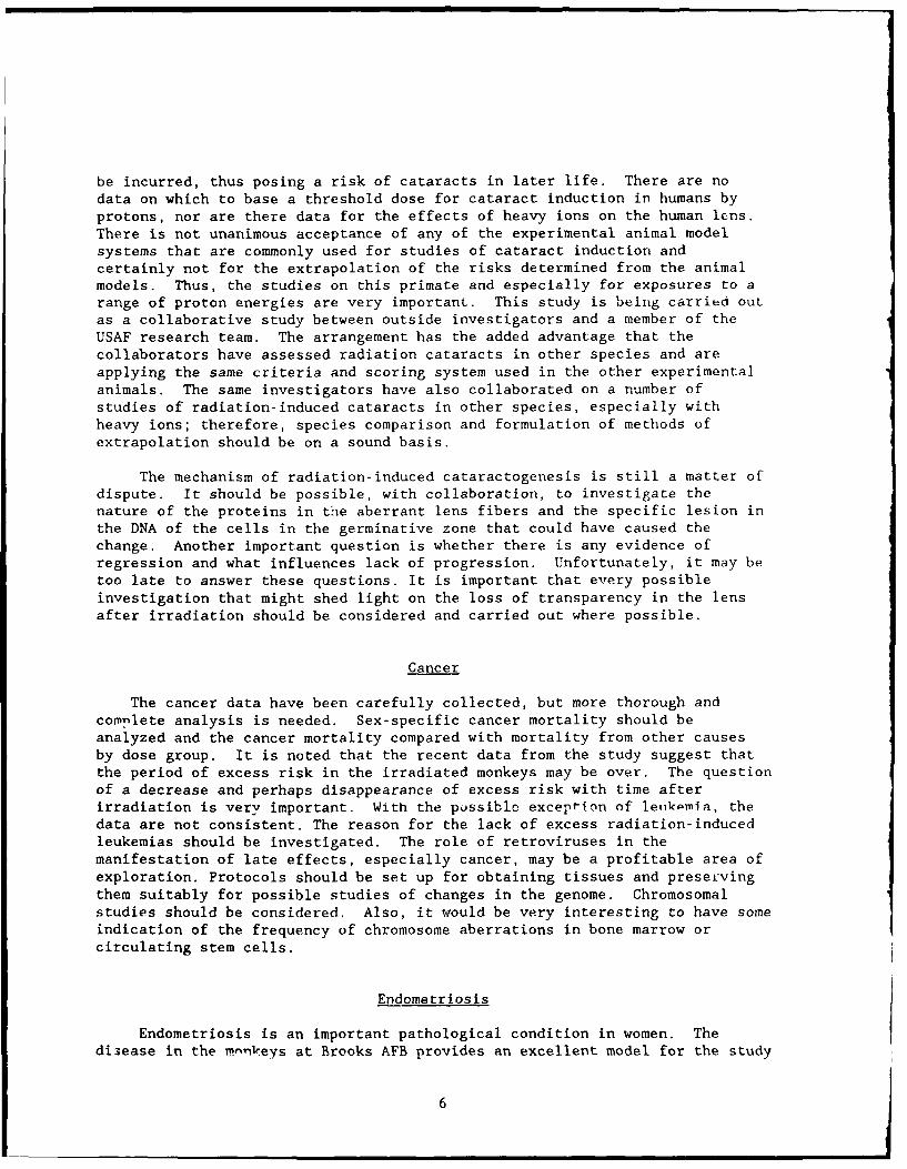
be incurred, thus posing a risk of cataracts in later life. There are nodata on which to base a threshold dose for cataract induction in humans byprotons, nor are there data for the effects of heavy ions on the human lens.There is not unanimous acceptance of any of the experimental animal modelsystems that are commonly used for studies of cataract induction andcertainly not for the extrapolation of the risks determined from the animalmodels. Thus, the studies on this primate and especially for exposures to arange of proton energies are very important. This study is being carried outas a collaborative study between outside investigators and a member of theUSAF research team. The arrangement has the added advantage that thecollaborators have assessed radiation cataracts in other species and areapplying the same criteria and scoring system used in the other experimentalanimals. The same investigators have also collaborated on a number ofstudies of radiation-induced cataracts in other species, especially withheavy ions; therefore, species comparison and formulation of methods ofextrapolation should be on a sound basis.
The mechanism of radiation-induced cataractogenesis is still a matter ofdispute. It should be possible, with collaboration, to investigate thenature of the proteins in the aberrant lens fibers and the specific lesion inthe DNA of the cells in the germinative zone that could have caused thechange. Another important question is whether there is any evidence ofregression and what influences lack of progression. Unfortunately, it may betoo late to answer these questions. It is important that every possibleinvestigation that might shed light on the loss of transparency in the lensafter irradiation should be considered and carried out where possible.
Cancer
The cancer data have been carefully collected, but more thorough andcomplete analysis is needed. Sex-specific cancer mortality should beanalyzed and the cancer mortality compared with mortality from other causesby dose group. It is noted that the recent data from the study suggest thatthe period of excess risk in the irradiated monkeys may be over. The questionof a decrease and perhaps disappearance of excess risk with time afterirradiation is very important. With the possiblc exception of leiikpmia, thedata are not consistent. The reason for the lack of excess radiation-inducedleukemias should be investigated. The role of retroviruses in themanifestation of late effects, especially cancer, may be a profitable area ofexploration. Protocols should be set up for obtaining tissues and preservingthem suitably for possible studies of changes in the genome. Chromosomalstudies should be considered. Also, it would be very interesting to have someindication of the frequency of chromosome aberrations in bone marrow orcirculating stem cells.
Endometriosis
Endometriosis is an important pathological condition in women. Thedi3ease in the mnkeys at Brooks AFB provides an excellent model for the study
6

of the et' iogy, pathogenesis, and therapy of endometriosis. The colony, whichis the largest known collection of monkeys with endometriosis, is a remarkablyvaluable resource and the research group at Brooks AFB has published a seriesof outstanding referenced articles on endometriosis. In fact, most of theinformation in the literature about endometriosis in monkeys has come fromthese studies.
The therapy of endometriosis is still a matter for investigation. Thesemonkeys provide a singular model for both the study of etiology and thetreatment of the disease. If it is possible to commit some of the monkeys tosuch research, the information gained could make an important contribution tothe medical community. Because endometriosis is becoming an increasingproblem in other monkey colonies, the USAFSAM group might consider preparinga concise but well referenced paper for the Journal of Medical Primatology.This paper would be in addition to that planned for Radiation Research. Thedecision of how to use the dwindling number of monkeys to best advantage willnot be easy, but some well designed studies of the pathogenesis and managementof endometriosis should be a carefully considered option.
Hematology and Biochemistry
There have been regular weighings of the monkeys and sampling of bloodfor routine hematological and biochemical analyses. It has been concludedthat white blood cell counts and protein and glucose levels were altered byproton irradiation in a dose-dependent way as both an acute and a lateeffect. The importance of unequivocal negative findings should not beforgotten. Presumably the protocol for this study was set up many years agowith tests that were standard at that time. If serum has been stored, othertests now in common use should be utilized to maximize the data obtained. Itwould appear worthwhile to consult with a number of people, especially thosenow involved in other studies, such as endometriosis, in order to includetests that would have specific use. Of particular importance to the futurework is the encouragement of the statistics group at USAFSAM to be involved inany analysis that is carried out. To make the analyses productive, certainquestions should be formulated. The question should be based on theexperience reported in other studies.
There are data for both the acute and late effects of irradiation formany hematological and biochemical end points in humans and also some datafor monkeys. The work at TNO in The Netherlands should definitely beexamined. The studies on the monkeys should be in the context of the humanstudies and, where possible, extensions of these studies. Obviously theexperimental design restricts the spectrum of tests that can be carried out,but tests of function should be considered in the higher dose survivors andcontrols.
Skin Effects
This part of the study is designed to test the hypothesis that exposureto proton radiation at a relatively young age reduces the proliferativecapacity of skin fibroblasts later in life. It has been noted that cells in
7
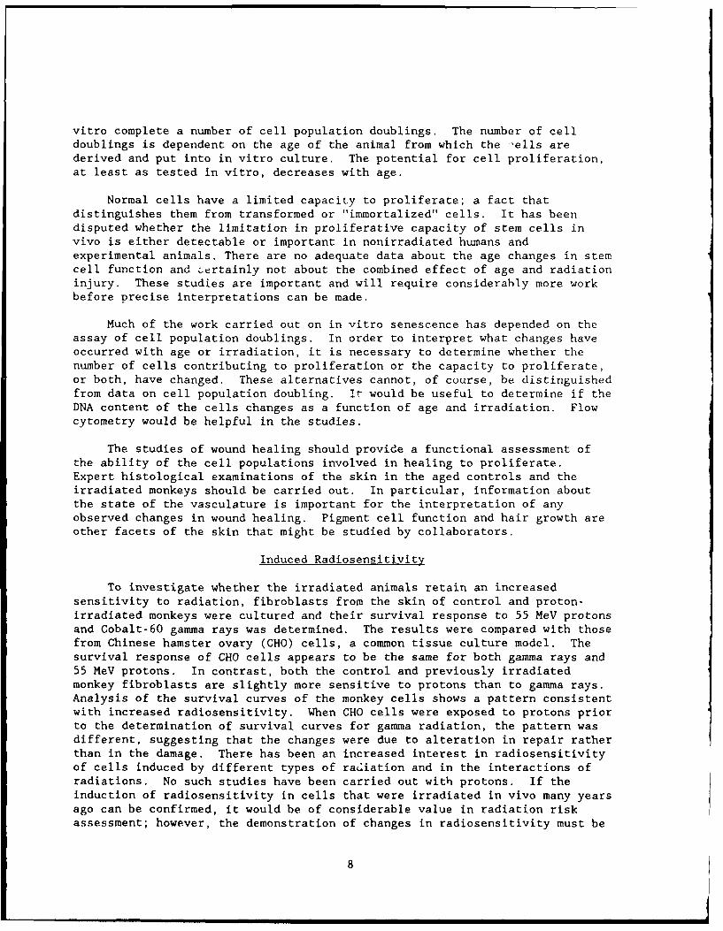
vitro complete a number of cell population doublings. The number of celldoublings is dependent on the age of the animal from which the -ells arederived and put into in vitro culture. The potential for cell proliferation,at least as tested in vitro, decreases with age.
Normal cells have a limited capacity to proliferate; a fact thatdistinguishes them from transformed or "immortalized" cells. It has beendisputed whether the limitation in proliferative capacity of stem cells invivo is either detectable or important in nonirradiated humans andexperimental animals. There are no adequate data about the age changes in stemcell function and certainly not about the combined effect of age and radiationinjury. These studies are important and will require considerably more work
before precise interpretations can be made.
Much of the work carried out on in vitro senescence has depended on theassay of cell population doublings. In order to interpret what changes haveoccurred with age or irradiation, it is necessary to determine whether thenumber of cells contributing to proliferation or the capacity to proliferate,or both, have changed. These alternatives cannot, of course, be distinguishedfrom data on cell population doubling. It would be useful to determine if theDNA content of the cells changes as a function of age and irradiation. Flowcytometry would be helpful in the studies.
The studies of wound healing should provide a functional assessment ofthe ability of the cell populations involved in healing to proliferate.Expert histological examinations of the skin in the aged controls and theirradiated monkeys should be carried out. In particular, information aboutthe state of the vasculature is important for the interpretation of anyobserved changes in wound healing. Pigment cell function and hair growth areother facets of the skin that might be studied by collaborators.
Induced Radiosensitivity
To investigate whether the irradiated animals retain an increasedsensitivity to radiation, fibroblasts from the skin of control and proton-irradiated monkeys were cultured and their survival response to 55 MeV protonsand Cobalt-60 gamma rays was determined. The results were compared with thosefrom Chinese hamster ovary (CHO) cells, a common tissue culture model. Thesurvival response of CHO cells appears to be the same for both gamma rays and55 MeV protons. In contrast, both the control and previously irradiatedmonkey fibroblasts are slightly more sensitive to protons than to gamma rays.Analysis of the survival curves of the monkey cells shows a pattern consistentwith increased radiosensitivity. When CHO cells were exposed to protons priorto the determination of survival curves for gamma radiation, the pattern wasdifferent, suggesting that the changes were due to alteration in repair ratherthan in the damage. There has been an increased interest in radiosensitivityof cells induced by different types of radiation and in the interactions ofradiations. No such studies have been carried out with protons. If theinduction of radiosensitivity in cells that were irradiated in vivo many yearsago can be confirmed, it would be of considerable value in radiation riskassessment; however, the demonstration of changes in radiosensitivity must be
8

carried out with precision because such changes are seldom large. The resultsmust be reproducible and the statistical analysis must be rigorous.
Immunocompetence
A collaborative study with Dr. William Stone of Trinity University, SanAntonio, and Dr. Michael Miller of the University of Texas Health ScienceCenter at San Antonio, has been underway for the past 4 years, but thes-observations on the immunocompetence of the aging control and irradiatedmonkeys have been limited by the restricted access to whole blood samples.Presumably this restriction has been considered necessary for the maintenanceof the original experimental design. The current plan is to includedeterminations of T and B lymphocyte populations, immunoglobulin levels, cellmediated immunity, complement levels, and autoantibodies.
Drs. Stone and Miller have found no significant differences inimmunoglobulin levels and complement activity between irradiated and controlmonkeys. A considerable variation in levels of IgG and IgA were found in boththe aged controls and irradiated monkeys. The investigators also noted thatthe frequency of autoantibodies, e.g., anti-reticulin, was higher in thecontrol monkey than in the irradiated animals. The number of monkeys studiedfor changes in cellular immunity has been small, but there is a suggestionthat the T cell responses are less in the irradiated animals compared to thecontrols. It is important that this finding be fully investigated. Othershave claimed that prolonged spaceflight may result in depressed immunefunction. The cause of the decreased function is thought to be reducedgravity, but the evidence is weak and the mechani.m unknown. It will beimportant to allay concerns about a potential reduction in immunocompetencewhether the potential cause is microgravity, radiation, or an interactionbetween the two.
It was the impression of the panel that the immunological studies werenot fully integrated into the overall study. For example, there was nomention of the possibilities of correlating the immunological findings withthe clinical and pathological findings. The correlation of changes inimmunoglobulins and the presence of amyloid and other degenerative changesshould be examined. The studies on the monkeys could give importantinformation about aging and the late effects of high total doses of protonson the immune system. Such studies should be carried out with vigor.
If the full potential of the monkey colony as a resource for informationabout aging and proton radiation on the immune system is to be exploited,some new protocols must be considered. More detailed evaluation of cellularimmunity should include evaluations of B and T cells, subsets of T cells, thehumoral response, and response to various mitogens on a larger number ofanimals than has been carried out so far. Perhaps some animals can bededicated to these studies.
9

Diabetes
Diabetes is another example of how the monkeys in the colony can providea model for study relevant to human disease. The colony provides a greatopportunity for an in-depth study of diabetes in a nonhuman primate model thatcan help in understanding the human disease. The resource is so valuable andhaE such potential for producing important information that funding foradditional studies should be sought. The opportunity for collaborativestudies with Dr. Charles F. Howard of the Oregon Regional Primate ResearchCenter should be explored.
Anatomic Pathology
Ore of the problems of a research project that lasts for decades andinvolves a number of changes in staffing is the maintenance of quality andconsistency. There has been remarkable continuity of the histopathologicalstudies, but, although they have become more detailed and systematic in thelast decade, they are not well integrated with the other aspects of the study.The description of the pathological findings is heavily weighted toward theanatomical and histological description of tumors. There was much lessinformation presented about the pathological and aging changes than aboutneoplasms. Here again, negative findings are of great importance. Whatshould eventually come from these studies is an overall assessment of theneoplastic and nonneoplastic changes with age and in relation to radiationexposure. Further information should be obtained on the correlation betweenhot spots and the origin of the CNS tumors.
Exposure to low energy protons occurs in space, and in the case of solarparticle events, astronauts in deep space could receive considerable doses tothe skin. Both the pathology and site of tumors of the skin and theradiation-induced changes in the dermis are of particular interest andrelevance to NASA. It was not clear how many of the denoted skin neoplasmswere dermal or subcutaneous in origin. The correlation of proton energy anddose with the skin lesions should be investigated.
The panel stressed the importance of the histopathological studies, andit is hoped that more can be accomplished in the investigation of both thetime- and dose-dependent changes in tissues. From the examination of thetissues of mrnkeys that died at different ages, it may be possible toinvestigate some of the pathogenesis of the disease processes involved.
SUMMARY AND RECOMMENDATIONS
The panel was unanimous in their support for the continuation andextension of the studies on this remarkable colony of monkeys. The value ofthe colony as a resource for studies, not only of the effects of protonradiation, but of aging, is very great. The colony should be maintained aslong as useful information can he obtained. It is impossible to predictprecisely how long that will be anc4 the panel suggests that the situationshould be reassessed in 5 years.
10

The colony has been maintained with great dedication and success; infact, with such success that the colony is now a unique resource. The aim ofdetermining the effects of proton irradiation has governed, to a large extent,the type of longitudinal studies carried out and therefore some of the studiesthat the committee considered important have not been undertaken. Now is thetime to take stock and assess what information can be obtained from theremaining animals and stored material. A close examination of the number ofanimals in the control and various radiation dose groups should be made and itshould be decided how the remaining animals can be used to the greatestadvantage. It is suggested that consultation with experts in each of theparticular research fields should be considered in order to make the bestchoices for possible future studies. The panel has made a number ofrecommendations about areas of investigations that should be pursued orexpanded.
A major concern of the panel is that the research program has been tooisolated. The program could have benefited from postdoctoral appointees andgraduate students doing their thesis work. Pcrhaps it is not too late tocoLIsider such a plan. The panel felt that there were a number of areas ofresearch that might have been considered by the USAF group but were notdiscussed and which might be included in the future. For example, the brainshave been examined for tumors, but much more might be learned about theeffects of aging and radiation on the nervous system if the modern techniquesused in neuroscience were applied. Behavior is another area that should beconsidered, in particular, behavior associated with motor function such asbalance, muscle coordination, etc. Yet another area are the changes with agein bone and muscle, especially in the females irradiated as juveniles.
There are now many techniques in molecular biology that could be appliedto the tissues of the monkeys, and experts that might become collaboratorsshould be contacted. We would reiterate the importance of storing cells andtissues for such studies.
A number of investigators at other institutions have been suggested aspossible collaborators or consultants for specific aspects of the program, anda list is appended of researchers in the various pertinent disciplines thatmight be considered. In both the mortality and cancer studies, and in factfor many of the other studies, the committee thought that more biostatisticalanalysis could help a great deal in the interpretation of the data.
11

BIBLIOGRAPHY
1. Dalrymple, G. V., I. R. Lindsay, and J. J. Ghidoni. The effects of 2-MeVwhole body x-irradiation on primates. Radiat Res 25:377-400 (1965).
2. Dalrymple, G. V., I. R. Lindsay, J. J. Ghidoni, H. L. Kundg9, and E. T.Still. The effect of massive doses oi jz MeV protons and Co gammaradiation on serum enzyme levels of whole body irradiated primates. JNuc Med 6:588-593 (1965).
3. Pickering, J. E. Training and operations--A concept. AMD-TR-65-2,Aerospace Medical Division, Brooks Air Force Base, Texas, 1965.
4. Dalrymple, G. V. and I. R. Lindsay. Protons and space travel--Anintroduction. Radiat Res 28:365-371 (1966).
5. Williams, G. H., J. D. Hall, and I. L. Morgan. Whole-body irradiation ofprimates with protons of energies to 400 MeV. Radiat Res 28:372-389(1966).
6. Mitchell, J. C., G. V. Dalrymple, G. H. Williams, J. D. Hall, and I. L.Morgan. Proton depth-dose dosimetry. Radiat Res 28:390-405 (1966).
7. Dalrymple, G. V., I. R. Lindsay, J. J. Ghidoni, H. L. Kundel, E. T. Still,R. Jacobs, and I. L. Morgan. Some effects of whole-body 32-MeV protonirradiations on primates. Radiat Res 28:406-433 (1966).
8. Dalrymple, G. V., J. J. Ghidoni, H. L. Kundel, T. L. Wolfe, and I. R.Lindsay. Edema-A delayed complication of total-body 32-MeV protonirradiation. Radiat Res 28:434-445 (1966).
9. Lindsay, I. R., G. V. Dalrymple, J. J. Ghidoni, J. C. Mitchell, and I. L.Morgan. Some effects of 55-MeV protons on primates. Radiat Res 28:446-464 (1966).
10. Dalrymple, G. V., I. R. Lindsay, J. C. Mitchell, J. D. Hall, and I. L.Morgan. The kinetics of recuperation following 55-MeV protonirradiation. Radiat Res 28:465-470 (1966).
11. Dalrymple, G. V., I. R. Lindsay, J. J. Ghidoni, J. D. Hall, J. C.Mitchell, H. I. Kundel, and I. L. Morgan. Some effects of 138-MeVprotons on primates. Radiat Res 28:471-488 (1966).
12. Dalrymple, G. V., I. R. Lindsay, J. D. Hall, J. C. Mitchell, J. J.Ghidoni, H. L. Kundel, and I. L. Morgan. The relative biologicaleffectiveness of 138-MeV protons as compared to Cobalt-60 gammaradiation. Radiat Res 28:489-506 (1966).
12

13. Dalrymple, G. V., I. R. Lindsay, J. J. Ghidoni, J. C. Mitchell, and I. L.Morgan. Some effects of 400-MeV protons on primates. Radiat Res 28:489-506 (1966).
14. Kundel, H. L, The effects of high-energy proton irradiation on thecardiovascular system of the rhesus monkey. Radiat Res 28:529-537(1966).
15. Hawrylewicz, E. J. and W. H. Blair. Effects of gamma and protonirradiaton on lactic dehydrogenase isoenzymes. Radiat Res 28:538-547(1966).
16. Dalrymple, G. V., I. R. Lindsay, J. J. Ghidoni, J. C, Mitchell, and I. L.Morgan. An estimate of the biological effects of the space protonenvironment. Radiat Res 28:548-566 (1966).
17. Zellmer, R., J. Culver, and J. E. Pickering. Proton-irradiation effectsin primates. Radiat Res Sup 7:325-329 (1967).
18. Lindsay, I. R, and G. V. Dalrymple. Acute somatic effects in primates ofprotons to 400-MeV. Radiat Res Sup 7:330-335 (1967).
19. Ghidoni, J. J., G. V. Dalrymple, I. R. Lindsay, H. L. Kundel, W.Haymaker, and E. Ballinger. Hepatic lesions produced by 32 and 55-MeVproton irradiation. Arch Path 83:370-376 (1967).
20. Siegal, A. M., H. W. Casey, R. W. Bowman, and J. E. Traynor. Leukemia ina rhesus monkey (Macaca mulatta) following exposure to whole-body protonirradiation. Blood 32 (6):989-996 (1968).
21. Blake, K. R., L. A. Boles, J. B. Nelson, and C. V. Parker, Jr. Largearea proton beams for radiobiologic research from the NASA-SREL Synchro-Cyclotron I. Investigation of the nominal 300-MeV primary beam II.Investigation of the nominal.600-MeV primary beam. USAFSAM-TR-68-79,1968.
22. Traynor, J. E. and A. M. Siegal. Early effects of 150-MeV protonirradiation in rhesus monkeys. USAFSAM-TR-68-87, 1968.
23. Woodruff, J. M. and D. K. Johnson. Hepatic hemangioendothelioma in arhesus monkey. Vet Path 5:328-332 (1968).
24. Hardy, K. A., J. C. Mitchell, and S. J. Allen. Measurements ofdepth-dose distributions in cylindrical phantoms exposed to 28-MeV,21-MeV, 14-MeV or 5-MeV protons. Radiat Res 37:272-282 (1969).
25. Siegal, A. M., H. W. Casey, and J. E. Traynor. Some biological effectsof low-energy protons on skin. Radiat Res 37:323-333 (1969).
13

26. Casey, H. W., P. S. Coogan, and J. E.:Traynor. Late effects of 32-MeVproton irradiation in primates. Radiat Res 39:634-642 (1969).
27. Hardy, K. A., R. E. Jorgensen, L. A. Boles, C. V. Parker, and K. R.Blake. Dosimetry for study of hematologic effects on primates of pulsedversus continuous radiation. USAFSAM-TR-70-31, 1970.
28. Traynor, J. E. and H. W. Casey. Five-year follow-up of primates exposedto 55-MeV protons. Radiat Res 47:143-148 (1971).
29. Kirk, J. H. Body weight decreases in some proton exposed primates.Space Sciences 3:271-277 (1971).
30. MacKenzie, W. F., C. A. Splitter, and M. G. Valerior. Endometriosis inprimates. Proceedings of the Third Conference on Experimental Medicineand Surgery of Primates. (E. Goldsmith and J. M. Janowski, Eds), MedicalPrimatology, 288-297, 1972.
31. Splitter, G. A., J. A. Kirk, W. F. MacKenzie, and C. A. Rawlings.Endometriosis in four irradiated rhesus monkeys. Vet Path 9:249-267(1967).
32. Haymaker, W., L. J. Rubinstein, and J. Miquel. Brain tumors inirradiated monkeys. Acta Neuropathol 20:267 (1972).
33. Kirk, J. H. Persistent pupillary membrane: Developmental review and anoccurrence in Macaca mulatta. Lab Animal Sci 22:245-248 (1972).
34. Kirk, J. H., H. W. Casey, and J. F. Harwell, Jr. Diabetes mellitus intwo rhesus monkeys. Lab Animal Sci 22: 245-248 (1972).
35. Kirk, J. H. General amyloidosisin a rhesus monkey. Lab Animal Sci 22:587-593 (1972).
36. Splitter, G. A., J. H. Kirk, and H. W. Casey. Radiation-related andspontaneous tumors in primates. Radionuclide Carcinogenesis, 12th HanfordBiology Symposium, Conf-720505, AEC Symp. 29, 55-89, Richland,WA, 1973.
37. Krupp, J. H. Nine-year mortality experience in proton-exposed Mncacamulatta. Radiat Res 67:244-252 (1976).
38. Kasa, T. J., G. D. Lathrop, H. J. Dupuy, C. H. Bonney, and J. D. ToftII. An epidemic focus of Trypanosoma cruzi infection in a subhumanprimate research colony. J Am Vet Med Assoc 171(9):850-854 (1977).
39. Boster, R. A. and R. E. Schmidt. What's your diagnosis? (Renaladenocarcinoma and diabetes mellitus). Lab Animal Sci 7(4):14-16 (1978).
14
40. Wood, D. H, R. A. Boster, R. L. Easoi, M. G. Yochmowitz, and Y. L.Salmon. Delayed effects of proton irradiation in Macaca mulatta I.
Endometriosis (15-Year Report). USAFSAM-TR-82-8, 1982.
41. Hubbard, G. B., D. H. Wood, and J. W. Fanton. Squamous cell carcinoma
with metastasis in a rhesus monkey (Macaca mulatta). Lab Animal Sci 33:
469-472 (1983).
42. Yochmowitz, M. G., D. H. Wood, and Y. L. Salmon. Delayed effects ofproton irradiation in Macaca mulatta II. Mortality (15-year report),
USAFSAM-TR-83-2, 1983.
43. Wood, D. H., M. G. Yochmowitz, Y. L. Salmon, R. L Eason, and R. A.
Boster. Proton irradiation and endometriosis. Aviat Space Environ
Med 54:718-724 (1983).
44. Bergtold, D. S., A. B. Cox, C. M. Su, and J. T. Lett. Late skin damage
in rabbits and monkeys after exposure to particulate radiations. Adv in
Space Res 3:221-229 (1983).
45. Fanton, J. W., G. B. Hubbard, W. M. Witt, and D. H. Wood. Adenocarcinoma
of the small intestine in two rhesus monkeys. J Am Vet Med Assoc 185:
1377 (1984).
46. Hubbard, C. B., J. W. Fanton, R. C. Harvey, and D. H. Wood. Paralysis
due to a glomangioma in a Macaca mulatta. Lab Animal Sci 34(6): 614-615
(1984).
47. Hubbard, C. B. and D. H. Wood. Glomangiomas in four irradiated Macaca
mulatta. Vet Path 21(6):609-610 (1984).
48. Hubbard, G. B., D. H. Wood, and W. I. Butcher. Mammary carcinoma with
metastasis in a rhesus monkey (Macaca mulatta). Brief Communication, VetPath 21:531-532 (1984).
49. Lett, J. T., A. B. Cox, D. S. Bergtold, A. C. Lee, and J. E. Pickering.
Late effects from particulate radiations in primate and rabbit tissues.
Adv in Space Res 4:251-256 (1984).
50. Salmon, Y. L., M. G. Yochmowitz, and D. H. Wood. Delayed effects ofproton irradiation in Macaca mulatta III. Glucose tolerance.
USAFSAM-TR-84-7, 1984.
51. Yochmowitz, M. G., D. H. Wood, and Y, L. Salmon. Seventeen-year
mortality experience of proton radiation in Macag_ Mulatt4. Radiat Res
102:14-34 (1985).
15

52. Wood, D. H., M. G. Yochmowitz, and Y. L. Salmon. Lifetime effects ofsingle-event proton exposures in rhesus monkeys. 22nd Hanford LifeSciences Symposium, CONF-830951. 219-236, Richland, WA, 1986.
53. Fanton, J. W., M. 0. Yothmowitz, D. H. Wood, and Y. L. Salmon. Surgicaltreatment of endometriosis in 50 rhesus monkeys. Am J Vet Res 47:1602-1604 (1986).
54. Wood, D. H., M. 0. Yochmowitz, K. A. Hardy, and Y. L. Salmon. Animalstudies of life shortening and cancer risk from space radiation. Adv inSpace Res 6(11):213-216 (1986).
55. Wood, D. H., M. G. Yochmowitz, K. A. Hardy, and Y. L. Salmon. Occurrenceof brain tumors in rhesus monkeys exposed to 55-MeV protons. Adv inSpace Res 6(11):275-283 (1986).
56. Cox, A. B., D. H. Wood, and J. T. Lett. Late radiation damage inprimate skin following exposures comparable to those from solar protons.Adv in Space Res 6(11):217-222 (1986).
57. Lett, J. T., A. B. Cox, and A. C. Lee. Cataractogenic potential ofionizing radiations in animal models that simulate man. Adv in SpaceRes 6(11):295-303 (1986)..
58. Fanton, J. W., R. E. Cordts, R. K. Harris, D. Romo, and D. H. Wood.Radioisotope imaging of brain, tumors in two rhesus monkeys. Lab AnimalSci 38(2):214-217 (1988).
59. Wood, D. H., J. E. Pickering, M. G. Yochmowitz, K. A. Hardy, and Y. L.Salmon. Radiation risk assessment for military space crews, MilMed 153(6):298-303 (1988).
60. Cox, A. B., A. C. Lee, and J. T. Lett. Delayed effects of protons on thelens and integument: A primate model. In Proc. NATO-ASI, Vol 154Terrestrial Space Radiation and Its Biological Effects (P.D. McCormackand C.E. Swenberg, eds.), New York, Plenum, 415-422, 1988.
61. Lett, J. T., A. B. Cox, and A. C. Lee. Selected examples of degenerativelate effects caused by particulate radiations in normal tissues. Proc.NATO-ASI, Vol. 154, Terrestrial Space Radiation and Its BiologicalEffects (P. D. McCormack and C. E. Swenberg and H. 3ucker, eds.),New York, Plenum, 393-413, 1988.
62. Wood, D. II., K. A. Hardy, A. B. Cox, Y. L. Salmon, M. G. Yochmowitz, andR. E. Cordts. Delayed effects of proton irradiation in Macaca mulatta(22 year summary). AIP Conference Proceedings 186, 381-392, 1988.
16

63. Cox, A. B., A. C. Lee, and J. T. Lett. Delayed effects of protons on thelens and integument: A primate model. In Proc. NATO-ASI, Vol 154,Terrestrial Space Radiation and Its Biological Effects (P. D.McCormack, C. E. Swenberg, and H. Bucker, eds.), New York, Plenum,393-413, 1988.
64. Cox, A. B. and J. T. Lett. The quantification of wound healing as amethod to assess late radiation damage in primate skin exposed tohigh-energy protons. Adv Space Res 9(10):125-130 (1989).
65. Lett, J. T., A. C. Lee, A. B. Cox, and D. H. Wood. Late cataractogenesiscaused by particulate radiations and photons in long-lived mammalianspecies. Adv Space Res 9(10):325-331 (1989).
66. Lett, J. T., A. B. Cox, M. D. Story, U. K. Ehmann, and E. A. Blakely.
Responses of synchronous L5178Y S/S cells to heavy ions and theirsignificance for radiobiological theory. Proc Roy Soc Lond (B) 237:27-42 (1989).
17

APPENDIX A
LIST OF PANEL MEMBERS
R. J. M. Fry, M.D. (Chairman) Warren K. Sinclair, Ph.D.
Oak Ridge National Laboratory PresidentBiology Division National Council for Radiation
P.O. Box 2009 Protection & Measurements
Bear Creek Road 7910 Woodmont Avenue, Suite 1016
Oak Ridge, TN 37831-8077 Bethesda, MD 20814
James A. Joseph, Ph.D. Philip Archer, Sc.D.
Gerontology Research Center Professor & Acting Chairman
National Institute of Aging Dept. of Preventive Medicine
Frances Scott Key Medical Center Univ. of Colorado School of Medicine
Baltimore, MD 21224 Box B119, 4200 E. 9th AvenueDenver, CO 80262
George W. Casarett, Ph.D.
Department of Biophysics Percival D. McCormack, M.D., Ph.D.University of Rochester Medical Life Science Division, Code EBM
Center NASA Headquarters
601 Elmwood Avenue Room 300, Federal Bldg. 10
Rochester, NY 14642 Washington, D.C. 20546
Harold M. McClure, D.V.M. Matt J. Kessler, D.V.M.
Associate Director Director
Yerkes Regional Primate Research Caribbean Primate Research Center
Center P.O. Box 1053
Emory University Sabana Seca, Puerto Rico 00749
Atlanta, GA 30322
Douglas Grahn, Ph.D.RetiredDivision of Biological & Medical
ResearchArgonne National Laboratory
9700 South Cass Avenue
Argonne, IL 60439
19

APPENDIX B
PRESENTATIONS AT THE CONFERENCE ON 9-10 MARCH 1989
1. OVERVIEWD. Wood, D.V.M., Ph.D., USAFSAM
2. EXPOSURE DATA & DOSE DISTRIBUTIONS
K. Hardy, M.S., USAFSAM
3. MORTALITY EXPERIENCE
D. Wood, D.V.M., Ph.D., USAFSAM
4. CATARACTOGENESIS
A. C. Lee, D.V.M., J. T. Lett, Ph.D., Colorado State University
5. CANCER RISKD. Wood, D.V.M., Ph.D., USAFSAM
6. ENDOMETRIOSISJ. Fanton, D.V.M., M.S., J. Golden, D.V.M., USAFSAM
7. HEMATOLOGY & BIOCHEMISTRYY. Salmon, M.A., P. Crump, Ph.D., USAFSAM
8. DIABETES
D. Wood, D.V.M., Ph.D., USAFSAM
9. INDUCED RADIOSENSITIVITYJ. Wigle, Ph.D., USAFSAM
10. IMMUNOCOMPETENCEW. Stone, Ph.D., Trinity University, M. Miller, M.D., University of
Texas Health Science Center at San Antonio
11. SKIN EFFECTS
A. B. Cox, Ph.D., USAFSAM
12. ANATOMIC PATHOLOGY
H. Davis, D.V.M., M.S., USAFSAM, G. Hubbard, D.V.M., M.S., SouthwestFoundation for Biomedical Research
21
APPENDIX C
LIST OF EXPERTS IN RESEARCH AREAS RELATED
TO THE LONG-TERM RHESUS MONKEY STUDY
Name Address Research Area
Dr. W.P. Dmowski Rush Presbyterian St. Luke's Endometriosis
Medical Center
600 S. Paulina Street
Chicago, IL 60612
Dr. D.M. Bowden Regional Primate Research Center Primate Gerontology
SJ-50 University of Washington
Seattle, WA 98195
Dr. K.R. Brizzee Delta Regional Primate Primate Gerontology
Research Center
Covington, LA 70433
Dr. M. Ordy Pennwalt Pharmaceuticals Primate Gerontology
Rochester, NY 14623
Dr. E.J. Masoro Department of Physiology Primate Gerontology
University of Texas Health
Science Center
San Antonio, TX 78284
Dr. M. Katz Audie L. Murphy Veterans Gerontology and
Memorial Hospital Endocrinology
7400 Morton Minter Dr
San Antonio, TX 78284
Dr. A. Harris Armed Forces Radiobiology Behavior
Research Institute
Bethesda, MD 20814-5415
Dr. M. Landauer Armed Forces Radiobiology Behavior
Research Institute
Bethesda, MD 20814-5415
Dr. P. Mele Armed Forces Radiobiology Behavior
Research Institute
Bethesda, MD 20814-5415
Dr. K. Ingram Gerontology Research Center Behavior
National Institute of Aging (NIA)
Francis Scott Key Medical Center
Baltimore, MD 21224
23

Name Address Research Area
Dr. J.A. Joseph Gerontology Research Center Behavior and
National Institute of Aging (NIA) neuroscienceFrancis Scott Key Medical Center
Baltimore, MD 21224
Dr. R. Wilcox University of Texas Behavior and
Austin, TX 78767 neuroscience
Dr. A. Mickley Armed Forces Radiobiology Neuroscience
Research InstituteBethesda, MD 20814-5415
Dr. W. Hunt Armed Forces Radiobiology NeuroscienceResearch Institute
Bethesda, MD 20814-5415
Dr. W. Blakely Armed Forces Radiobiology Molecular BiologyResearch InstituteBethesda, MD 20814-5415
Dr. T. Walden Armed Forces Radiobiology Molecular BiologyResearch Institute
Bethesda, MD 20814-5415
Dr. D. McClain Armed Forces Radiobiology Molecular BiologyResearch Institute
Bethesda, MD 20814-5415
Dr. N. Holbrook Gerontology Research Center GerontologyNational Institute of Aging (NIA)
Francis Scott Key Medical Center
Baltimore, MD 21224
Dr. T. MacVittie Armed Forces Radiobiology Hematology andResearch Institute ImmunologyBethesda, MD 20814-5415
Dr. M.B. Kay Olin E. Teague Veterans Center Immunology1901 South First St
Temple, TX 76504
Dr. G. Littlefield Medical Division, Oak Ridge CytogeneticsAssociated Universities (ORAU)
Oak Ridge, TN 37830
Dr. J. Preston Biology Division RadiobiologyOak Ridge National Laboratory (ORNL)
Oak Ridge, TN 37831
24

Name Address Research Area
Dr. A.L. Brooks Inhalation Toxicology Research Radiobiology
Institute
Lovelace Foundation, P.O. Box 5890
Albuquerque, NM 87115
Dr. C.E. Land and Radiation Epidemiology Branch Statistics
Dr. S. Jablon National Cancer InstituteBethesda, MD 20814
Dr. E. Frome Engineering Physics and Mathematics StatisticsOak Ridge National LaboratoryOak Ridge, TN 37831
Dr. E. Lakatta Gerontology Research Center CardiovascularNational Institute of Aging (NIA)Francis Scott Key Medical CenterBaltimore, MD 21224
Dr. H. Suit Department of Radiation Medicine Proton Effects
Massachusetts General Hospital
Boston, MA 02114
Dr. M. Goitein Department of Radiation Medicine Proton Dosimetry
Massachusetts General HospitalBoston, MA 02114
Dr. J. Hopewell University of Oxford Radiation Effects
Oxfordshire Health Authority on SkinResearch Institute
The Churchill Hospital
Headington, Oxford OX3 7LJ, England
Dr. C. Potten Department of Epithelial Kinetics Radiation Effects
Paterson Laboratory on SkinChristie Hospital & Holt Radium
InstituteManchester M20 9BX, England
Dr. C.F. Howard, Jr. Oregon Regional Primate Research Diabetes
Center505 N.W. 185th Avenue
Beaverton, OR 97006
Dr. E.A. Blakely Lawrence Berkeley Laboratory Radiobiology
University of CaliforniaBuilding 10-209
Berkeley, CA 94720
* U. S. GOVERNMENT PRINTING OPFICF: 1990--761-051/20010
25