Embed Size (px)
Citation preview
J Can Chiropr Assoc 2012; 56(3) 173
0008-3194/2012/173–178/$2.00/©JCCA 2012
Acute sciatica and progressive neurological deficit secondary to facet synovial cysts: A report of two cases.Brian Arthur, DC, MSc* Peter Lewkonia, MD, MSc FRCS C† Jeffrey A. Quon, DC, MHSc, PhD, FCCS(C)*‡ John Street, MD, PhD, FRCS(C)* Paul B. Bishop, DC, MD, PhD*
Objective: To describe two patients with lumbar facet synovial cysts causing sciatica and progressive neurological deficit. Clinical Features: A 52-year-old female with bilateral sciatica and a neurological deficit that progressed to a foot drop; and a 54-year-old female with worsening sciatica and progressive calf weakness were seen at a major tertiary care centre. Diagnostic imaging studies revealed the presence of spinal nerve root impingement by large facet synovial cysts. Interventions and Outcomes: Activity modification, gabapentinoid and non-steroidal anti-inflammatory medications were unsuccessful in ameliorating either patient’s symptoms. One patient had been receiving ongoing lumbar chiropractic spinal manipulative therapy despite the onset of a progressive neurological deficit. Both patients eventually required surgery to remove the cyst and decompress the affected spinal nerve roots.
* Combined Neurosurgical and Orthopaedic Spine Program, Vancouver General Hospital, Division of Spine, Department of Orthopaedics & I.C.O.R.D. (International Collaboration on Repair Discoveries), University of British Columbia, Vancouver, Canada
† Division of Orthopaedics, Department of Surgery, University of Calgary, Alberta, Canada‡ School of Population and Public Health, Faculty of Medicine, University of British Columbia, Vancouver, Canada
Address for Correspondence: Dr. Paul Bishop, ICORD Spine Centre, Blusson Pavilion, 6100- 818 West 10th Avenue, Vancouver, B.C. V5Z 1M9 CANADATel: (604) 875-4549 : Fax: (604) 875-8223Email: [email protected]
Funding Sources: None© 2012
Objectif : Description de deux patientes ayant des kystes synoviaux facettaires lombaires leur causant de la névralgie sciatique et un déficit neurologique progressif. Caractéristiques cliniques : Deux femmes ont été examinées à un grand centre de soins tertiaires. La première, âgée de 52 ans, est atteinte de névralgie sciatique bilatérale et d’un déficit neurologique qui a progressé jusqu’au pied tombant; la deuxième, âgée de 54 ans, souffre d’une névralgie sciatique qui s’empire et d’une faiblesse progressive au mollet. L’examen de l’imagerie diagnostique a révélé un coincement de la racine du nerf rachidien, causé par de grands kystes synoviaux facettaires. Interventions et résultats : La modification de l’activité et les médicaments gabapentinoïdes et anti-inflammatoires non stéroïdiens n’ont pas réussi à améliorer les symptômes des patientes. Une des patientes suivait une thérapie continue de manipulation rachidienne chiropratique malgré l’apparition d’un déficit neurologique progressif. En fin de compte, les deux patientes ont eu besoin de chirurgie pour extraire les kystes et décomprimer les racines du nerf rachidien affectées. Conclusion : Les patients atteints de névralgie sciatique aiguë qui développent un déficit neurologique progressif alors qu’ils reçoivent des soins doivent, sans
174 J Can Chiropr Assoc 2012; 56(3)
Acute sciatica and progressive neurological deficit secondary to facet synovial cysts: A report of two cases.
IntroductionThe etiology of lumbar facet joint synovial cysts (SC) is thought to be associated with vertebral segmental hyper-mobility and/or segmental instability.1 Patients with lum-bar facet joint SC typically present with acute sciatica, rather than lower back pain. When the cysts are large in size or atypical in morphology they can cause focal radiculopathy, neurogenic claudication and even cauda equina syndrome.1,2 However, unlike patients in whom spinal nerve root trauma is secondary to lumbar disc herniation, the natural history of acute sciatica second-ary to a SC is generally unfavourable and any associated neurologicaldeficitcanbecomeprogressive.3 Previous reports of the clinical management of acute radiculopathy secondary to SC have described a wide var-iety of therapeutic approaches. These include the use of differing forms of chiropractic spinal manipulative ther-apy4-6, image-guided injection therapies including aspira-tion or distension/rupture7-12 and surgical treatments such as either excision and fusion or excision alone2,13,14. This paper describes the diagnosis, clinical course and treatment of two patients with atypical forms of SC, one associated with an anterolisthesis and the other with SC calcification.
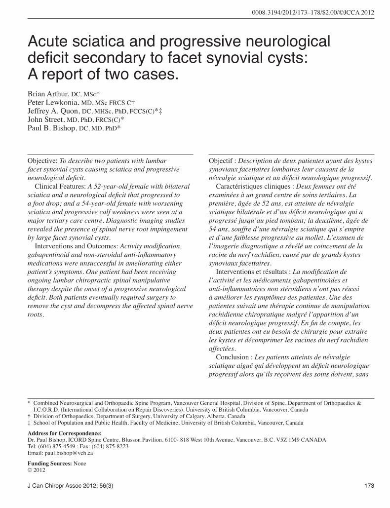
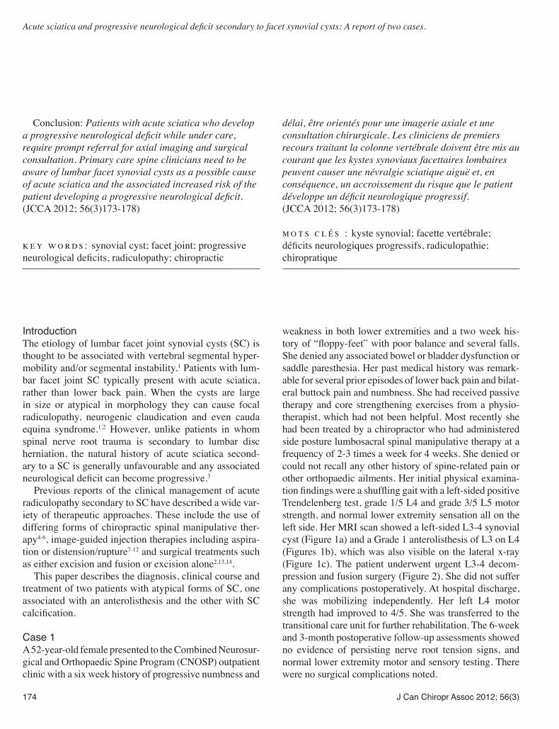
Case 1A 52-year-old female presented to the Combined Neurosur-gical and Orthopaedic Spine Program (CNOSP) outpatient clinic with a six week history of progressive numbness and
weakness in both lower extremities and a two week his-toryof“floppy-feet”withpoorbalanceandseveralfalls.She denied any associated bowel or bladder dysfunction or saddle paresthesia. Her past medical history was remark-able for several prior episodes of lower back pain and bilat-eral buttock pain and numbness. She had received passive therapy and core strengthening exercises from a physio-therapist, which had not been helpful. Most recently she had been treated by a chiropractor who had administered side posture lumbosacral spinal manipulative therapy at a frequency of 2-3 times a week for 4 weeks. She denied or could not recall any other history of spine-related pain or other orthopaedic ailments. Her initial physical examina-tionfindingswereashufflinggaitwithaleft-sidedpositiveTrendelenberg test, grade 1/5 L4 and grade 3/5 L5 motor strength, and normal lower extremity sensation all on the left side. Her MRI scan showed a left-sided L3-4 synovial cyst (Figure 1a) and a Grade 1 anterolisthesis of L3 on L4 (Figures 1b), which was also visible on the lateral x-ray (Figure 1c). The patient underwent urgent L3-4 decom-pression and fusion surgery (Figure 2). She did not suffer any complications postoperatively. At hospital discharge, she was mobilizing independently. Her left L4 motor strength had improved to 4/5. She was transferred to the transitional care unit for further rehabilitation. The 6-week and 3-month postoperative follow-up assessments showed no evidence of persisting nerve root tension signs, and normal lower extremity motor and sensory testing. There were no surgical complications noted.
Conclusion: Patients with acute sciatica who develop a progressive neurological deficit while under care, require prompt referral for axial imaging and surgical consultation. Primary care spine clinicians need to be aware of lumbar facet synovial cysts as a possible cause of acute sciatica and the associated increased risk of the patient developing a progressive neurological deficit. (JCCA 2012; 56(3)173-178) k e y w o r d s : synovial cyst; facet joint; progressive neurologicaldeficits,radiculopathy;chiropractic
délai, être orientés pour une imagerie axiale et une consultation chirurgicale. Les cliniciens de premiers recours traitant la colonne vertébrale doivent être mis au courant que les kystes synoviaux facettaires lombaires peuvent causer une névralgie sciatique aiguë et, en conséquence, un accroissement du risque que le patient développe un déficit neurologique progressif. (JCCA 2012; 56(3)173-178) m o t s c l é s : kyste synovial; facette vertébrale; déficitsneurologiquesprogressifs,radiculopathie;chiropratique
J Can Chiropr Assoc 2012; 56(3) 175
B Arthur, P Lewkonia, JA Quon, J Street, PB Bishop
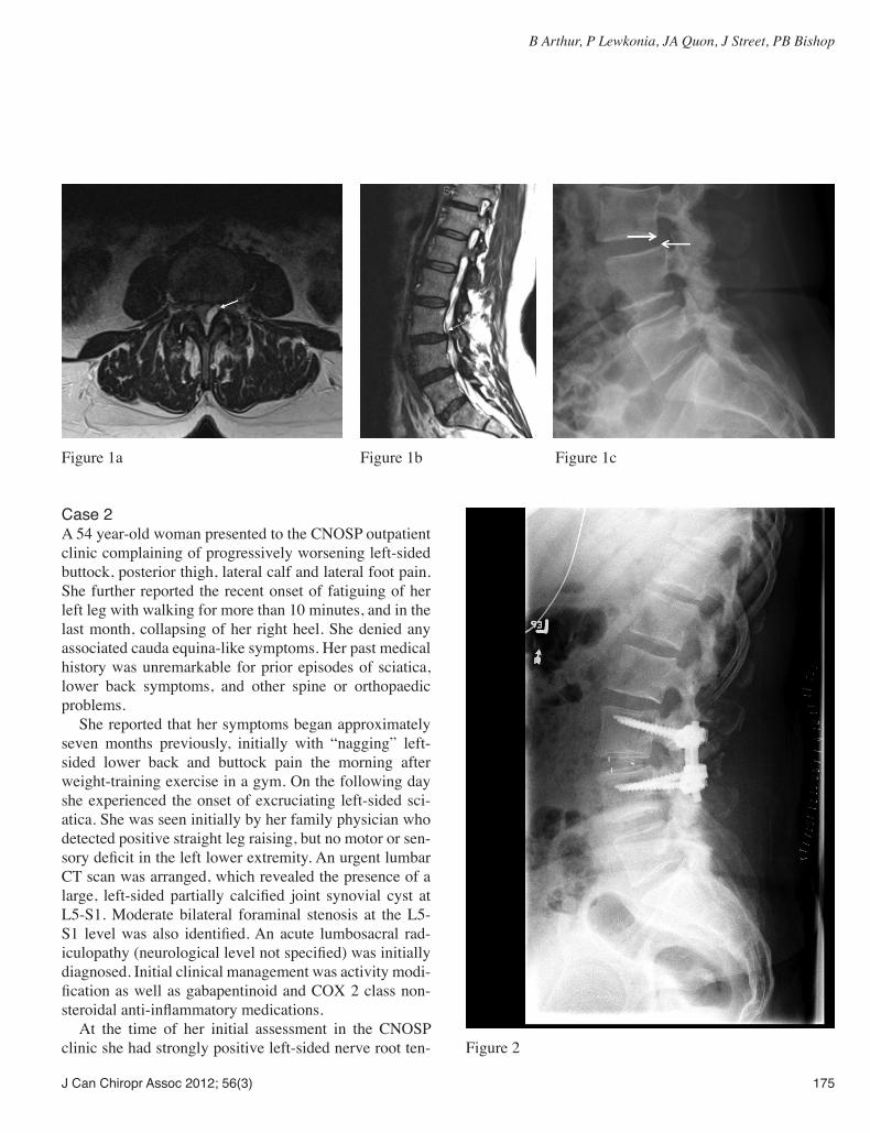
Case 2A 54 year-old woman presented to the CNOSP outpatient clinic complaining of progressively worsening left-sided buttock, posterior thigh, lateral calf and lateral foot pain. She further reported the recent onset of fatiguing of her left leg with walking for more than 10 minutes, and in the last month, collapsing of her right heel. She denied any associated cauda equina-like symptoms. Her past medical history was unremarkable for prior episodes of sciatica, lower back symptoms, and other spine or orthopaedic problems. She reported that her symptoms began approximately seven months previously, initially with “nagging” left-sided lower back and buttock pain the morning after weight-training exercise in a gym. On the following day she experienced the onset of excruciating left-sided sci-atica. She was seen initially by her family physician who detected positive straight leg raising, but no motor or sen-sorydeficitintheleftlowerextremity.AnurgentlumbarCT scan was arranged, which revealed the presence of a large, left-sidedpartially calcified joint synovial cyst atL5-S1. Moderate bilateral foraminal stenosis at the L5-S1 levelwas also identified.Anacute lumbosacral rad-iculopathy(neurologicallevelnotspecified)wasinitiallydiagnosed. Initial clinical management was activity modi-ficationaswellasgabapentinoidandCOX2classnon-steroidalanti-inflammatorymedications. At the time of her initial assessment in the CNOSP clinic she had strongly positive left-sided nerve root ten-
Figure 1a Figure 1b Figure 1c
Figure 2
176 J Can Chiropr Assoc 2012; 56(3)
Acute sciatica and progressive neurological deficit secondary to facet synovial cysts: A report of two cases.
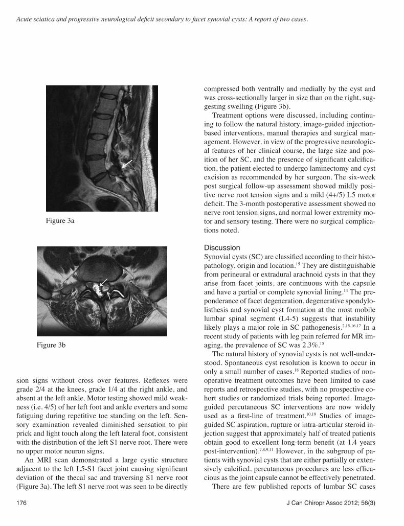
sion signs without cross over features. Reflexes weregrade 2/4 at the knees, grade 1/4 at the right ankle, and absent at the left ankle. Motor testing showed mild weak-ness (i.e. 4/5) of her left foot and ankle everters and some fatiguing during repetitive toe standing on the left. Sen-sory examination revealed diminished sensation to pin prick and light touch along the left lateral foot, consistent with the distribution of the left S1 nerve root. There were no upper motor neuron signs. An MRI scan demonstrated a large cystic structure adjacent to the leftL5-S1facet jointcausingsignificantdeviation of the thecal sac and traversing S1 nerve root (Figure 3a). The left S1 nerve root was seen to be directly
Figure 3a
Figure 3b
compressed both ventrally and medially by the cyst and was cross-sectionally larger in size than on the right, sug-gesting swelling (Figure 3b). Treatment options were discussed, including continu-ing to follow the natural history, image-guided injection-based interventions, manual therapies and surgical man-agement. However, in view of the progressive neurologic-al features of her clinical course, the large size and pos-itionofherSC,andthepresenceofsignificantcalcifica-tion, the patient elected to undergo laminectomy and cyst excision as recommended by her surgeon. The six-week post surgical follow-up assessment showed mildly posi-tive nerve root tension signs and a mild (4+/5) L5 motor deficit.The3-monthpostoperativeassessmentshowednonerve root tension signs, and normal lower extremity mo-tor and sensory testing. There were no surgical complica-tions noted.
DiscussionSynovialcysts(SC)areclassifiedaccordingtotheirhisto-pathology, origin and location.15 They are distinguishable from perineural or extradural arachnoid cysts in that they arise from facet joints, are continuous with the capsule and have a partial or complete synovial lining.14 The pre-ponderance of facet degeneration, degenerative spondylo-listhesis and synovial cyst formation at the most mobile lumbar spinal segment (L4-5) suggests that instability likely plays a major role in SC pathogenesis.2,15,16,17 In a recent study of patients with leg pain referred for MR im-aging, the prevalence of SC was 2.3%.15
The natural history of synovial cysts is not well-under-stood. Spontaneous cyst resolution is known to occur in only a small number of cases.18 Reported studies of non-operative treatment outcomes have been limited to case reports and retrospective studies, with no prospective co-hort studies or randomized trials being reported. Image-guided percutaneous SC interventions are now widely used as a first-line of treatment.10,19 Studies of image-guided SC aspiration, rupture or intra-articular steroid in-jection suggest that approximately half of treated patients obtaingood to excellent long-termbenefit (at1.4yearspost-intervention).7,8,9,11 However, in the subgroup of pa-tients with synovial cysts that are either partially or exten-sivelycalcified,percutaneousproceduresarelesseffica-cious as the joint capsule cannot be effectively penetrated. There are few published reports of lumbar SC cases
J Can Chiropr Assoc 2012; 56(3) 177
B Arthur, P Lewkonia, JA Quon, J Street, PB Bishop
treated by chiropractors. Cox, reported two cases that benefittedfromnon-thrustmanipulativetechniques(flex-ion distraction) combined with physical therapy modal-ities. However, long-term outcomes were not reported.4 Taylor, using Cox’s methods, reported similar benefitsin a single case, but the patient’s symptoms recurred and then necessitated ongoing visits and exercise therapy over a follow-up period of 4.5 years.5 In a third chiropractic casereport,thepatientreceivedfifteenadjustmentswithonlytemporarybenefitandeventuallyunderwentsurgicalexcisionwithgoodbenefit.6 Surgical cyst excision is considered the gold-standard treatment for successful long-term outcomes and is rec-ommendedinallcasesofneurologicdeficit.13,20 Concomi-tant lumbar fusion is required in cases with spondylol-isthesis and radiographically demonstrable segmental in-stability. In this regard, a systematic review of 82 studies showed that a substantial majority of surgically-treated SC patients obtained symptomatic resolution over the long-term (mean follow-up 25.4 months).20
The presentation of a patient with an acute radiculop-athy accompanied by a progressive neurological deficitshould raise concern for all spine clinicians. As primary spine healthcare providers, chiropractors may well be the portal of entry into the healthcare system for many of these patients. With the recent trend towards standardized treatment pathways in mainstream spine patient manage-ment, the diagnostic acumen of chiropractors is facing ever-increasing scrutiny, and in this environment the min-imum standard of care for spine patients must include diligent clinical monitoring and thorough record keeping. The two cases that have been presented highlight the importance of considering the presence of a SC in the differential diagnosis of patients with acute sciatica. In particular, the spine clinician’s index of suspicion for the presence of a SC should be raised when assessing a pa-tientwithaneurologicaldeficitincombinationwithade-generativespondylolisthesisand/orsignificantfacetjointdegenerationonplainfilmx-ray.
SummaryPrimary care spine clinicians treating patients with acute sciatica need to be aware of the possibility of a synovial cyst as an underlying cause. Careful monitoring of the patient’s lower extremity neurological function is pru-dent as the patient’s clinical course unfolds. In patients
whose symptoms are not improving or are progressively worsening, axial imaging and prompt surgical referral is indicated.
References1. Khan AM, Girardi F. Spinal lumbar synovial cysts.
Diagnosis and management challenge. Eur Spine J. 2006; 15: 1176–1182.
2. Boviatsis EJ, Staurinou LC, Kouyialis AT, Gavra MM, Stavrinou PC, Themistokleous M, Selviaridis P, Sakas DE. Spinal synovial cysts: pathogenesis, diagnosis and surgical treatment in a series of seven cases and literature review. Eur Spine J. 2008; 17: 831–837.
3. Shah RV, Lutz GE. Lumbar intraspinal synovial cysts: conservative management and review of the world’s literature. Spine J. 2003; 3: 479–488.
4. Cox JM, Cox JM. Chiropractic treatment of lumbar spine synovial cysts: a report of two cases. J Manipulative Physiol Ther. 2005; 28: 143–147.
5. Taylor DN. Spinal synovial cysts and intersegmental instability: a chiropractic case. J Manipulative Physiol Ther. 2007; 30: 152–157.
6. Firth RL. Lumbar intraspinal synovial cyst containing gas as a cause for low back pain. J Manipulative Physiol Ther. 2000; 23: 276–278.
7. Slipman CW, Lipetz JS, Wakeshima Y, Jackson HB. Nonsurgical treatment of zygapophyseal joint cyst-induced radicular pain. Arch Phys Med Rehabil. 2000; 81: 973–977.
8. Parlier-Cuau C, Wybier M, Nizard R, Champsaur P, Le Hir P, Laredo J-D. Symptomatic lumbar joint synovial cysts: Clinical assessment of facet joint steroid injection after 1 and 6 months and long-term follow-up in 30 patients. Radiology. 1999; 210: 510–513.
9. Sabers SR, Ross SR, Grogg, BE, Lauder, TD. Procedure-based nonsurgical management of lumbar zygapophyseal joint cyst-induced radicular pain. Arch Phys Med Rehabil. 2005; 86: 1767–1771.
10. Bureau NJ, Kaplan PA, Dussault RG. Lumbar facet joint synovial cyst: Percutaneous treatment with steroid injections and distension – clinical and imaging follow-up in 12 patients. Radiology. 2001; 221: 179–185.
11. Martha JF, Swaim B, Wang DA, Kim DH, Hill J, Bode R, Schwartz CE. Outcome of percutaneous rupture of lumbar synovial cysts: a case series of 101 patients. Spine J. 2009; 9: 899–904.
12. Allen TL, Tatli Y, Lutz GE. Fluoroscopic percutaneous lumbar zygapophyseal joint cyst rupture: a clinical outcome study. Spine J. 2009; 9: 387–395.
13. Lyons MK, Atkinson JL, Wharen RE, Deen HG, Zimmerman RS, Lemens SM. Surgical evaluation and management of lumbar synovial cysts: the Mayo Clinic Experience. J Neurosurg (Spine 1). 2000; 93: 53–57.
178 J Can Chiropr Assoc 2012; 56(3)
Acute sciatica and progressive neurological deficit secondary to facet synovial cysts: A report of two cases.
14. Ayberk G, Özveren F, Gök B, Yazgan A, Tosun H, Seçkin Z, Altundal N. Lumbar synovial cysts: experience with nine cases. Neurol Med Chir (Tokyo). 2008; 48: 298–303.
15. Doyle AJ, Merrilees M. Synovial cysts of lumbar facet joints in a symptomatic population. Prevalence on magnetic resonance imaging. Spine. 2004; 29: 874–878.
16. Tillich M, Trummer M, Lindbichler F, Flaschka G. Symptomatic intraspinal synovial cysts of the lumbar spine:correlationofMRandsurgicalfindings.Neuroradiology 2001; 43: 1070–1075.
17. Wilby MJ, Fraser RD, Vernon-Roberts B, Moore RJ. The prevalence and pathogenesis of synovial cysts within the ligamentumflavuminpatientswithlumbarspinalstenosisand radiculopathy. Spine 2009; 34: 2518–2524.
18. Houten JK, Sanderson SP, Cooper PR. Spontaneous regression of symptomatic lumbar synovial cysts. J Neurosurg (Spine 2) 2003; 99: 235–238.
19. Hong, Y, O’Grady T, Carlsson C, Casey J, Clements D. Percutaneous aspiration of lumbar facet synovial cysts. Anesthesiology 1995; 82: 1061–1062.
20.BydonA,XuR,ParkerSL,McGirtMJ,BydonM,Gokaslan ZL, Witham TF. Recurrent back and leg pain and cyst reformation after surgical resection of spinal synovial cysts: systematic review of reported postoperative outcomes. Spine J 2010; 10: 820–826.