Embed Size (px)
Citation preview
17K.I. Bland et al. (eds.), Surgery of the Pancreas and Spleen, DOI: 10.1007/978-1-84996-369-5_2, © Springer-Verlag London Limited 2011
2Acute PancreatitisJosé Eduardo M. Cunha, Marcel C.C. Machado, and José Jukemura
Pearls and Pitfalls
Acute pancreatitis (AP) represents a spectrum of disease •ranging from a mild, self-limited course to a rapidly pro-gressive, severe illness.Most patients have a mild disease and recover without •specific treatment, whereas the mortality rate of the severe form, which occurs in about 10–15% of patients, exceeds 20%. Clinical severity is determined by the degree of pan-creatic and peripancreatic necrosis defined by contrast-enhanced computed tomography (CT).The most common causes are gallstones and alcohol •abuse.The diagnosis of AP requires two of the following three •features: characteristic abdominal pain, hyperamylasemia, and findings of AP on CT.Management of mild AP is supportive – intravenous fluids •and analgesics; severe pancreatitis requires management in an intensive care unit.Early enteral nutrition is the preferred feeding method in •severe AP, because it preserves the intestinal mucosal bar-rier and does not predispose to fungal infections.Early endoscopic retrograde cholangiography with sphinc-•terotomy is indicated for severe biliary pancreatitis and cholangitis.
18 J.E.M. Cunha et al.
CT or US-guided fine-needle aspiration (FNA) for bacte-•riology should be performed in patients suspected of hav-ing infected pancreatic necrosis.Necrosectomy as opposed to catheter drainage is recom-•mended for infected pancreatic necrosis.Newer, minimal access methods (percutaneous, laparo-•scopic, endoscopic, and small incision, focused open necro-sectomy) have been introduced recently.Early operative intervention within 14 days of onset of AP •is discouraged for necrotizing pancreatitis (NP).Sterile pancreatic necrosis should be managed conservatively.•
Introduction
Acute pancreatitis (AP) is an acute inflammatory disease of the pancreas with an incidence that varies from 15 to 80 cases per year per 100,000 adults depending on the world region. According to recent data, the USA and Brazil are the countries with the highest occurrence rates. The clinical manifestations and evolution of AP depend on the various etiologic and morphologic aspects. Approximately 80–85% of patients have a mild, self-limiting process associated with edema in the interstitial pancreatic tissue or around the gland; this interstitial edematous pancreatitis resolves with supportive medical management. The remaining 15–20% of patients present with a severe, potentially life-threatening form of the disease; necrotizing pancreatitis is characterized by pancreatic parenchymal and/or peripancreatic necrosis which is generally associated with single or multiple organ failure (MODS). In these patients, the mortality rate approaches 30%.
Etiology
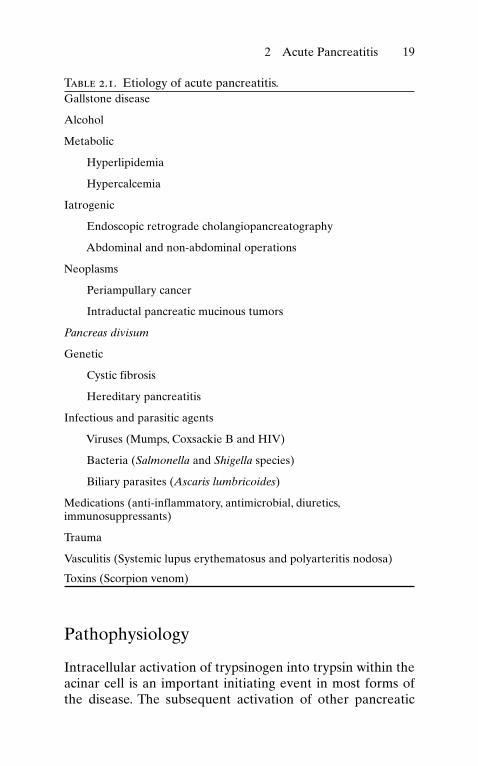
Many factors play an etiologic role in AP. The most common etiologic factors are outlined in Table 2.1, the most common of which are alcohol abuse and gallstones.
192 Acute Pancreatitis
Pathophysiology
Intracellular activation of trypsinogen into trypsin within the acinar cell is an important initiating event in most forms of the disease. The subsequent activation of other pancreatic
Table 2.1. Etiology of acute pancreatitis.Gallstone disease
Alcohol
Metabolic
Hyperlipidemia
Hypercalcemia
Iatrogenic
Endoscopic retrograde cholangiopancreatography
Abdominal and non-abdominal operations
Neoplasms
Periampullary cancer
Intraductal pancreatic mucinous tumors
Pancreas divisum
Genetic
Cystic fibrosis
Hereditary pancreatitis
Infectious and parasitic agents
Viruses (Mumps, Coxsackie B and HIV)
Bacteria (Salmonella and Shigella species)
Biliary parasites (Ascaris lumbricoides)
Medications (anti-inflammatory, antimicrobial, diuretics, immunosuppressants)
Trauma
Vasculitis (Systemic lupus erythematosus and polyarteritis nodosa)
Toxins (Scorpion venom)
20 J.E.M. Cunha et al.
proenzymes such as proelastase, chymotrypsinogen, procar-boxypeptidase and pyrophospholipase A2 by auto activation of trypsin leads to pancreatic acinar cell damage with spillage of activated enzymes into the pancreatic and peripancreatic tissues. Previous theories involving intraductal activation of enzymes within the pancreatic duct, reflux of bile, ductal hypertension, and a ductal rupture have been disproven. It appears that all etiologies of necrotizing pancreatitis work via this final common pathway, even alcohol abuse, biliary tract obstruction, and other etiologic factors. The prognosis of the AP depends on the intensity of the multiple organ dys-function syndrome (MODS) caused by the systemic inflam-matory response (SIRS). The pathogenesis of the systemic manifestations of complicated AP seems to result from release of multiple inflammatory mediators from activated leukocytes and macrophages, including the pro-inflammatory cytokines IL-1, IL-6, IL-8, PAF, and TNF-alpha, which appar-ently play the key roles in initiating the septic syndrome. The anti-inflammatory cytokines IL-10, TNF-soluble receptors, and IL1 receptor antagonist are also implicated in this inflammatory response to AP to some extent.
Infection of the necrotic tissue by enteric microorganisms represents a serious complication of AP that takes place in about 30% of patients with necrotizing pancreatitis.
Classification of Acute Pancreatitis
A classification system for AP based on clinical, pathologic, and radiologic criteria was proposed at an international sym-posium held in Atlanta in 1992. This “Atlanta classification” defined severe AP based on clinical manifestations, high scores in multiple factor scoring systems, as well as evidence of organ failure and intrapancreatic pathology. Although the Atlanta classification represented an important step forward in the understanding and management of AP, new knowledge achieved recently on pathophysiology, diagnosis, and man-agement of AP have made some of its aspects outdated.
212 Acute Pancreatitis
These factors motivated the organization of an international study group that is involved currently in revising and updat-ing the classification of AP.
Clinical Presentation
Abdominal pain of moderate to severe intensity is the initial symptom in the majority of the patients. Abdominal pain generally presents as sudden onset and persists for one or more days with nausea and vomiting present in about 90% of patients. Fever is a common finding over the first several days; however, it usually resolves thereafter. When present after the second week in patients with necrotizing pancreati-tis, fever is usually due to infection of the necrotic tissue. Jaundice is uncommon, although small increases in serum bilirubin may occur due to distal common bile duct compres-sion by the inflamed pancreatic head or when the AP is sec-ondary to a gallstone impacted at the ampulla of Vater.
Severe AP evolves over three variable phases. The first phase involves the first week or two after onset of AP. Clinical manifestations may include fever, leukocytosis, hemodynamic instability, respiratory failure, renal dysfunction, and senso-rial disturbances, related to SIRS; the pancreatic and peri-pancreatic necrosis is usually sterile during this first phase. The second phase often occurs after resolution of SIRS with a period of relative disease and symptom quiescence lasting one to two weeks. During this second phase, the necrosis becomes “superinfected” in up to 30% of patients with necrotizing AP. The third phase evolves along one of three forms. One form leads to liquefaction necrosis and eventual resolution of symptoms and the necrosis over the next 4–12 weeks. A second form involves the organization of sterile, non-infected pancreatic and peripancreatic liquefaction necrosis that persists, causing a feeling of unwell and inability to eat related to the mass effect of this “organized” necrosis which is termed “walled off pancreatic necrosis”; in some type of operative intervention is usually needed to resolve
22 J.E.M. Cunha et al.
this form. The third form is the most serious and involves progression of the superinfection of the necrosis into a pan-creatic abscess with both local and systemic symptoms. Active operative or, if the process is more localized, percutaneous or minimal access intervention with necrosectomy is necessary.
Risk Factors for Pancreatic Infection
Pancreatic infection occurs in up to 30% of all patients with severe AP. Infection is more likely to occur in patients with extensive pancreatic necrosis. Nevertheless, patients may still become infected even when lesser amounts of the gland are necrotic; indeed, about 20% of patients with necrotizing pan-creatitis have primarily peripancreatic necrosis which can become infected. Another risk factor for infection of the pan-creatic necrosis is the duration of the disease: the rate of infected necrosis usually increases from the second to the third week, declining to a lesser infection rate after the fourth week.
Biochemical Diagnosis
Diagnosis of AP is made on the basis of clinical presentation, characterized by the characteristic abdominal pain of pancre-atic origin combined with blood tests and imaging modalities. Determination of amylase and/or lipase in plasma remains the gold standard for the diagnosis of AP. Plasma levels of both enzymes peak within the first 24 h of symptoms. Because amylase and lipase have different half-lives, plasma amylase levels remain increased for approximately 36 h, whereas lipase will be sustained for a much longer period. Plasma levels of these enzymes do not correlate with severity of AP.
Imaging Diagnosis
Ultrasonography (US): Transabdominal US represents the first imaging technique usually utilized for evaluating patients
232 Acute Pancreatitis
with AP but plays only a limited role in the diagnosis or stag-ing, because the pancreas may not be visible in many patients with AP due to overlying abdominal gas. US can be of value in demonstrating biliary sludge or stones in the gallbladder, thereby implying an etiology of gallstone pancreatitis. US can detect an increased pancreatic volume, changes of the pan-creatic parenchyma, and presence of peripancreatic fluid collections. Because US cannot assess organ perfusion, detec-tion of pancreatic necrosis is difficult. This limitation has been addressed by the recent development of echo-enhanc-ers that have made possible a better evaluation of the pancre-atic blood supply.
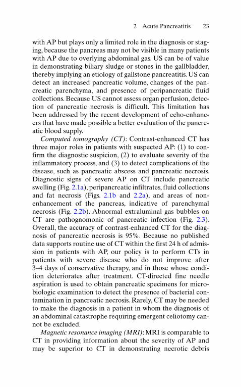
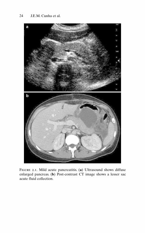
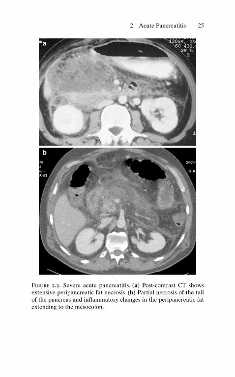
Computed tomography (CT): Contrast-enhanced CT has three major roles in patients with suspected AP: (1) to con-firm the diagnostic suspicion, (2) to evaluate severity of the inflammatory process, and (3) to detect complications of the disease, such as pancreatic abscess and pancreatic necrosis. Diagnostic signs of severe AP on CT include pancreatic swelling (Fig. 2.1a), peripancreatic infiltrates, fluid collections and fat necrosis (Figs. 2.1b and 2.2a), and areas of non-enhancement of the pancreas, indicative of parenchymal necrosis (Fig. 2.2b). Abnormal extraluminal gas bubbles on CT are pathognomonic of pancreatic infection (Fig. 2.3). Overall, the accuracy of contrast-enhanced CT for the diag-nosis of pancreatic necrosis is 95%. Because no published data supports routine use of CT within the first 24 h of admis-sion in patients with AP, our policy is to perform CTs in patients with severe disease who do not improve after 3–4 days of conservative therapy, and in those whose condi-tion deteriorates after treatment. CT-directed fine needle aspiration is used to obtain pancreatic specimens for micro-biologic examination to detect the presence of bacterial con-tamination in pancreatic necrosis. Rarely, CT may be needed to make the diagnosis in a patient in whom the diagnosis of an abdominal catastrophe requiring emergent celiotomy can-not be excluded.
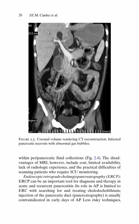
Magnetic resonance imaging (MRI): MRI is comparable to CT in providing information about the severity of AP and may be superior to CT in demonstrating necrotic debris
24 J.E.M. Cunha et al.
a
b
Figure 2.1. Mild acute pancreatitis. (a) Ultrasound shows diffuse enlarged pancreas. (b) Post-contrast CT image shows a lesser sac acute fluid collection.
252 Acute Pancreatitis
a
b
Figure 2.2. Severe acute pancreatitis. (a) Post-contrast CT shows extensive peripancreatic fat necrosis. (b) Partial necrosis of the tail of the pancreas and inflammatory changes in the peripancreatic fat extending to the mesocolon.
26 J.E.M. Cunha et al.
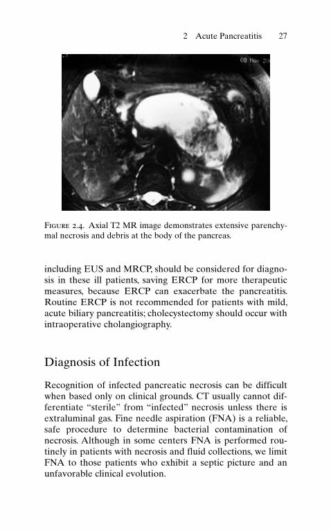
within peripancreatic fluid collections (Fig. 2.4). The disad-vantages of MRI, however, include cost, limited availability, lack of radiologic experience, and the practical difficulties of scanning patients who require ICU monitoring.
Endoscopic retrograde cholangiopancreatography (ERCP): ERCP can be an important tool for diagnosis and therapy in acute and recurrent pancreatitis. Its role in AP is limited to ERC with searching for and treating choledocholithiasis; injection of the pancreatic duct (pancreatography) is usually contraindicated in early days of AP. Less risky techniques,
Figure 2.3. Coronal volume rendering CT reconstruction. Infected pancreatic necrosis with abnormal gas bubbles.
272 Acute Pancreatitis
including EUS and MRCP, should be considered for diagno-sis in these ill patients, saving ERCP for more therapeutic measures, because ERCP can exacerbate the pancreatitis. Routine ERCP is not recommended for patients with mild, acute biliary pancreatitis; cholecystectomy should occur with intraoperative cholangiography.
Diagnosis of Infection
Recognition of infected pancreatic necrosis can be difficult when based only on clinical grounds. CT usually cannot dif-ferentiate “sterile” from “infected” necrosis unless there is extraluminal gas. Fine needle aspiration (FNA) is a reliable, safe procedure to determine bacterial contamination of necrosis. Although in some centers FNA is performed rou-tinely in patients with necrosis and fluid collections, we limit FNA to those patients who exhibit a septic picture and an unfavorable clinical evolution.
Figure 2.4. Axial T2 MR image demonstrates extensive parenchy-mal necrosis and debris at the body of the pancreas.
28 J.E.M. Cunha et al.
Severity Assessment
Most patients with AP have a self-limiting course and require only general support and intravenous fluids. In contrast, suc-cess of treatment of severely ill patients with AP depends on early identification and prompt institution of prophylactic antibiotic treatment. Early objective measures of severity are useful to recognize such patients.
Multiple factor scoring systems: The Ranson criteria, devel-oped in 1974, evaluate 11 clinical and laboratory parameters gathered during the first 48 h after the onset of AP. Although this scoring system selects patients who are at a high risk of life-threatening complications, it requires 48 h to complete. The Glasgow criteria, proposed to simplify the Ranson approach, was an equally effective predictor of mortality, regardless of etiology, but its accuracy was no better than the Ranson criteria and also requires 48 h for maximum effi-ciency. The APACHE II illness grading system has been used more successfully to stage the severity of illness in patients with AP. A score > 6 at admission tends to identify most of patients with severe pancreatitis and has proved useful in many randomized studies to select patients at highest risk. In addition, APACHE II can be employed throughout the course of the disease to follow progression and response to therapy. One disadvantage of APACHE II is that it gives too heavy a weighting for age. Because obesity is a known risk factor for patients with AP, proposal of the APACHE-O score, which scores one extra point for BMI 25–30 and two points for BMI > 30 enhances the accuracy of APACHE II grading of severity.
CT grading systems: Because non-enhancing, hypoper-fused areas within the pancreas on contrast-enhanced CT correlates with pancreatic necrosis, CT can also be used as a tool for grading of severity in AP. The morphologic severity of AP using the CT Severity Index (CTSI) was developed by Balthazar and coworkers. The CTSI is based on a combina-tion of peripancreatic inflammation, phlegmon, and degree of pancreatic necrosis seen on the initial CT. Large amounts
292 Acute Pancreatitis
of ischemic or infarcted pancreas obtain the highest scores. In our experience, CT findings of necrosis in the retropancre-atic, intercaval-aortic, and the peri-mesenteric vessels are associated with a higher mortality. One limitation is the high cost of repeated CTs related to the need for repeated CTs as the dynamic disease process unfolds.
Markers of pancreatic injury: Numerous studies have searched for a blood test that could be performed quickly to discriminate between mild and severe AP. Plasma levels of amylase and lipase have no value in prediction of severity. C-reactive protein (CRP) is the cheapest, most useful (albeit non-pancreas specific) marker to distinguish between edem-atous and necrotizing AP. Values > 150 mg/l are suggestive of severe AP. Although IL-6, IL-8, and procalcitonin are more sensitive tests than CRP, none of these blood studies are available widely and suspicion of the diagnosis of necrotizing pancreatitis is generally based on clinical and imaging assessment.
Medical Treatment
Although the majority of patients with AP have an unevent-ful course, every patient should be treated aggressively until the severity of the disease has been properly determined.
Supportive care: The goals of initial management include fluid and electrolyte replacement, nutritional support, and correction of hypovolemia. Some patients should be moni-tored by measurement of CVP, urine output, and placement of a Swan-Ganz catheter in patients with cardiopulmonary disease or cardiopulmonary decompensation. Good experi-mental evidence supports the value of aggressive resuscita-tion and hydration in treatment of AP.
Analgesic therapy: The treatment of pain in AP is essential. Moderate and severe pain requires narcotic analgesics, but not morphine, which may cause spasm at the sphincter of Oddi. Patient-controlled analgesia (PCA) via an epidural catheter can be very effective.

30 J.E.M. Cunha et al.
Inhibition of exocrine pancreatic secretion: Numerous clinical trials have failed to demonstrate any effectiveness of drugs used to inhibit exocrine pancreatic secretion. These data speak against the use of any medication to inhibit pan-creatic secretion. Oral intake is withheld by most surgeons during the initial course of the disease.
Antibiotics: The value of prophylactic antibiotics in AP remains very controversial. Reduction of infectious compli-cations and mortality has been demonstrated in several but certainly not all randomized studies. Recovery of microor-ganisms by FNA usually shows polymicrobial, Gram-negative bacteria of intestinal origin and on occasion (~10%) fungal infection, mainly of Candida species. Our current policy is to use imipenem and fluconazole in all patients with severe AP.
Nutritional support: Patients with severe AP have increased nutritional needs because of increases in energy expenditure and catabolism. Adequate provision of nutrition can be chal-lenging without stimulating pancreatic exocrine secretion. Early enteral feeding is associated with a reduction in the acute phase response and the severity of gut-derived infec-tious complications of AP.
Endoscopic treatment: Currently, most pancreatologists suggest early endoscopic treatment of patients with severe acute biliary pancreatitis. A meta-analysis of four random-ized controlled trials of endoscopic sphincterotomy (ES) showed reduced complications and mortality in those patients with biliary obstruction and/or cholangitis [6]. Our policy is to recommend early ES (within 72 h of hospital admission) in patients with severe AP when jaundice and/or cholangitis persists or when patients with initially mild disease deterio-rate clinically. ES is of no use in mild biliary AP.
Surgical Treatment
The role of surgical treatment of AP is to control local com-plications of the disease.
312 Acute Pancreatitis
Gallstone pancreatitis: Patients with gallstone pancreatitis not treated previously by ES should be offered same admis-sion cholecystectomy after their pain resolves; recurrent AP occurs in up to 70% of the patients within one year after AP if the gallbladder is not removed. Intraoperative cholangiog-raphy should be performed at the time of cholecystectomy. Cholecystectomy should be delayed 3–4 months in patients with severe AP, because early cholecystectomy has a higher complication rate and operative mortality. In patients too ill to undergo cholecystectomy, ES may be an alternative.
Infected pancreatic necrosis: Pancreatic infection consti-tutes the main indication for operative treatment of necrotiz-ing pancreatitis. Failure to recognize and treat these patients results in high mortality rates, whereas the mortality associ-ated with operative debridement and drainage is 10–20%.
Removal of infected pancreatic and peripancreatic necrotic tissue decreases the risks of local and systemic complications. Traditionally, surgical debridement has been performed via an open operative approach via laparotomy. The entire peri-pancreatic space, including the base of the small bowel mes-entery and the paracolic gutters, must be evaluated to effect a complete removal of the necrotic tissue. The CT serves as a guide to all suspect areas of necrosis. Repeat operations may be necessary. When the viability of the transverse colon is compromised, an extended right hemicolectomy with the construction of an ileostomy and mucous fistula is indicated. After completing the necrosectomy, some form of wide peri-pancreatic drainage should be performed. We have favored partial abdominal closure over multiple Penrose drains and repeat operations every 48–72 h until the completion of necrosis debridement, while others utilize closed irrigation/lavage of the peripancreatic space via inflow and outflow catheters left at the time of necrosectomy.
The results of surgical debridement depend on the timing of the operative procedure. Delaying the initial operation if possi-ble for 3 or 4 weeks after disease onset decreases the mortality, because the necrotic tissue demarcates from viable pancreas, leading to a safer, more complete necrosectomy. Recently,
32 J.E.M. Cunha et al.
necrosectomy performed by minimally invasive procedures such as laparoscopic and retroperitoneal percutaneous approaches, have been used in selected patients (see chapter 1).
Sterile pancreatic necrosis (SPN): In 60–70% of patients with necrotizing pancreatitis, the necrotic tissue remains ster-ile. The optimal approach in these patients who are symptom-atic is controversial. Some groups suggest that surgical debridement speeds recovery, but many patients can be suc-cessfully managed non-operatively. Furthermore, operative intervention may induce bacterial contamination of sterile necrosis with subsequent increase of mortality rates. Nevertheless, progressive deterioration from SIRS and ongo-ing sepsis are accepted indications for necrosectomy in patients with SPN.
Selected Readings
Bradley EL III (1991) Operative management of acute pancreatitis: ventral open packing. Hepatogastroenterology 38:134–138
Bradley EL III (1993) A clinically based classification system for acute pancreatitis. Summary of the International Symposium on Acute Pancreatitis, September 11–13, 1992, Atlanta, GA. Arch Surg 128: 586–590
Buchler MW, Gloor B, Muller CA, et al. (2000) Acute necrotizing pan-creatitis: treatment strategy according to the status of infection. Ann Surg 232:619–626
Cunha JEM, Machado MCC, Penteado S, et al. (1994) Pan-creatic necrosis in Brazil. In: Bradley EL III (ed) Acute pancreatitis: diagno-sis and treatment. Raven, New York, pp 121–125
Garg PK, Khanna S, Bohidar NP, et al. (2001) Incidence, spectrum and antibiotic sensitivity pattern of bacterial infections among patients with acute pancreatitis. J Gastroenterol Hepatol 16:1055–1059
Rau B, Pralle U, Mayer JM, Beger HG (1998) Role of ultra-sonograph-ically guided fine-needle aspiration cytology in the diagnosis of infected pancreatic necrosis. Br J Surg 85:179–184
Sharma VK, Howden CW (1999) Meta-analysis of randomized con-trolled trials of endoscopic retrograde cholangiography and endo-scopic sphincterotomy for the treatment of acute biliary pancreatitis. Am J Gastroenterol 94:3211–3214
Uomo G, Visconti M, Manes G, et al. (1996) Nonsurgical treatment of acute necrotizing pancreatitis. Pancreas 12:142–148