Embed Size (px)
Citation preview
Imaging in the ED 61
Acute Neck InfectionsBlair A. Winegar1, Wayne S. Kubal2
We present an overview of the imaging of acute neck infections with a focus on contrast-enhanced CT. The emphasis of this chap-ter is to enable the emergency radiologist to accurately diagnose neck infections, to effectively communicate imaging findings with emergency physicians, and to function as part of a team offering the best care to patients.
Patients with many types of head and neck infections may pres-ent in the emergency department. The causes of these disorders include dental infection, penetrating trauma, and upper respiratory infections. Neck infections continue to portend significant morbid-ity and mortality despite widespread access to antibiotics. Poten-tially life-threatening complications may occur in approximately 10–20% of acute neck infections, including airway obstruction, septic thrombophlebitis with septic emboli, arterial pseudoaneu-rysm, and mediastinitis [1]. As more and more patients use emer-gency departments for initial healthcare, emergency department physicians will increasingly encounter acute neoplastic and non-neoplastic conditions in the head and neck [2]. In this setting, CT is the preferred imaging modality [2].
Although acute neck infections are easily diagnosed by clini-cal examination, correct localization may be problematic without cross-sectional imaging. The extent of deep space involvement is particularly difficult to detect by clinical evaluation, with accu-rate localization of infection in only 42.9% of cases in one series [3]. In another series, the extent of deep neck space infection was underestimated in 70% of cases [4]. The anatomy of the cervical fascia affects the presentation, spread, and management of neck infection [5]. Therefore, radiologists must be familiar with the anatomy of the deep cervical fascia and the spaces bounded by these fascial planes. Because the layers of the deep cervical fascia are not well seen on contrast-enhanced CT, knowledge of their normal internal contents and expected boundaries is critical [6].
The Pharyngeal Mucosal SpaceThe pharyngeal mucosal space is bounded by the middle layer
of the deep cervical fascia and contains the mucosa of the upper aerodigestive tract and lymphoid tissue of the Waldeyer ring, in-cluding the palatine tonsils, lingual tonsils, and adenoids (Fig. 1). Upper aerodigestive tract infections begin within the pharyngeal
mucosal space and may spread to the deep spaces of the neck if not appropriately treated. Infections that involve the pharyngeal mucosal space include pharyngitis, tonsillitis, peritonsillar ab-scess, and epiglottis.
In patients with acute tonsillitis, the affected tonsillar tissue is enlarged and enhances after contrast material administration. The tonsils may display a striated enhancement pattern (tiger-stripe appearance), reflecting inflamed enhancing mucosa with underly-ing edematous submucosa. Uvulitis, enlargement and inflamma-tion involving the uvula may be an associated finding (Fig. 2A). Uncomplicated tonsillitis will not have a localized region of in-ternal hypoattenuation. As the infection progresses, an ill-defined region of hypoattenuation without a well-defined enhancing wall representing cellulitis or phlegmon may develop within the tonsil. This process may continue to evolve to abscess formation, defined by a low-density collection with attenuation similar to CSF sur-rounded by an enhancing wall. These abscesses may uncommonly remain within the tonsillar tissue, a true intratonsillar abscess. The majority of these abscesses will penetrate the fibrous capsule of the tonsil to involve the potential space located between the supe-rior constrictor muscle and tonsillar capsule, termed a peritonsillar abscess [2] (Fig. 2B).
Because treatment of peritonsillar abscess differs from acute tonsillitis and cellulitis or phlegmon, accurate characterization is paramount. The detection of a peritonsillar abscess requires incision and drainage or aspiration of the drainable fluid collec-tion in addition to antibiotic therapy. Clinical assessment has low specificity for differentiating tonsillar cellulitis from peritonsillar abscess. Even when strict imaging criteria are applied, accurate detection of drainable fluid collections on contrast-enhanced CT ranges from 63% to 77% in both adult and pediatric populations [7, 8]. An irregular or scalloped morphology of the enhancing wall of a fluid collection increases the specificity for abscess and sug-gests a more mature abscess [9] (Fig. 2C).
Epiglottitis and supraglottitis are inflammation of the epiglot-tis and adjacent supraglottic larynx, respectively, that is typically caused by bacteria. Historically, this condition was more frequent in children infected with Haemophilus influenzae type b (HIB). However, since widespread childhood vaccination to HIB was introduced in the 1990s, acute epiglottitis is now more frequent
1Both authors: Department of Medical Imaging, University of Arizona, 1501 Campbell Ave, PO Box 245067, Tucson, AZ 85724. Address correspondence to W. S. Kubal ([email protected]).
62 Imaging in the ED
Winegar and Kubal
in adults and is caused by a variety of bac-terial pathogens, including Streptococcus species and Staphylococcus aureus [10]. Symptoms include sore throat, fever, muf-fled voice, drooling, stridor or respiratory
compromise, and hoarseness. Definitive diagnosis is made by direct visualization, which may require laryngoscopy. Treat-ment includes maintaining a patent airway, corticosteroids, and IV antibiotics.
Historically, lateral radiographs have been used as a first-line imaging modal-ity for the detection of epiglottitis. Find-ings include thickening of the epiglottis (“thumb” sign), obliteration of the val-leculae (“vallecula” sign), thickening of the aryepiglottic folds, and dilatation of the hypopharynx. Contrast-enhanced CT is useful in diagnosis of epiglottis when direct visualization is not feasible. CT can differentiate epiglottitis from other causes of fever and sore throat (e.g., tonsillar ab-scess, retropharyngeal abscess) and allows characterization of complicating features (e.g., abscess formation, soft-tissue gas, airway compromise). The CT findings of epiglottitis include swelling of the epiglot-tis and supraglottic structures (e.g., aryepi-glottic folds, false vocal cords), inflamma-tory stranding of the preepiglottic fat, and thickening of the platysma and preverte-bral fascia (Fig. 3) [11].
The Retropharyngeal SpaceThe retropharyngeal space is a small
space containing predominantly fat situ-ated between the posterior aspect of the pharyngeal mucosal space and cervical esophagus and anterior to the preverte-bral musculature (Fig. 1). The suprahyoid retropharyngeal space contains medial
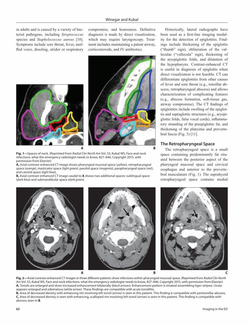
A CFig. 2—Axial contrast-enhanced CT images in three different patients show infections within pharyngeal mucosal space. (Reprinted from Radiol Clin North Am Vol. 53, Kubal WS, Face and neck infections: what the emergency radiologist needs to know, 827–846, Copyright 2015, with permission from Elsevier)A, Tonsils are enlarged and show increased enhancement bilaterally (black arrows). Enhancement pattern is striated (resembling tiger stripes). Uvula appears enlarged and edematous (white arrow). These findings are compatible with acute tonsillitis.B, Area of decreased density with enhancing rim involving left tonsil (arrow) is seen in this patient. This finding is compatible with peritonsillar abscess.C, Area of decreased density is seen with enhancing, scalloped rim involving left tonsil (arrow) is seen in this patient. This finding is compatible with abscess seen in B.
B
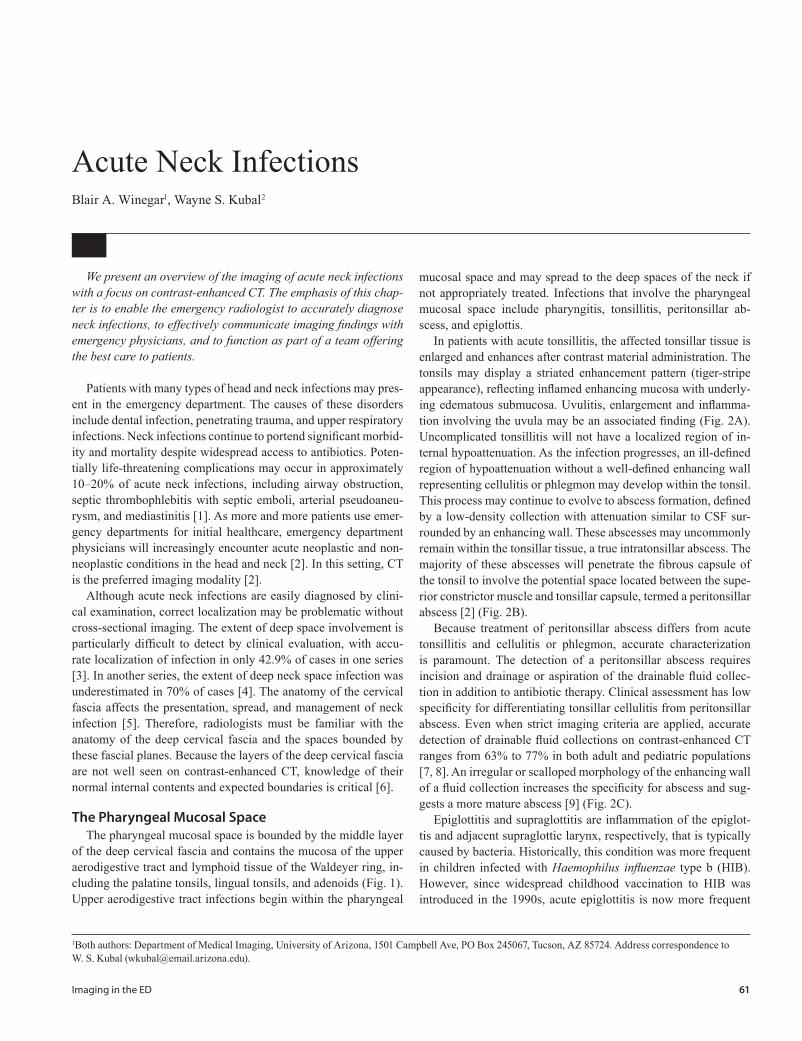
AFig. 1—Spaces of neck. (Reprinted from Radiol Clin North Am Vol. 53, Kubal WS, Face and neck infections: what the emergency radiologist needs to know, 827–846, Copyright 2015, with permission from Elsevier)A, Axial contrast-enhanced CT image shows pharyngeal mucosal space (yellow), retropharyngeal space (orange), masticator space (light green), parotid space (magenta), parapharyngeal space (red), and carotid space (light blue).B, Axial contrast-enhanced CT image caudal to A shows two additional spaces: sublingual space (dark blue) and submandibular space (dark green).
B
Imaging in the ED 63
Acute Neck Infections
and lateral retropharyngeal lymph nodes, which are more prominent in children less than 6 years old [12]. The retropha-ryngeal space has a bowtie configuration with variable presence of a median raphe, more often superiorly, which may confine disease to half of the space. The retropha-ryngeal space extends from the clivus to terminate between the T1 and T6 verte-bral levels. The danger space is a poten-tial space located posterior to the retro-pharyngeal space separated from it by the thin posterior slip of the alar fascia, which extends inferiorly to the diaphragm. Infec-tion within the retropharyngeal space or within the danger space may extend from the pharynx to the mediastinum [13]. It is therefore crucial to detect an infection within the retropharyngeal space and eval-uate the superior mediastinum for the po-tential complication of mediastinitis (Fig. 4). Infections within the retropharyngeal space may result from lymphatic spread of pharyngeal infections, cervical discogenic infections, or penetrating trauma.
On contrast-enhanced CT, retropharyn-geal abscess is defined by low-attenuation fluid filling the retropharyngeal space with a surrounding enhancing wall. The ab-scess typically shows mass effect on the
walls from the increased internal pressure. These findings contrast those of suppura-tive retropharyngeal lymphadenitis, typi-cally encountered in the pediatric popu-lation, which are focal, rim-enhancing fluid collections along the lateral margins of the suprahyoid retropharyngeal space. Retropharyngeal abscesses require surgi-cal drainage. Patients with suppurative retropharyngeal lymphadenitis typically receive a trial of IV antibiotics for 24–48 hours when the collection measures less than 2 cm3 in size. Surgical drainage is re-served for recalcitrant cases [14].
Calcific tendinitis of the longus colli muscle is an inflammatory condition that may mimic the clinical presentation (e.g., neck pain and fever) and some imaging findings of retropharyngeal abscess. On contrast-enhanced CT, this entity typically shows fluid or edema within the retropha-ryngeal space. In contrast to retropharyn-geal abscess, the walls of the retropharyn-geal space do not enhance in the setting of calcific tendinitis [15, 16]. In addition, amorphous calcification is present in the tendinous insertion of the longus colli mus-cle, typically seen along the inferior margin of the anterior arch of C1. Calcium hy-droxyapatite crystal deposition is respon-
sible for inciting the inflammation (Fig. 5). Differentiation of calcific tendinitis of the longus colli muscle from retropharyngeal abscess is essential, because treatment of the tendonitis is nonsurgical with nonste-roidal antiinflammatory medications.
The Masticator SpaceThe masticator space includes the mus-
cles of mastication and portions of the ad-jacent posterior mandible (Fig. 1). Because the masticator space contains the posterior mandible, infections are typically odon-togenic in origin, spreading from dental abscesses or following dental procedures involving the posterior mandibular molars. Because the deep cervical fascia surrounds all the muscles of mastication, infections can spread superiorly along the tempora-lis muscle above the zygomatic arch into the suprazygomatic masticator space to its attachment to the parietal calvaria. This fact may necessitate more superior imag-ing coverage to examine the entirety of the masticator space.
On contrast-enhanced CT, infections within the masticator space may show swelling of the affected muscles, variable enhancement, adjacent fat stranding, and possible rim-enhancing fluid collections. Careful evaluation of the adjacent poste-rior mandibular teeth on bone algorithm images can often identify the infectious source as evidenced by periapical lucency with or without cortical breach or the site of a recent dental procedure. Irregularity of the bone may reflect osteomyelitis, a com-plication of masticator space infection that requires aggressive antibiotic therapy.
Subtle deep masticator space infections involving the pterygoid muscles may be associated with inflammatory disease in the adjacent maxillary sinuses. Identifica-tion of infection in this location should be considered when inflammatory stranding is seen in the fat of the pterygopalatine fossa or retroantral fat pad situated be-tween the maxillary sinus and masticator space (Fig. 6).
The Parotid SpaceThe parotid gland and its intraparotid
lymph nodes are surrounded by the superfi-cial layer of the deep cervical fascia form-ing the parotid space (Fig. 1). The parotid
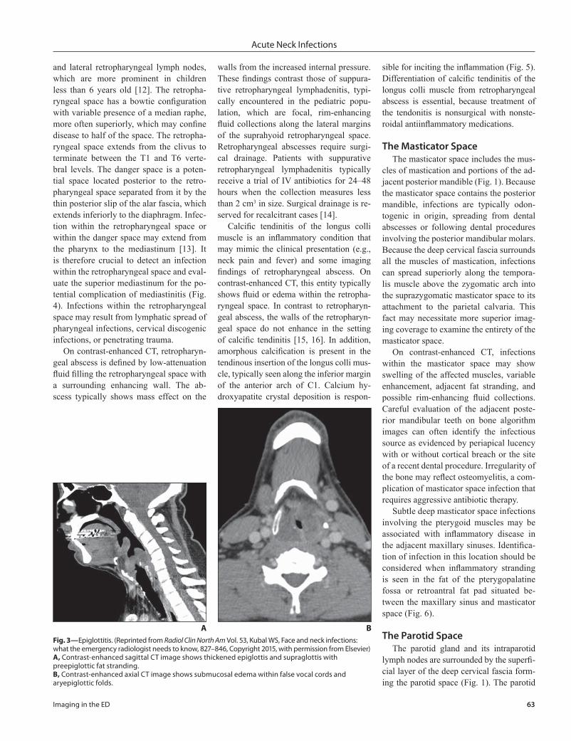
AFig. 3—Epiglottitis. (Reprinted from Radiol Clin North Am Vol. 53, Kubal WS, Face and neck infections: what the emergency radiologist needs to know, 827–846, Copyright 2015, with permission from Elsevier)A, Contrast-enhanced sagittal CT image shows thickened epiglottis and supraglottis with preepiglottic fat stranding.B, Contrast-enhanced axial CT image shows submucosal edema within false vocal cords and aryepiglottic folds.
B
64 Imaging in the ED
Winegar and Kubal
gland can vary in internal attenuation but normally has lower density than muscle and higher density than the subcutaneous fat on contrast-enhanced CT.
Parotid gland infections may result from bacterial or viral agents. Bacterial in-fections are often unilateral and assumed to result from an ascending infection from the oral cavity. These infections are easy to identify both clinically and on contrast-
enhanced CT because there is a normal parotid gland for comparison. Imaging shows unilateral enlargement and contrast enhancement of the affected parotid gland with surrounding inflammatory fat strand-ing. CT is particularly useful in the detec-tion of a focal rim-enhancing fluid collec-tion, compatible with intraparotid abscess, or a potential hyperattenuating calculus obstructing the parotid (Stensen) duct.
Viral parotitis is typically bilateral (e.g., mumps) and occurs in approximately 75% of cases [2]. These cases may be difficult to diagnose given the lack of a normal parotid gland for comparison. Increased attenua-tion of the parotid glands when compared with muscle and adjacent fat stranding will aid in the diagnosis.
The Carotid SpacePortions of the deep cervical fascia
loosely invest the internal jugular vein, carotid artery, portions of cranial nerves IX–XII, and adjacent internal jugular chain lymph nodes (Fig. 1). It is important to evaluate both the internal jugular vein and carotid arteries when assessing the carotid space. These vessels are readily identified on a contrast-enhanced examination. The internal jugular vein typically is located along the posterolateral margins of the ca-rotid arteries. Detection of inflammatory fat stranding along the margins of these vessels may indicate carotid space infection.
In 1936, Andre Lemierre [17] reported a case series in which septicemia resulted from anaerobic organisms found physiologi-cally in the human body. Lemierre syndrome (LS) describes septic thrombophlebitis of the internal jugular vein, typically the re-sult of oropharyngeal infection. This infec-tion may also result in septic emboli, most commonly to the lungs [18]. On contrast-enhanced CT, expansion and lack of contrast opacification of the internal jugular vein with surrounding inflammatory fat strand-ing is indicative of septic thrombophlebitis (Fig. 7A). CT may also show the causative oropharyngeal infection (e.g., peritonsillar abscess). When septic thrombophlebitis is detected, the lungs should be evaluated for cavitary nodules indicative of septic emboli (Fig. 7B). According to Weeks et al. [18], “given the relatively low incidence of LS and its potentially confusing clinical mani-festations, recognition of imaging findings consistent with the diagnosis may be crucial to rendering a timely diagnosis and institu-tion of appropriate therapy, and the radiolo-gist may be the initial physician to suggest or establish the diagnosis.”
Infectious arteritis affecting the cervi-cal portions of the carotid arteries is rare but may result in significant morbidity and mortality. This infection may be a direct
A
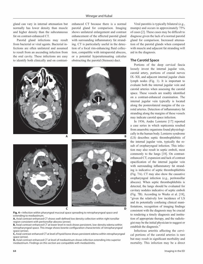
C DFig. 4—Infection within pharyngeal mucosal space spreading to retropharyngeal space and extending to mediastinum.A, Axial contrast-enhanced CT shows well-defined low density collection within right tonsillar region consistent with peritonsillar abscess (arrow).B, Axial contrast-enhanced CT at lower level in neck shows persistent, low-density edema within retropharyngeal space. This image shows bowtie configuration characteristic of retropharyngeal space (arrow).C, Axial contrast-enhanced CT at level of hyoid bone shows persistent edema within retropharyngeal space (arrow). D, Axial contrast-enhanced CT at level of mediastinum shows infection extending into superior mediastinum. Findings on this section are compatible with mediastinitis.
B
Imaging in the ED 65
Acute Neck Infections
extension from external infection (e.g., ret-ropharyngeal abscess) or of hematogenous origin (e.g., septic emboli). Weakening of the carotid wall by proteolytic enzymes or thrombosis of the vasa vasorum can lead to pseudoaneurysm formation (Fig. 8) or carotid rupture. Suspected infectious arte-ritis of the carotid artery, particularly with associated pseudoaneurysm, should be
emergently communicated to the treating physician given the dire consequences of a potential carotid rupture. Patients may be treated with stent placement, endovascular occlusion, or surgical bypass [19].
The Sublingual SpaceThe paired sublingual spaces are the
lateral aspects of the floor of mouth that
lie inferior to the intrinsic tongue muscles, superomedial to the mylohyoid muscle, and lateral to the geniohyoid-genioglos-sus complex (Fig. 1B). These spaces con-tain the sublingual glands, small portions of the superior submandibular glands, the submandibular ducts, tongue neurovascu-lar bundles, hyoglossus muscles, and fat. The roots of the second and third molars
A
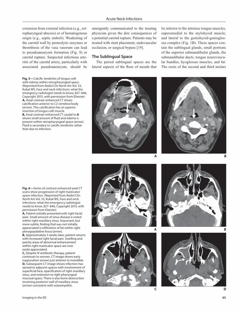
Fig. 5—Calcific tendinitis of longus colli with edema within retropharyngeal space. (Reprinted from Radiol Clin North Am Vol. 53, Kubal WS, Face and neck infections: what the emergency radiologist needs to know, 827–846, Copyright 2015, with permission from Elsevier)A, Axial contrast-enhanced CT shows calcification anterior to C2 vertebral body (arrow). This calcification lies at superior insertion of longus colli muscle.B, Axial contrast-enhanced CT caudal to A shows small amount of fluid and edema is present within retropharyngeal space (arrow). Fluid is secondary to calcific tendinitis rather than due to infection.
B
C
A
Fig. 6—Series of contrast-enhanced axial CT scans show progression of right masticator space infection. (Reprinted from Radiol Clin North Am Vol. 53, Kubal WS, Face and neck infections: what the emergency radiologist needs to know, 827–846, Copyright 2015, with permission from Elsevier)A, Patient initially presented with right facial pain. Small amount of sinus disease is noted within right maxillary sinus. Important, but more subtle, finding that was not initially appreciated is infiltration of fat within right pterygopalatine fossa (arrow).B, Approximately 3 weeks later, patient returns with increased right facial pain. Swelling and patchy areas of abnormal enhancement within right masticator space are now easily appreciated.C, Despite IV antibiotic therapy, patient continues to worsen. CT image shows early suppuration (arrow) just anterior to mandible.D, Subsequent CT image shows infection has spread to adjacent spaces with involvement of superficial face, opacification of right maxillary sinus, and extension to right pharyngeal mucosal space. There is also bone destruction involving posterior wall of maxillary sinus (arrow) consistent with osteomyelitis.
D
B
66 Imaging in the ED
Winegar and Kubal
extend below the insertion of the mylo-hyoid muscle, so infections of those teeth are likely to involve the submandibular space [2]. Because the roots of the teeth anterior to the second molar extend above the mylohyoid muscle, infections of those teeth typically are contained in the sublin-gual space [2].
Ludwig angina, a potentially life-threatening cellulitis involving the floor of mouth and adjacent submandibular spaces, is typically the consequence of odonto-genic infection. The infection extends to involve multiple spaces and may result in airway compromise. On contrast-enhanced CT, inflammatory stranding and edema af-fect the sublingual and submandibular spaces, which are particularly well visual-ized on coronal reformatted images. Soft-tissue gas and rim-enhancing fluid collec-tions may also be present.
The Submandibular SpaceThe submandibular space lies in-
ferolateral to the mylohyoid muscle and deep to the platysma (Fig. 1B) and con-tains predominantly the submandibular glands, lymph nodes, and fat. Posterior to the mylohyoid muscle attachment, the submandibular space communicates with the sublingual and parapharyngeal spaces. Odontogenic infections involv-ing the second or third mandibular mo-lars may extend into the submandibular spaces because these dental roots reside below the attachment of the mylohyoid sling. Submandibular space abscesses are rim-enhancing, low-density fluid collec-tions with surrounding inflammatory fat stranding that often connect to a posterior mandibular periapical lucency via an os-seous dehiscence.
In addition, infections may be cen-tered within the submandibular glands, resulting in sialadenitis. These bacte-rial infections will appear similar to their counterparts in the parotid glands, with asymmetric enlargement, enhancement, and surrounding inflammatory fat strand-ing of the affected submandibular gland. A hyperdense calculus may also be seen on CT along the expected course of the sub-mandibular (Wharton) duct and proximal ductal dilatation in the setting of obstruc-tive sialolithiasis.
Congenital LesionsSecond branchial cleft cysts are congen-
ital fluid-filled lesions within the subman-dibular spaces characteristically located posterior to the submandibular gland, deep to the sternocleidomastoid muscle, and anterolateral to the carotid artery. These
lesions may become clinically apparent when secondarily infected. CT shows a rim-enhancing fluid collection with sur-rounding stranding in the characteristic location. Metastatic cervical lymphade-nopathy in the setting of head and neck squamous cell carcinoma related to hu-
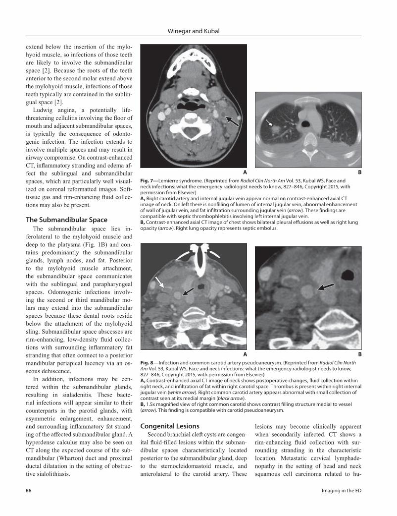
AFig. 8—Infection and common carotid artery pseudoaneurysm. (Reprinted from Radiol Clin North Am Vol. 53, Kubal WS, Face and neck infections: what the emergency radiologist needs to know, 827–846, Copyright 2015, with permission from Elsevier)A, Contrast-enhanced axial CT image of neck shows postoperative changes, fluid collection within right neck, and infiltration of fat within right carotid space. Thrombus is present within right internal jugular vein (white arrow). Right common carotid artery appears abnormal with small collection of contrast seen at its medial margin (black arrow).B, 1.5x magnified view of right common carotid shows contrast filling structure medial to vessel (arrow). This finding is compatible with carotid pseudoaneurysm.
B
AFig. 7—Lemierre syndrome. (Reprinted from Radiol Clin North Am Vol. 53, Kubal WS, Face and neck infections: what the emergency radiologist needs to know, 827–846, Copyright 2015, with permission from Elsevier)A, Right carotid artery and internal jugular vein appear normal on contrast-enhanced axial CT image of neck. On left there is nonfilling of lumen of internal jugular vein, abnormal enhancement of wall of jugular vein, and fat infiltration surrounding jugular vein (arrow). These findings are compatible with septic thrombophlebitis involving left internal jugular vein.B, Contrast-enhanced axial CT image of chest shows bilateral pleural effusions as well as right lung opacity (arrow). Right lung opacity represents septic embolus.
B
Imaging in the ED 67
Acute Neck Infections
man papillomavirus infection may have an identical imaging appearance and should be the top differential diagnostic consider-ation in patients outside of the pediatric or adolescent age groups.
Lymphatic malformations are congeni-tal slow-flow vascular malformations com-posed of abnormal lymphatic channels. These lesions may become clinically evi-dent when they are secondarily infected. On contrast-enhanced CT, lymphatic mal-formations are transspatial, multiseptated cystic lesions without wall enhancement. When infected, these malformations show wall and septal thickening and enhance-ment and can resemble a complex abscess.
SummaryThe detection, characterization, local-
ization, classification of potential compli-cations, and recognition of causes of head and neck infections are each critical roles of the emergency radiologist. Knowledge of normal head and neck anatomy and CT appearance will not only aid in detection of subtle infection and space of origin but also help in predicting potential patterns of spread, complications, and cause. Be-cause soft-tissue infections lie on a spec-trum from cellulitis to drainable abscess,
knowledge of the contrast-enhanced CT appearances of these different stages al-lows appropriate medical or surgical management for the underlying head and neck infection. The information provided should help the radiologist “to provide an accurate and prompt diagnosis, assess the extent of disease, evaluate for potential complications, and recommend definitive subspecialty evaluation” [2].
REFERENCES
1. Maroldi R, Farina D, Ravanelli M, et al. Emergency imaging assessment of deep neck space infections. Semin Ultrasound CT MR 2012; 33:432–442
2. Capps EF, Kinsella JJ, Gupta M, et al. Emergency imaging assessment of acute, nontraumatic con-ditions of the head and neck. RadioGraphics 2010; 30:1335–1352
3. Wang B, Gao BL, Xu GP, Xiang C. Images of deep neck space infection and the clinical significance. Acta Radiol 2014; 55:945–951
4. Crespo AN, Chone CT, Fonseca AS, et al. Clinical versus computed tomography evaluation in the di-agnosis and management of deep neck infection. Sao Paulo Med J 2004; 122:259–263
5. Scott BA, Stiernberg CM, Driscoll BP. Deep neck space infections. In: Bailey BJ, ed. Head and neck surgery: otolaryngology, 2nd ed. Philadelphia, PA: Lippincott-Raven, 1998:819–835
6. Kubal WS. Face and neck infections: what the emergency radiologist needs to know. Radiol Clin North Am 2015; 53:827–846
7. Vural C, Gungor A, Comerci S. Accuracy of computer-ized tomography in deep neck infections in the pedi-atric population. Am J Otolaryngol 2003; 24:143–148
8. Miller WD, Furst IM, Sàndor GKB, Keller MA. A pro-spective, blinded comparison of clinical examina-tion and computed tomography in deep neck in-fections. Laryngoscope 1999; 109:1873–1879
9. Kirse DJ, Roberson DW. Surgical management of retropharyngeal space infections in children. La-ryngoscope 2001; 111:1413–1422
10. Mayo-Smith MF, Spinale JW, Schiffman FJ, et al. Acute epiglottitis: an 18-year experience in Rhode Island. Chest 1995; 108:1640–1647
11. Smith MM, Mukherji SK, Thompson JE, et al. CT in adult supraglottitis. AJNR 1996; 17:1355–1358
12. Virk JS, Pang J, Okhovat S, Lingam RK, Singh A. An-alyzing lateral soft tissue neck radiographs. Emerg Radiol 2012; 19:255–260
13. Debnam JM, Guha-Thakurta N. Retropharyn-geal and prevertebral spaces: anatomic imaging and diagnosis. Otolaryngol Clin North Am 2012; 45:1293–1310
14. Shefelbine SE, Mancuso AA, Gajewski BJ, et al. Pe-diatric retropharyngeal lymphadenitis: differentia-tion from retropharyngeal abscess and treatment implications. Otolaryngol Head Neck Surg 2007; 136:182–188
15. Eastwood JD, Hudgins PA, Malone D. Retropharyn-geal effusion in acute calcific prevertebral tendini-tis: diagnosis with CT and MR imaging. AJNR 1998; 19:1789–1792
16. Chung T, Rebello R, Gooden EA. Retropharyngeal calcific tendinitis: case report and review of litera-ture. Emerg Radiol 2005; 11:375–380
17. Lemierre A. On certain septicæmias due to anaero-bic organisms. Lancet 1936; 227:701–703
18. Weeks DF, Katz DS, Saxon P, Kubal WS. Lemierre syndrome: report of five new cases and literature review. Emerg Radiol 2010; 17:323–328
19. Hirai T, Korogi Y, Sakamoto Y, et al. Emergency balloon embolization for carotid artery rupture secondary to postoperative infection. Cardiovasc Intervent Radiol 1996; 19:50–52